uterine luminal environment of the mare by tiina reilas
TRANSCRIPT

Department of Clinical Veterinary SciencesFaculty of Veterinary Medicine
University of Helsinki,Helsinki, Finland
andMTT Agrifood Research Finland
Animal Production ResearchEquines
Ypäjä, Finland
UTERINE LUMINAL ENVIRONMENT OF THE MARE
by
Tiina Reilas
ACADEMIC DISSERTATION
To be presented, with the permission of the Faculty of Veterinary Medicine,University of Helsinki, for public criticism
in the Auditorium Maximum, Hämeentie 57, Helsinki,on November 23rd, 2001, at 12 noon.
YPÄJÄ 2001

ISBN 952-91-4058-4ISBN 952-10-0194-1 (pdf)
Painosalama Oy - Turku, Finland 2001

To my family

4
CONTENTS
ABSTRACT ......................................................................................................................6LIST OF ORIGINAL ARTICLES ....................................................................................8ABBREVIATIONS...........................................................................................................9INTRODUCTION...........................................................................................................10REVIEW OF LITERATURE..........................................................................................11
1. Uterine luminal environment ..................................................................................111.1. Effect of steroid hormones...............................................................................111.2. Introduction of micro-organisms into the uterus .............................................131.3. Endometrial inflammation ...............................................................................141.4. Uterine involution ............................................................................................17
2. Examination methods of uterine luminal environment ...........................................202.1. Transrectal palpation and ultrasonography......................................................202.2. Endoscopy........................................................................................................202.3. Sampling of uterine luminal contents .............................................................20
3. Diagnostic and prognostic value of uterine samples ...............................................213.1. Uterine cytology ..............................................................................................213.2. Bacterial cultures .............................................................................................223.3. Uterine histology..............................................................................................233.4. Biochemical analysis of uterine fluid ..............................................................233.5. Predicting foal heat fertility .............................................................................27
AIMS OF THE STUDY..................................................................................................28MATERIALS AND METHODS ....................................................................................29
1. Animals ...................................................................................................................292. Experimental design................................................................................................29
2.1. Hormone treatments and bacterial inoculation (I) ...........................................292.2. Post-breeding study (II) ...................................................................................292.3. Fluid accumulation study (III) .........................................................................312.4. Involution study (IV) .......................................................................................312.5. Foal heat fertility study (V) .............................................................................31
3. Ovulation control and ultrasonography...................................................................314. Inseminations (II, III, V) .........................................................................................325. Hormone replacement therapy (I) ...........................................................................326. Intrauterine streptococcal inoculation (I) ................................................................327. Embryo recovery (III, V) ........................................................................................338. Sampling of the uterus ............................................................................................33
8.1. Endometrial swabs (I, IV)................................................................................338.2. Uterine lavage (I, II, IV, V) .............................................................................338.3. Tampon technique (III) ....................................................................................348.4. Endometrial biopsy (I, III) ...............................................................................34
9. Counting of neutrophils...........................................................................................3410. Bacteriological examination..................................................................................3511. Histological examination ......................................................................................3512. Biochemical analyses ............................................................................................35
12.1. Total protein (I, III-V)....................................................................................35

5
12.2. Trypsin-inhibitor capacity (TIC) (I, III-V) ....................................................3512.3. Plasmin (I, III-V) ...........................................................................................3612.4. Lysozyme (I, IV, V).......................................................................................3612.5. N-acetyl-ß-D-glucosaminidase (NAGase) (I, III-V)......................................3612.6. ß-glucuronidase (B-Gase) (I, III-V)...............................................................3612.7. Acid phosphatase (I, IV) ................................................................................37
13. In vitro motility studies of spermatozoa (III) ........................................................3714. Statistics ................................................................................................................37
RESULTS........................................................................................................................391. IUFA, endometrial oedema and biopsies ................................................................392. PMN numbers .........................................................................................................393. Bacteriological cultures...........................................................................................414. Biochemical analyses ..............................................................................................41
4.1. Total protein.....................................................................................................414.2. TIC...................................................................................................................494.3. Plasmin ............................................................................................................494.4. Lysozyme.........................................................................................................494.5. NAGase............................................................................................................504.6. B-Gase .............................................................................................................504.7. Acid phosphatase .............................................................................................51
5. Associations between PMNs, proteins and enzymes (I,IV) ....................................516. In vitro motility studies of spermatozoa..................................................................537. Embryo recovery and pregnancy results .................................................................53
DISCUSSION..................................................................................................................541. Collection methods of uterine fluid.........................................................................542. Composition of uterine lavage fluid in non-infected mares (I, IV).........................553. Composition of uterine lavage fluid after bacterial inoculation (I) .........................564. Intrauterine fluid accumulations during oestrus (II, III)..........................................585. Uterine involution and secretory function of the endometrium after parturition(IV)..............................................................................................................................606. Predicting fertility in foal heat (V)..........................................................................617. Neutrophilia after insemination (II) ........................................................................62
CONCLUSIONS .............................................................................................................65ACKNOWLEDGEMENTS ............................................................................................66REFERENCES................................................................................................................68
ORIGINAL ARTICLES

6
ABSTRACT
The uterine luminal environment, determined primarily by uterine secretions, mustoffer optimal conditions first for the fertilizing spermatozoa and later for thedevelopment of the embryo. Relatively few studies exist, however, concerning thebiochemical composition of equine uterine secretions. This thesis includesinvestigations on centrifuged uterine lavage fluids (ULFs) collected after hormonetreatments and bacterial inoculations of ovariectomized (OVX) mares. Uterine lavagefluids were also collected from non-parturient cyclic mares and from post-partum (p.p.)mares within 4 weeks of parturition, after the first p.p. ovulation and before embryorecovery 7 to 8 days later. Polymorphonuclear neutrophils (PMNs) were comparedwith total protein and lysozyme concentrations, trypsin-inhibitor capacity (TIC), andactivities of plasmin, N-acetyl-ß-D-glucosaminidase (NAGase), ß-glucuronidase (B-Gase) and acid phosphatase to determine whether more sensitive inflammatory markerscould be found to diagnose endometritis. In p.p. mares, these parameters were used todetermine the restoration of secretory activity after foaling and to investigate their relationto foal heat fertility. Undiluted uterine fluids were collected by tampon from oestrousmares to study whether composition of uterine fluid differs in mares with and withoutultrasonically detectable intrauterine fluid accumulations (IUFA). The intensity ofinflammatory reaction 6 h after different types of insemination was studied byanalysing PMNs from lavage fluid.
Seven days' treatment of OVX-mares with progestin increased accumulation ofNAGase, B-Gase and acid phosphatase (P<0.05) in ULF. PMNs, lysozymeconcentration, TIC and plasmin activity were mostly below detection limits. Afterbacterial inoculation, PMN numbers remained significantly elevated until 24 h incontrol mares and in mares treated with oestradiol, and until day 21 in mares treatedwith progestin plus oestradiol or progestin alone. Lysozyme activities were elevated inboth acute and persistent endometritis. TIC was a better indicator of acute than ofpersistent inflammation. Despite acute inflammation, total protein concentration, TICand plasmin activity did not increase significantly in oestradiol-treated mares.Progestin, not infection, was the main reason for increased activities of NAGase, B-Gase and acid phosphatase.
In non-parturient mares, activities of NAGase, B-Gase and acid phosphatase in ULFswere significantly higher in dioestrus than in oestrus (P<0.05). Lysozymeconcentration, TIC and plasmin activity were below detection limits. In p.p. mares,levels of total protein, NAGase, B-Gase and acid phosphatase were high soon afterfoaling but thereafter declined rapidly. In most mares, lysozyme and plasmin activitiesdecreased after foaling. TIC peaked around day 6 p.p.. Total protein and lysozymeconcentrations, TIC, and B-Gase (P<0.01) and acid phosphatase (P<0.05) activitieswere significantly higher in parturient mares during p.p. oestrus than in oestrous non-parturient mares. During dioestrus, differences between p.p. and non-parturient mareswere not statistically significant.
After the first p.p. ovulation but prior to insemination, PMNs, TIC and lysozyme levelsin ULF were elevated in 3/4 mares not producing embryos. However, only 1/5, 1/5 and

7
0/5 embryo-producing mares had elevated levels of PMNs, TIC and lysozyme,respectively. None of the measured parameters was significantly related to embryorecovery 7 days later (P≤0.10). At embryo recovery, NAGase was higher in mares notproducing embryos (P<0.05). The relation of this finding to embryo recovery isunclear.
IUFA were detected by ultrasonography in 39% of oestrous mares. None of the maresexhibited cytological or bacteriological evidence of acute endometritis. Total proteinconcentration, TIC and B-Gase activity in undiluted uterine fluid were significantlylower in mares with than in those without IUFA (P<0.01), whereas NAGase andplasmin activity did not differ between these groups. The presence of small fluidaccumulations at the time of insemination did not seem to affect fertility.
In the post-breeding study, mares infused with phosphate-buffered saline, seminalextenders or supernatant from centrifuged frozen-thawed semen exhibited only a mildneutrophil response. Insemination with frozen semen resulted in higher concentrations ofPMNs than insemination with extended fresh semen (P<0.05). Highest PMN counts werefound after insemination with frozen semen or concentrated fresh semen. Bacterialcontamination of uteri was negligible 6 h after breeding.
In conclusion, none of the examined parameters was superior to PMN counts indiagnosing endometritis. High total protein concentration, TIC and detectable lysozymeand plasmin activities during p.p. oestrus were associated with uterine inflammation.The endometrium of p.p. mares had presumably resumed normal secretory capacity bythe time of the first p.p. dioestrus. Endometrial inflammation at the time ofinsemination seems to be the best explanation for lowered foal heat fertility. Non-echogenic fluid accumulations during oestrus were associated with compositionalchanges in uterine secretions. Neutrophilia appears to be induced by spermatozoa ratherthan bacteria. Intensity of the neutrophil reaction apparently depends on concentrationand/or volume of inseminate.

8
LIST OF ORIGINAL ARTICLES
This thesis is based on the following original articles which are referred to in the textby their Roman numerals:
I. Reilas, T., Ristiniemi, M. & Katila, T. Influence of hormone replacementtherapy and bacterial inoculation on proteins and enzymes in uterine lavagefluid of ovariectomized mares. Reprod. Dom. Anim. 33, 1998: 11-19.
II. Kotilainen, T., Huhtinen, M. & Katila, T. Sperm-induced leukocytosis in theequine uterus. Theriogenology 41, 1994: 629-636.
III. Reilas, T., Katila, T., Mäkelä, O., Huhtinen, M. & Koskinen, E. Intrauterinefluid accumulation in oestrous mares. Acta vet. scand. 38, 1997: 69-78.
IV. Reilas, T. & Katila, T. Proteins and enzymes in uterine lavage fluid ofpostpartum and nonparturient mares. Reprod. Dom. Anim. (accepted).
V. Reilas, T., Huhtinen, M., Oksanen, M. & Katila, T. Relationship betweenembryo recovery rate and uterine lavage fluid composition in postpartummares. Reprod. Nutr. Dev. 40, 2000: 383-391.

9
ABBREVIATIONS
AI artificial inseminationB-Gase β-glucuronidaseBSA bovine serum albuminCV coefficient of variationDPBS Dulbecco's phosphate-buffered salineE oestrogen/oestradiolE. coli Escherichia coliFCS foetal calf serumGAGs glycosaminoglycansi inoculationIg immunoglobulin (A, G, M, T)i.m. intramusculari.u. intrauterineIUF intrauterine fluidIUFA intrauterine fluid accumulation (detected by ultrasonography)i.v. intravenousMHC major histocompatability complexMOT total motility4-MU 4-methylumbelliferoneNAGase N-acetyl-β-D-glucosaminidaseOVX ovariectomizedP progesterone/progestinPBS phosphate-buffered salinePG prostaglandinp.o. per osp.p. post-partumα1-PI alpha-1-proteinase inhibitorPMN polymorphonuclear neutrophilPMOT progressive motilityRT room temperatureS. aureus Staphylococcus aureusSD standard deviationSEM standard error of meanspp. speciesStrep. StreptococcusTIC trypsin-inhibitor capacityU µmol product/minULF uterine lavage fluidVAP average path velocity

10
INTRODUCTION
The uterus has many roles: it is the site of semen deposit in the horse and contributes tothe travel of spermatozoa on their way to the oviduct, it houses and nourishes therapidly growing foetal foal, and finally, the myometrium is essential in the expulsion ofthe foal during parturition. The pregnancy causes enormous changes in themyometrium, which has to adapt and stretch from dimensions of centimetres to metresduring the gestation of 11 months (Rossdale 1997). As amazing is the rapid return ofthe organ to its original size within 2 to 3 weeks and the ability of the mare to conceivein the first post-partum (p.p.) oestrus (known as foal heat) as early as only a week afterparturition. However, the pregnancy rates in foal heat are usually lower than insubsequent oestruses. Unfortunately, we have no reliable way of diagnosing whichmares are ready to be bred in foal heat and which ones are not.
The composition and quality of the uterine environment determine reproductivereadiness and performance of the female. The uterus is part of the endocrine system: atarget organ for steroids, but also the site of prostaglandin production. Hormonescontrol quantitative and qualitative secretory activity of the uterus. Synchrony betweenmaternal hormones and embryonic development is obligatory to establish pregnancy(Fischer & Beier 1986; Hinrichs & Kenney 1987). Before implantation, the conceptusis supported solely by secretions that accumulate in the uterine lumen; a phenomenoncalled histotrophic nutrition (Ashworth 1995). In the mare, implantation andplacentation take place considerably later than in other species, and therefore,histotrophe is particularly important in the horse. An inadequate uterine milieu causesembryonic mortality, which has a high incidence in horses. Changes in uterineenvironment during puerperium, inflammation and stages of the cycle are not very wellknown in horses.
The genitalia of stallions and mares harbour a microflora consisting of many bacterialspecies, the most frequently occurring of which are streptococci (Dimock & Snyder1924; Dimock & Edwards 1928). Dimock & Edwards (1928) recognized that stallionsmay infect mares and that infection of mares at the time of service is probablydependent upon some predisposition. Mares who are unable to clear bacteria afterbreeding continue to present a problem in studfarm practice. In mares, endometritis isoften not very obvious. Discharge from the vulva may alert the owner, but sometimesthe only sign is a failure to conceive. A cytological examination to identifypolymorphonuclear neutrophils (PMNs) is an easy and quick way to diagnose acuteendometritis and distinguish between bacterial contamination and significantendometritis (Knudsen 1964a; Wingfield Digby 1978); however, it does not detectchronic endometritis. In contrast, while histopathology of uterine biopsies reveals acuteand chronic inflammation in addition to degenerative glandular changes (Kenney1978), it is not a quick test. Detection of excessive intrauterine fluid is easy withultrasonography (Ginther & Pierson 1984), but it gives little information on thecomposition of the fluid. Thus, there is a need to find new tests for diagnosis of acuteand persistent endometritis which are sensitive, rapid and easy to perform.

11
REVIEW OF LITERATURE
1. Uterine luminal environment
The uterine environment has to offer optimal conditions first for the fertilizingspermatozoa and later for the development of the embryo. Uterine fluid consists ofhormones, prostaglandins, enzymes, energy substrates, ions, vitamins, amino acids,peptides, serum proteins and uterine proteins (Fischer & Beier 1986). The blood-uterine lumen barrier determines the rate at which matter is exchanged between thevascular and extravascular fluids of the uterus. Extracellular uterine fluid can bedivided into the following compartments: vascular, endometrial extracellular andluminal extracellular (McRae 1988).
1.1. Effect of steroid hormones
According to McRae (1988), the uterine luminal milieu is dynamic since thecomposition of the fluid varies between stages of the oestrous cycle and during thecourse of pregnancy. The secretion and release of proteins and enzymes are regulatedby steroids. Tight junctions between epithelial cells change with hormone status. Thenormal uterus contains small amounts of fluid that is produced locally by uterineglands or represents transudate from blood vessels. Uterine capillaries and venules arerelatively impermeable to plasma proteins except during transient responses tooestrogen. Epithelial cells are selectively permeable to substances primarily on thebasis of molecular size and lipid solubility. Uterine epithelium functions as a rate-limiting boundary between blood and uterine lumen even when vascular permeabilityis increased (McRae 1988).
Ovarian steroids cause structural and functional alterations to the endometrium. Theeffects of steroids can be studied in cyclic mares and in hormone-supplementedovariectomized (OVX) or seasonally acyclic mares. The hormone supplementations areoestrogen (E), progesterone (P) and combinations of oestrogen and progesterone (E +P).
It is commonly agreed that intrauterine fluid accumulation (IUFA) during dioestrusindicates an inflammatory process with negative effects on pregnancy (Adams et al.1987). In contrast, the significance of IUFA during oestrus is unclear. In anovulatorymares, IUFA was often detected, but the presence of fluid changed dynamically, andthe fluid was not associated with uterine or oviductal inflammation (Losinno et al.1997). Significant differences have been shown for uterine fluid concentrations of totalprotein (Zavy et al. 1979, 1982), acid phosphatase (Zavy et al. 1979, 1982; McDowellet al. 1987; LeBlanc et al. 1988) and prostaglandins (PGF and PGE2) (Watson et al.1988a), all being higher during progesterone dominance. Total protein concentrationsin uterine lavage fluids peaked at day 12 of dioestrus and decreased thereafter (Zavy etal. 1979, 1982). Similar findings have been reported by others, but the differencesbetween oestrus and dioestrus have not been statistically significant (Strzemienski &Kenney 1984; LeBlanc et al. 1988). Three progesterone-dependent proteins have beenseparated from uterine secretions of mares by Beier-Hellwig et al. (1995): uteroglobin

12
(<6.5 kDa), equine phospholipase A2 (17 kDa) and a third unidentified proteinmigrating at 22 kDa.
Asbury et al. (1980) reported that IgA levels were highest during oestrus, whereas IgGwas detected only from dioestrous samples of uterine fluid. However, Widders et al.(1985) did not notice cyclic changes in free Ig staining or plasma cell numbers usingimmunoperoxidase staining techniques. Similarly, hormone treatment (E or P) ofOVX-mares did not significantly affect the numbers of cells secreting IgA, IgG or IgM(Watson & Stokes 1988). More major histocompatability complex (MHC) Class II-positive cells (macrophages, monocytes, dendritic cells, epithelial cells and endothelialcells) were present in the epithelium and stratum compactum during oestrus thandioestrus, but numbers of T cells were unaffected by cycle stage (Watson & Dickson1993).
The pattern and activity of myometrial contractions have been shown to change duringthe cycle (Taverne et al. 1979; Cross & Ginther 1987; Griffin & Ginther 1990;Troedsson et al. 1993b). Synchronization of uterine electrical activity among differentsites in the uterus was more marked during oestrus than during dioestrus. This suggestsa hormone-dependent mechanism for the regulation of cell communication (Troedssonet al. 1993b). Progesterone inhibits and oestrogen stimulates formation of endometrialgap junctions in the myometrium (Garfield et al. 1980). Using transrectalultrasonography, the lowest uterine activity scores were associated with low steroidconcentrations (Cross & Ginther 1987; Griffin & Ginther 1990). The dailyadministration of oestradiol to anoestrous mares resulted in a gradual increase inuterine activity over 5 days (Cross & Ginther 1987). Griffin & Ginther (1990) observedin cyclic mares a progressive increase in activity between days 2 and 4 of dioestrus andagain between days 11 and 12, the activity being maximal on days 13 and 14.
Bacterial adherence to epithelial cells in vitro has been shown to be higher with P andlower with E (Watson et al. 1988b). Freeman et al. (1990) found mucins in the luminalepithelial cells of the equine endometrium. The secretion, elicited by hormonal orirritative/inflammatory stimuli, may be a protective response (Freeman et al. 1990).Tunón et al. (1995) demonstrated that endometrial epithelial cells on the uterinesurface and in the glands showed intense cellular vacuolation during oestrus. Thisindicates high secretory activity. Mucins synthesized actively during oestrus mayrender adherence of bacteria to the epithelium difficult.
It is common knowledge that acyclic mares treated with progesterone remain infectedafter bacterial challenge for an extended period, persisting as long as the treatmentcontinues (Washburn et al. 1982; Evans et al. 1986; Hinrichs et al. 1992). P-treatmentof acyclic mares has even been suggested as a model to study endometritis in mares(Colbern et al. 1987). Fluid accumulation in the uterine lumen has been shown to occurafter uterine inoculation of bacteria when mares are treated with P (Evans et al. 1986;Hinrichs et al. 1992). The cervix is relaxed during E-dominance and tight during P-dominance (Evans et al. 1986).

13
1.2. Introduction of micro-organisms into the uterus
The genitalia of the mare have a microflora that consists of harmless species andopportunistic organisms that can be pathogenic in susceptible animals (Dimock &Snyder 1924). Bacterial numbers decrease from the vagina to the uterus, and the feworganisms found in the cervix and uterus are probably transient (Scott et al. 1971;Hinrichs et al. 1988). Four organisms are responsible for the vast majority ofendometritis in the mare: Streptococcus zooepidemicus, Escherichia coli,Pseudomonas aeruginosa and Klebsiella pneumoniae. Alpha-haemolytic streptococci,Staphylococcus spp. and other bacteria may also be recovered but are usuallycontaminants. Yeast and fungi may cause endometritis as well; Candida spp.,Aspergillus spp. and Mucor spp. are most common. Taylorella equigenitalis is thecause of venereally transmitted contagious equine metritis (Blanchard et al. 1998).
Micro-organisms enter the uterus by ascending from the vagina. Breeding (Dimock &Snyder 1924) and foaling (Dimock & Edwards 1928; Dimock 1935), the two importantevents that are repeated annually in broodmares, are the most important ways ofintroducing bacteria into the uterus. Some procedures performed by veterinarians mayalso carry bacteria into the uterus, e.g. insemination, embryo transfer, uterine biopsiesor swabs, and infusion of medications (Hinrichs et al. 1992; McDonnell & Watson1992). Conformational abnormalities, such as pneumovagina (Caslick 1937a),urovagina and cervical lesions, facilitate the entrance of bacteria into the uterus (VanCamp 1986). Hinrichs et al. (1988) emphasized the importance of a properlyfunctioning vulvovaginal fold as barrier to ascending bacterial contamination. Theyalso found that clitoral fossa swab specimens had more bacterial growth (94% of thespecimens) than any other parts of the genital tract. This finding suggests the clitoris asa source of contamination during intrauterine (i.u.) manipulations (Hinrichs et al.1988).
Breeding
Breeding is commonly advocated as a cause for endometritis. All stallions harbourseveral species of bacteria on their external genitalia and in their semen (Dimock &Snyder 1924; Hughes et al. 1967; Millar & Francis 1974; Simpson et al. 1975). Anegative uterine pressure during coitus draws semen directly into the mare’s uterus(Millar 1952). As various bacteria are commonly cultured also from normal mares, andmost frequently from the caudal parts of the tract (Hinrichs et al. 1988), it is naturalthat representatives of the microflora move to the more cranial portion of thereproductive tract during breeding. More than 80% of mares showed bacteriologicallypositive cervical cultures 24 h after being bred (Bryans 1962). Today, the use ofartificial insemination (AI) and, particularly, semen collection by the open-endedartificial vagina has diminished bacterial contamination associated with breeding(Tischner et al. 1974).

14
Foaling
Management of foaling mares and events during foaling affect the degree of bacterialcontamination (Knudsen 1983). The incidence of bacteriologically positive cultures hasoften been lower 1 to 2 days p.p. (around 20-40% of mares) compared with 3 to 6 daysp.p. (70-90%) (König 1975; Gygax et al. 1979; Saltiel et al. 1987; Katila et al. 1988b).Mares with puerperal disturbances – dystocia or retained placenta – had more bacterialgrowth 3, 6 and 9 days p.p. than mares having normal deliveries (Glatzel & Belz1995). In all these studies, same mares had been sampled on several consecutive days.Therefore, uterine swabbing may have increased contamination of the uterus.
1.3. Endometrial inflammation
In acute inflammation, fluid moves from the blood into the tissues, causing oedemaand swelling. Increased permeability of dilated small blood vessels also allows largemolecules, including antibodies and complement components, to escape into tissues.PMNs adhere to the endothelial cells lining the vessels and migrate between the cellsinto the tissue. The migration of PMN is directed by chemotaxis. At the inflammatorysite, PMNs phagocytose and kill invading organisms. During phagocytosis lysosomalcontents are released. These include proteins and enzymes, as well as free oxygenradicals. Their main task is destruction of bacteria, but they may also be destructive tothe tissues (Tizard 1996). Endogenous neutral proteases are derived principally fromPMNs and macrophages and they hydrolyse proteins. The peptide products have anumber of physiological functions, including macrophage chemotaxis, and they are, inpart, responsible for perpetuating the inflammatory response (Matthews 1994).
Enzymes discharged upon cell death or during phagocytosis have been detected inuterine fluid of mares after experimental infection. Lactate dehydrogenase, acytoplasmic enzyme, has been detected in uterine flushings of oestrous mares. Afterthe inoculation of Streptococcus zooepidemicus, the concentrations increasedsignificantly within 4 h indicating death of PMNs, endometrial cells, or both (Pycock& Allen 1990). Alkaline phosphatase has been shown to be present in uterine fluid ofcyclic mares collected by tampons, but the concentrations did not differ during the daysof the cycle. After i.u. inoculation of streptococci, the alkaline phosphatase levelsincreased drastically and peaked at 12 h (Katila et al. 1990). Williamson et al. (1983)identified alkaline phosphatase in uterine lavage fluid and found no difference betweeninfected and non-infected mares. Pycock & Allen (1990) and Katila et al. (1990) havereported elevated lysozyme activities in uterine fluid of mares after bacterial challenge.
Inflammation associated with breeding
PMNs are known to be present in the endometrium of mares post-service (Brook1985). High numbers of leucocytes were detected in uterine lavage fluid of mares afterinsemination, and phagocytosis of spermatozoa was frequently observed (Bader &Krause 1980). It has been commonly believed – based on the study of Bryans (1962) -that bacteria cause the uterine inflammation associated with breeding. However, inrabbits (Tyler 1977) and in humans (Cohen 1984), leucocytosis occurs in the cervix as

15
a normal physiological response to spermatozoa. It is very likely that the PMNresponse in the mare’s uterus is provoked by sperm.
Inflammation associated with foaling
Uterine swabs taken from p.p. mares commonly show PMNs in the cytologicalexamination (Knudsen 1964a; Gygax et al. 1979; Saltiel et al. 1987; Katila et al.1988b). Neutrophils are needed to clear bacterial infections introduced during andimmediately after parturition (Brook 1985) and also to destroy microcaruncles (Gygaxet al. 1979; Katila 1988). Mares with puerperal disturbances, particularly with retainedplacenta, showed high numbers of PMNs in lochial secretions 3 to 6 days p.p. (Belz &Glatzel 1995). During foal heat approximately one-third of the mares showed no PMNson cytological examination, one-third had only a few PMNs and one-third hadmoderate to high numbers of PMNs (Shideler et al. 1987; Katila et al. 1988b; Huhtinenet al. 1996).
Inflammation associated with intrauterine manipulations
Any manipulation of the equine uterus easily causes increased numbers of neutrophils(Bennett et al. 1980). Intrauterine antibiotic treatments are irritant (Asbury 1982), buteven the infusion of physiological saline and the passing of an instrumenttranscervically into the uterus induce neutrophilia (Knudsen & Nydahl 1983).Stimulation of the cervix and uterus, but not of the vagina, provoke a PMN response(Williamson et al. 1987). Since collection of uterine fluid and other types of uterinesampling always involve the passing of a catheter into the uterus and othermanipulations, frequent samplings of the same mare cannot be done without causingerroneous results.
Elimination of uterine inflammation and infection
The ability of mares to eliminate uterine infections has been intensively studied. Afrequently used method is to infuse the uterus of mares with an irritating substance orwith bacteria and take uterine samples before infusion and at certain time intervalsafter the infusion. Experimental bacterial inoculations have commonly involvedintrauterine infusion of Streptococcus zooepidemicus. This has been the traditionalmethod of studying the course of endometritis and uterine defence mechanisms sincethe first reports in 1969 by Hughes & Loy and Peterson et al. These studies showed –and several subsequent ones have confirmed this – that there is an intense and early(<6h) PMN response in the endometrium, which starts to subside after 12 h. In younghealthy mares, the reproductive tract returns to normal appearance within 96 h,whereas persistently infected mares require more time to eliminate bacteria (Hughes &Loy 1969; Peterson et al. 1969).
Although Hughes & Loy (1969) and Peterson et al. (1969) noticed that mares aredifferent in their susceptibility to uterine infections, it is not clear how they differ.Numerous studies have attacked this problem using an intrauterine bacterial challenge.
16
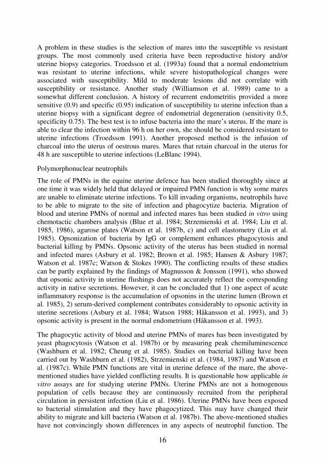
A problem in these studies is the selection of mares into the susceptible vs resistantgroups. The most commonly used criteria have been reproductive history and/oruterine biopsy categories. Troedsson et al. (1993a) found that a normal endometriumwas resistant to uterine infections, while severe histopathological changes wereassociated with susceptibility. Mild to moderate lesions did not correlate withsusceptibility or resistance. Another study (Williamson et al. 1989) came to asomewhat different conclusion. A history of recurrent endometritis provided a moresensitive (0.9) and specific (0.95) indication of susceptibility to uterine infection than auterine biopsy with a significant degree of endometrial degeneration (sensitivity 0.5,specificity 0.75). The best test is to infuse bacteria into the mare’s uterus. If the mare isable to clear the infection within 96 h on her own, she should be considered resistant touterine infections (Troedsson 1991). Another proposed method is the infusion ofcharcoal into the uterus of oestrous mares. Mares that retain charcoal in the uterus for48 h are susceptible to uterine infections (LeBlanc 1994).
Polymorphonuclear neutrophils
The role of PMNs in the equine uterine defence has been studied thoroughly since atone time it was widely held that delayed or impaired PMN function is why some maresare unable to eliminate uterine infections. To kill invading organisms, neutrophils haveto be able to migrate to the site of infection and phagocytize bacteria. Migration ofblood and uterine PMNs of normal and infected mares has been studied in vitro usingchemotactic chambers analysis (Blue et al. 1984; Strzemienski et al. 1984; Liu et al.1985, 1986), agarose plates (Watson et al. 1987b, c) and cell elastometry (Liu et al.1985). Opsonization of bacteria by IgG or complement enhances phagocytosis andbacterial killing by PMNs. Opsonic activity of the uterus has been studied in normaland infected mares (Asbury et al. 1982; Brown et al. 1985; Hansen & Asbury 1987;Watson et al. 1987c; Watson & Stokes 1990). The conflicting results of these studiescan be partly explained by the findings of Magnusson & Jonsson (1991), who showedthat opsonic activity in uterine flushings does not accurately reflect the correspondingactivity in native secretions. However, it can be concluded that 1) one aspect of acuteinflammatory response is the accumulation of opsonins in the uterine lumen (Brown etal. 1985), 2) serum-derived complement contributes considerably to opsonic activity inuterine secretions (Asbury et al. 1984; Watson 1988; Håkansson et al. 1993), and 3)opsonic activity is present in the normal endometrium (Håkansson et al. 1993).
The phagocytic activity of blood and uterine PMNs of mares has been investigated byyeast phagocytosis (Watson et al. 1987b) or by measuring peak chemiluminescence(Washburn et al. 1982; Cheung et al. 1985). Studies on bacterial killing have beencarried out by Washburn et al. (1982), Strzemienski et al. (1984, 1987) and Watson etal. (1987c). While PMN functions are vital in uterine defence of the mare, the above-mentioned studies have yielded conflicting results. It is questionable how applicable invitro assays are for studying uterine PMNs. Uterine PMNs are not a homogenouspopulation of cells because they are continuously recruited from the peripheralcirculation in persistent infection (Liu et al. 1986). Uterine PMNs have been exposedto bacterial stimulation and they have phagocytized. This may have changed theirability to migrate and kill bacteria (Watson et al. 1987b). The above-mentioned studieshave not convincingly shown differences in any aspects of neutrophil function. The

17
initial neutrophil response is neither delayed nor decreased in mares susceptible touterine infections (Couto & Hughes 1985; Williamson et al. 1987; Katila et al. 1990),but these mares continue to have PMNs in the uterus because of persistent infection.Mares with persistent endometritis had significantly elevated concentrations of PGFand total protein as well as higher percentage of PMNs and mononuclear cells inuterine washings than normal mares (Watson et al. 1987a).
Cellular and humoral immunity
Immunoglobulins IgG, IgT, IgA and IgM have been found in uterine secretions ofmares (Kenney & Khaleel 1975). Higher concentrations of immunoglobulins havebeen measured in uterine secretions than in serum, confirming the potential of theendometrium for local immunoglobulin production (Liu et al. 1981; Widders et al.1984). Immunoglobulin levels were elevated in the uterus in the presence of active(Asbury et al. 1980; Williamson et al. 1983) or chronic (Widders et al. 1984) infection.The presence of endometritis greatly increased MHC Class II expression and Tlymphocyte numbers (Watson & Dixon 1993). The densities of CD4+ (helper T-cells),CD8+ (cytotoxic T-cells) and B-cells were also significantly increased in mares withendometritis (Watson & Thomson 1996). However, no evidence exists that anydisturbances or deficiencies in either humoral or cellular immunity are present in maresthat are susceptible to uterine infections.
Mechanical clearance
The importance of physical clearance was for long overlooked despite the results of theelegant study carried out by Evans et al. (1986). Susceptible mares tend to accumulatefluid and retain bacteria or infused material in the uterus (Evans et al. 1986; Allen &Pycock 1988; LeBlanc et al. 1989, 1994, 1995; Troedsson & Liu 1991). Lymphaticdrainage from the uterine lumen is slower in susceptible than in resistant mares(LeBlanc et al. 1995). Myometrial contractions probably play a major role sincesignificant differences have been detected in electromyography between normal andsusceptible mares (Troedsson et al. 1993c), and since inhibition of uterine contractionsin normal mares decreases mechanical fluid clearance (Cadario et al. 1995;Nikolakopoulos & Watson 1999). The degree of cervical relaxation is also important inphysical clearance (Evans et al. 1986; LeBlanc et al. 1994).
1.4. Uterine involution
Involution is the restoration of the endometrium p.p. to a condition where conceptioncan take place again and new embryonic development can be carried out. Bearing inmind the great uterine changes during pregnancy, parturition and after parturition, it isamazing that some mares are able to conceive as early as one week after foaling.
When the villous epitheliochorial placenta separates at the maternal-foetal interface, itleaves the uterine epithelium relatively undamaged. During involution, degenerating ordetached epithelial cells above the maternal crypts are replaced and the uterineepithelium redifferentiates into ciliated and secretory cell types (Steven et al. 1979).
18
The numerous tiny microcaruncles disappear rapidly by degeneration and resorption.The major feature by days 6 and 7 p.p. are small focal remnants consisting ofcondensed stroma and crypts lined with contracted epithelial cells. In biopsy specimenstaken 10 and 15 days p.p., the former microcaruncles are detectable only by theoccurrence of siderophages and lymphocytes (Gygax et al. 1979; Bailey & Bristol1983; Katila 1988). Uterine glands are numerous, evenly distributed and dilated, andtheir lumen contains various amounts of cellular debris for 2 days p.p.. By day 4-5 p.p.,the glands in the upper layers of the stratum spongiosum have regenerated.Siderophages are present in large numbers around the glands and in the glandularepithelium. By day 15, almost all glands have returned to the pregravid size, butmitotic figures are still common in the epithelium (Bailey & Bristol 1983; Katila1988).
Biopsy specimens obtained 1 to 2 days p.p. show PMNs concentrated in the stratumcompactum, particularly around the microcaruncles. By day 5, lymphocytes appear andreplace PMNs. Lymphocytes are seen in the stratum compactum, particularly in thecell-dense areas, and also appear diffusely through the stratum spongiosum.Periglandular and –vascular lymphocytic infiltrations are a common finding in biopsyspecimens obtained 10 days p.p. (Shideler et al. 1987; Katila 1988).
Uterine size, tone and contents
A tremendous change in uterine size takes place after foaling, particularly during thefirst week after foaling (Gygax et al. 1979; Vandeplassche et al. 1983; Katila et al.1988a). Use of ultrasonography has shown that by 3 weeks the uterus has returned toits pregravid size (McKinnon et al. 1988b; Sertich & Watson 1992).
For several days following parturition, the uterus is extremely firm upon transrectalpalpation, particularly during the first week (Saltiel et al. 1987; Katila et al. 1988a;Griffin & Ginther 1991). This is probably due to extensive p.p. oedema. In humans,uteri 14 days p.p. show an increase in water content and this elevation is observed until5 weeks p.p. (Morrione & Seifter 1962). Prostaglandin F2α release, as measured by itsplasma metabolite concentration, is high immediately after foaling but returns tobaseline within 3 days (Sertich & Watson 1992). Very low levels of ultrasonicallydetectable uterine contractility have been observed between the day of parturition andthe first ovulation (Griffin & Ginther 1991). Both of these findings are somewhatsurprising since one would expect high contractile activity to facilitate emptying andcleansing of the uterus and involution.
Intrauterine fluid accumulations are commonly seen in the transrectal ultrasonographyof p.p. mares. During the first 2 days p.p. the number of mares with IUFA and thediameter of the collections were smaller than on days 3 to 5 p.p. This pattern of fluidaccumulation suggests that the collections were not residual placental fluids but insteaddeveloped in association with involution (Griffin & Ginther 1991). After this thenumber of mares with IUFA and the diameter of accumulations decreased over time(McKinnon et al. 1988b; Griffin & Ginther 1991; Arrott et al. 1994).

19
Foal heat
The first post-partum oestrus, commonly called foal heat, has some unique features ascompared with other species. The onset is very early, the heat is visible despite theabsence of a preceding progesterone phase and the heat culminates in ovulation. Themajority of mares (>90%) begin oestrus within 5 to 12 days p.p. (Ginther 1992). Thefirst ovulation usually takes place 9 to 12 days p.p. but is dependent on the time of theyear. For thoroughbreds in Kentucky, the average interval to first ovulation was 10.2 ±2.4 days; 43% of mares had ovulated by day 9, 93% by day 15 and 97% by day 20 p.p.(Loy 1980). In Finland, mares ovulated on average on 11.7 ± 3.4 days p.p. (Koskinen1991).
Because of the long gestation of 11 months, a mare would need to conceive within amonth after the previous parturition in order to foal every year. During this time shecan have only two oestrous periods. The short breeding season and the long gestationimpose pressures to breed in foal heat. The lowered pregnancy rate, the tendency ofincreased pregnancy loss and the risks in the transport of a young foal are the mostimportant reasons for postponing breeding. It is important to note that the time fromfoaling to conception is on average 18.5 days shorter in mares bred in foal heat than inthose bred in the 2nd p.p. oestrus (Loy 1980). Clearly, if any complications haveoccurred during or after parturition, breeding in foal heat should not be attempted.Breeding in foal heat does not diminish the mare’s chances of conceiving in the nextoestrus (Lieux 1980).
Pregnancy rates of mares bred in foal heat have been reported to be 10 to 20% lowerthan pregnancy rates at subsequent oestrous periods (Caslick 1937b: 39% vs 58%;Lieux 1973: 40% vs 51%; Sullivan et al. 1975: 53% vs 66%; Lieux 1980: 39% vs 55%;Fiolka et al. 1985: 52% vs 66%). Mares ovulating later in foal heat naturally havehigher pregnancy rates than mares ovulating earlier. The pregnancy rate of maresovulating on day 11 p.p. or later was 59% as compared with the rate of 45% for maresovulating <11 days (Loy 1980). Similarly, the pregnancy rate of mares that ovulated<15 days p.p. was 50%, whereas mares that ovulated >15 days p.p. had a significantlyhigher pregnancy rate of 82% (McKinnon et al. 1988b). On different stud farms,pregnancy rates vary markedly, and this can be explained by managemental factors(Loy 1980; Lenz 1986). Because of improved management and veterinary skills, andthe use of A.I. and ultrasonography, the pregnancy rates of all mares have increasedconsiderably. The same increase can be seen in the pregnancy rates of mares bred infoal heat (Arrott et al. 1994: 82%; Camillo et al. 1997: 72%).
Merkt & Günzel (1979) reported a higher pregnancy loss rate for lactating mares(17%) than for non-lactating mares (6%). In many other studies, however, thepregnancy loss rates of mares conceiving in foal heat as compared with foaling maresconceiving in subsequent oestruses have not been significantly different (Lieux 1980:16% vs 14%; Loy 1980: 13% vs 12%; Fiolka et al. 1985: 12% vs 7%; Woods et al.1987: 11% vs 12%). On the other hand, when the risk of early embryonic loss wasevaluated using the logistic regression model, mares bred during foal heat were 1.9times more likely to experience early embryonic mortality (Meyers et al. 1990).Futhermore, abortion rate has been reported to be higher for foaling mares bred 16

20
days p.p. or earlier than for mares bred >16 days p.p. (11.1% vs 7.6%, P<0.001)(Chevalier-Clément 1989).
2. Examination methods of uterine luminal environment
2.1. Transrectal palpation and ultrasonography
Rectal palpation is the oldest and still most commonly used method to evaluate thereproductive tract of the mare. The uterus is palpated for its size, symmetry, tone andendometrial folds. However, rectal palpation is somewhat subjective and requires a lotof experience to be reliable. While it is not very accurate in the diagnosis ofendometritis, large amounts of fluid in ventral dilatations of the uterus can be detected(Knudsen 1964b), and pyometra is easy to diagnose.
Transrectal ultrasonography allows the detection of even small fluid accumulations(Ginther & Pierson 1984), sometimes causing unnecessary worrying amongpractitioners. According to McKinnon et al. (1988a), prominent oedema of endometrialfolds and small (<3 mm in diameter) fluid collections are normal findings duringoestrus. The degree of echogenicity of fluid accumulations is correlated with theamount of debris or white blood cells. Clear fluid is non-echogenic (black) and exudateis echogenic (grey-white) (McKinnon et al. 1988a).
2.2. Endoscopy
Visualization of the uterine lumen is possible by endoscopy. The appearance of themucosal surface, fluid, pus, cysts, adhesions, etc. can be detected. Minor surgicalprocedures and small biopsies can be performed (Tunón et al. 1995). The relativelyhigh cost of the instrument has limited its widespread use.
2.3. Sampling of uterine luminal contents
Cells - PMNs, eosinophils, bacteria and fungi – are detached from the luminal wall bythe swabbing method. When uterine fluid is collected, the numbers of PMNs andbacteria can be counted, and various constituents of uterine fluid can be determined.Luminal contents, mucus, blood, PMNs and other cells are also seen in uterine biopsyspecimens, but the technique is most useful in examining the histology of theendometrium.
Swabs
Cervical swabs were originally used in diagnosing endometritis, but the more specificand less contaminated uterine swabs have replaced them. Over the last few decades,the simple unguarded swabbing rods have developed into double-guarded instruments,which prevent contamination of the swab during sampling. Contamination of the swabbefore, during or after passage through the reproductive tract may cause erroneousculture results (Blanchard et al. 1981). Swabs are most commonly taken manually by

21
guiding the rod through the cervix with the fingers. Another possibility is to introducethe rod through a vaginal speculum into the cervix and uterus. This is, however, onlypossible if the cervix is open. The swabs are routinely used for bacteriological culturesand for cytological examinations (Brook 1984). Uterine swabbing is a quick, easymethod to detect bacteria and neutrophils the uterus, but it is not a quantitativetechnique.
The swabs are usually cultured on blood agar and incubated for 24 to 48 h at 37oC inaerobic conditions. The use of enrichment media is not advised because they maystimulate overgrowth of certain bacteria. It is important to plate the swab soon aftersampling onto the appropriate agar. If this is not possible, a transport medium shouldbe used, although transport media may modify the number of bacteria (Brook 1984).
Cellular integrity may be lost in two hours on a cotton swab, so the swab should beplated on a slide immediately after it has been obtained (Brook 1984). The slide is air-dried and can be stained by a variety of methods. Epithelial cells should be seen incorrectly sampled and prepared smears. Some people evaluate the number of PMNs inrelation to epithelial cells, others use simple scoring systems. Sometimes also red bloodcells, macrophages or eosinophils are detected (Brook 1984).
Methods for uterine fluid collection
The most commonly practised method is the low-volume uterine lavage, where a smallamount (often 50 ml) of physiological or phosphate-buffered saline (PBS) is injectedinto the uterus through a Foley catheter, and after some minutes fluid is aspirated bysyringe (Zavy et al. 1978). This method does not take into consideration the dilutingeffect but allows the determination of the total amount of certain constituents, if all thefluid is recovered.
Undiluted uterine fluid can be absorbed into a tampon placed in the uterus for someminutes (Katila et al. 1990). Accurate determinations of concentrations of uterine fluidcomponents are possible, but the tampon method does not give any information on thetotal amount present in the uterus. Troedsson & Liu (1992) have combined the tamponand lavage methods to obtain more accurate assessments on uterine fluid composition.
If there is a sufficient amount of fluid in the uterus, fluid can be aspirated using acatheter and a syringe (Pycock & Allen 1990). A normal uterus is often too dry, andtherefore, the aspiration technique is most commonly applied after experimental orspontaneous infections.
3. Diagnostic and prognostic value of uterine samples
3.1. Uterine cytology
Cytological examination of uterine samples is focused primarily on the presence orabsence of neutrophils. PMNs are not present in normal mares at any stage of the

22
cycle, except for p.p. and post-service mares, and therefore, their presence indicatesendometritis.
Cytological examination of uterine swabs is a quick method to assess the presence anddegree of acute and persistent inflammation in uterine lumen and on the surface of thewalls. It can be considered a semi-quantitative method.
PMNs and other cells can be counted from a certain volume of uterine fluid in countingchambers or by cell counters. Native fluid obtained by tampons is used as such, bututerine lavage fluid (ULF) may need centrifuging to lower the detection limit.According to Ball et al. (1988), the uterine lavage technique allows the detection ofsmaller numbers of PMNs than the swabbing method.
3.2. Bacterial cultures
The relationship of endometritis with bacterial infection in mares, first reported in 1924by Dimock & Snyder, has continued to interest clinicians and researchers world-wide.In 1928, one-third of mares had bacteriologically positive cervical or uterine cultures(Dimock & Edwards 1928), and similar incidences of positive bacteriological cultureshave been reported in several surveys conducted in different countries over manydecades. The recovery of organisms from uterine swabs was considered to be a uterineinfection, leading to a practice of culturing more and more mares and treating thepositive ones with antibiotics. Gradually, practitioners and researchers began to realizethat contamination of samples is possible and that bacteriological examinationcomprises only part of the examination of the entire genital tract of the mare (Bryans1962). Since bacterial contamination of the swab is possible, the significance of anybacteria isolated should be assessed by cytological examination. An agreement of 86%between the presence of bacteria and PMNs in samples of p.p. mares has been reportedby Katila et al. (1988b). The occurrence of bacteria without PMNs is easily explainedby contamination during sampling. The detection of PMNs without bacteria probablyshows that bacteria have already been eliminated, but signs of the inflammatoryprocess remain (Brook 1984).
In post-partum mares, streptococci and coliforms are the most frequently foundorganisms; in some studies, they have been equally common in the early p.p. equineuterus (König 1975; Gygax et al. 1979; Bailey & Bristol 1983; Grammer 1989). E. coliis the most typically isolated micro-organism found in early p.p., being replaced bystreptococci during the advancing involution (König 1975; Gygax et al. 1979; Katila etal. 1988b). Anaerobic bacteria do not appear to be a problem in the p.p. mare (Purswellet al. 1989). Although uterine swabs obtained from mares in foal heat are oftenbacteriologically positive, the incidence of positive samples is lower than shortly afterfoaling (König 1975; Gygax et al. 1979; Bailey & Bristol 1983; Saltiel et al. 1987). Ifonly samples yielding heavy or moderate bacterial growth are considered to bemeaningful, the number of positive mares in foal heat is small (Katila et al. 1988b;Purswell et al. 1989; Sertich & Watson 1992). However, it is obvious that the processof bacterial elimination is not completed in all mares by the end of the foal heat.

23
The clearance of bacteria has been evaluated by sampling of the uterus at certainintervals. In some earlier studies, frequent uterine sampling of the same mares mayhave caused further bacterial contamination, particularly during the time ofprogesterone dominance. Hinrichs et al. (1992) and McDonnell & Watson (1992)showed that the population of bacteria changed during progesterone treatment, whichwas attributable to the introduction of organisms during transcervical manipulations.
Culturing of ULF allows the quantification of bacterial growth. A fixed volume ofuterine fluid is transferred with a loop onto an agar plate and spread evenly throughoutthe surface. After the incubation the colonies are counted. It is also possible tocentrifuge the lavage fluid before culturing, if bacterial numbers are low. The low-volume uterine flush technique has been shown to be more sensitive than uterineswabbing in detecting bacteria (Ball et al. 1988). It is also possible to culture uterinefluid obtained by tampons, but bacterial contamination during the introduction andremoval of the tampon is likely to occur, and therefore, the results are not verydependable.
3.3. Uterine histology
Histology of the endometrium is examined from uterine biopsy specimens, which areobtained by an endometrial biopsy punch. Acute and chronic inflammatory changes aredistinguished and their severity and spread can be assessed. Degenerative glandularchanges, such as periglandular fibrosis, cystic distention, can be diagnosed only by ahistological specimen. Lymphatic lacunae and vascular changes can also be seen(Kenney 1978).
On the basis of histological findings, Kenney (1978) divided mares into three biopsycategories. Mares placed in category I have a normal endometrium and a good chanceof conceiving. Mares in category III show widespread severe changes that are notcorrectable and lead to very low foaling rates (<10%). The initial category II, whichturned out to be quite large, was later divided into IIA (predicted foaling rate 50-80%)and to IIB, with a forecasted foaling rate of 10-50% (Kenney & Doig 1986).
Uterine biopsy specimens provide a more comprehensive picture than swabs. In 69%of p.p. samples, similar results for PMNs were given by cytology and histology (Katilaet al. 1988b).
3.4. Biochemical analysis of uterine fluid
Various constituents of uterine fluid (proteins, lysosomal enzymes, inflammatorymediators etc.) can be analysed from either native fluid or ULF. Lysosomal enzymeactivity has been found in the endometria and in uterine flushings of cows (Roberts &Parker 1974; Linford & Iosson 1975; Hussain et al. 1989), ewes (Roberts et al. 1976a;Hansen et al. 1985), gilts (Roberts et al. 1976b; Hansen et al. 1985) and mares (Hansenet al. 1985). These studies have shown that progesterone induces accumulation of

24
lysosomal enzymes in uterine lumen. Therefore, these enzymes might indicaterestoration of secretory activity of the endometrium in p.p. mares. In equine leucocytes,N-acetyl-β-D-glucosaminidase (NAGase) and acid phosphatase activities are high bothin mononuclear cells and in granulocytes, whereas β-glucuronidase (B-Gase) activity ishigher in mononuclear cells (Healy 1982). Measuring concentrations of lysosomalenzymes may provide a more accurate means of quantifying inflammation than bycounting leucocyte numbers.
In acute inflammation, the volume of uterine fluid increases mostly because ofincreased vascular permeability. All researchers agree that total protein levels inuterine lavage fluid are higher in spontaneously or experimentally infected mares thanin mares with healthy genitalia (Williamson et al. 1983; Blue et al. 1984; Strzemienski& Kenney 1984; Pycock & Allen 1990). In addition to protein, plasminogen andproteinase inhibitors leak into tissues during inflammatory reaction (Tizard 1996),possibly reflecting permeability changes.
Lysosomal enzymes
Lysozyme
Lysozyme is an enzyme that is present in all body fluids and in neutrophils. It splitspeptidoglycans in the cell wall of Gram-positive bacteria. Lysozyme is a potentopsonin, facilitating phagocytosis in the absence of specific antibodies. Lysozyme isfound in high concentrations in the lysosomes of PMNs, and therefore, it is present inhigh concentrations in areas of acute inflammation. Optimal pH for lysozyme activity(pH 3 to pH 6) is easily achieved in inflammatory sites as well as within phagosomes(Tizard 1996).
In cyclic mares, lysozyme concentrations in uterine fluid have varied from 0 to 47µg/ml, being on average 11 µg/ml. No significant changes have been observed duringthe oestrous cycle of mares (Katila et al. 1990), but in pigs, lysozyme activity appearsin uterine secretions in response to progesterone treatment (Roberts & Bazer 1988).While it is unclear which cells produce lysozyme, epithelial cells or migratory cells ofthe immune system have been suggested (Roberts & Bazer 1988).
Lysozyme levels have been reported to be very high 12 h after experimental bacterialinfection, in parallel with PMN numbers (Katila et al. 1990). Lysozyme concentrationin uterine lavage fluids of oestrous mares was in average 1.2 µg/ml (Pycock & Allen1990). After i.u. inoculation of streptococci, the highest lysozyme levels were obtainedat 4 h. The average concentration in uterine exudate was 71 µg/ml, and in uterinelavage fluid 6.3 µg/ml. The pH of the exudate decreased from 7.0 at 30 min post-inoculation to 6.5 at 4 h (Pycock & Allen 1990).
N-acetyl-β-D-glucosaminidase
NAGase hydrolyses glycosaminoglycans (GAGs). It attacks the non-reducingterminals of N-acetylglucosaminyl and N-acetylgalactosaminyl residues under acidicconditions. Transformation of GAGs is essential for cervical softening and dilatation

25
during parturition. In women, NAGase activity in the plasma gradually increases asgestation advances to reach a maximum 3-4 d before the onset of labour. NAGase isreleased to the maternal circulation mainly from the decidua and partly from theamnion (Takenaka et al. 1991).
Analysis of milk NAGase can be used to monitor inflammation within the mammarygland (Kitchen et al. 1978). In bovine mastitis, the somatic cell count of milk increasesmainly because of PMN influx from blood. The high activity of NAGase in bovinegranulocytes (Healy 1982; Dulin et al. 1985) and the finding that much of milkNAGase originates from the cellular components of milk (Kaartinen et al. 1988)explain the reported high correlation between NAGase and somatic cell count.
In cows, both uterine fluid and serum NAGase activities have been reported to beelevated after calving, declining gradually until the 32nd day p.p.. NAGase levels werehigher in uterine fluid than in serum, which suggests that the enzyme present in uterinefluid comes mainly from within the uterus. The highest levels of NAGase weredetected during the early p.p. period. The origin of the enzyme is mainly lochia, whichconsists of the remains of foetal membranes and fluids, blood leaking from rupturedumbilical and endometrial vessels, and sloughed surfaces of caruncles. Uterineleucocytes and bacterial infection probably contribute only marginally to the totaluterine activity of NAGase. Thus, NAGase levels have been suggested for use inmonitoring uterine involution (Hussain et al. 1989).
In the endometrium of cows, NAGase values were significantly higher in the epitheliallayers (composed of pseudostratified epithelial cells and secretory cells) than insubepithelial layers consisting of connective tissue. Further, the mean NAGase valuesfor the epithelial layer were significantly higher during the luteal phase and pregnancythan during the non-luteal phase. The authors thus concluded that the bovineendometrial epithelial tissue is active in the release of NAGase, that this is related toincreased progesterone concentration, and that the phagocytes are not the maincontributors to the total activity of NAGase in the uterine tissues (Hussain et al.1992b).
When NAGase was measured in uterine flushings of OVX-mares, simultaneousadministration of E and P stimulated accumulation of the enzyme. However, treatmentof mares with P or E alone did not significantly affect enzyme accumulation (Hansen etal. 1985).
β-glucuronidase
B-Gase has been analysed in uterine and oviductal fluids of hamsters. Theconcentration did not vary during the oestrous cycle (Tulsiani et al. 1996). In the uterusof pregnant rats, B-Gase activity was high in stromal tissues, glandular epithelial cellsand their secretions, and luminal epithelial cells. However, luminal epithelial cells incontact with the blastocyst showed marked depletion of B-Gase activity, suggestingthat in these regions the release of B-Gase was initited by the blastocyst (Roy et al.1983). B-Gase has also been used as a marker enzyme for detection of abnormal udder

26
secretions in cows (Nagahata et al. 1987). B-Gase has been found in equine leucocytes(Healy 1982) but it has not been analysed in the mare’s uterus.
Acid phosphatase
Acid phosphatase levels were higher in uterine lavage fluid of dioestrous mares than inthe fluid of oestrous mares (LeBlanc et al. 1988). The concentrations peaked 12-14 dafter ovulation and decreased to almost undetectable levels during oestrus, but werehigher in pregnant mares than in non-pregnant dioestrous mares (Zavy et al. 1979,1982). Treatment of OVX-mares with progesterone, and particularly with progesteroneand oestradiol simultaneously, increased acid phosphatase concentration and totalamounts in uterine flushes (McDowell et al. 1987). Acid phosphatase levels were veryhigh 4 days p.p. On day 8 p.p., the level was lower but was still elevated as comparedwith the values in cyclic mares (LeBlanc et al. 1988).
Uteroferrin, a cationic phosphatase containing two iron atoms, is a major component inacid phosphatase activity of progesterone-induced uterine secretions of mares (Zavy etal. 1982; McDowell et al. 1987; LeBlanc et al. 1988). Uteroferrin has an important roleas a transplacental iron transporter in pigs, and probably also in horses, which have asimilar placenta to pigs. Uteroferrin is synthesized and secreted by epithelial cells ofthe uterine glands for subsequent transport of iron to the foetal placental unit (Roberts& Bazer 1988).
Proteolytic and antiproteolytic activity
When fluid exudes from the bloodstream into the tissues, three enzyme cascades areactivated: the complement system, the coagulation system and the fibrinolytic system.Activation of the coagulation cascade also initiates the fibrinolytic system. This leadsto activation of plasminogen activator, which in turn generates plasmin (Tizard 1996).Activators of plasminogen are present in blood, vessel walls, body fluids and mosttissues (Kaneko et al. 1997). Plasmin is a serine protease with trypsin-like specificityand a potent fibrinolytic enzyme. In destroying fibrin, plasmin releases peptidefragments that are chemotactic for neutrophils (Tizard 1996). In contrast toplasminogen, plasmin is normally absent from blood and body fluids because a groupof circulating antiplasmins rapidly inactivates free plasmin (Kaneko et al. 1997).Plasminogen is present in uterine secretions, presumably as a serum transudate(Roberts & Bazer 1988). Proteolytic activity in the form of plasmin has not beenmeasured in the uterine fluid of mares.
Since acute inflammation can be destructive, it must be controlled. Plasma containsseveral molecules that either inactivate inflammatory mediators directly or inhibit theenzymes that generate the mediators (Tizard 1996). In horses, plasma antiproteaseactivity resides in two major electrophoretic fractions. The slower of these is the α-2macroglobulin, the faster being the α-1-protease inhibitor (α1-PI) (Matthews 1994).This is also called the α1-antitrypsin and its activity can be measured as trypsin-inhibitor capacity (TIC) (Berninger 1986). α1-PI or α1-antitrypsin inhibits proteolytic

27
action of serine proteinase elastase produced by neutrophils, thereby protecting tissuesfrom excessive proteolytic damage (Berninger 1986).
Scudamore et al. (1994) showed that α1-PI is not produced in the equine endometriumbut leaks from blood into the uterine lumen. A significant increase occurred in theconcentrations of α1-PI and albumin relative to total protein in uterine flushingsrecovered from mares during oestrus as compared with dioestrus. After inoculation ofbacteria, concentrations of α1-PI, albumin and total protein increased in uterineflushings. Thus, no difference was observed in the concentrations of α1-PI and albuminrelative to total protein before and after the induction of endometritis.
3.5. Predicting foal heat fertility
With the increased use of modern but more expensive breeding techniques, such asfrozen semen, imported fresh semen or embryo transfers, it has become more importantto know which mares are ready to be bred in foal heat and which ones are not. It seemsnatural to expect the size of the uterus to indicate the rate of uterine involution, andthus, the probability of the mare conceiving. Experienced practitioners have, however,shown that this is not the case (Loy 1980; Lenz 1986). None of the followingparameters examined – uterine size, tone and contents; palpability of endometrial folds;amount and character of vaginal contents; oedema and colour of portio vaginalis –were correlated with the outcome of foal heat insemination (Katila et al. 1988a).
The presence of bacteria or PMNs in uterine swabs had no correlation with foal heatpregnancy rate (Katila et al. 1988b) or embryo recovery rate (Huhtinen et al. 1996).Mares who had large amounts of PMNs in uterine biopsies on the 5th day p.p. showedsignificantly (P<0.05) decreased foal heat pregnancy rates (Katila et al. 1988b), butprocessing of biopsy samples takes too long for routine use in practice.
Transrectal ultrasonography is useful in the evaluation of foal heat mares, as shown byMcKinnon et al. (1988b). Fewer (P<0.005) mares became pregnant if they were bredwhen uterine fluid was detected during foal heat (33%), as compared with mares bredwhen fluid was not detected (84%). Mares with i.u. fluid accumulations duringbreeding did not have larger uteri than mares with no fluid. No correlation was foundbetween change in uterine size (from ovulation to 29-31 days p.p.) and pregnancy rates(McKinnon et al.1988b). In conclusion, traditional methods for examining mares(rectal palpation, vaginoscopy) have not provided any solutions for decision-makingwith regard to foal heat. Ultrasonography appears promising, but more sensitiveindicators are needed.

28
AIMS OF THE STUDY
Uterine lavage fluids or undiluted uterine fluids were collected from ovariectomized,cyclic or post-partum mares to:
1. determine whether proteolytic and antiproteolytic activities or various lysosomalenzymes could be used to diagnose uterine inflammation and monitor recoveryfrom endometritis, and whether these inflammatory markers were more sensitivethan PMNs,
2. study the inflammatory reaction of the uterus following insemination and comparethe effects of fresh and frozen semen,
3. determine whether accumulation of uterine fluid during oestrus is associated withcompositional changes in uterine secretions,
4. establish the time at which the secretory function of the endometrium is restoredafter parturition, and
5. determine the prognostic value of proteolytic and antiproteolytic activities andlysozyme concentrations, in particular, for predicting fertility in foal heat.

29
MATERIALS AND METHODS
Data were collected at the Agricultural Research Centre, Equine Research Station,Ypäjä, Finland (Studies II, III, IV and V), and at the College of Veterinary Medicine,Department of Obstetrics and Gynaecology, Hautjärvi, Finland (Studies I, II, IV). Asummary of materials is presented in Table 1.
1. Animals
One hundred and twelve different mares were used in 5 studies. The mares, weighing400 to 750 kg, were either Finnhorses (60), standardbreds (33), warmblood ridinghorses (18) or ponies (1). Their ages ranged from 3 to 18 years. They had foaled 0 to 8times, had no history of reproductive failure and were clinically normal. All 4 stallionsused for inseminations were known to be fertile.
2. Experimental design
2.1. Hormone treatments and bacterial inoculation (I)
Four OVX mares were treated with oestradiol benzoate on Days 1 to 7 (treatment E),oestradiol plus synthetic progestin on Days 8 to 14 (treatment EP) and progestin onDays 22 to 28 (treatment P). Mares were examined and sampled on Day 1 (C, controlsamples) and on Days 8, 15 and 29 (samples E, EP and P, respectively). In Experiment2, the mares were either treated or not treated (C) with hormones as in Experiment 1,but the hormone treatments continued for 4 wks (Days -7 to 21). On Day 0, the uteruswas swabbed and acute endometritis was induced by inoculation of bacteria. The mareswere examined and sampled at 6 and 24 h, and at 3, 7, 14 and 21 d after inoculation.
2.2. Post-breeding study (II)
Groups of 6 to 8 oestrous mares received the following treatments: 1) no treatment(control); 2) 30 ml of skim-milk extender; 3) 2 ml of egg yolk-glycerol extender(Martin et al. 1979); 4) 10 ml of frozen-thawed seminal plasma; 5) 10 ml of rawsemen; 6) 10 ml of raw semen diluted with 30 ml of skim-milk extender; 7) 2 ml offrozen semen; 8) 2 ml of frozen semen mixed with 10 ml of frozen-thawed seminalplasma; 9) 30 ml of phosphate-buffered saline (PBS); 10) raw semen concentrated bycentrifugation and resuspended in 2 ml of skim-milk extender; 11) 2 ml of frozensemen diluted with 30 ml of skim-milk extender; 12) 2 ml of supernatant from frozensemen centrifuged after thawing; 13) 2 ml of frozen semen centrifuged after thawingand resuspended in egg yolk-glycerol extender; and 14) natural breeding. The numberof spermatozoa was 800 million in treatments containing frozen semen or centrifugedraw semen, and 1100 to 2400 million in treatments containing raw semen.Insemination with fresh semen or natural breeding was repeated every second day andinsemination with frozen semen once daily until ovulation. Treatments which did notinclude spermatozoa were undertaken only once. Six hours after the first treatment,ultrasonography was used to estimate uterine fluid quantity and the mares weresampled.

30
Tab
le1.
Asu
mm
ary
ofan
imal
s,tr
eatm
ents
,sam
plin
gsan
dla
bora
tory
anal
yses
.St
udy
Num
ber
ofm
ares
Tre
atm
ents
Tim
ing
ofut
erin
esa
mpl
ings
Ute
rine
sam
ples
Exa
min
atio
nson
uter
ine
flui
dsO
ther
exam
inat
ions
I4
OV
XC
,E,E
P,P
;
C,E
,EP
,P+
bact
eria
lin
ocul
atio
n
afte
rtr
eatm
ents
;
befo
rein
ocul
atio
n;6
sam
plin
gd/
3w
ksaf
ter
inoc
ulat
ion
swab
,lav
age
swab
swab
,lav
age
PM
Ns,
bact
eria
,pro
tein
,TIC
,pl
asm
in,N
AG
ase,
B-G
ase,
acid
phos
phat
ase,
lyso
zym
e(n
=11
2)
biop
sy
II47
8ty
pes
ofA
I/br
eedi
ng,5
othe
rty
pes
ofin
fusi
ons,
cont
rols
inoe
stru
s,6
haf
ter
trea
tmen
tsla
vage
PM
Ns,
bact
eria
(n=
104)
endo
met
rial
oede
ma,
IUFA
III
57A
I(3
5m
ares
)in
mid
-oes
trus
(bef
ore
AI)
(57
mar
es)
tam
pon
PM
Ns,
bact
eria
(n=
57);
prot
ein,
TIC
,pla
smin
,N
AG
ase,
B-G
ase
(n=
27)
oede
ma,
IUF
A;
embr
yore
cove
ry(n
=35
);bi
opsy
(n=
53);
sper
mm
otil
ity
invi
tro
(n=
9)IV
16 12p.
p.
3O
VX
p.p.
5sa
mpl
ing
days
/cyc
le;
12sa
mpl
ing
days
/4w
ksp.
p.;
4sa
mpl
ing
days
/4w
ksp.
p.
lava
ge
swab
,lav
age
lava
ge
PM
Ns,
bact
eria
,pro
tein
,TIC
,pl
asm
in,N
AG
ase,
B-G
ase,
acid
phos
phat
ase,
lyso
zym
e(n
=95
)pr
oges
tero
ne
prog
este
rone
V15
p.p.
AI
afte
rth
efi
rstp
.p.
ovul
atio
n(b
efor
eA
Ian
dbe
fore
embr
yore
cove
ry
lava
geP
MN
s,ba
cter
ia,p
rote
in,T
IC,
plas
min
,NA
Gas
e,B
-Gas
e,ly
sozy
me
(n=
24)
embr
yore
cove
ry(n
=15
)
For
abbr
evia
tions
,see
the
listo
npa
ge9.

31
2.3. Fluid accumulation study (III)
The presence of fluid accumulation in the uterine lumen was determined byultrasonography every second day at the beginning of oestrus, daily after detection of afollicle ≥35 mm, and on the second day after ovulation. Mares were sampled when amature follicle was detected. Thirty-five of the mares were inseminated immediatelyafter or within 2 to 5 d of sampling. Embryos were recovered 6 d after detection ofovulation. Ultrasonography was performed and an endometrial biopsy specimen takenduring oestrus, after completion of the embryo transfer programme.
2.4. Involution study (IV)
After parturition, 3 groups of intact mares (4 mares/group) were sampled on days 1, 6,12 and 20 (group A), on days 2, 8, 14 and 24 (group B) and on days 4, 10, 16 and 28(group C). OVX mares were sampled on post-partum days 4, 10, 16 and 28. Sixteennon-parturient mares were sampled once or twice per cycle, either on the 2nd or 4th (or5th) day of oestrus, and on day 3, 8 or 14 after ovulation (7 mares/day).
2.5. Foal heat fertility study (V)
The first p.p. ovulation was determined by ultrasonographic examination every 12 h.Mares were inseminated within one hour of detection of ovulation. Collection of ULFswas attempted from 12 out of 15 mares immediately after ovulation detection butbefore insemination and from all 15 mares before embryo recovery 7 to 8 d later.
3. Ovulation control and ultrasonography
Before beginning studies II and III, blood samples for progesterone determination werecollected weekly to establish cyclicity and oestrus. Serum progesterone was measuredby direct radioimmunoassay (Farmos Diagnostica, Turku, Finland). Progesteronewas measured 3 times per wk to confirm the physiological status of foalingmares (IV, V).
Ultrasonography (Aloka SSD-210 Dx, 5 MHz probe) was used to monitor folliculargrowth and to detect ovulation, endometrial oedema and free fluid within uterinelumen. In foaling mares, rectal palpation and ultrasonography were started 5 d afterparturition (IV, V). Examinations were performed every second day at the beginning ofoestrus (II-V), daily (II, III, IV) or every 12 h (V) after detection of a follicle ≥35 mm,and on the second day after ovulation (III). In study I, ultrasonography was used todetect any intrauterine fluid accumulations (IUFA) before hormone treatments,bacterial inoculation and samplings of uterus. Endometrial oedema was graded from 0(no oedema) to 3 (marked endometrial oedema) (II, III). The maximum depth of freefluid was measured in one (I, II) or three places (III). IUFA scores (II) (no intrauterinefluid detected, score 0; maximum height ≤10 mm, score 1; height 11 to 39 mm, score2; height ≥40 mm, score 3) or sums of the three measurements (III) were used in the

32
analyses. The appearance of the fluid was also evaluated: non-echogenic (black),hypoechogenic (dark fluid with white spots) and hyperechogenic (light grey to white).
The degree of cervical opening was scored from 0 to 3 during sampling according tothe number of fingers that could easily be passed through the cervix (I, III).Examination for pregnancy was undertaken using ultrasonography 15 to 16 days afterovulation (II).
4. Inseminations (II, III, V)
Artificial inseminations with fresh semen were undertaken every second day untilovulation (II, III) or once within one hour after detection of ovulation (V). All semenwas collected using an open-ended artificial vagina. The insemination dose was ≥ 500x 106 progressively motile spermatozoa extended in skim milk-glucose extender(Kenney et al. 1975) (III, V) or 1100 to 2400 million spermatozoa (II). Naturalbreeding was repeated every second day until ovulation (II).
Insemination with frozen semen was repeated once daily until ovulation (II). Semenwas frozen using the procedure developed at the University of Hannover, WestGermany (Martin et al. 1979), but instead of 4-ml straws, 2-ml straws were used.Centrifugation was used to concentrate raw semen (1000 g, 10 min), to obtain seminalplasma from raw semen (2000 g, 15 min), to remove extender from frozen-thawedsemen (800 g, 10 min) and to obtain supernatant from frozen-thawed semen (1500 g,15 min) (II).
5. Hormone replacement therapy (I)
In Experiment 1, 4 OVX mares were treated daily with 5 mg of oestradiol benzoate(Estrogen, Orion-Farmos, Turku, Finland) i.m. on Days 1 to 7 (treatment E), with 5 mgof oestradiol benzoate i.m. plus 27.5 mg of allyltrenbolone (Regumate, Hoechst-Roussel, Paris, France) p.o. on Days 8 to 14 (treatment EP) and with 27.5 mg ofallyltrenbolone on Days 22 to 28 (treatment P). In Experiment 2, the mares receivedeither no hormones or E, EP or P treatment over a period of 4 wks (Days -7 to 21;treatment groups E + i, EP + i, and P + i). The day of bacterial inoculation (i) wasdesignated Day 0. Treatments were allocated by randomization to each of the 4 mares.
6. Intrauterine streptococcal inoculation (I)
Streptococcus zooepidemicus, cultured from the uterus of a mare clinically affectedwith endometritis, had been stored in liquid nitrogen. Bacteria were thawed, streakedonto a blood agar plate and incubated overnight at 37°C. Ten colonies were suspendedin 2 ml of Todd-Hewitt broth (BBL Microbiology Systems, Becton Dickinson and Co.,Cockeysville, MD, USA) and incubated for 6 h at 37°C. The suspension was diluted(1:10) with Todd-Hewitt broth, divided into 1-ml doses and frozen at -20°C. Theinoculum was prepared by rapidly thawing 1 dose in a water bath at 37°C andincubating it for 4 to 5 h at 37°C. This protocol consistently resulted in 108 to 109

33
colony-forming units per ml. Bacteria were suspended in 10 ml of PBS andinjected into the uterus with an insemination pipette.
7. Embryo recovery (III, V)
Embryos were recovered non-surgically 6 (III) or 7 to 8 d (V) after detection ofovulation by flushing the uterus three times with 1 l PBS (III) or three times withDulbecco's PBS (DPBS; Gibco BRL, Life Technologies, Paisley, Scotland ) (1 l, 2 land 2 l, successively, V). PBS was produced from laboratory quality salts anddistilled water, and supplemented with bovine serum albumin (BSA) (2 g/l) andkanamycin (25 mg/l). DPBS was supplemented with 1% heat-inactivated foetal calfserum (FCS; Gibco BRL, Life Technologies, Paisley, Scotland), 0.1% glucose and 25mg/l kanamycin. Flushing of the uterus was done via a two-way equine embryoflushing catheter (Embryonen-Spühlkatheter Ch 26, Klinika Medical-Vertriebs GmbH,Hannover, Germany) and the fluid collected in 1-l cylinders. The recovered fluid waseither passed through an embryo filter (Emcon - Immuno Systems, Spring Valley, WI,USA) (III) or all but 50 ml of the fluid was siphoned from the top of each cylinder aftera 20-min equilibration time (V). The fluid in the filter cup, and retained fluids and rinsefluids from glassware were examined for the presence of embryos using astereomicroscope. Once an embryo was located, it was transferred to flush mediumwith 5% FCS, photographed, classified according to McKinnon & Squires (1988) andmeasured with an eyepiece craticule (V). Immediately after embryo recovery, thedonor mares were given 7.5 mg of luprostiol i.m..
8. Sampling of the uterus
8.1. Endometrial swabs (I, IV)
The endometrium was swabbed with a guarded culture swab (Guarded CultureInstrument; Kalayijian Industries, Inc., Long Beach, CA, USA) using a proventechnique (Blanchard et al. 1981). The same swab was used for bacteriological andcytological studies.
8.2. Uterine lavage (I, II, IV, V)
Fifty millilitres of warm (37°C) saline (0.9% wt/vol NaCl) (I, IV), PBS (II) or DPBS(DPBS; Gibco BRL, Life Technologies, Paisley, Scotland ) (V) was infused into the uterusvia a modified 22 G Foley catheter (Bard Ltd., Sunderland, UK), and the uterus wasbriefly massaged per rectum. On post-partum days 1, 2, and 4, a rubber tube was usedfor uterine lavage (IV). After 5 min of equilibration, the lavage fluid was allowed toflow into sterile centrifuge tubes. When required, the recovery of fluid was assisted bymanipulation of the uterus via the rectum. Before embryo recovery (V), ULF wascollected using an embryo flushing catheter (Embryonen-Spühlkatheter Ch 26; KlinikaMedical-Vertriebs GmbH, Hannover, Germany) by flushing the uterus with 50 ml ofDPBS. Recovered fluid was examined for the presence of embryos before furtherhandling.

34
Microbiological and cytological analyses were performed on fresh samples. Twentymillilitres of each lavage fluid was centrifuged at 2000 g for 15 min (at 4°C) (II), andportions of the supernatant were stored at -20°C until analysis.
8.3. Tampon technique (III)
Intrauterine fluid (IUF) was collected using a regular size Tampax tampon (TampaxInc., Palmer, MA, USA). The technique was a modification of the procedure describedby Katila et al. (1990). A 24-inch piece of umbilical tape was attached to the tampon,which was then passed through the cervix into the uterus using a modified double-glove technique. The distal part of a rectal glove was cut off from the wrist to make aplastic tube. The gloved hand with the tampon was placed inside the tube, and the tubewas closed by gathering its end with one's fingers. The gloved hand was set free fromthe plastic tube before reaching the cervix. The tampon was allowed to absorb uterinefluid for 15 min and was then withdrawn through a sterile vaginal speculum. Afterremoval, each tampon was placed inside a 20-ml syringe, and the fluid absorbed by thetampon was squeezed out into a sterile plastic tube. Microbiological and cytologicalanalyses were performed on fresh samples. The remaining fluid was stored at -20°C forlater protein and enzyme analyses and in vitro semen motility studies.
8.4. Endometrial biopsy (I, III)
A uterine biopsy was taken according to the technique described by Kenney (1978),using an instrument with alligator-type jaws. The specimens were fixed in Bouin'ssolution for 24 h and then transferred to 70% alcohol. The fixed tissues were embeddedin paraffin, sectioned at a thickness of 6 µm and stained with haematoxylin and eosin.
9. Counting of neutrophils
Cytological smears were made by rotating the side of the swab (I, IV) or the tampon(III) on a slide. Smears were also prepared from ULFs (IV). The slides were air-dried,fixed with methanol and stained with 0.25% methyl violet or May-Grünewald-Giemsa.Numbers of PMNs per 10 fields at a magnification of 400 were scored as follows: none(no PMNs), +1 (1-10 PMNs), +2 (>10 but mostly isolated PMNs) and +3 (large clumps ofPMNs) (I, IV). In the fluid accumulation study (III), cytological examination results wereconsidered indicative of inflammation when more than 1 neutrophil was seen per high-power field (400X).
Neutrophils in ULF (I, II, IV, V) and in IUF (III) were counted using ahaemocytometer (25 µl sample + 475 µl 1% acetic acid or 0.8% NaCl). The detectionlimit was 100 000 PMNs/ml. To prevent inaccurate counts as a result of coagulation,hypotonic saline (0.3%) was used to dilute viscous samples (II). For samples with noneor only a few neutrophils, the detection limit was lowered to 10 000/ml by centrifuging10 ml of ULF and resuspending the pellet in 1 ml of PBS (II, IV) or in 1 ml of saline(0.9% wt/vol NaCl) (V).

35
10. Bacteriological examination
The tip of the swab was streaked onto a blood agar plate (I, IV). Uterine lavage fluid(10 µl) was plated onto blood agar using a loop (I, II, IV, V). Microbial growth after 48h incubation at 37ºC was scored as none, +1 (1-10 colonies), +2 (11-100 colonies), and+3 (>100 colonies). Bacterial growth was considered significant when more than 10colonies were recovered per plate.
One drop of uterine fluid (50 µl) absorbed by the tampon was squeezed out onto bloodagar (III). If no fluid was obtained from the tampon, blood agar was touched with thetip of the tampon and further streaking with a loop undertaken. Bacterial growth wasconsidered significant when more than 10 colonies of potential pathogens (ß-haemolytic streptococcus, Staphylococcus aureus, haemolytic Escherichia coli,Klebsiella pneumoniae, Pseudomonas aeruginosa) were recovered per plate.Identification was based on colony morphology, haemolysis, the catalase test andGram-stain. Eosin methylene blue agar was used when necessary.
11. Histological examination
The biopsies in study I were evaluated according to the principles outlined by Kenney& Doig (1986). In the fluid accumulation study (III), inflammatory (acute and chronic)and degenerative changes and the number of lymphatic lacunae were individuallyevaluated for each sample. Quantities of PMNs and mononuclear cells were scored asnone, slight, moderate or severe. Foci of gland nests or cystically distended glandssurrounded by periglandular fibrosis were counted and fibrosis scored from 0 to 4.Lymphatic lacunae were rated for size and frequency from 0 to 4.
12. Biochemical analyses
12.1. Total protein (I, III-V)
Protein concentrations (g/l) in ULF and in IUF were measured with the dye-bindingBio-Rad protein assay (Bio-Rad Laboratories, Richmond, CA, USA) (Bradford 1976)and a microtitration plate reader (Titertek Multiskan Photometer, Labsystems, Finland)using BSA as a standard.
12.2. Trypsin-inhibitor capacity (TIC) (I, III-V)
TIC was measured by means of a colorimetric assay, as described by Mattila et al.(1985). Analyses were performed on microtitration trays. The assay expressesantitrypsin activity in relative units, where a value of 1.0 represents the TIC of astandard bovine milk sample. The α1-proteinase inhibitor (α1-PI) content of thisstandard was 9 mg/l. The detection limit of the assay is 0.1. Intra- and inter-assaycoefficient of variation (CV) is 6% (Mattila et al. 1985).

36
12.3. Plasmin (I, III-V)
A fluorogenic coumaryl peptide substrate was used for the analysis of plasmin(Richardson & Pearce 1981). The determinations were performed on microtitrationtrays at room temperature (RT) (Mattila & Sandholm 1986). The sensitivity of theassay, when translated to enzyme activity/min, was approximately 0.03 µmolproduct/min/l. The intra-assay CV was about 2%.
12.4. Lysozyme (I, IV, V)
A modification of the turbidimetric method described by Mörsky (1983) was used todetermine lysozyme. Hen-egg-white lysozyme (Sigma L-6876; Sigma Chemical Co.,St. Louis, MO, USA) working standards were prepared in 0.9% saline with 1 g/l BSAand 0.1 g/l sodium azide. The substrate suspension contained 210 mg of Micrococcuslysodeikticus cells (Sigma M-3770) in 100 ml of working buffer (67 mM phosphatebuffer, pH 6.2 with 0.1 g/l BSA). Determinations were performed at 37°C. Sixtymicrolitres of standards and samples were mixed with 440 µl of working buffer indisposable FP-901 plastic cuvettes (Labsystems, Helsinki, Finland). The reaction wasstarted by adding 100 µl of bacterial suspension. The cuvette block was shaken brieflyin a mixer, and absorbance was measured at 620 nm by a FP-901 Chemistry Analyser(Labsystems), which measures 9 cuvettes simultaneously. Decreases in absorbancewere monitored for 2 min. The detection limit of the assay was 0.3 mg/l. The intra- andinter-assay CV were about 6% and 12%, respectively.
12.5. N-acetyl-ß-D-glucosaminidase (NAGase) (I, III-V)
Analysis of NAGase was carried out using a method developed for microtitration platefluorometry (Linko-Löppönen & Mäkinen 1985). Ten microlitres of lavage fluid orIUF was mixed with 50 µl of substrate solution [2.25 mM 4-methyl-umbelliferyl-N-acetyl-ß-D-glucosaminide (Sigma M-2133; Sigma Chemical Co., St. Louis, MO, USA)in 0.25 M citrate buffer, pH 4.6]. After 15 min of incubation in the dark at RT, thereaction was stopped by adding 100 µl of 0.2 M glycine-NaOH buffer, pH 10.7. Thefluorescence of the released 4-methyl-umbelliferone (4-MU) was measured (excitation355 nm, emission 480 nm) using a Fluoroskan-1 fluorometer (Labsystems, Helsinki,Finland). NAGase activities of samples (µmol product/min/l at RT) were interpolatedfrom a standard curve obtained using serial dilutions of 4-MU (Sigma M-1381) in 0.2M glycine-NaOH buffer (pH 10.7). The sensitivity of the assay was 0.02 µmolproduct/min/l. The intra- and inter-assay CV for NAGase were about 2% and 5%,respectively.
12.6. ß-glucuronidase (B-Gase) (I, III-V)
The method for determining B-Gase activity, a modification of the procedure describedby Nagahata et al. (1987), was essentially the same as that used for determiningNAGase activity. Four-methyl-umbelliferyl-ß-glucuronide (Sigma M-9130; SigmaChemical Co., St. Louis, MO, USA) was used as substrate, and the incubation period

37
was increased to 1 h. The sensitivity of the assay was 0.005 µmol product/min/l. Theintra- and inter-assay CV for B-Gase were about 2% and 5%, respectively.
12.7. Acid phosphatase (I, IV)
Determination of acid phosphatase was similar to that used for determining NAGase.Four-methyl-umbelliferyl-phosphate (Sigma M-8883; Sigma Chemical Co., St. Louis,MO, USA) was used as substrate, and the incubation period was 15 min. Thesensitivity of the assay was 0.02 µmol product/min/l. The intra- and inter-assay CV foracid phosphatase were about 2% and 5%, respectively.
13. In vitro motility studies of spermatozoa (III)
Intrauterine fluid collected from 9 mares in mid-oestrus and semen from 1 stallionwere used to investigate the effects of IUF on spermatozoal motion. Semen wascollected using an open-ended artificial vagina. Spermatozoal concentration wasdetermined, and the semen was extended 1:1 with Kenney's extender (Kenney et al.1975). The extended semen was added to 9 test tubes containing a) 0.4 ml of IUF, b)0.2 ml of IUF + 0.2 ml of Kenney's extender and c) 0.4 ml of Kenney's extender. Thefinal sperm concentration in each tube was 40 x 106/ml. The tubes were transferred intoa water bath (37oC). Spermatozoal motility was measured at 15 min, 2 h 15 min and 5h 15 min. The motion characteristics of spermatozoa were measured using a Hamilton-Thorn motility analyser (HTM-S version 7.2) with Makler chambers. The basicparameter settings had been determined earlier (Andersson & Katila 1992).
14. Statistics
Because of right-skewed distributions and heterogeneity in variances, the values werefirst transformed logarithmically. Statistical analyses were performed with theStatgraphics program (STSC 1986) (II, IV, V) and with the SAS/STAT (SAS InstituteInc. 1988) (I, III). Significance was set at P<0.05. Effects of hormone replacementtherapy, bacterial inoculation and time on proteins, enzymes and PMNs were testedusing repeated measures analyses of variance (I). Effects of oestrous cycle on totalprotein concentration and lysosomal enzyme activities were tested using one-wayanalysis of variance followed by Tukey’s test (IV).
Differences in total protein concentration, TIC and enzyme activities in IUF betweenmares with or without IUFA were analysed using general linear model procedures foranalysis of variance (III). The Mann-Whitney test was used to compare medians ofproteins and enzymes in lavage fluids between post-partum and non-parturient maresin oestrus or in dioestrus, and between ovariectomized and intact p.p. mares sampledon the same days (IV). T-tests for paired samples were used to compare ULFscollected within 13 h after the first p.p. ovulation with those collected 7 to 8 days later,and t-tests for two independent samples were used to compare lavage fluids frommares producing an embryo with those from mares not producing an embryo (V).Associations between total protein, NAGase, B-Gase and plasmin in lavage fluid

38
collected 7 to 8 days after the first p.p. ovulation were subjected to a Pearsoncorrelation analysis. In the post-breeding study (II), PMNs in lavage fluid wereanalysed using one-way analysis of variance, employing the least significant difference(LSD) statistic for mean separation. A paired-sample t-test was used when appropriate.
Associations between age, numbers of foalings, IUFA scores, degrees of endometrialoedema and PMNs in lavage fluid were subjected to Spearman rank correlationanalysis (II). Chi-square analysis or Fisher's exact test was used to assess significanceof associations between IUFA and endometrial oedema, degree of cervical opening,age of mares and endometrial biopsies (III). One-way analysis of variance was used todetermine the significances of effects of IUF on spermatozoal motility (III).

39
RESULTS
The effects of exogenous hormones and the stage of the cycle on the composition ofULF are presented in studies I and IV, respectively. The effects of experimentalinfection on the composition of lavage fluid are also described (I). Numbers of PMNsafter AI (II) and composition of IUF in oestrus (III) are described. The results ofanalyses of uterine lavage fluids collected during uterine involution (IV) and after thefirst p.p. ovulation and during the subsequent dioestrus (V) are presented. Results willbe shown in connection with the examination method used.
1. IUFA, endometrial oedema and biopsies
Most mares had echogenic fluid accumulations in the uterus 6 and 24 h afterinoculation of bacteria (I). In treatments EP+i and P+i, the amount of free fluidincreased with time from inoculation. In the post-breeding study (II), IUFA weredetected before treatments in 21% (22/104) of oestrous cycles. All fluid accumulationsappeared completely black (non-echogenic). Pretreatment fluid scores correlated withthe degrees of endometrial oedema (r=0.29; P<0.01). Positive correlations were foundbetween age and free uterine fluid before (r=0.36; P<0.001) and 6 h after treatment(r=0.24; P<0.05). Age also correlated positively with degree of oedema (r=0.22;P<0.05).
IUFA were detected during oestrus on one or more occasions in 22 of 57 mares (39%)(III). Fifteen mares (26%) exhibited accumulation of non-echogenic fluid on thesampling day. Fluid accumulations were detected in 11/33 (33%) mares aged >7 yearsand in 4/24 (17%) mares ≤7 years (P=0.16). In these mares, the sums of maximaldepths of fluid at three locations were 1 to 15 mm (n=9), 16 to 30 mm (n=4) and >30mm (n=2). At the time of sampling, 6/15 (40%) mares with IUFA and 14/42 (33%)mares without IUFA had moderate to marked endometrial oedema (scores 2 to 3). Onendometrial biopsy, fibrosis was more prominent (P<0.05) in mares with IUFA (n=9)than in mares without IUFA (n=44), and in mares >7 years than in mares ≤7 years ofage. Neutrophils, mononuclear cells and lymphatic lacunae were not associated withfluid accumulation (P>0.05).
At the time of ovulation detection, non-echogenic intrauterine fluid accumulations ofless than 20 mm in maximum depth were detected by ultrasonography in 2/15 p.p.mares (V).
2. PMN numbers
After bacterial inoculation (I), PMN numbers in ULF rose sharply within 6 h (P<0.01)in all treatments (Figure 1). Numbers decreased significantly between 6 and 24 h intreatments C+i and E+i (P<0.05). PMN numbers were significantly elevated until 24 hin treatments C+i and E+i (P<0.05), and until Day 21 in treatments EP+i and P+i(P<0.05).

40
Table 2. Number of polymorphonuclear leucocytes (PMNs) in uterine lavage fluids 6 h aftertreatment (II).
Treatment group PMNs 106/ml(mean ± SEM)
Multiple-range analysis forPMNx by group (LSD)
1912324614115131087
No treatmentPBSSupernatantEgg yolk extenderSkim milk extenderFrozen seminal plasmaExtended fresh semenNatural breedingExtended frozen semenRaw semen"Washed" frozen semenConcentrated fresh semenFrozen semen + seminal plasmaFrozen semen
0.0 ± 0.01.4 ± 1.30.3 ± 0.20.3 ± 0.10.3 ± 0.19.5 ± 5.95.0 ± 4.4
19.4 ± 12.08.7 ± 4.47.4 ± 2.5
29.2 ± 13.538.3 ± 23.953.7 ± 34.658.6 ± 19.8
abbbcbccdcdecdefdefdefgdefgefgfgg
x: Log-transformed data; a-g means with different letters are significantly different (P<0.05).
In the post-breeding study (II), all lavage fluids collected from control mares werecytologically negative. All infusions elicited PMN influx into the uterus (Table 2).Insemination with frozen semen resulted in a greater neutrophil response thaninsemination with fresh extended semen (P<0.01) or with raw semen (P<0.05).However, when all groups were compared, no significant difference (P>0.05) wasfound between frozen and raw semen (Table 2). Neutrophil concentrations did notdiffer between mares inseminated with frozen semen and those inseminated withconcentrated fresh semen (P>0.05). Significantly more neutrophils were recoveredfrom ULFs after insemination with frozen semen than after infusion of egg yolk-glycerol extender (P<0.001) or supernatant from centrifuged frozen semen (P<0.01). Anegative correlation existed between PMNs in ULF and volumes of inseminate (r = -
PMNs
0,01
0,1
1
10
100
1000
-1 1 3 5 7 9 11 13 15 17 19 21
Days after inoculation
106 /m
l
C E EP P
Figure 1. Effects of exogenous hormonesand intrauterine inoculation ofstreptococci on neutrophil numbers inuterine lavage fluid (I). Values are means± SEM of four ovariectomized mares. Themares received either no hormones (C),oestradiol (E), oestradiol and progestin(EP) or progestin alone (P).

41
0.38; P<0.01). Age and numbers of foalings had no correlation with neutrophilnumbers in the lavage fluid. In mares inseminated with fresh semen, PMN numbers didnot correlate with pregnancy rates.
In intact post-partum mares, 15/24 (63%) and 5/23 (22%) of ULFs collected 1 to 10days and 12 to 28 days p.p., respectively, were cytologically positive (IV). All ULFsfrom non-parturient mares (n=35) were cytologically negative (IV). At the time of thefirst p.p. ovulation, PMN numbers were elevated in 3/4 nonembryo-producing maresand in 1/5 embryo-producing mares (P=0.10; Table 5) (V). At embryo recovery 7 to 8days after ovulation detection, PMN numbers exceeded the detection limit in 1 out of15 samples.
3. Bacteriological cultures
Six hours after bacterial inoculation (I), all mares had streptococcal endometritis basedon results of uterine swabs and/or flushings. On Day 3 post-inoculation, 2/4, 1/4, 3/4and 4/4 of the mares in treatments C+i, E+i, EP+i and P+i, respectively, hadstreptococcal endometritis. On Day 21, 1/4, 1/4, 3/4 and 4/4 of the mares in treatmentsC+i, E+i, EP+i and P+i, respectively, exhibited pathogenic organisms and PMNs intheir uterine swabs/lavage fluids. Pure growths of Strep. zooepidemicus, E. coli andKlebsiella spp. were cultured on 3, 3 and 1 occasion, respectively; on 2 occasions, themares established a mixed bacterial growth (Strep. zooepidemicus with Klebsiella spp.or Staphylococcus aureus). In progestin treatments (EP+i and P+i), proportions ofsamples with invading organisms increased with time from inoculation of Strep.zooepidemicus (to 1/8, 3/8, 4/8, 4/8 and 5/8 on Days 1, 3, 7, 14 and 21, respectively).
In the post-breeding study (II), 17/104 samples (16%) were bacteriologically positive,but the growth was considered insignificant (score in all cases +1). Positive samplesoccurred sporadically among the groups.
Bacteria were a common finding in lavage fluids collected 1 to 4 days p.p. (9/12, 75%)(IV). Two out of nine lavage fluids collected after detection of the first p.p. ovulationwere bacteriologically positive, but the growth was considered insignificant (<10colonies or +2 growth of mixed bacteria) (V).
4. Biochemical analyses
4.1. Total protein
Hormone replacement therapy of OVX-mares (I) had no statistically significant effect ontotal protein concentrations in ULFs (Figure 2). However, in non-parturient cyclic mares(IV), total protein concentration was significantly lower (P<0.05) during early oestrusthan during mid- and late dioestrus (Figure 2). Total protein concentration in IUF wassignificantly lower in mares with fluid accumulation than in those without fluidaccumulation (P<0.001) (Table 3) (III).

42
After bacterial inoculation (I), total protein concentration increased significantly within 6h in treatments C+i, EP+i and P+i (P<0.05), but not in treatment E+i (Figure 2). Intreatments EP+i and P+i, values remained significantly elevated until Day 14 and Day 21,respectively (P<0.05).
After foaling (IV), total protein concentrations in ULFs were high during the first 4days p.p. and declined steadily thereafter (Figure 2). Differences between intact andOVX p.p. mares were not statistically significant. The mean concentration during foalheat was significantly higher than during subsequent dioestrus (P<0.001) or duringoestrus of non-parturient mares (P<0.01) (Table 4). During dioestrus, differencesbetween lavage fluids from p.p. and non-parturient mares were statisticallyinsignificant. No significant differences were detected between post-ovulatory samplesfrom embryo-producing and nonembryo-producing mares (Table 5) (V). Duringdioestrus, total protein concentration in lavage fluid tended to be higher in nonembryo-producing mares (P<0.10) (Table 6).
Table 3. Protein concentrations and enzyme activities in uterine fluid in mares with and withoutintrauterine fluid accumulation detected using ultrasonography (III).
No fluid detected(n=13)
Fluid detected(n=14)
P value
Total protein (g/l)TIC (relative units)Plasmin (U/l)NAGase (U/l)B-Gase (U/l)
25.2 ± 12.971.4 ± 72.80.89 ± 0.8457.4 ± 43.54.66 ± 2.94
8.2 ± 8.79.4 ± 14.7
1.39 ± 1.4098.8 ± 147.41.89 ± 0.99
0.00050.00450.27010.34050.0047
Values are expressed as means ± SD. TIC: trypsin-inhibitor capacity; NAGase: N-acetyl-ß-D-glucosaminidase; B-Gase: β-glucuronidase; U: µmol product/min.
Table 4. Composition of uterine lavage fluids from post-partum and non-parturient mares (IV).
Oestrus Dioestrus
Post-partummares(n=8)
Non-parturient
mares(n=14)
Post-partummares(n=8)
Non-parturient
mares(n=14)
Total protein (g/l)TIC (relative units)Plasmin (U/l)Lysozyme (mg/l)NAGase (U/l)B-Gase (U/l)Acid phosphatase (U/l)
4.500 ± 4.14017.48 ± 21.520.378 ± 0.699
1.26 ± 2.363.74 ± 3.63
0.046 ± 0.032146.5 ± 244.8
0.113 ± 0.056ND
0.012 ± 0.006ND
2.48 ± 3.090.009 ± 0.01827.0 ± 36.6
0.298 ± 0.0860.20 ± 0.14
NDND
23.60 ± 15.530.088 ± 0.052
78.6 ± 37.7
0.253 ± 0.1060.140 ± 0.113
NDND
35.27 ± 25.460.060 ± 0.055153.7 ± 245.6
Values are expressed as means ± SD. TIC: trypsin-inhibitor capacity; NAGase: N-acetyl-β-D-glucosaminidase; B-Gase: β-glucuronidase; U:µmol product/min; ND: not detected.

43
Table 5. Comparison of protein concentrations, enzyme activities and PMN numbers in uterinelavage fluids collected after detection of the first p.p. ovulation from mares producing or notproducing embryos 7 to 8 days later (V).
Embryo recovered(n=5)
No embryo recovered(n=4)
P value
Total protein (g/l)TIC (relative units)Plasmin (U/l)Lysozyme (mg/l)NAGase (U/l)B-Gase (U/l)PMNs (106 /ml)
0.496 ± 0.0810.096 ± 0.1920.058 ± 0.018
ND2.28 ± 1.17
0.002 ± 0.0010.04 ± 0.08
0.892 ± 0.6090.730 ± 0.7870.104 ± 0.064
0.89 ± 0.912.90 ± 1.46
0.015 ± 0.0160.85 ± 1.43
0.2340.0970.3050.0690.5150.1680.101
Values are expressed as means ± SD. TIC: trypsin-inhibitor capacity; NAGase: N-acetyl-β-D-glucosaminidase; B-Gase: β-glucuronidase; PMNs: neutrophils; U: µmol product/min; ND: notdetected.
Table 6. Comparison of protein concentrations, enzyme activities and PMN numbers in uterinelavage fluids collected before embryo recovery 7 to 8 days after ovulation detection from maresproducing or not producing embryos (V).
Embryo recovered(n=8)
No embryo recovered(n=7)
P value
Total protein (g/l)TIC (relative units)Plasmin (U/l)Lysozyme (mg/l)NAGase (U/l)B-Gase (U/l)PMNs (106/ml)
0.390 ± 0.173ND
0.114 ± 0.094ND
11.57 ± 4.510.074 ± 0.093
ND
0.617 ± 0.2440.07 ± 0.15
0.164 ± 0.1340.11 ± 0.26
17.78 ± 5.420.226 ± 0.3010.005 ± 0.011
0.0700.3500.8740.3500.0350.0540.350
Values are expressed as means ± SD. TIC: trypsin-inhibitor capacity; NAGase: N-acetyl-β-D-glucosaminidase; B-Gase: β-glucuronidase; PMNs: neutrophils; U: µmol product/min; ND: notdetected.

44
Total protein
0
0,1
0,2
0,3
0,4
C E EP P
Hormone treatments
g/l
Total protein
0
0,1
0,2
0,3
0,4
-4 -2 3 8 14
Days from ovulation
Total protein
0,1
1
10
100
-1 1 3 5 7 9 11 13 15 17 19 21
Days after inoculation
g/l
C E EP P
Total protein
0,1
1
10
100
1 4 8 12 16 20 24 28
Days after parturition
Intact A Intact B
Intact C OVX
Figure 2. Total protein concentration (mean ± SEM) in centrifuged uterine lavage fluidcollected from a) OVX-mares before and after hormone treatments (I), b) cyclic mareson different days of the oestrous cycle (IV), c) hormone-treated OVX-mares afterintrauterine inoculation of streptococci (I) and d) intact and OVX-mares on differentdays post-partum (IV). OVX-mares received no hormones (C, p.p. mares), oestradiol(E), oestradiol and progestin (EP) or progestin alone (P). Different letters above thecolumns indicate statistical differences (P<0.05).
a
aa a
a ab a
bb
a) b)
c) d)

45
Trypsin-inhibitor capacity
0,1
1
10
100
-1 1 3 5 7 9 11 13 15 17 19 21
Days after inoculation
Rel
ativ
eun
its
Trypsin-inhibitor capacity
0,1
1
10
100
1 4 8 12 16 20 24 28
Days after parturition
Plasmin
0,01
0,1
1
10
-1 1 3 5 7 9 11 13 15 17 19 21
Days after inoculation
U/l
Plasmin
0,01
0,1
1
10
1 4 8 12 16 20 24 28
Days after parturition
Lysozyme
0,1
1
10
100
1000
-1 1 3 5 7 9 11 13 15 17 19 21
Days after inoculation
mg/
l
C E EP P
Lysozyme
0,1
1
10
100
1000
1 4 8 12 16 20 24 28
Days after parturition
Intact A Intact B
Intact C OVX
Figure 3. TIC, plasmin activity and lysozyme concentration in centrifuged uterinelavage fluid collected from hormone-treated OVX-mares after intrauterine inoculationof streptococci (I), and from intact and OVX-mares on different days post-partum (IV).OVX-mares received no hormones (C, p.p. mares), oestradiol (E), oestradiol andprogestin (EP) or progestin alone (P). Values are means ± SEM. U = µmolproduct/min.

46
NAGase
0102030405060
C E EP P
Hormone treatments
U/l
NAGase
0102030405060
-4 -2 3 8 14
Days from ovulation
NAGase
0
1
10
100
1 000
10 000
-1 1 3 5 7 9 11 13 15 17 19 21
Days after inoculation
U/l
C E EP P
NAGase
0
1
10
100
1 000
10 000
1 4 8 12 16 20 24 28
Days after parturition
Intact A Intact B
Intact C OVX
Figure 4. NAGase activity (mean ± SEM) in centrifuged uterine lavage fluid collectedfrom a) OVX-mares before and after hormone treatments (I), b) cyclic mares ondifferent days of the oestrous cycle (IV), c) hormone-treated OVX-mares afterintrauterine inoculation of streptococci (I) and d) intact and OVX-mares on differentdays post-partum (IV). OVX-mares received no hormones (C, p.p. mares), oestradiol(E), oestradiol and progestin (EP) or progestin alone (P). Different letters above thecolumns indicate statistical differences (P<0.05). U = µmol product/min.
a a
b b
a a a
b
b
c)
b)
d)
a)

47
B-Gase
00,020,040,060,08
0,10,12
C E EP P
Hormone treatments
U/l
B-Gase
00,020,040,060,08
0,10,12
-4 -2 3 8 14
Days from ovulation
B-Gase
0,01
0,1
1
10
-1 1 3 5 7 9 11 13 15 17 19 21
Days after inoculation
U/l
C E EP P
B-Gase
0,01
0,1
1
10
1 4 8 12 16 20 24 28
Days after parturitionIntact A Intact B
Intact C OVX
Figure 5. B-Gase activity (mean ± SEM) in centrifuged uterine lavage fluid collectedfrom a) OVX-mares before and after hormone treatments (I), b) cyclic mares ondifferent days of the oestrous cycle (IV), c) hormone-treated OVX-mares afterintrauterine inoculation of streptococci (I) and d) intact and OVX-mares on differentdays post-partum (IV). OVX-mares received no hormones (C, p.p. mares), oestradiol(E), oestradiol and progestin (EP) or progestin alone (P). Different letters above thecolumns indicate statistical differences (P<0.05). U = µmol product/min.
a a
b b
aba
ab
bb
a) b)
c) d)

48
Acid phosphatase
1
10
100
1000
C E EP P
Hormone treatments
U/l
Acid phosphatase
1
10
100
1000
-4 -2 3 8 14
Days from ovulation
Acid phosphatase
1
10
100
1 000
10 000
100 000
1 000 000
-1 1 3 5 7 9 11 13 15 17 19 21
Days after inoculation
U/l
C E EP P
Acid phosphatase
1
10
100
1 000
10 000
100 000
1 000 000
1 4 8 12 16 20 24 28
Days after parturition
Intact A Intact B
Intact C OVX
Figure 6. Acid phosphatase activity (mean ± SEM) in centrifuged uterine lavage fluidcollected from a) OVX-mares before and after hormone treatments (I), b) cyclic mareson different days of the oestrous cycle (IV), c) hormone-treated OVX-mares afterintrauterine inoculation of streptococci (I) and d) intact and OVX-mares on differentdays post-partum (IV). OVX-mares received no hormones (C, p.p. mares), oestradiol(E), oestradiol and progestin (EP) or progestin alone (P). Different letters above thecolumns indicate statistical differences (P<0.05). U = µmol product/min.
a
bc
c aba a
bcc
a) b)
c) d)

49
4.2. TIC
In hormone-treated OVX-mares (I) and in non-parturient cyclic mares (IV), TIC wasbelow the detection limit in all but 1 and 2 lavage fluids, respectively. TIC in IUF wassignificantly lower (P<0.01) in mares with fluid accumulation than in those withoutaccumulation (Table 3) (III).
After bacterial inoculation (I), TIC rose sharply within 6 h in all treatments (P<0.05)except treatment E+i (Figure 3). TIC exceeded the pre-inoculation value until 24 h intreatment C+i, and until Day 7 in treatments EP+i and P+i (P<0.05).
In post-partum mares (IV), TIC in lavage fluid increased gradually, reaching peakvalues within approximately 1 wk of foaling. Thereafter, TIC declined steadily to lowvalues by day 16 p.p. (Figure 3). Differences between intact and OVX p.p. maressampled on the same days were not statistically significant. Mean TIC in lavage fluidwas significantly higher during foal heat (P<0.01) than during oestrus of non-parturientmares (Table 4). Immediately after the first p.p. ovulation (V), TIC was slightlyelevated in 1/5 embryo-producing mares and in 3/4 nonembryo-producing mares(P<0.10; Table 5).
4.3. Plasmin
In hormone-treated OVX-mares (I) and in non-parturient cyclic mares (IV), plasminactivity was below the detection limit in all but 1 lavage fluid. Plasmin activity in IUFdid not differ statistically between mares with fluid accumulation and mares withoutfluid accumulation (Table 3) (III).
After bacterial inoculation (I), plasmin activity in ULF rose sharply within 6 h in alltreatments (P<0.01), except treatment E+i (Figure 3). Plasmin exceeded the pre-inocu-lation value until 24 h in treatments C+i and P+i, and until Day 7 in treatment EP+i(P<0.01).
In post-partum mares (IV), plasmin activities in lavage fluid were high during the firstweek after foaling but declined to low levels thereafter (Figure 3). No significantdifferences were detected between intact and OVX-mares sampled on the same days(P>0.05). No significant differences (P>0.05) were detected between post-ovulatorysamples, or dioestrous samples, from embryo-producing and nonembryo-producingmares (Tables 5 and 6) (V). Nor did post-ovulatory samples differ significantly fromdioestrous samples in these two groups.
4.4. Lysozyme
In hormone-treated OVX-mares (I), lysozyme concentrations exceeded the detectionlimit in 2 lavage fluids. In non-parturient mares (IV), lysozyme concentration wasbelow the detection limit in all lavage fluids.
After bacterial inoculation (I), lysozyme concentrations increased significantly within 6 hin all treatments (P<0.05) (Figure 3). Lysozyme concentrations were significantly higher

50
than pre-inoculation values until 6 h in treatment E+i, until 24 h in treatment C+i, untilDay 7 in treatment EP+i and until Day 21 in treatment P+i (P<0.05).
In post-partum mares (IV), the highest lysozyme concentrations were found on day 2p.p. (Figure 3). After high values on days 1 to 6 p.p., lysozyme concentration declinedrapidly. During foal heat, lysozyme concentration in lavage fluid was significantlyhigher (P<0.01) than during oestrus of non-parturient mares (Table 4). Duringdioestrus 16 to 28 d p.p., lysozyme concentration remained below the detection limit inall samples. At the time of the first p.p. ovulation (V), lysozyme concentrations wereelevated in 3/4 nonembryo-producing mares. None of the 5 embryo-producing mareshad elevated levels of lysozyme (Table 5). At embryo recovery 7 to 8 days afterovulation detection, lysozyme concentration exceeded the detection limit in 1 out of 15samples (Table 6).
4.5. NAGase
Treatment of OVX-mares (I) with progestin significantly increased NAGase activity inULF (P<0.01) (Figure 4); mean activity was 37 times higher in treatment P than intreatment C. A slight interaction effect (P*E) was detected for NAGase (P<0.05). Innon-parturient cyclic mares (IV), NAGase activities in lavage fluids were significantlyhigher (P<0.05) during mid- to late dioestrus than during oestrus (Figure 4). Nodifference in NAGase activity of IUF was found between oestrous mares with fluidaccumulation and mares without fluid accumulation (Table 3) (III).
Bacterial inoculation (I) did not increase NAGase activity in lavage fluid in treatmentsC+i and E+i (Figure 4). In treatments EP+i and P+i, activities increased gradually up toDay 3. From Day 3 to Day 21, NAGase values were for the most part significantlyhigher (P<0.05) than pre-inoculation values.
In post-partum mares (IV), peak values in lavage fluid occurred on day 4 p.p. (Figure4). Thereafter, activities declined rapidly to low values. During foal heat, NAGaseactivity was significantly lower (P<0.001) than during subsequent dioestrus, and didnot differ from activities of oestrous non-parturient mares (Table 4). On day 16 p.p.,the median value of NAGase was 18 times as high in intact dioestrous mares as inOVX-mares (P<0.10) (Figure 4). NAGase activities in post-ovulatory samples (V)from embryo- and nonembryo-producing mares did not differ (P>0.05) (Table 5). Atembryo recovery 7 to 8 days after ovulation detection, NAGase activity was lower(P<0.05) in embryo-producing mares (Table 6).
4.6. B-Gase
In hormone-treated OVX-mares (I), progestin significantly increased activities of B-Gase (P<0.05) in ULF (Figure 5); mean activity was 15 times higher in treatment Pthan in treatment C. In non-parturient cyclic mares (IV), B-Gase activity wassignificantly higher (P<0.05) during mid- and late dioestrus than in mid- to late oestrus(Figure 5). B-Gase activity in IUF was significantly lower (P<0.01) in oestrous mareswith IUFA than in mares without IUFA (Table 3) (III).

51
Bacterial inoculation (I) did not increase B-Gase activity in lavage fluid in treatmentsC+i and E+i (Figure 5). In treatments EP+i and P+i, activities were significantly higher(P<0.05) than pre-inoculation values on Days 7 and 21, and on Days 1, 3 and 21,respectively.
In post-partum mares (IV), activities in lavage fluid declined rapidly after peak valueson day 4 p.p. (Figure 5). However, during the first p.p. oestrus, B-Gase activity wassignificantly higher (P<0.01) than in non-parturient mares during oestrus (Table 4).During dioestrus, differences between p.p. and non-parturient mares were notstatistically significant (Table 4).
Both in embryo- and nonembryo-producing mares, B-Gase activities were significantlylower (P<0.05) soon after the first p.p. ovulation than in dioestrus (V). No significantdifferences were detected between post-ovulatory samples from these two groups(P>0.05) (Table 5). At the time of embryo recovery, B-Gase activities tended to behigher (P<0.10) in mares not producing embryos (Table 6).
4.7. Acid phosphatase
Treatment with progestin significantly increased activities of acid phosphatase (P<0.01) inULF (Figure 6); mean activity was 13 times higher in treatment P than in treatment C (I).In non-parturient mares (IV), acid phosphatase activity was significantly higher(P<0.05) during mid- and late dioestrus than in mid- to late oestrus (Figure 6).
Bacterial inoculation (I) did not increase acid phosphatase activity in treatments C+i andE+i (Figure 6). In treatments EP+i and P+i, activities exceeded the pre-inoculation valuesonly on Day 7, and on Days 3 and 21, respectively (P<0.05).
In post-partum mares (IV), peak activities occurred on day 4 p.p. (Figure 6).Thereafter, activities declined rapidly, but during foal heat, they remained significantlyhigher (P<0.05) than in oestrous non-parturient mares (Table 4). During dioestrus,differences between post-partum and non-parturient mares were not statisticallysignificant (Table 4). Differences detected between ovariectomized and intact p.p.mares were not statistically significant (Figure 6).
5. Associations between PMNs, proteins and enzymes (I,IV)
PMN numbers, total protein concentration and lysozyme activity in uterine lavage fluidparallelled each other after bacterial challenge (Figure 7). TIC relative to total proteinwas higher in acute inflammation than in persistent endometritis.
NAGase, B-Gase and acid phosphatase activities in lavage fluid followed a similarpattern of change after parturition (Figure 8). After day 14 p.p., NAGase activitiesincreased because of rising serum progesterone concentrations.

52
No hormones
0,01
0,1
1
10
100
1000
0,25 1 3 7 14 21
Days after inoculation
Progestin treatment
0,01
0,1
1
10
100
1000
0,25 1 3 7 14 21
Days after inoculation
PMN PROT TIC LYS PL
Figure 7. Parallelism of PMN numbers (billions/l), total protein concentration (g/l),TIC (relative units), lysozyme concentration (mg/l), and plasmin activity (U/l) inuterine lavage fluid after bacterial inoculation (I). Values are means of four OVX-mares.
0,010,1
110
1001000
10000100000
1000000
0 2 4 6 8 10121416182022242628
Days after parturition
Acid phosphatase U/l)
NAGase (U/l)
B-Gase (U/l)
0,01
0,1
1
10
100
1000
0 2 4 6 8 10121416182022242628
Days after parturition
Total protein (g/l)
TIC (relative units)
Plasmin (U/l)
Lysozyme (mg/l)
Figure 8. Combined data of intact post-partum mares (IV). Values are means of fourmares.

53
6. In vitro motility studies of spermatozoa
Addition of undiluted IUF to extended semen significantly reduced progressivemotility (PMOT), average path velocity (VAP) and percentage of rapid spermatozoa(P<0.01; Figure 1. in III). The effect on total motility (MOT) was less pronounced(P<0.05). Addition of diluted uterine fluid to extended semen had no effect on MOT,PMOT, VAP or percentage of rapid spermatozoa.
7. Embryo recovery and pregnancy results
In the post-breeding study (II), pregnancy rates were 86% (6/7) for raw semen, 83%(5/6) for extended fresh semen, 50% (4/8) for concentrated raw semen, 50% (3/6) fornatural breeding, 25% (2/8) for frozen semen, 38% (3/8) for frozen semen plus seminalplasma, 0% (0/8) for diluted frozen semen and 25% (2/8) for centrifuged resuspendedfrozen semen.
In non-parturient mares (III), the embryo recovery rate was 66% (23/35). Twenty-fiveembryos (including 2 sets of twins) were recovered. Embryo recovery rate in maresinseminated after the first p.p. ovulation was 53% (8/15) (V). All embryos wereconsidered normal and of excellent or good quality.

54
DISCUSSION
1. Collection methods of uterine fluid
In endometritis (I, II) and post-partum studies (IV, V), uterine lavage was chosen as thecollection method of uterine secretions for several reasons. Firstly, lavage techniqueenables sufficient fluid recovery also from non-infected mares with small volumes ofuterine secretions. Secondly, uterine flush technique has been shown to be superior to theswab technique for the diagnosis of endometritis, on the basis of quantitative cytologicaland microbiological findings (Ball et al. 1988). Thirdly, uterine lavage is the only way tocollect uterine secretions at embryo recovery without compromising embryo recoveryresults. Accordingly, uterine lavage technique allowed comparison between the differentstudies. However, it should be pointed out that this technique does result in dilution ofnative secretions to an unknown extent. Consequently, the flushing technique does notdistinguish mares with small amounts of concentrated uterine fluid from mares with largeramounts of less concentrated uterine fluid. Despite the drawbacks of uterine lavage, thistechnique has diagnostic value. In the present studies, steroid hormones, bacterialinoculation, different types of breeding, and foaling had significant effects on ULFcomposition. Troedsson & Liu (1992) emphasized the importance of using undiluteduterine secretions or dilution markers in washings because of the different concentrationsof uterine secretions in washings from susceptible and resistant mares. Looking at thisfrom a different point of view, while their analysis of undiluted uterine secretions obtainedafter bacterial challenge failed to show differences in protein concentration betweensusceptible and resistant mares, the different course of endometritis in these two groups ofmares was detected using uterine lavage (Troedsson & Liu 1992).
In the post-breeding study (II), the use of uterine lavage enabled better quantitation ofbacteria and PMNs than the swab or the tampon. The flush technique allows a largerarea of the endometrium to be sampled. This is an advantage, particularly whennumbers of bacteria are low. In our experience, this technique also results in morepositive and fewer false-negative diagnoses than the swab technique in diagnosingacute endometritis. Bacteriological examination of uterine secretions obtained bytampon yields unreliable results. A non-guarded tampon is likely to becomecontaminated especially during withdrawal through a vaginal speculum. This wasclearly shown in the fluid accumulation study (III), where 54% of the tamponscollected from normal mares without cytological evidence of endometrialinflammation yielded some bacteria. However, in that study, bacteriologicalexamination was of secondary importance; more importantly, the tampon techniqueallowed comparison of uterine secretions in oestrous mares with and withoutultrasonically visible fluid accumulations. Analysis of undiluted uterine fluid providedan opportunity to speculate about the causes of uterine fluid accumulations. In addition,use of native secretions permitted detection of those parameters that fell below thedetection limits of assays when lavage fluids from non-infected oestrous mares wereanalysed.
The collection of IUF with a tampon proved satisfactory in mares with fluidaccumulation (93%). Good recovery rates have also been reported by others after
55
bacterial challenge (Katila et al. 1990; Troedsson & Liu 1992). However, in mareswithout fluid accumulation, our fluid recovery rate of 31% was much poorer than the96% reported by Tunón et al. (2000). Moreover, the average volume of IUF recoveredwas remarkably lower (0.5 ml) than the amount of fluid (3.5 ml) obtained by Tunón etal. (1998). The 20-ml syringe used in our study for squeezing the fluid from thetampon may have had a weaker pressure system than the manual press used by Tunónet al. (2000). Another possible reason for the low recovery rate in our study may havebeen an inadequate absorption time. Instead of 30 min, the tampon was only allowed toabsorb fluid for 15 min to minimize irritation of the endometrial mucosa. The speed ofinflammatory response to physical stimulation is unknown. Leucocyte and proteinconcentrations in uterine flushings were significantly elevated 6 h after uterine andcervical stimulation, but the inflammatory response was not studied earlier(Williamson et al. 1987). The first neutrophils have been shown to enter the uteruswithin an hour after intrauterine infusion of oyster glycogen (Watson et al. 1987a), andwithin half an hour after AI (Katila 1995) and bacterial infusion (Pycock & Allen1990). Presumably, an excessive tampon absorption time will distort findings.
2. Composition of uterine lavage fluid in non-infected mares (I, IV)
The dosage of oestradiol benzoate (5 mg/day) used in OVX-mares was sufficientlyhigh to produce an endometrium typical of an oestrous mare. Doses as high as 10mg/day have also been used (McDowell et al. 1987). However, 1 mg/day (Watson etal. 1988b) or 1.5 mg/day (Hamer et al. 1985) have been reported to induce biopsychanges consistent with oestrus. To produce the endometrial changes characteristic ofdioestrus, synthetic progestin was used at the recommended dose for suppression ofoestrus.
Seven days' treatment of ovariectomized mares with oestradiol, oestradiol plus progestinor progestin alone had no significant effect on total protein concentrations of ULF. Totalprotein concentration in ULF was significantly higher during mid- and late dioestrusthan during early oestrus. In some studies, no significant differences have beenobserved in total protein concentration between dioestrus and oestrus (Blue et al. 1984,Strzemienski & Kenney 1984). Others have reported that progression of dioestrus(Zavy et al. 1978) and increasing dosage and duration of progesterone treatmentincreased total recoverable protein (McDowell et al. 1987).
Lavage fluids diluted the uterine fluid so much that lysozyme concentration, TIC andplasmin activity fell below the detection limits of the assays. Scudamore et al. (1994)measured α1-PI, also known as α1-antitrypsin, in equine uterine flushings using enzymelinked immunosorbent assay. In their study, α1-PI concentrations relative to total proteinwere higher during oestrus than during dioestrus. Immunological analyses measure theconcentration of total α1-PI (complexed α1-PI and the native α1-PI isoforms), whichprobably differs from that of functionally active α1-PI determined in our study. Althoughlysozyme activity in porcine uterine flushings appeared to be induced by progesterone(Roberts et al. 1976b), its activity in undiluted equine uterine fluid did not differ statisti-cally between dioestrus and oestrus (Katila et al. 1990). This may represent either adifference between species or between sample collection techniques. The results indicate

56
that with the methods used no TIC, lysozyme or plasmin activity should be detected inULF from non-inflamed mares at any stage of the cycle. To detect differences caused bysteroid hormones, undiluted uterine fluids would need to be sampled.
Treatment with progestin significantly increased activities of NAGase, B-Gase andacid phosphatase in ULF. A slight interaction effect (progestin*oestradiol) wasdetected only for NAGase. In contrast to an earlier study (McDowell et al. 1987), asynergistic effect of oestradiol and progesterone on acid phosphatase was not observed.Hansen et al. (1985) reported 10 times higher NAGase activities in 3 mares treatedwith progesterone for 28 days than in mares treated with oestradiol benzoate or sesameoil. Accumulation of NAGase was significantly stimulated only by a combination ofprogesterone and oestrogen. In study IV, activities of acid phosphatase, B-Gase andNAGase were significantly higher during dioestrus than during oestrus. Acidphosphatase activities in uterine flushings of the mare have been shown to reach amaximum at days 12-14 of the luteal phase and then to decline to almost undetectablelevels by day 20 (Zavy et al. 1982).
3. Composition of uterine lavage fluid after bacterial inoculation (I)
The well-known problems associated with endometritis studies using progesterone-treated mares (Hinrichs et al. 1992; McDonnell & Watson 1992) were also encounteredin the present study. Increasingly more progestin-treated mares became infected withcontaminant bacteria during the 3 weeks' treatment period. Re-infection with coliformsis likely to have affected the composition of uterine fluid. The different duration ofhormone treatments at each sampling time should also be noted. Concentrationsobtained after bacterial challenge may have been a combination of the effects ofhormones and uterine inflammation.
Neutrophil numbers in ULF rose sharply within 6 h in all treatment groups, and totalprotein concentrations increased in all treatment groups other than E. Total proteinconcentrations in uterine washings have been shown to peak within 6 h after bacterialinfusion (Williamson et al. 1987). In our study, protein concentration wasapproximately 20 times higher in progestin-treated mares than in oestrogen-treatedmares 6 h after bacterial challenge. A high concentration of protein in lavage fluid mayindicate a severe inflammatory reaction with large amounts of exudate and/orprevention of drainage.
During oestrus or oestrogen dominance, lowered adhesion of bacteria to endometrialcells (Watson et al. 1988b) and effective uterine drainage through an open cervix(Evans et al. 1986) facilitated by myometrial contractions (Troedsson et al. 1993b)assist removal of bacteria, semen, inflammatory by-products and accumulated uterinefluid from the mare. Removal of material may be so effective during oestrogentreatment that the rapidly decreasing numbers of bacteria only provoke a mildinflammatory reaction. This may indicate adjustment of inflammatory reaction to fulfila need, while avoiding becoming excessively pronounced since inflammation itself isdestructive.

57
TIC was measured to determine whether it reflects permeability changes in the uterus,as it does in bovine mastitis (Honkanen-Buzalski & Sandholm 1981). In the mare, α1-PI is not produced in the endometrium but leaks from the blood into the uterine lumen(Scudamore et al. 1994). Thus, an enhanced accumulation of α1-PI is expected whenvascular permeability is increased. TIC and plasmin activity rose sharply within 6 h inall other treatments except treatment E. When infection persisted, TIC and plasminactivities remained elevated but were markedly lower than during the initial phases ofinflammation. Reasons for the lowered activities may have been reducedvasopermeability during persistent infection, proteolytic inactivation or inactivationbecause of antiproteinase-proteinase complex formation. Purified equine α1-PI andneutrophil elastase have been shown to form a complex in vitro (Scudamore et al.1993). In our study, TIC relative to total protein was higher in the 6- and 24-h samplesthan in other samples in progestin-treated mares. Vascular leakage, which occursduring inflammation, is probably greatest in the early stages of inflammation. Part ofthe elevated TIC relative to total protein may have been due to this high permeability,and the blood-uterine lumen barrier actively selecting certain substances over others(McRae 1988).
Lysosomal enzymes seem to be normal secretory products of the endometrium.Progesterone, not infection, is the main reason for increased activities. In our study,acid phosphatase, NAGase and B-Gase activities parallelled each other after bacterialchallenge. Although these enzymes can be found in equine leucocytes (Healy 1982),their activities in uterine flushings were not significantly elevated in acuteendometritis. It should be noted that in our studies analyses were performed oncentrifuged samples. Studies on experimental mastitis have shown that much of themilk NAGase is within the cells (Kaartinen et al. 1988, 1990). The degree of cellularNAGase sequestration was related to different mastitis pathogens (Kaartinen et al.1990). Streptococcus agalactiae, in particular, was able to block the release of NAGasefrom phagocytes. Thus, non-centrifuged frozen-thawed samples are perhaps moresuitable for analysis of these lysosomal enzymes. Future research could investigate theeffects of acute endometritis-producing organisms on NAGase release. Lysozyme, onthe other hand, turned out to be a reliable indicator of both acute and persistentendometritis. Activities closely correlated with neutrophil numbers and total proteinconcentrations. Another study has reported that neutrophil numbers and lysozymefollowed the same pattern of change after bacterial inoculation in oestrous mares(Katila et al. 1990).
The functions of lysosomal enzymes are unclear. They are believed to play importantroles in several processes of reproduction. The in vitro study by Hussain et al. (1992)demonstrated that NAGase has bactericidal activity on some Gram-positive pathogensof the cow. NAGase treatment significantly decreased numbers of viable Actinomycespyogenes, S. aureus and Strep. agalactiae, but had no effect on E. coli andEnterobacter aerogenes. Consequently, the blockage of NAGase release from PMNsseems to be a counter-strategy of some bacteria against antibacterial effects ofNAGase. In our study, streptococci were often replaced by coliforms in maresreceiving progestin. Resistance to NAGase or to other bactericidal enzymes likelysozyme may have facilitated colonization of coliforms.

58
4. Intrauterine fluid accumulations during oestrus (II, III)
Intrauterine fluid was detected by ultrasonography during oestrus in 21% of cycles inthe post-breeding study (II) and in 39% of mares in the fluid accumulation study (III).This reflects the incidence of IUFA in normal oestrous mares. Small amounts of fluidare not an unusual finding (McKinnon et al. 1988a). Secretions from the uterine glandsand transudate from the blood are generally believed to be sources of uterine fluid inthe mammalian uterus. Accumulation of fluid in the uterus can be caused by increasedproduction or decreased elimination. Slower lymphatic drainage caused by deficientmyometrial contractions has been suggested as one of the factors impairing drainage(LeBlanc et al. 1995). An inadequate relaxation of the cervix has been proposed topredispose a mare to fluid accumulation (Evans et al. 1986; LeBlanc et al. 1994). Ifimpaired mechanical clearance was the only cause of fluid accumulation, thecomposition of uterine fluid would probably be the same in mares with and withoutIUFA. However, in our study (III), compositional changes in IUF suggest differentlevels of fluid production.
The mean total protein concentration in undiluted uterine fluid of mares with IUFAwas one-third of that found in mares without IUFA. The low protein content resembledthat of a transudate. The mean concentration in mares without IUFA (25.2 g/l) wassomewhat lower than in a subsequent study (34.5 g/l; Tunón et al. 1998). In bothstudies, variation among mares was high. High albumin concentration has been foundin uterine secretions from healthy oestrous mares (Tunón et al. 1998). Since albumin isnot produced by the endometrium, the authors concluded that it must have resultedfrom transudation. The high similarity between protein profiles of uterine fluid andthose of serum further supported their theory that serum transudation contributes to theaccumulation of intrauterine fluid during oestrus. The diluting effect of the transudateupon the specific secretion from the uterine glands has also been proposed by Tunón etal. (2000).
An increased TIC was expected in IUF of mares with IUFA. However, the mean TICof uterine fluid was lower in mares with fluid accumulations than in mares without.One possible explanation is that TIC was derived from selective instead of passivetransudation. The proteolytic activity of the uterine fluid in the form of plasmin did notdiffer between mares with and without IUFA. Plasmin, formed from blood-derivedplasminogen, is inhibited by α1-PI. Because of possible antiproteinase-proteinasecomplex formation, analyses of plasmin activity and TIC are not good measures oftransudation.
Mean NAGase activities did not differ in mares with and without fluid accumulation.NAGase is produced by the endometrium, the secretory activity of which is highlydependent on oestrogen and progesterone stimulation. Non-ciliated cells on the uterinesurface and in the mid-deep glandular section present numerous secretory vesicles,indicating a high secretory activity (Tunón et al. 1995). Uterine fluid NAGase activityof our mares was about ten times higher than equine plasma NAGase activity found byRaulo et al. (1996). If a transudate had constituted a substantial portion of the fluidaccumulations, secretions of uterine glands would have been diluted. Our resultssuggest that secretory activity of the endometrium is high in mares with fluid

59
accumulations. Owing to larger fluid volumes, the total NAGase activity in the uterusis likely to be higher in mares with than without IUFA. Unlike NAGase, the mean B-Gase activity was significantly lower in mares with fluid accumulation. AlthoughNAGase and B-Gase are both lysosomal enzymes, their origin, secretion rates orinactivation rates may differ. However, since the results show the activities not theconcentrations, it is difficult to draw conclusions about their secretion rates.
In study II, IUFA scores correlated with the degree of endometrial oedema. In studyIII, 40% of mares with and 33% of mares without IUFA had moderate to markedoedema of endometrial folds, but this difference was not significant. It would seemlogical that mares with marked oedema might also have more fluid in the uterinelumen. Tunón et al. (2000) have suggested that transudation is a major mechanism forthe formation of intraluminal uterine fluid in the mare. Causey et al. (2000) observedthat the extracellular mucus blanket stained less intensely in biopsy samples takenduring oestrus than during dioestrus. Increased hydration due to greater endometrialoedema at oestrus was suspected as the cause of less intense staining.
In study II, positive correlations were found between age and occurrence of free uterinefluid, but in study III, the incidence of IUFA did not differ in mares ≤7 years and >7years. The cut-off point in study III was probably too low. Different results might havebeen obtained if mares older than 12 or 14 years would have been categorized as old.Carnevale & Ginther (1992) reported that endometrial biopsies of old mares (≥15years) had more inflammatory cell infiltrations, more fibrotic changes and less denseglands than biopsies of young mares. In their study, IUF collections were moreabundant in old than in young mares, and pregnancy rate on day 12 was lower in oldmares. In study III, fluid accumulation was associated with increased fibrosis of theendometrium, but not with lymphatic lacunae in endometrial biopsy specimens.Endometrial fibrosis is known to increase with age (Kenney 1978; Carnevale &Ginther 1992). Endometrial fibrosis has been suspected of blocking uterine drainagethrough the lymph vessels (LeBlanc 1994). Fibrosis may also be involved in increasedmucus production of the endometrium. Causey et al. (2000) observed hypersecretion ofmucus in fibrotic nests and inflamed endometrial samples.
Neither of our studies showed any negative effects of IUFA during oestrus onpregnancy rates. Pycock & Newcombe (1996) reported higher pregnancy rates formares with no fluid (62%) than for mares with a small amount of fluid (27%) or thosewith >20 mm of fluid (13%). In McKinnon et al. (1987), embryo recovery rates wereunaffected when the estimated fluid quantity was <100 ml. Fluid quality may be moreimportant for fertility than quantity (Squires et al. 1989). In our study, the fluid wasnon-echogenic and contained neither PMNs nor significant numbers of bacteria. It wasconsidered not to be of inflammatory origin, and therefore, negative effects on fertilitywere unlikely. The addition of undiluted IUF to extended semen did significantlyreduce sperm motility in vitro, but no effect was observed when uterine fluid wasdiluted. Similar results have been published by Squires et al. (1989) and recently byAlghamdi et al. (2001). Further studies are needed to clarify the relationship ofintraluminal fluid at oestrus to fertility.

60
5. Uterine involution and secretory function of the endometrium afterparturition (IV)
During the post-partum oestrus, 5 out of 8 mares showed cytological evidence ofinflammation. In most cases, neutrophils were found only in the lavage fluid. Inprevious studies, 31% (Ricketts & Mackintosh 1987) and 23% (Purswell et al. 1989) offoal heat mares yielded cytologically positive swabs. The high number of cytologicallypositive mares in our study was probably related to the sampling technique. The flushtechnique has been shown to be superior to the swab technique for the diagnosis ofendometritis (Ball et al. 1988). Mares cytologically positive during foal heat were freefrom neutrophils during the first post-partum dioestrus.
One to four days post-partum, total protein concentration in ULF was high but declinedrapidly thereafter. As expected, total protein concentration during oestrus wassignificantly higher in samples from post-partum mares than in those from non-parturient mares. The concentration in p.p. mares was similar to that obtained byLeBlanc et al. (1988) at 8 days post-partum. The highest protein concentrations andTIC, as well as increased activities of lysozyme and plasmin, were associated withuterine inflammation. In mares with clear cytologic evidence of uterine inflammation,TIC and plasmin were higher than in samples collected 6 days earlier. In cytologicallynegative mares and in mares with only a few neutrophils, a downward trend wasevident in TIC and plasmin. It seems that both proteolytic and antiproteolytic activitiesof ULF decrease during the normal involutionary process but increase in mares withacute inflammation. These results suggest that TIC and plasmin activity in ULF mayhave some diagnostic value in assessing the course of endometrial inflammation inpost-partum mares.
Lysozyme concentration in ULF was high soon after foaling but decreased rapidlythereafter. During the p.p. oestrus, lysozyme was detectable in 4 of the 5 mares withcytological evidence of inflammation. In contrast to TIC and plasmin, lysozymefollowed a downward trend also in these mares. Our results (I), like the results ofKatila et al. (1990), show that lysozyme concentrations correlate closely with PMNnumbers. Factors other than neutrophils may have contributed to the elevated lysozymeactivity immediately post-partum. Lysozyme has been shown to accumulate inallantoic fluid in the pig (Roberts et al. 1976b) and in amniotic fluid in humans(Barling et al. 1985). Whether this applies to the horse remains to be seen.
One to four days p.p., acid phosphatase, B-Gase and NAGase activities in ULF werehigh but thereafter declined rapidly both in intact and ovariectomized mares. The highactivities were most likely due to residual placental fluids. Cell death resulting inenzyme release may have contributed to enzyme activity. Karyorrhexis andcytoplasmic vacuolization with subsequent cellular lysis have been shown to beinvolved in removal of some microcaruncles (Gygax et al. 1979). Acid phosphataseand B-Gase activities were still significantly higher in parturient mares during the p.p.oestrus than in oestrous non-parturient mares. Our results confirm the findings ofLeBlanc et al. (1988), who observed a decrease in the acid phosphatase activity ofuterine lavage fluid between days 4 and 8 after parturition.

61
After the first post-partum ovulation, activities of acid phosphatase, B-Gase andNAGase increased in individual mares because of rising serum progesterone. Thisincrease was most obvious for NAGase, the activity of which had already returned tonormal levels by foal heat. As expected, this rise in enzyme activities was not observedin ovariectomized post-partum mares. In cows, NAGase activity in undiluted uterinefluid peaked at day 5 p.p. and then gradually declined, reaching its lowest value at day32 (Hussain et al. 1989). The more rapid return of NAGase activities in the mare wasanticipated because of a different involution process in this species. Extensive changestake place in the mare’s uterus during the first week p.p.. Changes have been reportedin uterine size (Gygax et al. 1979; Katila et al. 1988a; McKinnon et al. 1988b; Sertich& Watson 1992; Arrott et al. 1994), uterine histology (Gygax et al. 1979; Bailey &Bristol 1983; Katila 1988), elimination of bacteria and inflammation (König 1975;Gygax et al. 1979; Katila et al. 1988b; McCue & Hughes 1990) and secretory activity(LeBlanc et al. 1988). Ultrasonographic studies on uterine size have shown that uterineinvolution is completed in a mean of 23 days (McKinnon et al. 1988b) or by 27 to 31days (Griffin & Ginther 1991). Repair of uterine tissues occurs even faster. Resorptionof microcaruncles was completed by 7 days, dilated glands had returned to normal sizeby 11 days and the endometrium had resumed its pregravid appearance by 14 days(Gygax et al. 1979). Sertich et al. (1988) have shown that uterine involution isindependent of ovarian function.
The completion of uterine involution in the present study, as determined bybiochemical evaluation, was consistent with the results reported by others usingultrasonographic and histological evaluation. After day 16, protein and enzyme levelsin ULF of post-partum mares had decreased to normal levels, showing that thesecretory function of the endometrium had presumably been restored to its pregravidstate.
6. Predicting fertility in foal heat (V)
None of the parameters measured at the time of AI were significantly related to theembryo recovery rate 7 days later, possibly because the number of mares was so small.Embryo recovery rates were 1/4 and 4/5 in mares with and without PMNs in theiruterine lavage fluid at the time of AI, respectively. Neutrophils in uterine biopsyspecimens obtained at 5 days p.p. have been shown to correlate significantly with foalheat pregnancy rate (Katila et al. 1988b), whereas those obtained by uterine swabsshowed no similar correlation (Gygax et al. 1979; Katila et al. 1988b; McCue &Hughes 1990). Embryo recovery rates were 0/3 and 5/6 in mares with lysozymepresent or absent, and 1/4 and 4/5 in mares having TIC present or absent in ULF at thetime of AI, respectively. Determinations of lysozyme and TIC in ULF appear to besensitive methods for quantifying uterine inflammation in p.p. mares. Further studiesare needed to establish whether PMNs, lysozyme or TIC in ULF can be used inpredicting conception. The good foal heat pregnancy rates reported during 1990's(Arrott et al. 1994; Camillo et al. 1997) challenge the traditional belief of low foal heatfertility. Consequently, the need for prognostic indicators of foal heat fertility hasdiminished.

62
At the time of embryo recovery, NAGase activities in ULF were significantly higher,and B-Gase activities tended to be higher in mares not producing embryos than inembryo-producing mares. In mares not producing embryos, B-Gase activity showed nocorrelation with total protein concentration, as it did in embryo-producing mares, butinstead correlated negatively with plasmin activity. The relation of these findings toembryo recovery is unclear. Although these lysosomal enzymes accumulate in theuterine lumen in response to progesterone (I, IV), other factors seem to have beenresponsible for higher activities in mares not producing embryos. Whether endometrialinflammation during oestrus is expressed as increased NAGase or B-Gase activityduring the subsequent dioestrus has yet to be determined.
7. Neutrophilia after insemination (II)
The reason for initiating the post-breeding study was an observation made on maresthat had been inseminated with frozen semen: purulent discharge from the vulva wasfrequently seen 6 to 12 h after insemination. One of our hypotheses was that glyceroland egg yolk in the freezing extender cause the strong neutrophilia. It turned out thatthis was not the case. All extenders used (PBS, egg yolk-glyserol extender, skim milk)caused a very mild neutrophil response, which was significantly different from thereaction caused by frozen semen or by concentrated fresh semen. These results havelater been substantiated by others (Bergman & deKruif 1997; Parlevliet et al. 1997).
Another hypothesis was that the inflammatory reaction was due to destructive orirritating enzymes released from damaged or dead spermatozoa that are always presentin high numbers in frozen-thawed stallion semen. However, supernatant obtained bycentrifuging frozen-thawed semen elicited a very mild neutrophilia, of the samemagnitude as that obtained using the extenders. When frozen semen was centrifugedand the spermatozoa resuspended in new egg yolk-glycerol extender (washed frozensemen), a strong neutrophilia followed the insemination. This shows that spermatozoathemselves provoke neutrophilia. Later, it was shown that the PMN numbers in uterinefluid 5 h after AI were not different for mares inseminated with live or deadspermatozoa (Katila 1997).
The third hypothesis was that marked neutrophilia was the result of high numbers ofspermatozoa being present in a small volume of frozen semen. Our findings supportthis assumption. The highest neutrophil counts were found after insemination withfrozen semen or concentrated fresh semen. Inflammatory reaction after natural matingand AI with fresh semen was not significantly different, which was later confirmed byNikolakopoulos & Watson (1997). A negative correlation was observed between PMNnumbers and volumes of inseminate (P<0.01). Nikolakopoulos & Watson (2000) alsoconcluded that large inseminate volumes decrease the inflammatory response. Thiscould be related to improved mechanical drainage when large inseminate volumes areused.
Bacterial contamination of the uterus 6 h after breeding or insemination wasinsignificant. For more than 30 years - since the study of Bryans (1962) - it wasbelieved that massive numbers of bacteria are introduced into the uterus of the mare

63
during breeding (Asbury 1986). At the same time, it was well known that PMNsrapidly invade the uterus after breeding, but this was considered to be caused bybacterial insult. In our study, the samples were taken only once, at 6 h, so it remainedunclear whether bacteria had already been eliminated or whether they had hadinsufficient time to multiply. In a subsequent study, mares were sampled from 0.5 to 48h after insemination (Katila 1995). At 1-h post-insemination, uterine fluid of all maresexhibited growth of corynebacteria after a 48-h incubation. The same organism wascultured from all ejaculates of the stallion. In subsequent samplings, the number ofpositive mares decreased rapidly, showing that the bacteria had not been established inthe uterus.
One explanation for the discrepancy between the study of Bryans (1962) and ourscould be the difference between natural cover and AI. AI, particularly when semen isdiluted with extenders that contain antibiotics, is believed to be a more hygienic way ofbreeding mares than natural service (Kenney et al. 1975). In the semen collection, weused an open-ended artificial vagina, which results in semen with very low numbers ofbacteria as compared with the traditional Missouri model artificial vagina (Lindeberget al. 1999). However, the mares that were bred naturally did not show more bacterialgrowth in uterine samples than inseminated mares. It seems, therefore, that bacterialcontamination during breeding and AI is much smaller than has been expected and isnot the cause for the massive neutrophilia.
Our conclusion was that spermatozoa, not bacteria, induce the strong, transientneutrophilia in the equine uterus in association with breeding. The leucocyte responseafter insemination with frozen semen was of the same magnitude as after bacterialinoculation in study I. Troedsson (1995) reported that the inflammatory response to700 x 106 spermatozoa freed from seminal plasma and bacteria was comparable withuterine response to Streptococcus zooepidemicus observed in a previous study(Troedsson et al. 1993d). Sperm-induced leucocytosis has also been shown in manyother species, e.g. in rats and mice (Austin 1957), goat and cattle (Mattner 1968),rabbits (Tyler 1977), women (Cohen 1984) and swine (Rozeboom et al. 1999).Troedsson et al. (2001) suggested that spermatozoa initiate chemotaxis of PMN byactivation of complement, which results in cleavage of factors C5a and C3b. C5a ischemotactic for PMN, causing it to migrate from the blood to the uterine lumen. C3bopsonises spermatozoa, thus facilitating their phagocytosis by PMN.
Seminal plasma has been shown to have a dose-dependent suppressive effect on PMNchemotaxis, phagocytosis of spermatozoa and complement-induced cytolysis in vitro(Troedsson et al. 2000). In rabbits (Tyler 1977) and in women (Cohen 1984), seminalplasma without spermatozoa did not induce neutrophilia. In our study, addition offrozen-thawed seminal plasma to frozen-thawed semen did not reduce theinflammatory reaction of the uterus 6 h after insemination. Contrary to expectations,seminal plasma also induced neutrophilia. Presumably, the centrifugation was notsufficiently effective to remove spermatozoa. Instead, semen should have been filtrated(Magistrini et al. 2000). The role of seminal plasma, especially in the use of frozensemen, requires further studies. Seminal plasma is removed before semen processingand storage because it is detrimental to the survival of sperm (Pruitt et al. 1993).

64
However, it may be important in the female genital tract in the regulation of breeding-induced endometritis with regard to duration and magnitude of inflammation(Rozeboom et al. 1999; Troedsson et al. 2001) and in facilitating sperm transport intothe oviducts (Einarsson & Viring 1973; Overstreet & Tom 1982).
After our results had been published, further discussion has taken place on the possibledetrimental effects of marked neutrophilia. Since frozen semen induced the strongestneutrophilia, this has been interpreted as one reason for the low pregnancy rates afterAI with frozen semen. Because insemination with frozen semen is usually performed atthe time of ovulation, time left for uterine clearance before the cervix closes is fairlyshort. Mares that are susceptible to endometritis may fail to clear the post-breedingendometritis. Generally, the inflammatory response should be considered a normalphysiological phenomenon that is necessary to eliminate excessive spermatozoa andany contaminating bacteria.
Another aspect is the possible negative effect of the inflammatory response to sperm. Ithas been shown that uterine secretions obtained from mares after inseminationdecreased sperm motility in vitro (Alghamdi et al. 2001). On the other hand, a double-mating study in rabbits has shown that spermatozoa destined to fertilize can passundamaged through large numbers of PMNs that had resulted from an earlier mating(Taylor 1982). Daily inseminations of normal mares with frozen semen seemed to haveno negative effects, with the pregnancy rate per cycle being 50% (Love et al. 1989).

65
CONCLUSIONS
1. NAGase, B-Gase and acid phosphatase accumulated in the uterine lumen inresponse to progestin treatment and during natural dioestrus. During acuteinflammation, measuring activities of these lysosomal enzymes from centrifugeduterine lavage fluid apparently has no diagnostic value. Lysozyme activities wereelevated in ULF in both acute and persistent endometritis. TIC and plasmin activityseemed to be better indicators of acute than persistent inflammation. None of theexamined parameters was superior to PMN counts in diagnosing endometritis.
2. The occurrence of intrauterine fluid accumulations in clinically normal mares wasnot accompanied by cytological, histological or bacteriological evidence of acuteendometritis. Accumulation of uterine fluid during oestrus was associated withcompositional changes in the uterine secretions. This presumably indicates that thefluid is composed both of glandular secretions and of a transudate.
3. Protein and enzyme levels were high in ULFs of early p.p. mares. During oestrus,total protein and lysozyme concentrations, TIC, and B-Gase and acid phosphataseactivities in ULF were significantly higher in parturient mares than in non-parturient mares. During dioestrus, differences were not statistically significant.The results suggest that the endometrial inflammation present at foal heat subsides,and the endometrium resumes normal secretory capacity by the time of the firstp.p. dioestrus.
4. None of the parameters examined could reliably predict conception in foal heat.However, detection of PMNs, TIC and lysozyme activity in ULF of most maresnot producing embryos suggests that endometrial inflammation at the time ofinsemination is probably the best explanation for lowered pregnancy rates in foalheat.
5. Intrauterine post-breeding neutrophilia is induced by spermatozoa rather thanbacteria. The high neutrophil counts found after insemination with frozen semen orconcentrated fresh semen suggest that the intensity of the neutrophil reactiondepends on concentration and/or volume of the inseminate.

66
ACKNOWLEDGEMENTS
The studies were carried out in 1989-1994 initially at the College of VeterinaryMedicine, Department of Obstetrics and Gynaecology, Hautjärvi, Finland, and theAgricultural Research Centre, Equine Research Station, Ypäjä, Finland. Over time, thenames changed to the Faculty of Veterinary Medicine, Department of ClinicalVeterinary Sciences, University of Helsinki, Finland, and MTT Agrifood ResearchFinland, Animal Production Research, Equines, Ypäjä, Finland.
I wish to express my deepest gratitude to my supervisor Professor Terttu Katila for herenthusiastic attitude, encouragement and support, invaluable help in writing this thesis,and for being a good friend. She has never lost her patience, although supervision ofmy work has undoubtedly sometimes felt like carrying dead weight. I am sure that sheis at least as relieved as I now that this thesis is finally finished.
My gratitude also to Professor Markus Sandholm, my second supervisor, who died in1999, for graciously allowing me to use the facilities of Pharmacologyand Toxicology, Faculty of Veterinary Medicine, University of Helsinki.
I am grateful to Helena Jansson, MSc (Agr), and Markku Saastamoinen, PhD (Agr), directorsof the Equine Research Station, and to Professor Matti Alanko, the former head of theDepartment of Obstetrics and Gynaecology, for placing facilities and resources at mydisposal.
I thank Docent Ulf Magnusson and Docent Liisa Kaartinen, for pre-examining themanuscript and providing valuable comments and constructive criticism.
Special thanks are reserved for my colleague Mirja Huhtinen, DVM, PhD, forcollaboration and friendship during these trying years. Also being the mother of threechildren while writing her thesis, she has encouraged me when I almost lost faith incompleting this thesis.
I am deeply grateful to my co-author Professor Erkki Koskinen for valuable advice andsupport, and for his friendship. I am indebted to co-authors Olli Mäkelä, DVM, fordoing a great job in collecting undiluted uterine fluids, and Minna Ristiniemi, DVM,and Mari Oksanen, DVM, for helping me in the early stages of my studies. Thanks arealso extended to Erja Kuusela, DVM, and Kaisa Virtanen, DVM, who as veterinarystudents collected the first uterine lavage fluids from post-partum mares.
My thanks to Varpu Ruponen, Marja-Liisa Tasanko and Raili Mäkipää for always beingkind and helpful, and performing excellent work in the laboratory, first at Hautjärviand later at the Saari unit of the Faculty of Veterinary Medicine. Technicians MarjaMiskala and Marjatta Lehtisaari at Ypäjä are also thanked for skilful assistance. Thetechnical assistance and advice provided by Kaisa Nyholm and Matti Salonen fromPharmacology and Toxicology is gratefully acknowledged. The stablemastersPirkko and Martti Lampola at Hautjärvi, and Arja Mansala, Liisa Taskinenand Niilo Lyytikäinen at Ypäjä are thanked for their enormous help in examiningthe mares.

67
Appreciation is expressed to several other people who facilitated my work. I amgrateful to Arto Ketola, MSc (Soc), for helping with the statistics. I am especially thankful tomy veterinary colleagues Professors Satu Pyörälä and Magnus Andersson, and ErkkiPyörälä, DVM, for their help and encouragement. Thanks are extended to RiittaReinikainen, DVM, for taking over my duties during the last intensive periods ofpreparation of the manuscript. I would also like to thank Carol Ann Pelli, HonBSc, forediting the English language of my thesis.
Finally, I would like to thank my husband, Esa, and children, Ville, Aki and Heidi, forbeing understanding and for support and love. A heartfelt thanks also to my parents fortheir unremitting support throughout my life.
Ypäjä, November 2001
Tiina Reilas

68
REFERENCES
Adams, G.P., Kastelic, J.P., Bergfelt, D.R. & Ginther, O.J. Effect of uterine inflammation andultrasonically-detected uterine pathology on fertility in the mare. J. Reprod. Fert., Suppl. 35,1987: 445-454.
Alghamdi, A., Troedsson, M.H.T., Laschkewitsch, T. & Xue, J.L. Uterine secretion from mareswith post-breeding endometritis alters sperm motion characteristics in vitro. Theriogenology 55,2001: 1019-1028.
Allen, W.E. & Pycock, J.F. Cyclical accumulation of uterine fluid in mares with loweredresistance to endometritis. Vet. Rec. 122, 1988: 489-490.
Andersson, M. & Katila, T. Evaluation of frozen-thawed stallion semen with a motilityanalyser. Congr. Proc. 12th Int. Congr. Anim. Reprod. & A.I. 4, 1992: 1837-1938.
Arrott, C., Macpherson, M., Blanchard, T., Varner, D., Thompson, J., Simpson, B., Bruemmer,J., Vogelsang, S., Fernandez, M., Fleet, T. & Burns, P. Biodegradable estradiol microspheres donot affect uterine involution or characteristics of postpartum estrus in mares. Theriogenology42, 1994: 371-384.
Asbury, A.C. Some observations on the relationship of histologic inflammation in theendometrium of mares to fertility. Proc. 28th Ann. Conf. Am. Ass. Equine Pract. 1982: 401-404.
Asbury, A.C. Endometritis in the mare. In: Morrow, D.A. (ed) Current therapy intheriogenology 2. W.B.Saunders Company, 1986: 718-722.
Asbury, A.C., Halliwell, R.E.W., Foster, G.W. & Longino, S.J. Immunoglobulins in uterinesecretions of mares with differing resistance to endometritis. Theriogenology 14, 1980: 299-308.
Asbury, A.C., Schultz, K.T., Klesius, P.H., Foster, G.W. & Washburn, S.M. Factors affectingphagocytosis of bacteria by neutrophils in the mare’s uterus. J. Reprod. Fert., Suppl. 32, 1982:151-159.
Asbury, A.C., Gorman, N.T. & Foster, G.W. Uterine defense mechanisms in the mare: serumopsonins affecting phagocytosis of Streptococcus zooepidemicus by equine neutrophils.Theriogenology 21, 1984: 375-385.
Ashworth, C.J. Maternal and conceptur factors affecting histotrophic nutrition and survival ofembryos. Livestock Prod. Sci. 44, 1995: 99-105.
Austin, C.R. Fate of spermatozoa in the uterus of the mouse and rat. J. Endocr. 14, 1957: 335-343.
Bader, H. & Krause, A. Investigations about the transport, distribution and the fate ofspermatozoa in the genital tract of the mare. 9th Int. Congr. Anim. Reprod. & A.I., vol 5, 1980:197-200.
Bailey, J.V. & Bristol, F.M. Uterine involution in the mare after induced parturition. Am. J.Vet. Res. 44, 1983: 793-797.
Ball, B.A., Shin, S.J., Patten, V.H., Lein, D.H. & Woods, G.L. Use of a low-volume uterineflush for microbiologic and cytologic examination of the mare’s endometrium. Theriogenology29, 1988: 1269-1283.

69
Barling, P.M., John, M.J., Walsh, J.R. & Niall, H.D. The isolation and characterization oflysozyme from human foetal membranes: a comparison with the enzyme from other sources.Comp. Biochem. Physiol. [B] 81, 1985: 509-513.
Beier-Hellwig, K., Kremer, H., Bonn, B., Linder, D., Bader, H. & Beier, H.M. Partialsequencing and identification of three proteins from equine uterine secretion regulated byprogesterone. Reprod. Dom. Anim. 30, 1995: 295-298.
Belz, J.P. & Glatzel, P.S. Fruchtbarkeit bei Stuten nach gestörtem bzw. UngestörtemPuerperium. Aussagekraft histologischer und zytologischer Untersuchungen (Fertility in maresafter disturbed or undisturbed puerperal periods: Significance of histological and cytologicalexaminations of the uterus). Tierärztl. Prax. 23, 1995: 267-272.
Bennett, D.G., Poland, H.J., Kaneps, A.J. & Snyder, S.P. Reaction of the equine endometriumto intrauterine infusion. Proc. Ann. Conf. Am. Ass. Equine Pract. 1980: 135-139.
Bergman, H.J. & deKruif, A. Preliminary evaluation of the inflammatory response of theendometrium on semen, extender and its components in mares. Pferdeheilkunde 13, 1997: 543.
Berninger, R.W. Alpha1-antitrypsin. In: Murano, G. (ed.) Protease inhibitors of human plasma.Biochemistry and pathophysiology. PJD Publications, New York, 1986: 23-99.
Blanchard, T.L., Garcia, M.C., Hurtgen, J.P. & Kenney, R.M. Comparison of two techniquesfor obtaining endometrial bacteriologic cultures in the mare. Theriogenology 16, 1981: 85-93.
Blanchard, T.L., Varner, D.D. & Schumacher, J. Manual of equine reproduction. Mosby-Year-Book, Inc. 1998, 209 pages.
Blue, H.B., Blue, M.G., Kenney, R.M. & Merritt, T.L. Chemotactic properties and protein ofequine uterine fluid. Am. J. Vet. Res. 45, 1984: 1205-1208.
Bradford, M. A rapid and sensitive method for the quantitation of microgram quantities ofprotein utilizing the principle of protein-dye binding. Anal. Biochem. 72, 1976: 248-254.
Brown, A.E., Hansen, P.J. & Asbury, A. Opsonization of bacteria by uterine secretions of cyclicmares. Am. J. Reprod. Immun. Microb. 9, 1985: 119-123.
Brook, D. The diagnosis of equine bacterial endometritis. Comp. Contin. Educ. 6, 1984: S300-306.
Brook, D. Cytological and bacteriological examination of the mare’s endometrium. Eq. Vet.Sci. 5, 1985: 16-22.
Bryans, J.T. Research on bacterial diseases of horses. Lectures for Stud Managers’ Course,Lexington, Kentucky, 1962: 153-160.
Cadario, M.E., Thatcher, M.-J.D. & LeBlanc, M.M. Relationship between prostaglandin anduterine clearance of radiocolloid in the mare. Biol. Reprod. Mono 1, 1995: 495-500.
Camillo, F., Marmorini, P., Romagnoli, S., Vannozzi, I. & Bagliacca, M. Fertility at the firstpost partum estrous compared with fertility at the following estrous cycles in foaling mares andwith fertility in nonfoaling mares. J. Eq. Vet. Sci. 17, 1997: 612-616.
Carnevale, E.M. & Ginther, O.J. Relationships of age to uterine function and reproductiveefficiency in mares. Theriogenology 37, 1992: 1101-1115.
Caslick, E.A. The vulva and the vulvo-vaginal orifice and its relations to genital tracts of theThoroughbred mare. Cornell Vet. 27, 1937: 178-187 (a).
70
Caslick, E.A. The sexual cycle and its relation to ovulation with breeding records of thethoroughbred mare. Cornell Vet. 27, 1937: 187-206 (b).
Causey, R.C., Ginn, P.S., Katz, B.P., Hall, B.J., Anderson, K.J. & LeBlanc, M.M. Mucusproduction by endometrium of reproductively healthy mares and mares with delayed uterineclearance. J. Reprod. Fert., Suppl. 56, 2000: 333-339.
Cheung, T.W., Liu, I.K.M., Walsh, E.M. & Miller, M.E. Phagocytic and killing capacities ofuterine-derived polymorphonuclear leukocytes from mares resistant and susceptible to chronicendometritis. Am. J. Vet. Res. 46, 1985: 1938-1940.
Chevalier-Clément, F. Pregnancy loss in the mare. Anim. Reprod. Sci. 20, 1989: 231-244.
Cohen, J. Immunological aspects of sperm selection and transport. In: Crighton, D.B. (ed.)Immunological aspects of reproduction in mammals. Kent, Butterworths, 1984: 77-89.
Colbern, G.T., Voss, J.L., Squires, E.L., Ellis, R.P., Shideler, R.K. & McChesney, A.E.Development of a model to study endometritis in mares. Theriogenology 7, 1987: 73-76.
Couto, M.A. & Hughes, J.P. Intrauterine inoculation of a bacteria-free filtrate of Streptococcuszooepidemicus in clinically normal and infected mares. Theriogenology 5, 1985: 81-86.
Cross, D.T. & Ginther, O.J. The effect of estrogen, progesterone and prostaglandin F2α onuterine contractions in seasonally anovulatory mares. Dom. Anim. Endocr. 4, 1987: 271-278.Dimock, W.W. Breeding problems in mares. Cornell Vet. 25, 1935: 165-176.
Dimock, W.W. & Edwards, P.R. The pathology and bacteriology of the reproductive organs ofmares in relation to sterility. Ky. Agric. Exp. Sta. Bull. 286, 1928: 157-237.
Dimock, W.W. & Snyder, E. Bacteria of the genital tract of mares and the semen of stallionsand their relation to breeding efficiency. J. Am. Vet. Med. Assoc. 64, 1924: 288-297.
Dulin, A.M., Paape, M.J. & Miller, R.H. NAGase activity of neutrophils, macrophages andlymphocytes. J. Dairy Sci. 68, Suppl. 1, 1985: 207.
Einarsson, S. & Viring, S. Distribution of frozen-thawed spermatozoa in the reproductive tractof gilts at different time intervals after insemination. J. Reprod. Fert. 32, 1973;117-120.
Evans, M.J., Hamer, J.M., Gason, L.M., Graham, C.S., Asbury, A.C. & Irvine, C.H.G.Clearance of bacteria and non-antigenic markers following intra-uterine oculation into maidenmares: effect of steroid hormone environment. Theriogenology 26, 1986: 37-50.
Fiolka, G., Kuller, H.-J. & Lender, S. Embryonale Mortalität beim Pferd (Embryonic mortalityof horses). Mh. Vet.-Med. 40, 1985: 835-838.
Fischer, B. & Beier, H.M. Uterine environment in early pregnancy. In: Sreenan, J.M. & Diskin,M.G. (eds). Embryonic mortality in farm animals. Martinus Nijhoff Publishers, TheNetherlands 1986: 93-108.
Freeman, K.P., Roszel, J.F., Slusher, S.H. & Castro, M. Variation in glycogen and mucins in theequine uterus related to physiologic and pathologic conditions. Theriogenology 33, 1990: 799-808.
Garfield, R.E., Kannan, M.S. & Daniel, E.E. Gap junction formation in myometrium: control byestrogens, progesterone, and prostaglandins. Am. J. Physiol. 238, 1980: C81-C89.
Ginther, O.J. Reproductive biology of the mare. Basic and applied aspects. 2nd ed. Equiservices,Wisconsin 1992. 642 pages.

71
Ginther, O.J. & Pierson, R.A. Ultrasonic anatomy and pathology of the equine uterus.Theriogenology 21, 1984: 505-516.
Glatzel, P.S. & Belz, J.P. Fruchtbarkeit bei Stuten nach gestörtem bzw. ungestörtemPuerperium. Aussagekraft klinischer, mikrobiologischer und hormonanalytischerUntersuchungen (Fertility in mares after disturbed or undisturbed puerperal periods:significance of clinical, microbiological and hormonal examinations). Berl. Münch. Tierärztl.Wschr. 108, 1995: 367-372.
Grammer, H. Untersuchung der klinischen Uterusinvolution und des intrauterinen Keimmilieusder Stute post partum. (A study on clinical uterine involution and of intrauterine bacterial milieuin post partum mares). Wien. Tierärztl. Mschr. 76, 1989: 355.
Griffin, P.G. & Ginther, O.J. Uterine contractile activity in mares during the estrous cycle andearly pregnancy. Theriogenology 34, 1990: 47-56.
Griffin, P.G. & Ginther, O.J. Uterine morphology and function in postpartum mares. Eq. Vet.Sci. 11, 1991: 330-339.
Gygax, A.P., Ganjam, V.K. & Kenney, R.M. Clinical, microbiological and histological changesassociated with uterine involution in the mare. J. Reprod. Fert., Suppl. 27, 1979: 571-578.
Håkansson, A., Albihn, A. & Magnusson, U. The contribution of complement on opsonicactivity in the uterine secretions of mares free of endometritis. Theriogenology 39, 1993: 601-609.
Hamer, J.M., Taylor, T.B., Evans, M.J., Gason, L.M. & Irvine, C.H.G. Effect of administrationof estradiol and progesterone, and bacterial contamination, on endometrial morphology ofacyclic mares. Anim. Reprod. Sci 9, 1985:317-322.
Hansen, P.J. & Asbury, A.C. Opsonins of Streptococcus in uterine flushings of maressusceptible and resistant to endometritis: Control of secretion and partial characterization. Am.J. Vet. Res. 48, 1987: 646-650.
Hansen, P.J., Bazer, F.W. & Roberts, R.M. Appearance of β-hexosaminidase and otherlysosomal-like enzymes in the uterine lumen of gilts, ewes and mares in response toprogesterone and oestrogens. J. Reprod. Fert. 73, 1985: 411-424.
Healy, P.J. Lysosomal hydrolase activity in leucocytes from cattle, sheep, goats, horses andpigs. Res Vet Sci 33, 1982: 275-279.
Hinrichs, K. & Kenney, R.M. Effect of timing of progesterone administration on pregnancy rateafter embryo transfer in ovariectomised mares. J. Reprod. Fert., Suppl. 35, 1987: 439-443.
Hinrichs, K., Cummings, M.R., Sertich, P.L. & Kenney, R.M. Clinical significance of aerobicbacterial flora of the uterus, vagina, vestibule, and clitoral fossa of clinically normal mares. J.Am. Vet. Med. Ass. 193, 1988: 72-75.
Hinrichs, K., Spensley, M.S. & McDonough, P.L. Evaluation of progesterone treatment tocreate a model for equine endometritis. Equine vet. J. 23, 1992: 457-461.
Honkanen-Buzalski, T. & Sandholm, M. Trypsin-inhibitor in mastitic milk and colostrum:correlation between trypsin-inhibitor capacity, bovine serum albumin and somatic cell contents.J. Dairy Res. 48, 1981: 213-223.
Hughes, J.P. & Loy, R.G. Investigations on the effect of intrauterine inoculations ofStreptococcus zooepidemicus in the mare. Proc. 15th Ann. Conv. Am. Ass. Eq. Prac. 1969: 289-292.

72
Hughes, J.P., Asbury, A.C., Loy, R.G. & Burd, H.E. The occurrence of Pseudomonas in thegenital tract of stallions and its effects on fertility. Cornell Vet. 57, 1967: 53-69.
Huhtinen, M., Reilas, T. & Katila, T. Recovery rate and quality of embryos from maresinseminated at the first post-partum oestrus. Acta vet. Scand. 37, 1996: 343-350.
Hussain, A.M., Daniel, R.C.W. & O’Boyle, D. N-acetyl-β-D-glucosaminidase activity inbovine uterine fluid and blood serum durign the postpartum period. Theriogenology 32, 1989:507-514.
Hussain, A.M., Daniel, R.C.V. & Frost, A.J. The bactericidal effect of N-acetyl-ß-D-glucosaminidase on bacteria. Vet Microbiol 32, 1992: 75-80 (a).
Hussain, A.M., Daniel, R.C.W., O’Boyle, D.O. & Frost, A.J. N-acetyl-β-D-glucosaminidaseactivity in the endometrial epithelium of the cow’s uterus during the oestrous cycle andpregnancy. N.Z. Vet. J. 40, 1992: 4-7(b).
Kaartinen, L., Kuosa, P.L., Veijalainen, K. & Sandholm, M. Compartmentalization of milk N-acetyl-β-D-glucosaminidase (NAGase). Release of NAGase from the cellular compartment bystorage, freezing and thawing, detergent and using cell stimulants. J. Vet. Med. B 35, 1988:408-414.
Kaartinen, L., Mattila, T., Frost, A. & Sandholm, M. Sequestration of N-acetyl-β-D-glucosaminidase in somatic cells during experimental bovine mastitis induced by endotoxin,Staphylococcus aureus or Streptococcus agalactiae. Res. Vet. Sci 48, 1990: 306-309.
Kaneko, J.J., Harvey, J.W. & Bruss, M.L. (eds). Clinical biochemistry of domestic animals. 5th
ed. Academic Press, San Diego, CA 1997. 932 pages.
Katila, T. Histology of the post partum equine uterus as determined by endometrial biopsies.Acta vet. Scand. 29, 1988: 173-180.
Katila, T. Onset and duration of uterine inflammatory response of mares after insemination withfresh semen. Biol. Reprod. Mono 1, 1995: 515-517.
Katila, T. Neutrophils in uterine fluid after insemination with fresh live spermatozoa or withkilled spermatozoa. Pferdeheilkunde 13, 1997: 540.
Katila, T., Koskinen, E. & Oijala, M. Evaluation of the post partum mare in relation to foal heatbreeding. I. Rectal palpation, vaginoscopy and ultrasound scanning. J. Vet. Med. 35, 1988: 92-100 (a).
Katila, T., Koskinen, E., Oijala, M. & Parviainen, P. Evaluation of the post-patum mare inrelation to foal heat breeding. II. Uterine swabbing and biopsies. J. Vet. Med. A 35, 1988: 331-339(b).
Katila, T., Lock, T.F., Hoffmann, W.E. & Smith, A.R. Lysozyme, alkaline phosphatase andneutrophils in uterine secretions of mares with differing resistance to endometritis.Theriogenology 33, 1990: 723-732.
Kenney, R.M. Cyclic and pathologic changes of the mare endometrium as detected by biopsy,with a note on early embryonic death. J. Am. Vet. Med. Ass. 172, 1978: 241-261.
Kenney, R.M. & Khaleel, S.A. Bacteriostatic activity of the mare uterus: A progress report onimmunoglobulins. J. Reprod. Fert., Suppl. 23, 1975: 357-358.

73
Kenney, R.M. & Doig, P.A. Equine endometrial biopsy. In: Morrow, D.A. (ed.) CurrentTherapy in Theriogenology. WB Saunders Co, Philadelphia, 1986: 723-729.
Kenney, R.M., Bergman, R.V., Cooper, W.L. & Morse G.W. Minimal contamination techni-ques for breeding mares: technique and preliminary findings. Proc. 21st Ann. Conv. Amer.Assoc. Equine Pract., 1975: 289-292.
Kitchen, B.J., Middleton, G. & Salmon, M. Bovine milk N-acetyl-β-D-glucosaminidase and itssignificance in the detection of abnormal udder secretions. J. Dairy Res. 45, 1978: 15-20.
Knudsen, O. Endometrial cytology as a diagnostic aid in mares. Cornell Vet. 54, 1964: 415-422(a).
Knudsen. O. Partial dilatation of the uterus as a cause of sterility in the mare. Cornell Vet. 54,1964: 423-438(b).
Knudsen, O. Gynekologiska sjukdomar under den tidiga postpartumperioden hos häst.(Gynecologic diseases during the early post partum period in the horse). Allmänt Veterinärmöte1983: 206-211.
Knudsen, O. & Nydahl, C. Praktikerns diagnostik av endometrit hos sto – bättre och enklaremetodik. (Practitioner’s diagnostics of endometritis in the mare – a better and simpler method)Svensk Veterinärtidning 35, 1983: 321-326.
König, K.K. Klinische und bakteriologische Untersuchung des Genitaltraktes der Stute währenddes Puerperiums, Vergleich der Untersuchungsergebnisse hinsichtlich des Alters und derweiteren Fruchtbarkeit der Stuten sowie Beurteilung der Besamungstauglichkeit derFohlenrosse aus bakteriologischer Sicht (Clinical and bacteriological examination of the genitaltract of the mare during the puerperium, relation of results with age and subsequent fertility ofthe mare as well as evaluation of suitability for artificial insemination in foal heat from thebacteriological point of view). Diss. Hannover 1975.
Koskinen, E. Reproductive performance in mares in Finland. A study of seasonality, ovulationand postovulatory breeding. Diss., College of Veterinary Medicine, Helsinki 1991. 38 pages.
LeBlanc, M.M. Breakdown of uterine defense mechanisms in the mare: Is a delay in physicalclearance the culprit? Proc. Ann. Meet. Soc. Theriogenology, Kansas City, Missouri, 1994:121-129.
LeBlanc, M.M., Hansen, P.J. & Buhi, W.C. Uterine protein secretion in postpartum and cyclicmares. Theriogenology 29, 1988: 1303-1316.
LeBlanc, M.M., Asbury, A.C. & Lyle, S.K. Uterine clearance mechanisms during the earlypostovulatory period in mares. Am. J. Vet. Res. 50, 1989: 864-867.
LeBlanc, M.M., Neuwirth, L., Asbury, A.C., Tran, T., Mauragis, D. & Klapstein, E.Scintigraphic measurement of uterine clearance in normal mares and mares with recurrentendometritis. Equine vet. J. 26, 1994: 109-113.
LeBlanc, M.M., Johnson, R.D., Calderwood Mays, M.B. & Valderrama, C. Lymphaticclearance of India ink in reproductively normal mares and mares susceptible to endometritis.Biol. Reprod. Mono 1, 1995: 501-506.
Lenz, T.R. The practitioner’s approach to foal heat breeding. Proc. Ann. Meeting Soc.Theriogenology 1986: 111-119.
Lieux, P. Computerized results of a breeding practice. Proc. 19th Ann. Conv. Am. Ass. Eq.Practitioners 1973: 55-62.

74
Lieux, P. Comparative results of breeding on first and second post-foaling heat periods. Proc.26th Ann. Conv. Am. Ass. Eq. Practitioners 1980: 129-132.
Lindeberg, H., Karjalainen, H., Koskinen, E. & Katila, T. Quality of stallion semen obtained bya new semen collection phantom (Equidame) versus a Missouri artificial vagina.Theriogenology 51, 1999: 1157-1173.
Linford, E. & Iosson, J.M. A quantitative study of some lysosomal enzymes in the bovineendometrium during early pregnancy. J. Reprod. Fert. 44, 1975: 249-260.
Linko-Löppönen, S. & Mäkinen, M. A microtiter plate assay for N-acetyl-ß-D-glucosaminidaseusing a fluorogenic substrate. Anal. Biochem. 148, 1985: 50-53.
Liu, I.K.M., Mitchell, G., Perryman, L.E. & Stewart, E.W. Immunological defence mechanismsof the uterus in the mare. Proc. Ann. Meeting of Soc. Theriogenology, 1981: 265-267.
Liu, I.K.M., Cheung, A.T.W., Walsh, E.M., Miller, M.E. & Lindenberg, P.M. Comparison ofperipheral blood and uterine-derived polymorphonuclear leukocytes from mares resistatn andsusceptible to chronic endometritis: Chemotactic and cell elastimetry analysis. Am. J. Vet. Res.46, 1985: 917-920.
Liu, I.K.M., Cheung, A.T.W., Walsh, E.M. & Ayin, S. The functional competence of uterine-derived polymorphonuclear neutrophils (PMN) from mares resistant and susceptible to chronicuterine infection: A sequential migration analysis. Biol. Reprod. 35, 1986: 1168-1174.
Losinno, L., Woods, G.L. & Miragaya, M.H. Uterine and oviductal inflammation inanovulatory mares with ultrasonographically-detected intrauterine luminal fluid.Theriogenology 48, 1997: 1361-1368.
Love, C.C., Loch, W.L., Bristol, F., Garcia, M.C. & Kenney, R.M. Comparison of pregnancyrates achieved with frozen semen using two packaging methods. Theriogenology 31, 1989: 613-622.
Loy, R.G. Characteristics of postpartum reproduction in mares. Vet. Clin. N.A: Large AnimalPractice 2 (2), 1980: 345-359.
Magistrini, M., Lindeberg, H., Koskinen, E., Beau, P. & Seguin, F. Biophysical and 1Hmagnetic resonance spectroscopy characteristics of fractionated stallion ejaculates. J. Reprod.Fert. Supplement 56, 2000: 101-110.
Magnusson, U. & Jonsson, K. A method for the accurate measurement of opsonic activity inuterine secretions of the mare. Theriogenology 36, 1991: 737-747.
Martin, J.C., Klug, E. & Günzel A. Centrifugation of stallion semen and its storage in largevolume straws. J. Reprod. Fertil., Suppl 27, 1979: 47-51.
Matthews, A.G. Proteolysis and antiproteolysis – a delicate balance. Equine vet. J. 26, 1994:89-90.
Mattila, T., Saari, S., Vartiala, H. & Sandholm M. Milk antitrypsin as a marker of bovinemastitis - correlation with bacteriology. J. Dairy Sci. 68, 1985: 114-122.
Mattila, T. & Sandholm M. Milk Plasmin, N-Acetyl-ß-D-glucosaminidase, and antitrypsin asdeterminants of bacterial replication rates in whey. J. Dairy Sci. 69, 1986: 670-675.
Mattner, P.E. The distribution of spermatozoa and leucocytes in the female genital tract in goatsand cattle. J. Reprod. Fert. 17, 1968: 253-261.
75
McCue, P.M. & Hughes, J.P. The effect of post-partum uterine lavage on foal heat pregnancyrate. Theriogenology 33, 1990: 1121-1129.
McDonnell, A.M. & Watson, E.D. The effect of transcervical uterine manipulations onestablishment of uterine infection in mares under the influence of progesterone. Theriogenology38, 1992: 945-950.
McDowell, K.J., Dharp, D.C. & Grubaugh, W. Comparison of progesterone and progesterone +oestrogen on total and specific uterine proteins in pony mares. J. Reprod. Fert., Suppl. 35, 1987:335-342.
McKinnon, A.O., Squires, E.L., Carnevale, E.M., Harrison, L.A., Frantz, D.D., McChesney,A.E. & Shideler, R.K. Diagnostic ultrasonography of uterine pathology in the mare. Proc. 33rd
Ann. Conv. Am. Ass. Equine Pract. 1987: 605-620.
McKinnon, A.O. & Squires, E.L. Morphologic assessment of the equine embryo. J. Am. Vet.Med. Assoc. 192, 1988: 401-406.
McKinnon, A.O., Squires, E.L. & Shideler, R.K. Diagnosic ultrasonography of the mare’sreproductive tract. Eq. Vet. Sci. 8, 1988: 329-333(a).
McKinnon, A.O., Squires, E.L., Harrison, L.A., Blach, E.L. & Shideler, R.K. Ultrasonographicstudies on the reproductive tract of mares after parturition: Effect of involution and uterine fluidon pregnancy rates in mares with normal and delayed first postpartum ovulatory cycles. J. Am.Vet. Med. Ass. 192, 1988: 350-353(b).
McRae, A.C. The blood-uterine lumen barrier and exchange between extracellular fluids. J.Reprod. Fert. 82, 1988: 857-873.
Merkt, H., & Günzel, A.-R. A survey of early pregnancy losses in West German Thoroughbredmares. Eq. Vet. J. 11, 1979: 256-258.
Meyers, P.J., Bonnett, B.N. & McKee, S.L. An epidemiological approach to the investigation ofearly embryonic mortality in mares. Proc. Ann. Meet. Soc. Theriogenology 1990: 152-168.
Millar, R. Observations on the bacterial flora of the vulvo-vaginal tract of mares. Aust. Vet. J.28, 1952: 171-173.
Millar, R. & Francis, J. The relation of clinical and bacteriological findings to fertility inthoroughbred mares. Aust. Vet. J. 50, 1974: 351-355.
Morrione, T.G. & Seifter, S. Alteration in the collagen content of the human uterus duringpregnancy and post partum involution. J. Exp. Med. 115, 1962: 357-365.
Mörsky, P. Turbidimetric determination of lysozyme with Micrococcus lysodeikticus cells:Reexamination of reaction conditions. Anal. Biochem. 128, 1983: 77-85.
Nagahata, H., Saito, S. & Noda, H. Changes in N-acetyl-β-D-glucosaminidase and B-glucuronidase activities in milk during bovine mastitis. Can. J. Vet. Res. 51, 1987: 126-134.
Nikolakopoulos, E. & Watson, E.D. Does artificial insemination with chilled, extended semenreduce the antigenic challenge to the mare's uterus compared with natural service?Theriogenology 47, 1997:583-590.
Nikolakopoulos, E. & Watson, E.D. Uterine contractility is necessary for the clearance ofintrauterine fluid but not bacteria after bacterial infusion in the mare. Theriogenology 52, 1999:413-423.

76
Nikolakopoulos, E. & Watson, E.D. Effect of infusion volume and sperm numbers onpersistence of uterine inflammation in mares. Equine vet. J. 32, 2000: 164-166.
Overstreet, J.W. & Tom, R.A. Experimental studies of rapid sperm transport in rabbits. J.Reprod. Fert. 66, 1982: 601-606.
Parlevliet, J.M., Tremoleda, J.M., Cheng, K.P., Pycock, J.F. & Colenbrander, B. Influence ofsemen, extender and seminal plasma on the defence mechanism of the mare’s uterus.Pferdeheilkunde 13, 1997: 540.
Peterson, F.B., McFeely, R.A. & David, J.S.E. Studies on the pathogenesis of endometritis inthe mare. Proc. 15th Ann. Conv. Am. Ass. Eq, Prac., 1969: 279-287.
Pruitt, J.A., Arns, M.J. & Pool, K.C. Seminal plasma influences recovery of equinespermatozoa following in vitro culture (37°C) and cold-storage (5°C). Theriogenology 39,1993: 291.
Purswell, B.J., Ley, W.B., Sriranganathan, N. & Bowen, J.M. Aerobic and anaerobic bacterialflora in the postpartum mare. Eq. Vet. Sci. 9, 1989: 141-144.
Pycock, J.F. & Allen, W.E. Inflammatory components in uterine fluid from mares withexperimentally induced bacterial endometritis. Equine vet. J. 22, 1990: 422-425.
Pycock, J.F. & Newcombe, J.R. The relationship between intraluminal uterine fluid,endometritis, and pregnancy rates in the mare. Eq. Pract. 18, 1996: 19-22.
Raulo, S.M., Hyyppä, S., Räsänen, L.A. & Pösö, A.R. Exercise-induced changes in theactivities of β-glucuronidase and N-acetyl-β-D-glucosaminidase in plasma and muscle ofstandardbred trotters. J. Vet. Med. A. 43, 1996: 119-126.
Richardson, B.C. & Pearce, K.N. The determination of plasmin in dairy products. N Z J DairySci Technol 16, 1981: 209-220.
Ricketts, S. & Mackintosh, M. Role of anaerobic bacteria in equine endometritis. J. Reprod.Fertil. Suppl. 35, 1987: 343-351.
Roberts, R.M. & Bazer, F.W. The functions of uterine secretions. J. Reprod. Fert. 82, 1988:875-892.
Roberts, G.P. & Parker, J.M. An investigation of enzymes and hormone-binding proteins in theluminal fluid of the bovine uterus. N.Z.J. Dairy Sci. Technol. 16, 1974: 305-313.
Roberts, G.P., Parker, J.M. & Symonds, H.W. Macromolecular components of genital tractfluids from the sheep. J. Reprod. Fert. 48, 1976: 99-107 (a).
Roberts, G.P., Bazer, F.W., Baldwin, N. & Pollard, W.E. Progesterone induction of lysozymeand peptidase activities in the porcine uterus. Archs. Biochem. Biophys. 177, 1976: 499-507(b).
Rossdale, P.D. The uterus, an organ of many roles. Pferdeheilkunde 13, 1997: 427-430.
Roy, S.K., Sengupta, J. & Manchanda, S.K. Histological study of β-glucuronidase in the ratuterus during implantation and pseudopregnancy. J. Reprod. Fert. 68, 1983: 161-164.
Rozeboom, K.J., Troedsson, M.H., Molitor, T.W. & Crabo, B.G. The effect of spermatozoa andseminal plasma on leukocyte migration into the uterus of gilts. J. Anim. Sci. 77, 1999: 2201-2206.
Saltiel, A., Gutierrez, A., de Buen-Llado, N. & Sosa, C. Cervico-endometrial cytology andphysiological aspects of the post-partum mare. J. Reprod. Fert., Suppl. 35, 1987: 305-309.

77
SAS Institute Inc. SAS/STAT User's Guide, Release 6.03 Edition. Cary, NC: SAS Institute,Inc. 1988.
Scott, P., Daley, P., Gidley Baird, G., Sturgess, S. & Frost, A.J. The aerobic bacterial flora ofthe reproductive tract of the mare. Vet. Rec. 88, 1971: 58-61.
Scudamore, C.L., Pemberton, A.D., Watson, E.D. & Miller, H.R.P. Neutrophil chemotaxis inthe horse is not mediated by a complex of equine neutrophil elastase and equine alpha-1-proteinase inhibitor. Br. vet. J. 149, 1993: 331-338.
Scudamore, C.L., Pemberton, A.D., Miller, H.R.P., McDonnel, A.M., Thomson, S.R.M.,Dawson, A., & Watson, E.D. Measurement by ELISA of equine alpha-1-proteinase inhibitor inuterine flushings of mares. Res. Vet. Sci. 57, 1994: 45-52.
Sertich, P.L. & Watson, E.D. Plasma concentrations of 13, 14-dihydro-15-ketoprostaglandin F2αin mares during uterine involution. J. Am. Vet. Med. Ass. 201, 1992: 434-437.
Sertich, P., Hinrichs, K., Schiereck, D. & Kenney, R. Periparturient evets in ovariectomizedembryo transfer recipient mares. Theriogenology 30, 1988: 401-409.
Shideler, R.K., McChesney, A.E., Squires, E.L. & Osborne, M. Effect of uterine lavage onclinical and laboratory parameters in postpartum mares. Eq. Pract. 9, 1987: 20-26.
Simpson, R.B., Burns, S.J. & Snell, J.R. Microflora in stallion semen and their control with asemen extender. Proc. 21st Ann. Conv. Am. Ass. Eq. Practitioners, 1975: 255-261.
Squires, E.L., Barnes, C.K., Rowley, H.S., McKinnon, A.O., Pickett, B.W. & Shideler, R.K.Effect of uterine fluid and volume of extender on fertility. Proc. 35th Ann. Conv. Am. Ass.Equine Pract. 1989: 25-30.
Steven, D.H., Jeffcott, L.B., Mallon, K.A., Ricketts, S.W., Rossdale, P.D. & Samuel, C.A.Ultrastructural studies of the equine uterus and placenta following parturition. J. Reprod. Fert.,Suppl. 27, 1979: 579-586.
Strzemienski, P.J. & Kenney, R.M. Effect of stage of cycle, sampling frequency and recoveryof micro-organisms on total protein content of mare uterine flushings. J. Reprod. Fert. 70, 1984:327-332.
Strzemienski, P.J., Do, D. & Kenney, R.M. Antibacterial activity of mare uterine fluid. Biol.Reprod. 31, 1984: 303-311.
Strzemienski, P.J., Dyer, R.M., Sertich, P.L., Garcia, M.C. & Kenney, R.M. Bactericidalactivity of peripheral blood neutrophils during the oestrous cycle and early pregnancy in themare. J. Reprod. Fert. 80, 1987: 289-293.
STSC, Inc. Statgraphics® Statistical Graphics System, 2115 East Jefferson Street, Rockville,Maryland, USA, 1986.
Sullivan, J.J., Turner, P.C., Self, L.C., Gutteridge, H.B. & Bartlett, D.E. Survey of reproductiveefficiency in the Quarter-horse and Thoroughbred. J. Reprod. Fert., Suppl. 23, 1975: 315-318.
Takenaka, A., Soga, H., Miyamoto, K., Kasahara, K. & Yoshida, Y. An enzyme (β-D-N-acetylglucosaminidase) released from the decidua to maternal plasma prior to the onset of labor.Acta Obstet. Gynecol. Scand. 70, 1991: 469-473.
Taverne, M.A.M., van der Weyden, G.C., Fontijne, P., Dieleman, S.J., Pashen, R.L. & Allen,W.R. In-vivo myometrial electrical activity in the cyclic mare. J. Reprod. Fert. 56, 1979: 521-532.

78
Taylor, N.J. Investigation of sperm-induced cervical leucocytosis by a double mating study inrabbits. J. Reprod. Fert. 66, 1982: 157-160.
Tischner, M., Kosiniak, K. & Bielanski, W. Analysis of the pattern of ejaculation in stallions. J.Reprod. Fert. 41, 1974: 329-335.
Tizard, I.R. Veterinary immunology. An introduction. 5th ed. W.B.Saunders , Philadelphia, PA1996. 531 pages.
Troedsson, M.H.T. Uterine defense mechanisms in the mare. Ph.D. Thesis. University ofCalifornia, Davis, USA, 1991.
Troedsson, M.H.T. Uterine response to semen deposition in the mare. Proc. Ann. Meet. Soc.Theriogenology, San Antonio, TX, 1995: 130-135.
Troedsson, M.H.T.& Liu, I.K.M. Uterine clearance of non-antigenic markers (51Cr) in responseto a bacterial challenge in mares potentially susceptible and resistant to chronic uterineinfections. J. Reprod. Fert., Suppl. 44, 1991: 283-288.
Troedsson, M.H.T. & Liu, I.K.M. Measurement of total volume and protein concentration ofintrauterine secretion after intrauterine inoculation of bacteria in mares that were either resistantor susceptible to chronic uterine infection. Am. J. Vet. Res. 53, 1992: 1641-1644.
Troedsson, M.H.T., de Moraes, M.J. & Liu, I.K.M. Correlations between histologic endometriallesions in mares and clinical response to intrauterine exposure with Streptococcuszooepidemicus. Am. J. Vet. Res. 54, 1993: 570-572(a).
Troedsson, M.H.T., Wikström, A.O.G., Liu, I.K.M., Ing, M., Pascoe, J. & Thurmond, M.Registration of myometrial activity using multiple site electromyography in cyclic mares. J.Reprod. Fert. 99, 1993: 299-306(b).
Troedsson, M.H.T., Liu, I.K.M., Ing, M., Pascoe, J. & Thurmond, M. Multiple siteelectromyography recordings of uterine activity following an intrauterine bacterial challenge inmares susceptible and resistant to chronic uterine infection. J. Reprod. Fert. 99, 1993: 307-313(c).
Troedsson, M.H.T., Liu, I.K.M & Thurmond, M. Function of uterine and blood derivedpolymorphonuclear neutrophils (PMN) in mares susceptible and resistant to chronic uterineinfection (CUI): phagocytosis and chemotaxis. Biol. Reprod. 49, 1993: 507-514 (d).
Troedsson, M.H.T., Lee, C-S., Franklin, R.D. & Crabo, B.G. The role of seminal plasma inpost-breeding uterine inflammation. J. Reprod. Fert. Supplement 56, 2000:341-349.
Troedsson, M.H.T., Loseth, K., Alghamdi, A.M. & Crabo, B.G. Interaction between equinesemen and the endometrium: the inflammatory response to semen. Proc. 3rd Int. Symp. StallionReproduction, Fort Collins, Colorado, 2001: 27.
Tulsiani, D.R., Chayko, C.A., Orgebin-Crist, M.C. & Araki, Y. Temporal surge ofglycosyltransferase activities in the genital tract of the hamster during the estrous cycle. Biol.Reprod. 54, 1996: 1032-1037.
Tunón, A-M., Rodriguez-Martinez, H., Haglund, A., Albihn, A., Magnusson, U. & Einarsson,S. Ultrastructure of the secretory endometrium during oestrus in young maiden and foaledmares. Equine vet. J. 27, 1995: 382-388.
Tunón, A-M., Rodriguez-Martinez, H., Hultén, C., Nummijärvi, A. & Magnusson, U.Concentrations of total protein, albumin and immunoglobulins in undiluted uterine fluid ofgynecologically healthy mares. Theriogenology 50, 1998: 821-831.

79
Tunón, A-M., Ekwall, H., Nummijärvi A. and Rodriguez-Martinez, H. X-ray microanalysis ofthe secretory epithelium of the endometrial glands and intraluminal uterine fluid in oestrusmares. Reprod. Dom. Anim. 35, 2000: 221-227.
Tyler, K.R. Histological changes in the cervix of the rabbit after coitus. J. Reprod. Fert. 49,1977: 341-345.
Van Camp, S.D. Breeding soundness examination of the mare and common genitalabnormalities encountered. In: Morrow, D.A. (ed.) Current therapy in theriogenology 2. W.B.Saunders Company, Philadelphia, PA 1986. p. 654-661.
Vandeplassche, M., Bouters, R., Spincemaille, J., Bonte, P. & Coryn, M. Observations oninvolution and puerperal endometritis in mares. Irish Vet. J. 37, 1983: 126-132.
Washburn, S.M., Klesius, P.H. & Venkataseshu, K.G. Effect of estrogen and progesterone onthe phagocytic response of ovariectomized mares infected in utero with β-hemolyticstreptococci. Am. J. Vet. Res. 43, 1982: 1367-1370.
Watson, E.D. Opsonins in uterine washings influencing in vitro activity of equine neutrophils.Equine vet. J. 20, 1988: 435-437.
Watson, E.D. & Dixon, C.E. An immunohistological study of MHC Class II expression and Tlymphocytes in the endometrium of the mare. Equine vet. J. 25, 1993: 120-124.
Watson, E.D. & Stokes, C.R. Plasma cell numbers in uteri of mares with persistent endometritisand in ovariectomised mares treated with ovarian steroids. Equine vet. J. 20, 1988: 424-425.
Watson, E.D. & Stokes, C.R. Effect of susceptibility to endometritis on specific antibody in theendometria of mares. Theriogenology 34, 1990: 39-45.
Watson, E.D. & Thomson, S.R.M. Lymphocyte subsets in the endometrium of genitally normalmares and mares susceptible to endometritis. Equine vet. J. 28, 1996: 106-110.
Watson, E.D., Stokes, C.R., David, J.S.E., Bourne, F.J. & Ricketts, S.W. Concentrations ofuterine luminal prostaglandins in mares with acute and persistent endometritis. Equine vet. J.19, 1987: 31-37(a).
Watson, E.D., Stokes, C.R. & Bourne, F.J. Influence of administration of ovarian steroids onthe function of neutrophils isolated from the blood and uterus of ovariectomized mares. J.Endocr. 112, 1987: 443-448(b).
Watson, E.D., Stokes, C.R. & Bourne, F.J. Cellular and humoral defence mechanisms in maressusceptible and resistant to persistent endometritis. Vet. Imm. Immunopath. 16, 1987: 107-121(c).
Watson, E.D., Stokes, C.R. & Bourne, F.J. Effect of exogenous ovarian steroids on the uterineluminal prostaglandins in ovariectomised mares with experimental endometritis. Res. Vet. Sci.44, 1988: 361-365(a).
Watson, E.D., Stokes, C.R. & Bourne, F.J. Influence of ovarian steroids on adherence (in vitro)of Streptococcus zooepidemicus to endometrial epithelial cells. Equine vet. J. 20, 1988: 371-372(b).
Widders, P.R., Stokes, C.R., David, J.S.E. & Bourne, F.J. Quantitation of the immunoglobulinsin reproductive tract secretions of the mare. Res. Vet. Sci. 37, 1984: 324-330.
Widders, P.R., Stokes, C.R., David, J.S.E. & Bourne, F.J. Immunohistological studies of thelocal immune system in the reproductive tract of the mare. Res. Vet. Sci. 38, 1985: 88-95.
80
Williamson, P., Dunning, A., O’Connor, J. & Penhale, W.J. Immunoglobulin levels, proteinconcentrations and alkaline phosphatase activity in uterine flushings from mares withendometritis. Theriogenology, 19, 1983: 441-448.
Williamson, P., Munyua, S., Martin, R. & Penhale, W.J. Dynamics of acute uterine response toinfection, endotoxin infusion and physical manipulation of the reproductive tract in the mare. J.Reprod. Fert., Suppl.35, 1987: 317-325.
Williamson, P., Munyua, S.J.M. & Penhale, J. Endometritis in the mare: a comparison betweenreproductive history and uterine biopsy as techniques for predicting susceptibility of mares touterine infection. Theriogenology 32, 1989: 351-357.
Wingfield Digby, N.J. The technique and clinical application of endometrial cytology in mares.Equine Vet. J. 10, 1978: 167-170.
Woods, G.L., Baker, C.B., Baldwin, J.L., Ball, B.A., Bilinski, J., Cooper, W.L., Ley, W.B.,Mank, E.C. & Erb, H.N. Early pregnancy loss in brood mares. J. Reprod. Fert., Suppl. 35, 1987:455-459.
Zavy, M.T., Bazer, F.W. & Sharp, D.C. A non-surgical technique for the collection of uterinefluid from the mare. J. Anim. Sci. 47, 1978: 672-676.
Zavy, M.T., Mayer, R., Vernon, M.W., Bazer, F.W. & Sharp, D.C. An investigation of theuterine luminal environment of non-pregnant and pregnant pony mares. J. Reprod. Fert., Suppl.27, 1979: 403-411.
Zavy, M.T., Sharp, D.C., Bazer, F.W., Fazleabas, A., Sessions, F. & Roberts, R.M.Identifications of stage-specific and hormonally induced polypeltides in the uterine proteinsecretions of the mare during the oestrous cycle and pregnancy. J. Reprod. Fert. 64, 1982: 199-207.