utilizing a restorative approach to correct an adult ... · an adult skeletal class iii...
TRANSCRIPT

Continuing Education
Utilizing a RestorativeApproach to Correct
an Adult Skeletal Class III Malocclusion
Authored by Gerard J. Lemongello, DMD
Upon successful completion of this CE activity 1 CE credit hour may be awarded
A Peer-Reviewed CE Activity by
Opinions expressed by CE authors are their own and may not reflect those of Dentistry Today. Mention of
specific product names does not infer endorsement by Dentistry Today. Information contained in CE articles and
courses is not a substitute for sound clinical judgment and accepted standards of care. Participants are urged
to contact their state dental boards for continuing education requirements.
Dentistry Today is an ADA CERPRecognized Provider.
Approved PACE Program ProviderFAGD/MAGD Credit Approvaldoes not imply acceptanceby a state or provincial board ofdentistry or AGD endorsement.June 1, 2006 to May 31, 2009AGD Pace approval number: 309062

ABOUT THE AUTHORDr. Lemongello graduated from theUniversity of Florida, College ofDentistry and has been in privatepractice since 1987. His focus is oncosmetic and comprehensive restorativedentistry. He can be reached at
(561) 627-9000 or [email protected].
INTRODUCTION
Edward H. Angle described class III malocclusion asone in which the mandibular first molar is positionedmesially relative to the maxillary first molar.1 A class IIIskeletal relationship can occur as a result of a normalmaxilla with mandibular protrusion, maxillary retrusion with anormal mandible, or a combination of maxillary retrusionand mandibular protrusion. A class III dental relationship canexist when the maxillary/mandibular relationship is normal.
A pseudo class III malocclusion is caused by a forwardshift of the mandible to avoid incisal interferences.2 Formany class III malocclusions, both surgical and orthodontictreatment are required. Depending on the amount of
skeletal discrepancy, surgical correction may consist ofmandibular retraction, maxillary protraction, or acombination of both procedures. For some minor class IIImalocclusions, or in the case of a pseudo class IIImalocclusion, surgical intervention may not be necessary.
Treatment objectives, whether utilizing surgery,orthodontic treatment, or restorative treatment, are thesame: to correct the class III crossbite, create an idealoverjet/overbite relationship, achieve a dental class Iocclusion, correct the occlusal/incisal plane, correct themidline, and restore the teeth to proper size and proportion.The objective is to provide the patient with an acceptablefunctional-occlusal relationship and an aesthetic dental/facial appearance.
Malocclusions are common. Patients with crowded and rotated teeth, spacing, or a crossbite who areunsatisfied with their appearance may not be interested intraditional orthodontic treatment or surgical correction.Their objections can be related to the length of time neededto complete treatment, or fear of extensive surgery withextended recuperation. When deciding upon treatment, theclinician must understand how the malocclusion affects thepatient aesthetically, functionally, and biologically, and thelong-term impact of treatment. Many patients may notrequire treatment. Others may need treatment to improvefunctions as well as improve the long-term prognosis of theteeth and stomatognathic system. Still others may requesttreatment based solely on the desire to improve aesthetics.The practitioner must determine the benefits andconsequences of each treatment option. It is important tospeak with the patient, and determine when a noninvasivetreatment plan may be optimal.
Once the patient understands and is fully informed ofthe treatment options, their benefits, and disadvantages,some in-dividuals may desire treatment that does notinvolve orthodontics. In some cases, restorative techniqueswith veneers, crowns, or fixed prosthetics can provideexceptional strength, function, and aesthetics. The decisionto proceed with restorative alignment of the teeth ratherthan orthodontic alignment is dependent on full disclosureand understanding of the treatment options, and theclinician’s understanding of preparation design, aesthetics,and occlusion.
Continuing Education
1
Recommendations for Fluoride Varnish Use in Caries Management
Utilizing a RestorativeApproach to Correct anAdult Skeletal Class IIIMalocclusion LEARNING OBJECTIVES:
After reading this article, the individual will learn: • How malocclusion affects the patient aesthetically,
functionally, and biologically, and the long-term impactof treatment.
• A treatment protocol that provided a patient with aclass III malocclusion with an acceptable functional-occlusal relationship and an aesthetic dental/facialappearance.

CASE REPORT
History
A 47-year-old man presented with multiple dentalproblems ranging from recurrent caries, compromisedperiodontal health, occlusal trauma, and aestheticconcerns. He had begun to experience discomfort and hadbecome concerned about the health of his teeth. In his 20sthe patient had discussed orthodontic treatment and jawsurgery to correct his malocclusion, but elected not toreceive treatment. Now in his 40s, the patient was unhappywith the appearance of his teeth and was interested inrestoring his mouth to proper health without orthognathicsurgery and orthodontics.
Clinical Data
The patient was seen for a comprehensive examinationincluding a full set of radiographs and digital photographs(Figures 1 to 7). The medical history was noncontributory.Evaluation of the temporomandibular joint revealed no historyof previous problems and no current pathology. Jaw openingand range of motion were within normal limits. No joint sounds,signs, or symptoms of instability were evident. Head and neck,and muscles of mastication, were normal to palpation. Hardtissue examination revealed multiple restorations withrecurrent caries. Tooth wear was evident throughout botharches. Occlusal examination revealed an anterior crossbiteextending to a posterior crossbite on the left side. A class IIIcuspid and first molar relationship was present. Skeletalexamination revealed a re-trusive maxilla and protruded man-dible (Figures 1 to 4) Examination of the face and profilerevealed a shortened mid-face height and longer lower facelength suggestive of a class III malocclusion.
The periodontal examination revealed generalizedinflammation. Much of the inflammation was associatedwith the failing restorations. Aesthetically, the centralincisors were not visible with the resting lip position, but themandibular teeth were evident. A flat to reverse smile linewas present, with the incisal plane being shorter than theoc-clusal plane. The length of the central incisors was short(measuring approximately 8 to 9 mm), these teeth weremisshapen from wear, and not commensurate with thegolden proportion.3 The color of the teeth did not
complement the smile and were of low value.
Diagnosis
The diagnosis was a mutilated class III malocclusionwith an asymmet-rical anterior/posterior crossbite, agingrestorations with recurrent caries that were in need ofreplacement, occlusal wear with possible loss of verticaldimension, and an unaesthetic smile.
Continuing Education
2
Utilizing a Restorative Approach to Correct an Adult Skeletal Class III Malocclusion
Figure 1. Smile View.
Figure 2. Retractedfrontal view in occlusion.
Figure 3. Retracted rightlateral view in occlusion.
Figure 4. Retracted leftlateral view in occlusion.

TREATMENT APPROACHES
Prior to development of the definitive treatment plan thebenefits and limitations of the 2 main treatment optionswere discussed with the patient: 1. orthodontic treatmentfollowed by restorative dentistry, or 2. restorative dentistryalone. The benefits of orthodontic treatment with a restorative component would in-clude less invasiverestoration of the teeth. Nevertheless, it was obvious thatonce orthodontic treatment was complete, the patientwould still require considerable restorative dentistry,specifically addressing recurrent caries in all 4 posteriorsextants. The anterior dentition would require restorationdue to wear and need to re-establish anterior/cuspidguidance. Lastly, with orthodontic treatment the shape andcolor of the existing dentition would remain the same,therefore not addressing one of the patient’s maintreatment goals—to improve the appearance of his smile.To achieve this goal the anterior teeth would requirerestoration, most likely porcelain veneers. Orthodontictreatment would also re-quire an extended treatment timeof at least 9 to 12 months, and at that point the result wouldbe limited to preprosthetic aesthetics.
The benefits of the restorative dentistry option wouldaddress the failing restorations in all 4 posterior sextants. Itwould also allow restoration of the worn anterior dentition,which would also re-establish the anterior/cuspid guidance.The color of the dentition could be improved, addressingthe goal of improving the color and shape of the teeth, andthereby the patient’s smile. An extended treatment timewould not be necessary with this option, with treatmentcompleted in 3 to 6 weeks. The compromise with thistreatment option would be the need for a more aggressiveapproach to tooth preparation, and all teeth would requirerestoration to correct the malocclusion. Financially, bothoptions were equivalent, and therefore not an issue.
After consideration of both op-tions, the patient electedto restore all teeth without orthodontic treatment.
DISCUSSION
The treatment plan had 4 specific goals: 1. optimal oralhealth, 2. occlusal stability, 3. comfort when functioning,and 4. acceptable aesthetics. The relationship of the jaws
and teeth should be analyzed to determine whichsegment/teeth is/are properly related to the cranial baseand skeletal facial profile. The treatment goal is to maintainwhat is correctly aligned and change what is not. Analysisof the mounted casts is an important step. An important out-come is occlusal stability, with a fo-cus on stable holdingcontacts for each tooth.4 Radiographic examination playsan important role as well, establishing biological health ofthe periodontium relative to pulpal, osseous, and structuralconcerns. Radiographic exam also provides analysis ofskeletal relationships to aid in diagnosis and treatment.
When properly treated, crossbite relationships can bevery stable, predictable, and maintainable. This is possiblebecause the teeth are not being bodily moved throughosseous tissue with retained memory of the periodontalligament and other structures. Further, stability and main-tainability are achieved through stable centric occlusioncontacts. Cros-sbites can be divided into 2 categories:anterior crossbite and posterior crossbite, each with adifferent set of challenges and considerations. They may or
Continuing Education
3
Utilizing a Restorative Approach to Correct an Adult Skeletal Class III Malocclusion
Figure 5. Pretreatmentretracted view.
Figure 6. Maxillaryocclusal view.
Figure 7. Mandibularocclusal view.
may not occur together, and should be analyzed separately.4
Anterior and posterior crossbites are analyzed separatelybecause they are evaluated by different criteria. Anteriorcrossbites are evaluated with regard to aesthetics, anteriorcentric contacts, and anterior guidance. Posterior crossbitesare evaluated based on the teeth in relationship to the bone,tongue, and cheeks, and the occlusal relationship of maxillaryteeth to mandibular teeth. A posterior crossbite may be afunctional, stable relationship similar to a normal archrelationship, and may not require treatment. Evaluatinganterior and posterior crossbites separately may revealsituations where correction of the crossbite (anterior orposterior) is not necessary to achieve the desired goal.
The potential problems associated with anteriorcrossbites are: aesthetics, absence of centric contact onanterior teeth or reversed anterior contacts, and lack ofanterior guidance. Anterior crossbites do not provideanterior guidance in protrusive or lateral excursions. ClassIII malocclusions do not have traditional anterior/cuspidguidance, while class I and II occlusions do have thisguidance. The class III patient does not use protrusivemovements in a similar way to class I and class II patientswho use these movements. Most class III patients limit theirfunction to vertical movements and have a verticalfunctional pattern. They are vertical chewers with a verticalenvelope of function because the class III malocclusiondoes not al-low forward movement. Most crossbite patientsdo not use lateral functional movements similar to class Iand class II occlusions. Regarding vertical movement, thegoal is to maintain the posterior centric stop position fromthe previous class III in the new class I position relative tothe vertical axis of the root. After treatment, the new class Iocclusion should be designed and restored with minimaloverjet and overbite, and minimal anterior guidance.
Additional consideration must be given to changes thatoccur in proprioception of the teeth and lips. With an anteriorcrossbite, when moving maxillary anterior teeth forward, theremust be sufficient alveolar bone to support the new toothposition. The stresses exerted are reversed, so it may taketime for the alveolar bone and periodontal ligament to realignto the new stresses. The teeth may be tender when functioningduring the period of realignment, or just after.4
Another consideration when treating a crossbite, whichis also a concern during rehabilitation, is the possibility ofincreasing the vertical dimension.5 Evaluation is required todetermine if the vertical dimension should change.Changes in the vertical dimension may be required tocorrect a deep bite, level the occlusal plane, meet theprosthetic requirements for the selected restorativematerial(s), or change the anterior-posterior relationship ofthe anterior teeth when restoring anterior tooth position (aspresent in a class II or class III malocclusion).
Increasing the vertical dimension can help accomplish2 goals when attempting to correct an anterior crossbite.First, increasing the vertical dimension causes the man-dibular anterior teeth to move down and away from thelingual of the maxillary anterior teeth along the arc ofopening and closing path while the condyles are in centricrelation. This will allow the mandibular incisors to be morein line with the maxillary anterior teeth, helping to correctthe anterior crossbite. The second is improved aesthetics.Many patients with an anterior crossbite have short clinicalcrowns. By increasing the vertical dimension, room iscreated to lengthen the teeth and improve aesthetics.When establishing the occlusal plane it is better to keep theCurve of Wilson and Curve of Spee relatively flat and on aneven plane (one that is more shallow).6
TREATMENT PLAN
The treatment plan would be a full-mouth restorationof all remaining teeth with crowns, bridges, onlays, onlayveneers, and porcelain veneers to correct the class IIIcrossbite, restore carious and worn teeth, restoreanterior/cuspid guidance, and improve aesthetics. Initialtreatment would consist of a diagnostic work-up,including models mounted by face-bow transfer to asemi-adjustable articulator in centric relation. Occlusalanalysis of the mounted models would be performed toidentify the skeletal and dental relationship. This wouldallow determination of how much (if any) the verticaldimension of occlusion would need to be opened torestore the maxillary and mandibular arch form, andcorrect the crossbite.
Continuing Education
4
Utilizing a Restorative Approach to Correct an Adult Skeletal Class III Malocclusion

The Challenges
The challenges discussed with the patient prior totreatment included: change in speech, change in sensationof the upper lip as a result of the new position of the teeth,the effect of the new jaw position on the TMJ and musclesof mastication, increased vertical dimension, and sensationof centric stops on the anterior teeth (the patient had neverexperienced these contacts). When treating any full mouthrestorative case where vertical dimension is to be changed,caution should be made not to increase vertical dimensionmore than is necessary. In this case the goal was toincrease the posterior vertical dimension no more than onemm. Opening the vertical dimension by this minimalamount should not have an adverse effect on the TMJ. Ifthe joint is comfortable at the existing vertical dimension, itis unlikely that the joint will experience any discomfort at analtered vertical di-mension.7-9 Also, it has been shown thataltering the vertical dimension in this manner does notproduce muscle pain.10-12 Alteration of the ver-ticaldimension is generally measured at the anterior teeth. Ithas been shown that a 3 mm change in the verticaldimension in the anterior region results in a one-mmchange in the length of the masseter muscles. This is welltolerated.13 It is advisable to discuss with the patient theanticipated changes in speech, altered tooth sensation, andbite sen-sation. The period of adjustment may be a fewmonths with restorative dentistry, and may be more easilytolerated than the adjustment following orthognathicsurgery and orthodontic treatment.
PRETREATMENT PHASE
A complete pretreatment analysis is essential whenrestoring an anterior crossbite. A wax-up of all anterior teethshould be accomplished that represents the final contoursand tooth position.
Instructions were forwarded to the laboratory with alldiagnostic materials including photographs and mountedmodels in centric relation. The instructions included adescription of soft-tissue changes, desired length of thecentral incisors, maxillary and mandibular arch formchanges, anterior tooth proportions, molar relationships,overjet and overbite dimensions, anterior/cuspid guidancerequirements, and amount of increase in the vertical
dimension (Figure 8). A diagnostic wax-up would berequired to visualize the outcome (Figure 9).
In addition to the diagnostic wax-up, fabrication of anacrylic anterior centric relation jig capturing the new verticaldimension of the maxillary and mandibular teeth wasrequested. This jig would become the preparation guide,providing a vertical stop at the new vertical dimension incentric re-lation (Figure 10). The diagnostic wax-up wouldserve as the restorative blueprint for progression of thecase through the preparation, provisional, and restorativephases. A putty matrix of the diagnostic wax-up would bemade of both arches, to be used in the fabrication of theprovisional restorations. With the diagnostic wax-up,anterior vertical dimension/centric relation jig, and puttymatrices fabricated, the patient could be appointed toprepare and provisionalize both the maxillary andmandibular arches simultaneously. The pa-tient’s toleranceto the new vertical dimension and occlusal scheme wouldbe evaluated, which would then be followed by thedefinitive restorative phase.
Continuing Education
5
Utilizing a Restorative Approach to Correct an Adult Skeletal Class III Malocclusion
Figure 8. Pretreatmentmodels mounted incentric relation atpredetermined desirednew vertical dimension.
Figure 9. Pretreatmentdiagnostic wax-up.
Figure 10. Pretreatmentacrylic anterior centricrelation jig preparationguide at desired newvertical dimension onmounted models.

TREATMENT PREPARATION
Before administering anesthesia, the anterior verticaldimension/centric relation jig was tried in place to evaluatethe planned new vertical dimension. With the jig secure, adigital caliper was used to verify that the increase in verticaldimension was within the defined limits initially decidedupon at the diagnostic work-up phase6,14 (Figure 11).
This measurement was compared and verified againstthe existing vertical dimension to verify the increase invertical dimension within defined limits that was determinedat the diagnostic work-up phase.
Once the new vertical dimension was confirmed,anesthetic was administered. Preparation of all maxillary andmandibular posterior teeth was accomplished utilizing theanterior vertical dimension/centric relation jig on the maxillaryand man-dibular anterior teeth as a guide (Figure 12).
With preparation of the posterior teeth complete, andthe anterior teeth completely seated in the jig, a biterelationship of the maxillary to mandibular posteriorprepared teeth was taken in a stiff polyvinyl bite registrationmaterial (Figure 13). The bilateral bite registration materialwould become the posterior guide for re-storing the case.The anterior jig was removed and all remaining maxillaryand man-dibular anterior teeth were prepared (Figure 14).
Once this was completed, all posterior teeth were seatedinto the bite registration. An anterior bite registration was thencreated, indexing the prepared maxillary and mandibularteeth (Figure 15). With both the anterior and posterior biteregistrations in place, a measurement was made with thedigital caliper verifying the vertical dimension measurement.
A polyether impression material was then used tocapture both the maxillary and mandibular prepared teeth. Aface-bow transfer was taken of the maxillary arch to allowmounting of the maxillary master cast to the articulator.Digital photographs of the prepared teeth and stump shadewere taken. The provisional restoration was then prepared.
PROVISIONALIZATION
The provisional restoration was fabricated utilizing a puttymatrix made from the diagnostic wax-up. The provisional wasremoved from the matrix and separated into 2 posteriorsegments and 1 anterior segment for both the maxillary and
mandibular arches, and trimmed appropriately. The maxillaryand mandibular anterior segments were returned to the mouth. With both segments in place, an initial equilibration wasperformed on the provisional. Adjustment in this way acts as
Continuing Education
6
Utilizing a Restorative Approach to Correct an Adult Skeletal Class III Malocclusion
Figure 11. Anteriorvertical dimension/centricrelation jig. Digital caliperused to verify increase invertical dimension withindefined limits determinedat diagnostic work-upphase.
Figure 12. Preparationof the posterior teethwith the anterior teethcompletely seated inthe jig.
Figure 13. Biterelationship of themaxillary to mandibularposterior prepared teeth.
Figure 14. Maxillaryand mandibular toothpreparations.
Figure 15. Posteriorteeth seated into biteregistration. An anteriorbite registration is thencreated, indexing theprepared maxillary andmandibular teeth.
an anterior jig, allowing the condyle to position in centricrelation at this vertical dimension. Measurement with thedigital caliper was made, verifying that the vertical dimensionhad remained the same. With the vertical dimension andcentric relation verified, the maxillary and mandibularposterior provisional segments were tried in. The posteriorprovisional segments were equilibrated until equal centricholding contacts were recorded on all posterior and anteriorteeth. Anterior and cuspid guidance were then established,which completed the oc-clusal adjustment.
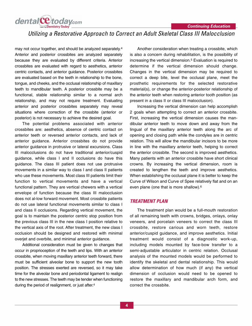
All segments of the provisional restoration wereremoved and prepared for cementation. The maxillary andmandibular anterior veneer segments would be tacked byspot etching with 35% phosphoric acid, bonded with unfilledresin and flowable composite, cleaned to satisfaction, andlight-cured into place. The posterior provisionals werecemented with provisional cement. With the occlusaladjustments complete, aesthetic recontouring of theprovisional was performed. The provisional was thenpolished and glazed to satisfaction (Figures 16 and 17).
The patient was dismissed and reappointed the nextday for evaluation of the provisional restoration. The finalimpressions, face-bow transfer, and bite registration wereforwarded to the laboratory with in-structions to create andmount the master models. The laboratory was advised thatmodels and photographs of the provisional would beforwarded in the future, after patient acceptance of the newvertical dimension and occlusal scheme could be verified.
The patient presented for evaluation of the provisionalin regard to aesthetics, phonetics, and function. Thepatient was very satisfied with the aesthetics. Regardingspeech, the “S” position was verified and the patient wasable to accommodate to the new incisal edge position withminimal phonetic problems.13,15,16 The patient was awareof the maxillary teeth against the inside of the upper lip,and accommodated well. The patient expressed that timewas needed to adjust to the new vertical dimension andocclusal scheme, as this felt foreign. With the aestheticsand phonetics acceptable, the occlusion was re-evaluatedutilizing the Tekscan. Ad-justments were made with theTekscan to provide equal intensity centric contacts.17,18
The patient was then released and scheduled for anotherevaluation in one week.
The patient presented for the one-week follow-upappointment. The vertical dimension was comfortable but thepatient still expressed difficulty adjusting to the new occlusalscheme. The evaluation revealed that cuspid guidance wasnot comfortable. Adjustments were made, converting cuspidguidance to group function with lateral excursive movementsdistributed across the first molar, bicuspids, and cuspid. TheTek-scan was utilized to assist with the occlusal adjustments.Once these adjustments were completed the pa-tient wascomfortable. The patient was scheduled 2 weeks later to verifycomfort with the new occlusion. This was accomplished, andmodels and photographs of the provisional were forwarded tothe laboratory with instructions for fabrication of the finalrestorations. Instruction included fabrication of porcelain togold crowns for all molars, zirconia crowns for all bicuspids,and feldspathic porcelain veneers for all cuspids and incisors.
FINAL RESTORATION EVALUATION AND CEMENTATION
All restorations were re-turned from the ceramist andinspected for accuracy prior to final delivery. All restorationswere acceptable in regard to fit and contact on the mastermodel. The patient presented for the cementationappointment. Anesthesia was administered and theprovisional restorations were removed. The veneers wereinitially tried on teeth Nos. 8 and 9 to check fit and accuracy.
Continuing Education
7
Utilizing a Restorative Approach to Correct an Adult Skeletal Class III Malocclusion
Figure 16. Provisional,frontal retracted view inocclusion.
Figure 17. Provisional,left lateral view inocclusion.
The remaining veneers, crowns, and fixed partial denturewere also tried in to inspect the fit of all restorations. Thepatient was asked to bite to-gether gently to verify occlusionand vertical dimension. Although occlusal adjustments wererequired, the occlusal position and vertical dimension wereacceptable. The veneers were tried in with a veneer try-inpaste and inspected for shade match to the otherrestorations. The veneers and crowns were then removedfrom the mouth. All posterior restorations were placed first ona quadrant-by-quadrant basis.
After appropriate setting time all excess cement wasremoved. W2 rubber dam clamps were placed on themaxillary first bicuspids and a rubber dam was placedutilizing the split rubber dam technique.19,20 The maxillaryveneers were bonded utilizing acceptable wet bondingtechniques, maintaining a moist surface.21-23 The “rapidcementation” technique as described by Rosenthal19 wasused to bond all veneers. The W2 rubber dam clamps wereplaced on the mandibular first bicuspids and the rubber damwas placed on the mandibular teeth. The mandibularveneers were bonded utilizing the same technique andsequence as described for the maxillary veneers.19,20-23
Once all restorations were secure the occlusion wasadjusted to achieve proper centric contacts and establishproper anterior and lateral guidance.24 Lateral excursionswere established as group function with excursive contact onthe first molar to cuspids.
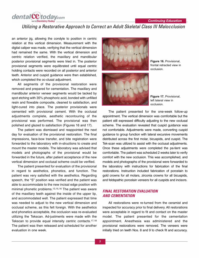
The patient was given postoperative instructions, andan appointment was made for evaluation in 24 hours. Thenext day the teeth were inspected for aesthetics, phonetics,and function. With the aesthetics and phonetics acceptable,the occlusion was re-evaluated utilizing the Tekscan.Adjustments were made to provide equal time-intensitycentric contacts.17,18 The patient was scheduled forevaluation in one week, at which impressions would bemade to fabricate a night guard. The final result wasexcellent and well accepted by the patient (Figures 18-20).
SUMMARY AND CONCLUSION
When determining if treatment for malocclusion isindicated, the clinician must understand how themalocclusion affects the patient aesthetically, functionally,
and biologically, as well as any impact of treatment. It isimportant to consult the patient and advise when a lessinvasive orthodontic treatment plan may be optimal. Oncethe patient is fully informed of the treatment options anddesires treatment without orthodontics, a restorative/prosthodontic approach using veneers, crowns, or fixedprosthetics can provide exceptional strength, function, andaesthetics. As in all cases, thorough evaluation andplanning are essential.
The decision to proceed with restorative alignment ofthe teeth rather than orthodontic alignment is dependent onfull disclosure to the patient and the clinician’sunderstanding of preparation design, aesthetics, andocclusion. As demonstrated in this class III case, withproper examination, diagnosis, treatment planning, andcommunication, excellent aesthetic, phonetic, andfunctional results can be achieved and maintained. As withall full-mouth restorative cases, periodic occlusal evaluationwill be necessary at normal hygiene intervals.
Continuing Education
8
Utilizing a Restorative Approach to Correct an Adult Skeletal Class III Malocclusion
Figure 18. Finalrestoration, smile view.
Figure 19. Finalrestoration, retractedview.
Figure 20. Finalrestoration, retractedleft lateral view.
REFERENCES1. Angle EH. Treatment of Malocclusion of the Teeth and
Fractures of the Maxillae, Angle’s System. 6th ed. Philadelphia,PA: SS White Dental Manufacturing; 1900:5-15.
2. Proffit WR. Contemporary Orthodontics. St Louis, MO: Mosby;1986:47-49.
3. Rufenacht C. Fundamentals of Esthetics. Chicago, IL:Quintessence; 1990.
4. Dawson PE. Evaluation, Diagnosis, and Treatment of OcclusalProblems. 2nd ed. St Louis, MO: Mosby; 1989:555-567.
5. Kois JC, Phillips KM. Occlusal vertical dimension: alterationconcerns. Compend Contin Educ Dent. 1997;18:1169-1177.
6. Spear FM. Approaches to vertical dimension. AdvancedEsthetics and Interdisciplinary Dentistry. 2006;2(3): 2-12.
7. Rivera-Morales WC, Mohl ND. Relationship of occlusal verticaldimension to the health of the masticatory system. J Prosthet Dent. 1991;65:547-553.
8. Kovaleski WC, DeBoever J. Influence of occlusal splints on jawposition and musculature in patients with temporomandibularjoint dysfunction. J Prosthet Dent. 1975;33:321-327.
9. Manns A, Miralles R, Santander H, et al. Influence of thevertical dimension in the treatment of myofascial pain-dysfunction syndrome. J Prosthet Dent. 1983;50:700-709.
10. Hellsing G. Functional adaptation to changes in verticaldimension. J Prosthet Dent. 1984; 52:867-870.
11. Gamon JA, Wright SM. Perception of vertical dimension. J Oral Rehabil. 1982;9:307-316.
12. Tryde G, Stoltze K, Fujii H, et al. Short-term changes in theperception of comfortable man-dibular occlusal positions. J Oral Rehabil.1977; 4:17-21.
13. Spear FM. Occlusion in clinical practice. Seminar presented at:Seattle Institute for Advanced Dental Education; October 2001;Orlando, FL.
14. Lee RL. Esthetics and its relationship to function. In: RufenachtCR, ed. Fundamentals of Esthetics. Chicago, IL: Quintessence;1990: 137-209.
15. Hammond RJ, Beder OE. Increased vertical dimension andspeech articulation errors. J Prosthet Dent. 1984;52:401-406.
16. Howell PG. Incisal relationships during speech. J ProsthetDent. 1986;56:93-99.
17. Kerstein RB, Grundset K. Obtaining bilateral simultaneousocclusal contacts with computer analyzed and guided occlusaladjustments. Quintessence Int. 2001;32:7-18.
18. Carey JP, Craig M, Kerstein RB, et al. Determining arelationship between applied occlusal load and articulatingpaper mark area. The Open Dentistry Journal. 2007;1:1-7.
19. Rosenthal L. Syllabus for the Aesthetic Advantage Continuum.Paper presented at: Postgraduate seminar at the Atlantic CoastResearch Clinic. March 1998; West Palm Beach, FL.
20. Rosenthal L. The state of the art in advanced porcelainveneering: part 2. Esthetic Dentistry Update 1991;2(6):96-101.
21. Kanca J III. Improving bond strength through acid etching ofdentin and bonding to wet dentin surfaces. J Am Dent Assoc.1992 123:35-43.
22. Kanca J III. Resin bonding to wet substrate. 1. Bonding todentin. Quintessence Int. 1992;23: 39-41.
23. Gwinnett AJ. Moist versus dry dentin: its effect on shear bondstrength. Am J Dent. 1992;5:127-129.
24. Dawson PE. Evaluation, Diagnosis, and Treatment ofOcclusal Problems. 2nd ed.St Louis, MO: Mosby; 1989:555-567; 592-607.
Continuing Education
9
Utilizing a Restorative Approach to Correct an Adult Skeletal Class III Malocclusion

POST EXAMINATION INFORMATION
To receive continuing education credit for participation inthis educational activity you must complete the programpost examination and receive a score of 70% or better.
Traditional Completion Option:
You may fax or mail your answers with payment to Dentistry Today(see Traditional Completion Information on following page). Allinformation requested must be provided in order to process theprogram for credit. Be sure to complete your “Payment”, “PersonalCertification Information”, “Answers” and “Evaluation” forms, Yourexam will be graded within 72 hours of receipt.. Upon successfulcompletion of the post-exam (70% or higher), a “letter ofcompletion” will be mailed to the address provided.
Online Completion Option:
Use this page to review the questions and mark your answers.Return to dentalCEtoday.com and signin. If you have notpreviously purchased the program select it from the “OnlineCourses” listing and complete the online purchase process. Oncepurchased the program will be added to your User History pagewhere a Take Exam link will be provided directly across from theprogram title. Select the Take Exam link, complete all the programquestions and Submit your answers. An immediate grade reportwill be provided. Upon receiving a passing grade complete theonline evaluation form. Upon submitting the form your Letter OfCompletion will be provided immediately for printing.
General Program Information:
Online users may login to dentalCEtoday.com anytime in thefuture to access previously purchased programs and view or print“letters of completion” and results.
POST EXAMINATION QUESTIONS
1. A class III skeletal relationship can occur as aresult of:a. a normal maxilla with mandibular protrusion.b. maxillary retrusion with a normal mandible.c. a combination of maxillary retrusion and
mandibular protrusion. d. all of the above.
2. The decision to proceed with restorativealignment of the teeth rather than orthodontic alignment is dependent on:a. full disclosure and understanding of the treatment
options.b. the clinician’s understanding of preparation design.c. aesthetics, and occlusion.d. all of the above.
3. What compromise exists when utilizing arestorative option to correct a malocclusion vs.orthodontic treatment?a. the need for a more aggressive approach to tooth
preparation.b. there is no compromise to restorative treatment.c. restorative treatment takes longer to complete.d. all of the above.
4. The treatment plan in this article had 4 specificgoals: Which goal is NOT correct?a. optimal oral health.b. improvement in speech.c. comfort when functioning.d. acceptable aesthetics.
5. The potential problems associated with anteriorcrossbites are: a. aesthetics. b. absence of centric contact on anterior teeth, or
reversed anterior contacts.c. lack of anterior guidance.d. all of the above.
6. When converting a class III mal occlusion to anew class I the occlusion should be designed and restored with:a. minimal overjet and overbite.b. minimal anterior guidance.c. 5 mm of overjet and 90% overbite.d. a and b.
7. In the case presented, the challenges discussedwith the patient prior to treatment included: a. change in speech.b. change in sensation of the upper lip as a result of
the new position of the teeth.c. the effect of the new jaw position on the muscles
of mastication, increased vertical dimension, and sensation of centric stops on the anterior teeth.
d. all of the above.
8. Caution should be made not to increase verticaldimension more than is necessary. A 3 mmopening of the vertical in the anterior segment will: a. have an adverse effect on the TMJ.b. produce muscle tension. c. result in a 1 mm change in the length of the
masseter muscles, which is well tolerated.d. all of the above.
Continuing Education
10
Utilizing a Restorative Approach to Correct an Adult Skeletal Class III Malocclusion

PROGRAM COMPLETION INFORMATION
If you wish to purchase and complete this activitytraditionally (mail or fax) rather than Online, you mustprovide the information requested below. Please be sure toselect your answers carefully and complete the evaluationinformation. To receive credit you must answer at least sixof the eight questions correctly.
COMPLETE ONLINE AT: www.dentalcetoday.com
TRADITIONAL COMPLETION INFORMATION:Mail or Fax this completed form with payment to:
Dentistry TodayDepartment of Continuing Education100 Passaic AvenueFairfield, NJ 07004
Fax: 973-882-3662
PAYMENT & CREDIT INFORMATION:
Examination Fee: $20.00 Credit Hours: 1.0
Note: There is a $10 surcharge to process a check drawn on any bank other than a US bank. Should you have additionalquestions, please contact us at (973) 882-4700.
o I have enclosed a check or money order.
o I am using a credit card.
My Credit Card information is provided below.
o American Express o Visa o MC o Discover
Please provide the following (please print clearly):
Exact Name on Credit Card
Credit Card # Expiration Date
Signature
PROGRAM EVAUATION FORMPlease complete the following activity evaluation questions.
Rating Scale: Excellent = 5 and Poor = 0
Course objectives were achieved.
Content was useful and benefited your clinical practice.
Review questions were clear and relevant to the editorial.
Illustrations and photographs were clear and relevant.
Written presentation was informative and concise.
How much time did you spend reading the activity & completing the test?
Continuing Education
Utilizing a Restorative Approach to Correct an Adult Skeletal Class III Malocclusion
ANSWER FORM:Please check the correct box for each question below.
1. o a o b o c o d 5. o a o b o c o d
2. o a o b o c o d 6. o a o b o c o d
3. o a o b o c o d 7. o a o b o c o d
4. o a o b o c o d 8. o a o b o c o d
PERSONAL CERTIFICATION INFORMATION:
Last Name (PLEASE PRINT CLEARLY OR TYPE)
First Name
Profession / Credentials License Number
Street Address
Suite or Apartment Number
City State Zip Code
Daytime Telephone Number With Area Code
Fax Number With Area Code
E-mail Address
/
Dentistry Today is an ADA CERPRecognized Provider.
Approved PACE Program ProviderFAGD/MAGD Credit Approvaldoes not imply acceptanceby a state or provincial board ofdentistry or AGD endorsement.June 1, 2006 to May 31, 2009AGD Pace approval number: 309062