validity of the finger tapping test in parkinson’s disease ... · h i g h l i g h t s • finger...
TRANSCRIPT

Clinical Neurophysiology 123 (2012) 2034–2041
Validity of the finger tapping test in Parkinson’s disease, elderly and young healthy subjects: Is there a role for central fatigue?
Pablo Arias, Verónica Robles-García, Nelson Espinosa, Yoanna Corral, Javier Cudeiro
Neuroscience and Motor Control Group (NEUROcom), Department of Medicine-INEF Galicia and INIBIC (Institute of Biomedical Research of A Coruña), University of A Coruña, Spain
H I G H L I G H T S
• Finger tapping test at comfort rate is a valid and reliable test used to detect alterations in rhythm formation in parkinsonian and elderly subjects.
• In the young subjects fatigue induces a fast slowing of the tapping rate if the finger tapping test is performed as fast as possible.
• Due to this fatigue, using the finger tapping test to evaluate alteration in rhythm formation clinically across populations appears invalid if it is performed at the fastest rate.
A B S T R A C T
Objective: The main goal of this work is to evaluate the validity of the finger tapping test (FT) to detect alterations in rhythm formation. Methods: We use FT to study the alterations in motor rhythm in three different groups: Parkinson’s patients, elderly healthy controls, and young healthy control subjects (HY). The test was performed in COMFORT and FAST tapping modes and repeated on two different days. Results: For the variables analyzed (frequency and variability) both modes were repeatable in all groups. Also, intra-class correlation coefficients showed excellent levels of consistency between days. The test clearly differentiated the groups in both FAST and COMFORT modes. However, when fatigue was analyzed, a decrease in the tapping frequency was observed in HY during the FAST mode only. The amplitude of motor evoked potentials (MEPs) induced by transcranial magnetic stimulation (TMS) was early-potentiated but not delayed-depressed, both for COMFORT and FAST modes. This suggests that fatigue was not of cortico-spinal origin. Other forms of central fatigue are discussed. Conclusions: FT at FAST mode is not a valid test to detect differences in rhythm formation across the groups studied; fatigue is a confounding variable in some groups if the test is performed as fast as possible. Significance: COMFORT mode is recommended in protocols including the FT for evaluating rhythm formation.
1. Introduction
The finger tapping test (FT) is a basic tool for evaluating rhythmic movement patterns. It is commonly used in clinical assessments and as part of costly research protocols, including brain imaging studies (Stoodley et al., 2010; Foki et al., 2010; Stavrinou et al., 2007) and neurophysiological examinations (Leijnse et al., 2008; Astolfi et al., 2004). The test allows the study of several key elements to be subsequently used as complementary data for characterizing disease profiles, for instance arrythmokinesis (Nakamura et al., 1976; Wertham, 1929), which can include hastening, faltering, or freezing in the tapping pattern (Nakamura et al., 1978) and hypokinesia, in Parkinson’s disease (PD). The test is also a sensitive marker for the detection of alterations in rhythm formation due to aging (Shimoyama et al., 1990). Despite their conceptual simplicity, FT provides highly valuable information useful in the characterization of a number of diseases which make the test a widely used protocol. Nevertheless, there are several methodological aspects which remain unaddressed and should be taken into account, such as the relation between duration, tapping rate protocol and fatigue.

In the classic work by Shimoyama et al. (1990) subjects were asked to tap for 15 s at each subject’s maximum tapping rate. The protocol allowed the characterization of healthy subjects of different ages, PD and other pathologies. Later on, however, it was reported that tapping rate decreases after a few seconds (about 7 s) if tapping was as fast as possible (Aoki et al., 2003). This questions whether the results obtained previously (Shimoyama et al., 1990) were due to central rhythm formation impairment. An alternative version of the test is tapping at a rate comfortable for each subject (Collyer et al., 1994; Del Olmo et al., 2006). This test probably has some advantages: the effect of fatigue seems not to be a limiting factor and the test allows a clear depiction of movement amplitude, permitting the characterization of hypokinetic movement profiles.
In addition, there is another important methodological point to be considered: determining test duration. Most experiments (Shimoyama et al., 1990; Del Olmo et al., 2006; Farkas et al., 2006; Gill et al., 1986) set a fixed duration for the test. This means that the number of events recorded varies from one subject to another, depending on the tapping frequency executed. It is plausible that the different number of events may impact on some of the variables usually measured, for instance, the coefficient of variability (CV = (sd/mean) x 100). This variable depends on the difference of each point from the mean and it seems reasonable to standardize the use of the variable by using the same number of events in each sample, which is complicated if duration of the test is fixed in time.
In this framework, the aim of this study is to re-appraise the role of the finger tapping test in evaluating rhythm formation in PD, healthy elderly controls (HE), and young healthy controls (HY). We will perform the test at both fastest and comfort rates to assess a potential effect of fatigue on the validity of the test to detect alteration of rhythm formation in PD and aging. We will control the effect of the number of events, by determining test duration on the basis of number of tapping events rather than using a given time. We will also evaluate the reliability of the each testing mode by performing each protocol twice (one week apart) under the same conditions. Interpretation of results will be always tempered by controlling for the possible role of fatigue by analyzing any change in tapping frequency (e.g. depressed responsiveness resulting from continuous tapping) at the beginning and the end of the test, along with any evidence of cortico-spinal fatigue. Fatigue has been defined as an exercise-induced reduction in maximal voluntary muscle force which may have central origin (Gandevia, 2001). Amongst its forms, cortico-spinal (CS) fatigue is defined as an adaptation in the cerebral cortex or spinal cord following a period of prolonged effort which leads to lack of the ability of voluntary command to recruit spinal motor-neurons fully, in fully motivated subjects (Di Lazzaro et al., 2003). CS fatigue is manifest by an initial enhancement followed by a depression below baseline levels of the motor evoked potentials induced by TMS, evaluated at rest (Brasil-Neto et al., 1993; Samii et al., 1996; Liepert et al., 1996).
We hypothesized a fatigue-related slowing of tapping rate at FAST rate with CS origin, which questions the validity of the test as a means to characterize rhythm formation across groups.
2. Methods
2.1. Ethical approval
This project was approved by the University of A Coruña Ethics Committee. The protocol conformed to the Helsinki declaration and subjects signed consent forms.
2.2. Subjects
We tested three groups of subjects:
2.2.1. PD PD patients belonging to local associations of patients, Asociación Parkinson Galicia (A Coruña), and
Asociación Parkinson Bueu (both in Spain), were recruited as participants. 17 PD diagnosed with idiopathic PD were evaluated whilst OFF-dose (12-h after withdrawing medication). PD were on average 69.47 yrs old (SEM 2.33), and had mean UDPRS-III scores of 28.25 points (SEM 2.70).
2.2.2. HE 20 HE were also included in the study. Their ages were on average 70.55 yrs (SEM 2.69). They were
approached through relatives of the staff of our institution.
2.2.3. HY 21 HY, staff working and students taking classes in our institution, were recruited for the study. They
were on average 23.90 yrs old (SEM 1.93). In experiments specifically aimed at studying cortico-spinal fatigue, another group of 24 HY was included (23.37 yrs old; SEM 1.13).
General inclusion criteria included: Lack of dementia (Mini-Mental Examination State score >25); lack of arthro-muscular impairment such as arm prosthesis, arthritis, etc.; or any kind of disease affecting the execution of the task (apart from PD in the patients group). Specific criteria for the PD were diagnosis of idiopathic PD, and ability to cope with OFF-period after withdrawing medication for 12 h.
All subjects were right-handed (Oldfield, 1971), and performed the task with their dominant hands.
2.3. Materials
Inter-tap intervals were recorded by means of an electronic system which included a metal plate and a metal ring adapted to the distal phalange of the index finger. The system was connected to a laptop PC and registered the time (sampled at 1 KHz) during which the metal pieces were in contact and the inter-touch time, allowing the calculation of inter-tap interval.
We used a Magstim 2002 stimulator to deliver monophasic pulses through a 70 mm figure of 8 coil to evaluate CS fatigue. The coil was oriented tangential to the skull with the handle pointing 45º backwards thereby inducing currents in a postero-anterior direction. The coil was positioned over the hot-spot for the first dorsal interosseous muscle (FDI) of executing hand. Motor evoked potentials (MEPs) were recorded over FDI, and extensor digitorum (EXT) through a belly-tendon montage using surface electrodes Ag–AgCI). MEPs were recorded by means of D360 amplifiers (Digitimer, Welwyn Garden City, Herts) band-width filtered between 3–3000 Hz. Data was sampled at 10KHz and stored in the computer by means of a CED 1401 Power mkII A-D converter (Cambridge Electronic Design, Cambridge, UK). A customized Matlab (Mathworks, Spain) program was used to process data.
2.4. Procedure
Subjects were comfortably seated with forearms laid on a table in front of them, so that both elbows were flexed at about 90–100º. Seat height was adapted so that subjects were in an optimal comfort position to perform the test. Subjects were asked to perform FT with their index finger by flexing-extending the metacarpo-phalangeal joint while staring at the hand executing the task.
Two condition modes were included: Tapping at their fastest rate (FAST), and tapping at their preferred, comfortable rate (COMFORT). Before the FAST condition subjects were reminded to tap as fast as they could from the very beginning of the test. Because intra-session variability in tapping protocols in PD has been reported previously (Wu et al., 1999, who recommended a minimum of two sets of evaluations per session), each condition was repeated three times, allowing 3 min rest between repetitions. Before starting each of the FAST trials, subjects were asked if they felt any fatigue; if they had, they would have been given extra rest time, but no subject reported feeling fatigued. The first 3 taps of each trial were
discarded in order to reach a steady rate. Each of the trials lasted until subjects performed 50 finger tapping cycles, apart from the 3 initial discarded taps. Between modes subjects rested for 6 min. The whole protocol, 3 sets of 50 taps at COMFORT, and 3 sets of 50 taps at FAST, was repeated twice (DAY1 & DAY2), a week apart from each other, under the same conditions.
The order of conditions was randomized (FAST-COMFORT or COMFORT-FAST) between subjects, and for each subject the order was the same for the first and the second evaluation days.
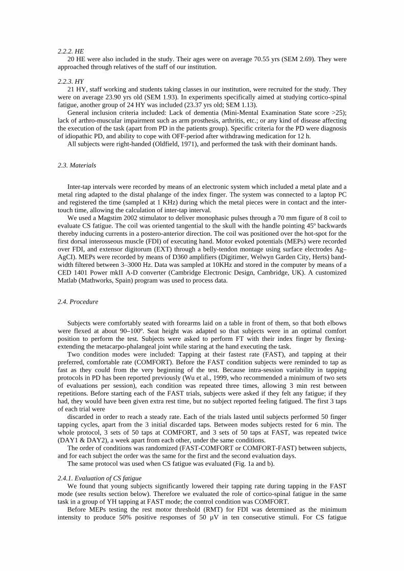
The same protocol was used when CS fatigue was evaluated (Fig. 1a and b).
2.4.1. Evaluation of CS fatigue We found that young subjects significantly lowered their tapping rate during tapping in the FAST
mode (see results section below). Therefore we evaluated the role of cortico-spinal fatigue in the same task in a group of YH tapping at FAST mode; the control condition was COMFORT.
Before MEPs testing the rest motor threshold (RMT) for FDI was determined as the minimum intensity to produce 50% positive responses of 50 µV in ten consecutive stimuli. For CS fatigue
evaluation sets of 10 consecutive stimuli at 120% RMT (Di Lazzaro et al., 2003) were delivered at 0.16 Hz over the marked hot-spot of the FDI. The protocol is represented in Fig. 1b. MEPs amplitudes evoked by TMS pulses after fatiguing contractions have been shown to be initially enhanced, right at the end of activity, and subsequently depressed below baseline levels at about 30 s after activity cessation (and on up to minutes) (Brasil-Neto et al., 1993; Samii et al., 1996; Liepert et al., 1996). Therefore we evaluated changes in MEPs along 1 min before tapping, immediately after tapping, and again 2 min (for 1 min) after tapping in order to evaluate the MEPs modulation profile induced by FAST and COMFORT tapping.
Fig. 1. (a) Finger tapping protocol. Each Subject performed 3 sets of 50 cycles of finger tapping (FT) at COMFORT and other 3 sets at FAST (modes). The order of modes was randomized, and there was a rest of 3 min between sets and 6 min between modes. (b) During the central fatigue protocol an overlapping sequence of corticoespinal excitability evaluations was also included: (i) PRE; (ii) FT, (iii) POST; (rest periods); (iv) POST-2. Each TMS block (TMS1, TMS2. . .) comprised the recording of 10 MEPs, performed just before, immediately after, and again after 1 min, with respect to the 50 finger tapping cycles. Averaging of the 3 sets of evaluations allowed a total recording of 30 MEPs for each of the three sets of PRE, POST, & POST-2.
2.5. Analyzed variables
The following variables were analyzed: (i) The FREQUENCY of tapping (in Hz). (ii) The coefficient of variation (CV) of inter-tap interval, defined as: CV(%) = (sd/mean) x 100. (iii) Tapping fatigue: Reduction in the FREQUENCY from the first 10 (FREQUENCY1–10) to the last 10
taps of the sequence (FREQUENCY41–50). Complementary analysis was performed taking all intervals of ten taps from 1 to 50 (FREQUENCY1–10; FREQUENCY11–20; FREQUENCY21–30; FREQUENCY31–40; FREQUENCY41–50).
(iv) Central fatigue: This was defined as a significant decrease in the MEP amplitude after tapping. Since modifications of EMG-background activity might be a fatigue-related phenomenon, its impact on the MEPs was controlled. Therefore the level of EMG-background (area (mV x ms) in the period from -80 ms to -10 ms with respect to TMS trigger) was evaluated.
2.6. Statistical analysis
The following analyses were performed:
(i) The sensibility of the test to differentiate groups was analyzed by means of an Analysis of Variance with repeated measures (ANOVA-RM). One between-subject factor was set: GROUP (with three levels: PD, HE, HY). Two within subject factors were also defined, DAY (with two levels, DAY1 and

DAY2, each of the days of evaluation) and TAPPINGRATE (FAST & COMFORT modes). This analysis was done for both the CV and the frequency of tapping.
(ii) To evaluate how performance was influenced by disease severity the 8 PD with the lower scores in the motor section of the UPDRS were compared to the 8 PD with the higher scores. This was done by means of a Student t-test for independent samples, applied to the variables CV and tappingfrequency at the COMFORT and FAST tapping modes.
(iii) Consistency in execution was evaluated by means of the Intra-class Correlation Coefficient (ICC) between DAY1 & DAY2, for both CV and FREQUENCY at the two tapping rates, FAST & COMFORT. The mean difference between days was also analyzed, (Day 1 minus Day 2 for each subject). This was done using a one sample t-test and was evaluated for each group separately; it reflects if the difference in execution between days was significantly different from 0.
(iv) An ANOVA-RM was performed to analyze the effect of fatigue in the tapping frequency. Two within-subjects factor were set, factor FREQUENCY with two levels (FREQUENCY1–10 & FREQUENCY41–50) and factor TAPPING_RATE with two levels (COMFORT & FAST). A subsequent analysis on those subjects showing fatigue was done, using the same model but analyzing the drop of frequency at intervals of 10 taps, then factor FATIGUE had 5 levels (FREQUENCY1–10, FREQUENCY11–20. . . FREQUENCY41–50).
(v) CS fatigue was analyzed with another ANOVA-RM, reflecting how the MEP size changes after tapping. Three within-subject factors were included:
(vi) TAPPING_RATE, with two levels (COMFORT & FAST). (vii) Factor PRE_POST_POST2 with three levels. PRE: MEP amplitude averaged at TMS sets 1, 3, and 5
(all recorded immediately before tapping, TMS1, TMS3, TMS5); POST: MEP amplitude averaged at TMS sets 2, 4, and 6 (all recorded immediately after tapping); and POST2: MEP amplitude averaged at TMS sets 3, 5, and 7 (all recorded immediately after 10 rest). Each PRE-POST-POST2 outcome came from averaging 30 MEPs in each subject.
(viii) MUSCLE with 2 levels (FDI, EXT).
This model was used for both, the EMG-background, and MEP size. Results are expressed as Mean and the Standard Error of the Mean. Normality of distributions was
assessed by means of Kolgomorov–Smirnov test of one sample. During ANOVA analysis the degrees of freedom were corrected with Greenhouse Coefficients (ɛ) in case of sphericity violation; and significance was set at p < 0.05.
3. Results
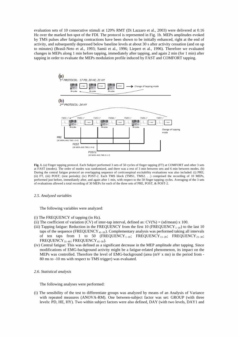
3.1. CV at COMFORT and FAST rate
Using the finger tapping test, groups were differentiated based on their CV (F(2,55) = 9.765 p < 0.001GROUP), and this difference was dependent on the tapping rate (F(2,55) = 7.869 p = 0.001GROUP_TAPPING_RATE). CV obtained during COMFORT mode detected differences among groups (Fig. 2a), and PD severity (Fig. 3). On the other hand, FAST mode also differentiated the healthy groups from the PD (Fig. 2b) but, importantly, not between HY and HE. It is worth noting that performance between the 2 days did not differ significantly. ICC showed high consistency in execution between days, with the exception of the HE at fast rate; also the difference in execution between days was never significantly different from 0 (Fig. 4a and b; and Table 1).
3.2. Frequency at COMFORT and FAST rate
Frequency of tapping did not vary between days and across all groups. Unsurprisingly, frequency was affected by the tapping mode (COMFORT or FAST) F(1,55) = 592.945 p < 0.001TAPPING_RATE, and the test allowed us to differentiate the groups by means of this variable, but this was dependent on the tapping rate F(2,55) = 17.342 p < 0.001TAPPING_RATE_GROUP. Follow-up analysis (Fig. 2) shows that HY tapped significantly faster than HE and PD at Fast rate (p < 0.001 both), while PD vs. HE showed no significant difference. Interestingly, PD tapped faster at COMFORT than the other two groups (p = 0.028 vs. HC and p = 0.017 vs. YC). Likewise ICC showed high consistency between days, both at COMFORT and at
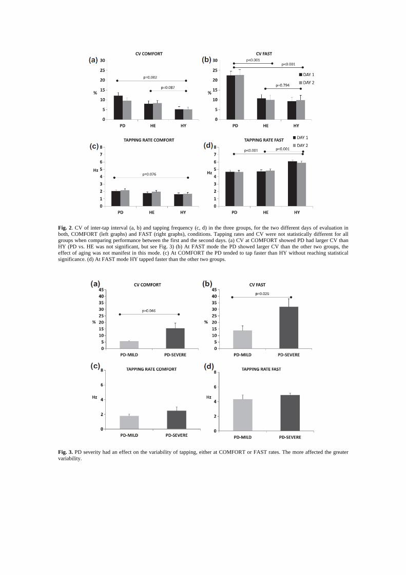
Fig. 2. CV of inter-tap interval (a, b) and tapping frequency (c, d) in the three groups, for the two different days of evaluation in both, COMFORT (left graphs) and FAST (right graphs), conditions. Tapping rates and CV were not statistically different for all groups when comparing performance between the first and the second days. (a) CV at COMFORT showed PD had larger CV than HY (PD vs. HE was not significant, but see Fig. 3) (b) At FAST mode the PD showed larger CV than the other two groups, the effect of aging was not manifest in this mode. (c) At COMFORT the PD tended to tap faster than HY without reaching statistical significance. (d) At FAST mode HY tapped faster than the other two groups.
Fig. 3. PD severity had an effect on the variability of tapping, either at COMFORT or FAST rates. The more affected the greater variability.

FAST tapping rate, and, in addition, difference in execution between days was never significant different from 0 (p > 0.100 in all cases; Fig. 4c and d; and Table 1).
3.3. Fatigue at COMFORT and FAST tapping rates
The ANOVA showed a strong interaction indicating that groups behaved differently in their response to FATIGUE, F(2,55) = 8.903 p < 0.001FATIGUE_TAPPING_RATE_GROUP. Follow-up with groups Split showed that it was the YC group that behaved differently. Whereas no sign of fatigue appeared in the PD & HE (Fig. 5a and b), the HY showed an obvious drop of the tapping frequency dependent on the tapping mode F(1,20) = 26.918 p = 0.001FATIGUE_TAPPING_RATE. No slowing of tapping rate was observed at the end of the test at COMFORT but it was clear at FAST mode (p < 0.001; Fig. 5a–c).
3.4. Cortico-spinal fatigue
A new group of 24 HY was evaluated to assess CS fatigue. There was again a clear drop in the tapping rate when executing the test, again observed only at FAST mode F(4,92) = 16.123 p < 0.001FATIGUE_TAPPING_RATE. In this mode, frequency was maintained for the 20th first taps and then dropped from initial values (p < 0.001; Fig. 6a). At the end of the task there was an increase in the CS excitability (augmented MEP amplitude), F(2,46) = 34.689 p < 0.001PRE-POST-POST2 (Fig 6b black line); however this change appeared regardless tapping mode (Fig 6c and d), and was observed in the two evaluated muscles. If restricted to the first five MEPs (up to the vertical dashed line in Fig 6b–d), the analysis showed that CS excitability was increased right at the end of tapping in the first of the MEPs F(8,184)ɛ =0.481 = 5.076 p = 0.001PRE-POST-POST2xEVENT; again this did not depend on the rate of tapping (Fig 6c and d). There was no delayed depression in MEPs from 30 s onwards, after tapping (Fig 6c and d). The analysis of the EMG-background activity showed the same profile that MEPs; in no case the different tapping modes had differential effect on pre-activity F(1,23) = 0.330 p = 0.571TAPPING-RATE; interactions were never significant.
4. Discussion
The aim of this study was to examine the validity and reliability of the finger tapping test in different modes, FAST or COMFORT, and to re-appraise some methodological aspects of its usage. Chiefly, we have focused on the capacity of the test to detect differences in rhythm formation amongst three different groups of subjects. We deemed that this is a critical aspect, assuming that the test might be valid for one population but not for another. Indeed, our data now seem to suggest that is the case for the young group at FAST rate.
Results proved that the test performed at COMFORT rate is reproducible across different testing days, and can detect differences in rhythm formation between the three groups of subjects. In this mode, the test is also sensitive to alterations in rhythm formation as the disease progresses. This has been shown previously in more complex movements (Arias and Cudeiro, 2008), indicating (as observed here also) that early PD have a similar pattern of variability to elderly subjects. At FAST rate, however, an effect of fatigue appears in the HY group. Therefore the observed drift in frequency, which clearly affects inter-tap variability, may not reflect an alteration in rhythm formation. Further, when variability is considered in the case of the elderly, consistency between days is also questioned at FAST rate. This seems to disagree with previous work by Gill et al. (1986) who showed good association between execution in different days. It is worth saying, however, that the Pearson’s coefficients used in that study do not assess consistency (Bland and Altman, 1986). The appearance of fatigue (a drop in tapping rate) at FAST rate has been previously reported (Aoki et al., 2003) questioning the study of Shimoyama et al. (1990) in which subjects tapped as fast as they could for 15 s. Therefore tapping at COMFORT mode not only avoids fatigue and permits better characterization of rhythm formation, it also allows a closer investigation of the hypokinetic elements of the movement, as a frequency-amplitude trade-off at faster rates will require all subjects to perform short-amplitude movements to get faster frequencies.
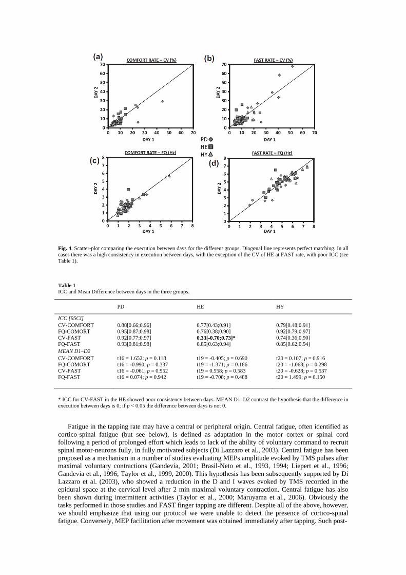
Fig. 4. Scatter-plot comparing the execution between days for the different groups. Diagonal line represents perfect matching. In all cases there was a high consistency in execution between days, with the exception of the CV of HE at FAST rate, with poor ICC (see Table 1).
Table 1 ICC and Mean Difference between days in the three groups.
PD HE HY
ICC [95CI] CV-COMFORT 0.88[0.66;0.96] 0.77[0.43;0.91] 0.79[0.48;0.91] FQ-COMORT 0.95[0.87;0.98] 0.76[0.38;0.90] 0.92[0.79;0.97] CV-FAST 0.92[0.77;0.97] 0.33[-0.70;0.73]* 0.74[0.36;0.90] FQ-FAST 0.93[0.81;0.98] 0.85[0.63;0.94] 0.85[0.62;0.94] MEAN D1–D2 CV-COMFORT t16 = 1.652; p = 0.118 t19 = -0.405; p = 0.690 t20 = 0.107; p = 0.916 FQ-COMORT t16 = -0.990; p = 0.337 t19 = -1.371; p = 0.186 t20 = -1.068; p = 0.298 CV-FAST t16 = -0.061; p = 0.952 t19 = 0.558; p = 0.583 t20 = -0.628; p = 0.537 FQ-FAST t16 = 0.074; p = 0.942 t19 = -0.708; p = 0.488 t20 = 1.499; p = 0.150 * ICC for CV-FAST in the HE showed poor consistency between days. MEAN D1–D2 contrast the hypothesis that the difference in execution between days is 0; if p < 0.05 the difference between days is not 0.
Fatigue in the tapping rate may have a central or peripheral origin. Central fatigue, often identified as cortico-spinal fatigue (but see below), is defined as adaptation in the motor cortex or spinal cord following a period of prolonged effort which leads to lack of the ability of voluntary command to recruit spinal motor-neurons fully, in fully motivated subjects (Di Lazzaro et al., 2003). Central fatigue has been proposed as a mechanism in a number of studies evaluating MEPs amplitude evoked by TMS pulses after maximal voluntary contractions (Gandevia, 2001; Brasil-Neto et al., 1993, 1994; Liepert et al., 1996; Gandevia et al., 1996; Taylor et al., 1999, 2000). This hypothesis has been subsequently supported by Di Lazzaro et al. (2003), who showed a reduction in the D and I waves evoked by TMS recorded in the epidural space at the cervical level after 2 min maximal voluntary contraction. Central fatigue has also been shown during intermittent activities (Taylor et al., 2000; Maruyama et al., 2006). Obviously the tasks performed in those studies and FAST finger tapping are different. Despite all of the above, however, we should emphasize that using our protocol we were unable to detect the presence of cortico-spinal fatigue. Conversely, MEP facilitation after movement was obtained immediately after tapping. Such post-

Fig. 5. Decrease in tapping frequency at the FAST rate in the HY. The figure shows the change in the tapping rate at the last 10 taps of the sequence vs. the first 10 taps, either at FAST (a), or COMFORT (b) tapping modes. In the HY group there was a significant decrease in the tapping rate at FAST mode at the end of the sequence. (c) Example of such a decrease in a representative subject; the horizontal dashed lines represent the mean rate for the sequences. The drift in frequency has a clear impact on the CV which increases due to fatigue rather than due to rhythm formation impairment.
Fig. 6. Cortico-spinal excitability changes related to tapping drift in the HY. (a) Slowing in the tapping rate along the sequence of 50 taps, observed only at the FAST mode. (b) Associated to tapping (either COMFORT or FAST) there was an increase in the cortico spinal excitability right after tapping (b; black line) which returned to baseline levels (grey diamonds) after the rest period (grey triangles). Sections (c, d) present data split by tapping rate mode, and by muscle. The same profile is observed at FAST (c) and COMFORT (d) tapping modes, therefore the changes observed in cortico spinal excitability do not explain the drop in the tapping rate, since during COMFORT no decrease in tapping frequency was observed. By the 5th MEP (vertical dashed lines) POST values overlapped PRE and POST-2; therefore MEP by MEP ANOVA analysis was restricted to the 5 first MEPs. Values are mean and SEM; X axis represents interval of 10 taps in (a) and the MEP number and its delay (s) in relation to the end of the tapping activity in (b–d).

exercise facilitation is in agreement with previous work and might have been related to fatigue if it were accompanied by a subsequent depression in the MEP amplitude at about 30 s and on (Samii et al., 1996), which was not observed in our study. Interestingly such facilitation was not dependent on tapping rate, (COMFORT or FAST, which was about 3 times faster). Therefore fatigue of M1 or spinal motorneurons seems not have been responsible for the decrease in the tapping frequency, unless it has a faster form with a faster recovery profile than the frequency of stimulation used. This might be possible as fatigue has been related to duration of exercise (Samii et al., 1997), so that very fast tapping might induce very fast forms of fatigue with very fast recovery profile; this will be addressed in the future, since our MEP testing protocol did not allow us to detect fatiguing during the execution of the task or lasting only few seconds after the task ended. This has to be acknowledged as a limitation of this study. Of course, some other forms of central fatigue might be responsible for our findings, ranging from central homeostatic regulation (Hilty et al., 2011) to adaptations of afferent feedback to the spinal cord (Gandevia, 2001; Duchateau and Hainaut, 1993) in response to tapping at the fastest rate.
Another mechanism to explain drop of frequency in the tapping rate at FAST may be related to peripheral fatigue, or perhaps, to a combination of central and peripheral. Peripheral fatigue has a muscular origin and involves bioenergetics or alterations in the contraction-relaxation cycle (Davis and Walsh, 2010); for instance, slowing of relaxation (Edwards et al., 1975; Dutka and Lamb, 2004). The observed drop in frequency was detected at about second 6 (end of third 10-taps interval). This is in agreement with several known features of the metabolic process of isometric muscle contraction, for instance the maximal ATP re-synthesis from the phosphocreatine system, which decays at about 6–8 s (Wells et al., 2009). Notably, the metabolic cost in human adductor pollicis is larger for dynamic than for isometric contractions (Newham et al., 1995), which could explain why in our data fatigue appears earlier.
Conversely to the effects on HY, HC and PD showed no sign of fatigue during tapping in FAST mode. This may be because their tapping frequency at FAST was significantly lower than that of the HY. There is evidence that sarcopenia leads to muscle atrophy of IIb fibers (Lee et al., 2006), thus it is conceivable that these fastest fibers are needed for tapping rates like those displayed by the young; given their scarcity due to age, maximum possible tapping frequencies are reduced, but the frequency is more easily maintained. This, beside central mechanisms altered by aging might account for the slower frequency obtained at FAST in HC and PD (McGinley et al., 2010; Oliviero et al., 2006).
Summarizing, our results show that FT in FAST mode is not a suitable test to evaluate alteration in rhythm formation across populations of different ages, as it might induce a drift in the tapping rate not due to impairment in central rhythm formation but due to fatigue. However the value of the FAST mode as a test cannot be denied, since there may be some objectives which cannot be properly achieved with COMFORT mode. This is the case, for instance, in bradykinetic profiles, because the test duration is (usually) very short. Nevertheless, some other protocols in which many trials are needed with larger periods of execution (fMRI, EEG, etc.), are better candidates for FT at COMFORT mode if avoiding fatigue is required. Our study also shows an age-dependent induction of fatigue when performing an easy and common test at FAST rate. The underlying mechanism is not straight-forwardly explainable by cortico-spinal fatigue and should be further investigated. In conclusion, the finger tapping test at FAST mode induces fatigue, questioning its validity as a means to evaluate rhythm formation across populations.
Acknowledgements
This work was supported by Xunta de Galicia (Conselleria de Educación-2007/000140-0 and Dirección Xeral de I+D+i; 2010–2012), Spain. We are indebted to Dr. Kenneth L. Grieve for his helpful advice and corrections in the manuscript. All authors declare no competing interests exist.
References
Aoki T, Francis PR, Kinoshita H. Differences in the abilities of individual fingers during the performance of fast, repetitive tapping movements. Exp Brain Res 2003;152(2):270–80.
Arias P, Cudeiro J. Effects of rhythmic sensory stimulation (auditory, visual) on gait in Parkinson’s disease patients. Exp Brain Res 2008;186(4):589–601.
Astolfi L, Babiloni F, Babiloni C, Carducci F, Cincotti F, Basilisco A, et al. Time-varying cortical connectivity by high resolution EEG and directed transfer function: simulations and application to finger tapping data. Conf Proc IEEE Eng Med Biol Soc 2004;6:4405–8.
Bland JM, Altman DG. Statistical methods for assessing agreement between two methods of clinical measurement. Lancet 1986;1(8476):307–10.
Brasil-Neto JP, Cohen LG, Hallett M. Central fatigue as revealed by postexercise decrement of motor evoked potentials. Muscle Nerve 1994;17(7):713–9.
Brasil-Neto JP, Pascual-Leone A, Valls-Sole J, Cammarota A, Cohen LG, Hallett M. Postexercise depression of motor evoked potentials: a measure of central nervous system fatigue. Exp Brain Res 1993;93(1):181–4.
Collyer CE, Broadbent HA, Church RM. Preferred rates of repetitive tapping and categorical time production. Percept Psychophys 1994;55(4):443–53.
Davis MP, Walsh D. Mechanisms of fatigue. J Support Oncol 2010;8(4):164–74. Del Olmo M, Arias P, Furio MC, Pozo MA, Cudeiro J. Evaluation of the effect of training using auditory stimulation
on rhythmic movement in Parkinsonian patients – a combined motor and F-18 -FDG PET study. Parkinsonism Relat Disord 2006;12(3):155–64.
Di Lazzaro V, Oliviero A, Tonali PA, Mazzone P, Insola A, Pilato F, et al. Direct demonstration of reduction of the output of the human motor cortex induced by a fatiguing muscle contraction. Exp Brain Res 2003;149(4):535–8.
Duchateau J, Hainaut K. Behaviour of short and long latency reflexes in fatigued human muscles. J Physiol 1993;471:787–99.
Dutka TL, Lamb GD. Effect of low cytoplasmic [ATP] on excitation–contraction coupling in fast-twitch muscle fibres of the rat. J Physiol 2004;560(Pt.2):451–68.
Edwards R, Hill D, Jones D. Metabolic changes associated with the slowing of relaxation in fatigued mouse muscle. J Physiol 1975;251(1):287–301.
Farkas Z, Szirmai I, Kamondi A. Impaired rhythm generation in essential tremor. Mov Disord 2006;21(8):1196–9. Foki T, Pirker W, Klinger N, Geissler A, Rath J, Steinkellner T, et al. FMRI correlates of apraxia in Parkinson’s
disease patients OFF medication. Exp Neurol 2010;225(2):416–22. Gandevia SC, Allen GM, Butler JE, Taylor JL. Supraspinal factors in human muscle fatigue: evidence for suboptimal
output from the motor cortex. J Physiol 1996;490(2):529–36. Gandevia SC. Spinal and supraspinal factors in human muscle fatigue. Physiol Rev 2001;81(4):1725–89. Gill DM, Reddon JR, Stefanyk WO, Hans HS. Finger tapping: effects of trials and sessions. Percept Mot Skills
1986;62(2):675–8. Hilty L, Jancke L, Luechinger R, Boutellier U, Lutz K. Limitation of physical performance in a muscle fatiguing
handgrip exercise is mediated by thalamoinsular activity. Hum Brain Mapp 2011;32(12):2151–60. Lee WS, Cheung WH, Qin L, Tang N, Leung KS. Age-associated decrease of type IIA/B human skeletal muscle
fibers. Clin Orthop Relat Res 2006;450:231–7. Leijnse JN, Campbell-Kyureghyan NH, Spektor D, Quesada PM. Assessment of individual finger muscle activity in
the extensor digitorum communis by surface EMG. J Neurophysiol 2008;100(6):3225–35. Liepert J, Kotterba S, Tegenthoff M, Malin JP. Central fatigue assessed by transcranial magnetic stimulation. Muscle
Nerve 1996;19(11):1429–34. Maruyama A, Matsunaga K, Tanaka N, Rothwell JC. Muscle fatigue decreases shortinterval intracortical inhibition
after exhaustive intermittent tasks. Clin Neurophysiol 2006;117(4):864–70. McGinley M, Hoffman RL, Russ DW, Thomas JS, Clark BC. Older adults exhibit more intracortical inhibition and
less intracortical facilitation than young adults. Exp Gerontol 2010;45(9):671–8. Nakamura R, Nagasaki H, Narabayashi H. Arrhythmokinesia in parkinsonism. In: Birkmayer W, Hornykiewicz O,
editors. Advances in parkinsonism. Hoffmann-LaRoche; 1976. p. 258–68. Nakamura R, Nagasaki H, Narabayashi H. Disturbances of rhythm formation in patients with Parkinson’s disease:
part I. Characteristics of tapping response to the periodic signals. Percept Mot Skills 1978;46(1):63–75. Newham DJ, Jones DA, Turner DL, McIntyre D. The metabolic costs of different types of contractile activity of the
human adductor pollicis muscle. J Physiol 1995;488(3):815–9. Oldfield RC. The assessment and analysis of handedness: the Edinburgh inventory. Neuropsychologia 1971;9(1):97–
113. Oliviero A, Profice P, Tonali PA, Pilato F, Saturno E, Dileone M, et al. Effects of aging on motor cortex excitability.
Neurosci Res 2006;55(1):74–7. Samii A, Wassermann EM, Hallett M. Post-exercise depression of motor evoked potentials as a function of exercise
duration. Electromyogr Motor Control: Electroencephalogr Clin Neurophysiol 1997;105(5):352–6. Samii A, Wassermann EM, Ikoma K, Mercuri B, Hallett M. Characterization of postexercise facilitation and
depression of motor evoked potentials to transcranial magnetic stimulation. Neurology 1996;46(5):1376–82. Shimoyama I, Ninchoji T, Uemura K. The finger-tapping test. A quantitative analysis. Arch Neurol 1990;47(6):681–
4.

Stavrinou ML, Moraru L, Cimponeriu L, Della Penna S, Bezerianos A. Evaluation of cortical connectivity during real and imagined rhythmic finger tapping. Brain Topogr 2007;19(3):137–45.
Stoodley CJ, Valera EM, Schmahmann JD. An fMRI study of intra-individual functional topography in the human cerebellum. Behav Neurol 2010;23(1–2):65–79.
Taylor JL, Allen GM, Butler JE, Gandevia SC. Supraspinal fatigue during intermittent maximal voluntary contractions of the human elbow flexors. J Appl Physiol 2000;89(1):305–13.
Taylor JL, Butler JE, Gandevia SC. Altered responses of human elbow flexors to peripheral-nerve and cortical stimulation during a sustained maximal voluntary contraction. Exp Brain Res 1999;127(1):108–15.
Wells GD, Selvadurai H, Tein I. Bioenergetic provision of energy for muscular activity. Paediatr Respir Rev 2009;10(3):83–90.
Wertham F. A new sign of cerebellar diseases. J Nerv Ment Dis 1929;69:486–93. Wu G, Baraldo M, Furlanut M. Inter-patient and intra-patient variations in the baseline tapping test in patients with
Parkinson’s disease. Acta Neurol Belg 1999;99:182–4.