vascular anatomy of the lower limb · its main function: is to supply the head & neck of femur....
TRANSCRIPT

Musculoskeletal Block - Lecture 18
Vascular anatomy of the lower limb
Objective:✓List the main arteries of the lower limb.✓Describe their origin, course distribution & branches✓List the main arterial anastomosis.✓List the sites where you feel the arterial pulse.✓Differentiate the veins of LL into superficial & deep � Describe their origin, course & termination andtributaries
Editing file
Contact us:[email protected]
Color index:ImportantIn male’s slides onlyIn female’s slides onlyExtra information, explanation

➔ Is the main arterial supply to the lower limb.➔ It is the continuation of the External Iliac artery.
Beginning Relations Termination*In girls slide
Branches
It enters the thigh behind the inguinal ligament at the Mid Inguinal Point (Midway between the anterior superior iliac spine and the symphysis pubis)
Anterior:In the femoral triangle the artery is superficial covered only by Skin & fascia(Upper part)Lower part: passes behind the Sartorius.
terminates by passing through the Adductor Canal (deep to sartorius)
supplies: Lower abdominal wall, Thigh & External Genitalia
Posterior:Hip joint , separatedfrom it �by Psoas muscle, Pectineus &Adductor longus.
through the following branches:
1.Superficial Epigastric.2.Superficial Circumflex Iliac. �3.Superficial External Pudendal.4.Deep External Pudendal.5.Profunda Femoris (Deep Artery of Thigh)
Medial:Femoral vein.
It exits the canal by passing through the Adductor Hiatus and becomes the Popliteal artery.
Lateral:� Femoral nerve and its Branches
Femoral A. & Femoral V.*in boys slides
At the inguinal ligament:The vein lies medial to the artery.
At the apex of the femoral triangle:The vein lies posterior to the artery.
At the opening in the adductor magnus:The vein lies lateral to the artery.
Arteries of the lower limb:● Femoral artery
Helpful video
Helpful video
*For a clearer view of the pictures just zoom in as possible as you can
Extra notes:-any injury in the Femoral triangle is so dangerous due to the superficiality of the Femoral Artery.- the femoral artery and vein are enclosed within the Femoral sheath while the Femoral nerve lies outside the sheath.-the Superficial External Pudendal and Deep External Pudendal. Arteries supply the genitals-At the Adductor Hiatus the Artery will descend downward and the Vein will ascend upward .

CRUCIATE ANASTOMOSIS:Where? At the gluteal region
�function: It Provides blood supply to the lower limb in case of ligation of the femoral artery.
�Anatomical postition: It lies at the level of the lesser trochanter.
Formed by what?: the union of �Medial & Lateral circumflexfemoral arteries + the Inferior gluteal artery + the First perforating artery.
Where does the Lateral circumflex femoral arteries descend? Around the knee
Trochanteric anastomosis:
Its main function: is to supply the head & neck of femur.
Applied anatomy
Cannulation of The Femoral
Artery *In girls slide
Femoral Pulse
How to Stop bleeding from
the femoral artery?
*In girls slide
-Because of the superficial position of the femoral artery,it is used for left cardiac angiography.-A long catheter is inserted percutaneously into the artery and passed up the external iliac artery, common iliac artery , aorta to the left ventricle.
It can be palpated just inferior to the Midinguinal point.-midway between the anterior superior iliacspine and symphysis pubis.
By pressing the artery directly posterior against the superior pubic ramus and the femoral head. (Against the bone)
The Cruciate & Trochanteric anastomosis
provide a connection
between the internal iliac &
femoral arteries
● ARTERIAL ANASTOMOSIS IN THE LOWER LIMB
What is it & Where does Arise from?
Where does it pass?
What does it Give
(Branches) ?
Where does it End?
medially behind the femoral vessels.
It is the main arterial supply to the thigh.It is an important, large artery.�It arises from the lateral side of the femoral artery. (4cm below the inguinal ligament)
➢ Profunda Femoris Artery● Medial (Behind) & lateral (Front)
circumflex femoral arteries.
● Three perforating arteries.
It ends by becoming the 4th perforating artery.

POPLITEAL ARTERY● The continuation of the femoral artery.● the deepest structure in the Popliteal Fossa (posterior to the Popliteal
Vein & Tibial Nerve)● it runs close to the capsule of the knee joint.
● It enters the Popliteal fossa through an opening in the Adductor magnus.
● It Ends at the lower border of popliteus muscle ● by dividing into Anterior and Posterior Tibial arteries .
BRANCHES
❖ Anterior:● Popliteal surface of the femur.● Knee joint.● Popliteus muscle.
● Muscular● Five Genicular branches to the articular
capsule and ligaments of the knee joint Genicular:anatomy of or relating to the knee
genicular anastomosis:
● it is an important anastomosis around the knee. ● It compensates for the narrowing of the Popliteal
artery during prolonged flexion of the knee.● Formed from the genicular branches of the
popliteal artery.● Anastomoses provide blood supply when popliteal artery is
compressed
Popliteal Pulse:
Because of the deep position its pulsations are best felt in the inferior part of the popliteal fossa ( here the artery is related to the tibia)
Weakening or loss of the popliteal pulse is a sign of femoral artery obstruction.
RELATIONS
❖ Posterior:● Popliteal vein.● Tibial nerve.● skin and fascia.

ANTERIOR TIBIAL ARTERY
● It is the smaller terminal branch of the popliteal artery.
● It enters the anterior compartment of the leg with the Deep Peroneal nerve.(anterior tibial nerve)
● It supplies structures in the Anterior Compartment of the Leg & Dorsum of foot.
● It ends at the ankle joint midway between the malleoli
● where it becomes the Dorsalis Pedis artery (dorsal artery of the foot).
DORSALIS PEDIS ARTERY● It is the main source of blood supply to the
toes.
● Begins in front of ankle joint as the direct continuation of the Anterior Tibial artery.
● It is superficial.
● It passes to the 1st interosseous space where it divides into
- deep plantar artery (to the sole to join the plantar arch)
- and the first dorsal metatarsal artery.
DORSALIS PEDIS PULSEIt is easy to be felt being subcutaneous, over the tarsal bones between the tendons of Extensor hallucis longus and Extensor digitorum longus
- Some people have congenitally non palpable DP pulse, the anomaly is usually bilateral.
- A diminished or absent dorsalis pedis pulse usually suggests vascular insufficiency resulting from arterial disease.

POSTERIOR TIBIAL ARTERY● It is the larger terminal branch of the popliteal
artery and provides the main blood supply to the Posterior compartment of the Leg & Sole of the Foot.
● Its lower part is covered by skin & fascia only● It Terminates by dividing into: Medial & Lateral
plantar arteries.
.
● Nutrient artery to the tibia (the largest nutrient artery of the body).
● Calcaneal arteries: supply the Heel.
● Peroneal (Fibular) artery:The largest and most important branch.
It supplies a nutrient artery to the fibula & Muscular branches to the muscles of the lateral and posterior compartments of the leg and sole of the foot.
● Anastomotic branches to anastomosis around ankle joint.● Medial & Lateral plantar arteries.
BRANCHES
POSTERIOR TIBIAL PULSETaken Postero- inferior to the medial malleolus (in the groove between the malleolus and the heel).
The flexor retinaculum must be relaxed by inverting the foot.
Palpation of PT pulse is essential for examining patients with occlusive peripheral arterial diseases.

The larger branch
At the base of the 5th metatarsal bone, it curves medially to form the Deep Plantar Arch.Joins the Dorsalis pedis artery at the proximal end of the 1st intermetatarsal space.
Gives: Muscular, Articular & Cutaneous branches.
The Plantar Arch gives Plantar Digital Arteries.
PLANTAR ARTERIES
● The smaller terminal branch of the posterior tibial artery.
● It supplies mainly the muscles of the great toe and gives most of plantar digital arteries.
● Its superficial branch supplies the skin of the medial side of the sole.
● Arises beneath the Flexor Retinaculum.
● Gives: Muscular, Articular and Cutaneous branches
● Ends by supplying the medial side of the big toe.
Medial plantar artery :
Lateral plantar artery :
Plantar Arch
● completed by the medial plantar artery and branch from dorsalis pedis artery.
● The arch supplies the skin, fascia and muscles in the sole and plantar digital arteries to the adjacent digits .

Veins of the lower limb
The blood passes from the superficial to the deep veins. The veins of the lower
limb are classified into superficial and deep veins. The superficial and deep veins have valves which are
more numerous in the deep veins.
Superficial veins Deep veinslie in the subcutaneous tissue ● Classified into:
1- GSV (Great saphenous vein)2- SSV (short saphenous vein)
deep to the deep fascia and accompany(join) all major arteries:
- femoral vein- popliteal vein
-Formed by the union of venae comitantes around the anterior and posterior tibial arteries.- Lies posterior to popliteal artery.
Popliteal vein
Deep Veins
-It enters the thigh by passing through the opening in the adductor magnus.-It leaves the thigh in the intermediate compartment of the femoral sheath.-Passes behind the inguinal ligament to become the External iliac vein.
Femoral veins
-Deep veins, usually they are paired and accompany arteries.-They are contained within the vascular sheath of the arteries, so the arterial pulsations help to compress and move blood in the veins especially during exercise.
Venae comitantes
Helpful video
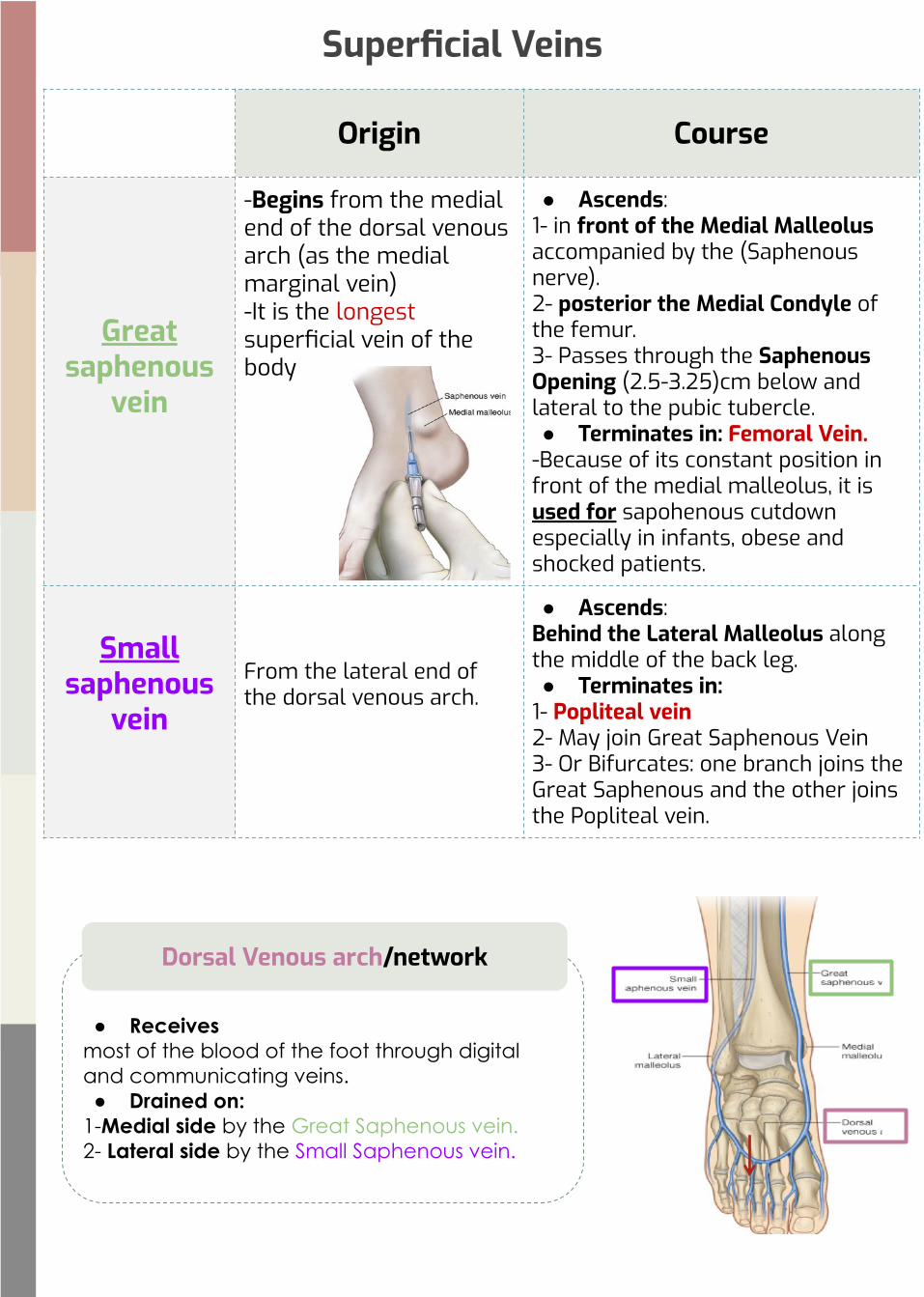
Origin Course
Great saphenous
vein
-Begins from the medial end of the dorsal venous arch (as the medial marginal vein)-It is the longest superficial vein of the body
● Ascends: 1- in front of the Medial Malleolus accompanied by the (Saphenous nerve).2- posterior the Medial Condyle of the femur.3- Passes through the Saphenous Opening (2.5-3.25)cm below and lateral to the pubic tubercle.● Terminates in: Femoral Vein.
-Because of its constant position in front of the medial malleolus, it is used for sapohenous cutdown especially in infants, obese and shocked patients.
Small saphenous
veinFrom the lateral end of the dorsal venous arch.
● Ascends: Behind the Lateral Malleolus along the middle of the back leg.● Terminates in:
1- Popliteal vein2- May join Great Saphenous Vein3- Or Bifurcates: one branch joins the Great Saphenous and the other joins the Popliteal vein.
Superficial Veins
● Receives most of the blood of the foot through digital and communicating veins.● Drained on:
1-Medial side by the Great Saphenous vein.2- Lateral side by the Small Saphenous vein.
Dorsal Venous arch/network

Varicose Veins-It is dilation and degeneration of the superficial veins that may be complicated by ulcers.-More common in the postero medial part of the lower limb.-Results because of incompetence of the valves in the perforating veins, or valves within the great saphenous itself.-This allows the passage of high pressure blood from the deep to the superficial veins.
-Penetrate the deep fascia close to their origin from the superficial veins. They contain valves which normally allow the blood to flow from the superficial to the deep veins-The perforating veins pass through the deep fascia at an oblique angle so during muscular contraction, they are compressed. This also prevents blood flowing from the deep to superficial veins.-Connect the Great Saphenous vein with the deep veins along the medial side of the calfNote: it is originated from superficial veins and inserted in deep veins, acts as a connector.
Perforating Veins
Deep Vein Thrombosis (DVT)-The veins of the lower limb are subject to venous thrombosis after a bone fracture.- Venous stasis is the main cause by pressure on the veins from the bedding during prolonged hospital stay and aggravated by muscular inactivity.-Thrombophlebitis may develop around the vein.-Pulmonary thromboembolism may occur when a thrombus breaks free from the lower limb vein and passes to the lungs.

1)A2)B3)C4)D5)C6)A
Q1:What is the most important, main arterial supply to the thigh? A.Profunda femorisB.Superficial Epigastric.C.Superficial Circumflex iliac.D.Deep External Pudendal.
Q2: which of the following has a Medial rotation with the Femoral artery?
A.Adductor longus.B.Femoral vein.C.Femoral nerveD.Pectineus
Q3:How does the Femoral Artery enters the thigh?A.Medial to the inguinal ligamentB.In front of the inguinal ligamentC.Behind the inguinal ligamentD.lateral to the inguinal ligament
Q4:Where can you feel the Femoral pulse?A.Lateral to the lingual ligamentB.Posterior to the lingual ligamentC.Medial to the lingual ligamentD.Inferior to the lingual ligament
Q5:popliteal artery continuation of what ?A. dorsalis pedis arteryB.anterior tibial arteryC. femoral arteryD.tibial artery
Q6: genicular anastomosis compensates for what artery?A.popliteal arteryB.femoral arteryC. anterior tibial arteryD.posterior tibial artery
Q7:what is the main source of blood supply to the toes. the main arterial supply of the toe?
A.dorsalis pedis arteryB. femoral arteryC.posterior tibial arteryD.anterior tibial artery
Q8: dorsalis pedis artery pass to which interosseous spaceA. 2ndB. 1stC.3rdD.5th
Q9:wich of the following accompany all major arteries?A.perforating veinsB.venae comitantesC.deep veinsD. Superficial veins
Q10:the popliteal veins is …. to the popliteal artery.A.anteriorB.posteriorC.superiorD.inferior
Q11:the great saphenous vein is accompanied by?A.deep veinsB.superficial veinsC.saphenous nerveD.peroneal nerve
Q12:where is the site of varicose veins?A.anterior part of lower limbB.lateral part of lower limbC.posteromedial part of lower limb
MCQs7)A8)B9)C10)B11)C12)C

SAQs
Q1) List The two types of Arterial Anastomoses found in the lower limb and what they mainly supply?
Q2) What are the terminal branch of the popliteal artery?
This lecture is done by:
Shayma AlghanoumSarah AlaidaroosFaisal Alnashwan
Special thanks to Manal Altwaim
Team leaders:Mayasem Alhazmi
Fahad Alajmi
Q1) 1-the Cruciate anastomosis, It Provides blood supply to the lower limb in case of ligation of the femoral artery. 2-the Trochanteric anastomosis, supply the head & neck of femur.Q2) anterior and posterior tibial arteries