view - development - the company of biologists
TRANSCRIPT

INTRODUCTION
The
Enhancer of split gene complex (E(SPL)-C) is one of theneurogenic loci of Drosophila melanogaster (Lehmann et al.,1983; Knust et al., 1987a). It comprises seven, partiallyredundant genes, which encode transcription factors of thebasic helix-loop-helix (bHLH) family (Knust et al., 1987b,1992; Klämbt et al., 1989; Delidakis and Artavanis-Tsakonas,1992; Schrons et al., 1992). Products of the neurogenic and ofthe proneural genes (Ghysen and Dambly-Chaudière, 1989;Romani et al., 1989) form a functional network that contributesto the correct segregation of neural and epidermal cell lineages(see Campos-Ortega, 1993, and Ghysen et al., 1993, forreviews). Lineage segregation (Hartenstein and Campos-Ortega, 1984) occurs within neuroectodermal equivalencegroups, which express proneural genes. By analogy with theclusters from which sensory organ mother cells are selected inthe epidermis (see Jan and Jan, 1990, for a review), the neu-roectodermal equivalence groups are called proneural clusters(Ghysen and Dambly-Chaudière, 1989; Romani et al., 1989;Simpson, 1990). One cell in each cluster is selected to take onneural fate and develops as a neuroblast. Selection as a neuro-blast is associated with continued expression of the proneuralgenes of the achaete-scute complex AS-C (Cabrera et al.,1987; Romani et al., 1987; Brand and Campos-Ortega, 1988;Cabrera, 1990; Martin-Bermudo et al., 1991; Skeath andCarroll, 1992; Ruiz-Gómez and Ghysen, 1993) and is accom-panied by lateral inhibition. This ensures that the remainingcells of the cluster cease to express proneural genes, continue
transcribing the genes of the E(SPL)-C (Knust et al., 1987b,1992) and adopt epidermal fate.
A formal genetic model (Campos-Ortega, 1993) proposesthat cell lineage segregation is regulated by reciprocal interac-tions of the products of the E(SPL)-C and the proneural genes,all of which are transcription factors of the bHLH family (seeVillares and Cabrera, 1987, Alonso and Cabrera, 1988, Caudyet al., 1988, and González et al., 1989, for proneural genes; andKlämbt et al., 1989, Delidakis and Artavanis-Tsakonas, 1992,and Knust et al., 1992, for the E(SPL)-C). In embryos carryingneurogenic mutations, expression of the proneural genesachaete (ac) and lethal of scute (l’sc) fails to become restrictedto single cells in each cluster (Brand and Campos-Ortega,1988; Cabrera, 1990; Skeath and Carroll, 1992; Ruiz-Gómezand Ghysen, 1993). This suggests that the neurogenic genessuppress proneural gene expression within the non-segregatingcells of the neuroectoderm, thereby permitting their epidermaldevelopment. Since the E(SPL)-C is the last link in an epistaticchain of interactions among neurogenic genes (de la Conchaet al., 1988), this function could be mediated by the proteinsencoded by the gene complex. Neuroblast development,however, requires that the genes of the E(SPL)-C are inactive.This inactivation might be brought about by the products of theproneural genes (Campos-Ortega, 1993).
To advance our understanding of the role played by thegenes of the E(SPL)-C during lineage segregation, we analysedcis-acting regions necessary for neuroectodermal expression oftwo of these genes, E(spl) and HLH-m5. Control regions foractivation of transcription in the neuroectoderm and repression
815Development 120, 815-826 (1994)Printed in Great Britain © The Company of Biologists Limited 1994
The
Enhancer of split gene complex (E(SPL)-C) ofDrosophila comprises seven genes encoding bHLHproteins, which are required by neuroectodermal cells forepidermal development. Using promoter and gene fusionswith the lacZ gene, we determined the location of cis-actingsequences necessary for normal expression of two of thegenes of the E(SPL)-C, E(spl) and HLH-m5. About 0.46 kbof E(spl) and 1.9 kb of HLH-m5 upstream sequences arenecessary to reproduce the normal transcription pattern ofthese genes. The gene products of achaete, scute and lethalof scute, together with that of ventral nervous system con-densation defective, act synergistically to specify the neu-
roectodermal E(spl) and HLH-m5 expression domains.Negative cross- and autoregulatory interactions of theE(SPL)-C on E(spl) contribute, directly or indirectly, to thisregulation. Interactions involve DNA binding, since muta-genesis of binding sites for bHLH proteins in the E(spl)promoter abolishes neuroectodermal expression andactivates ectopic expression in neuroblasts. A model foractivation and repression of E(spl) in the neuroectodermand neuroblasts, respectively, is proposed.
Key words: Drosophila, neurogenesis, gene regulation, E(SPL)-C,cis-acting elements, trans-regulatory factors
SUMMARY
Neuroectodermal transcription of the
Drosophila neurogenic genes E(spl) and
HLH-m5 is regulated by proneural genes
Bernd Kramatschek and José A. Campos-Ortega*
Institut für Entwicklungsbiologie, Universität zu Köln, 50931 Köln, Germany
*Author for correspondence
816
of transcription in neuroblasts were identified within 0.46 kbof E(spl); regulatory regions of HLH-m5 are distributedthroughout 1.9 kb upstream sequences. Furthermore, we foundthat proteins encoded by proneural genes act as trans-regula-tors for both E(spl) and HLH-m5. Thus, ac, scute (sc) and l’sc,together with ventral nervous system condensation defective(vnd) act synergistically to generate the neuroectodermalexpression domains of E(spl) and HLH-m5. We also shownegative autoregulation of E(spl) and cross-regulatory interac-tions of genes of the E(SPL)-C with E(spl). Most, if not all ofthese interactions involve binding of regulatory proteins tospecific DNA sequences, since mutation of bHLH binding sitesin the E(spl) promoter abolishes expression. We propose amodel for activation of E(spl) in the neuroectoderm and itsrepression in neuroblasts.
MATERIALS AND METHODS
For the analysis of lacZ expression in mutant backgrounds, we usedthe following mutations and deficiencies: Df(1)RT184 (Mason et al.,1986); Df(1)260.1, Df(1)sc19, In(1)y3PLsc4R, In(1)y3PLsc8R,In(1)sc4Lsc9R (Garcia-Bellido, 1979); vnd6 (White et al., 1983);Df(3R)boss16 (Hart et al., 1990); Df(3R)gror72.1, Df(3R)gror8.1,Df(3R)gror171.1 (Schrons et al., 1992). They were maintained overbalancer chromosomes carrying lacZ insertions (FM7c, lacZ ftz orTM3, lacZ ftz) to allow distinction of the genotypes of mutantembryos.
Construction of fusion genesCloning techniques applied here are described in Sambrook et al.(1989). For in-frame fusions between E(spl) or HLH-m5 and the lacZgene, we constructed P-transformation vectors (pWlacB) as modifi-cations of pWlacI (Molsberger et al., 1988). For construction of het-erologous E(spl)-hsp70 promoter fusions, the vector pWHL was used(Hinz et al., 1992). It contains the hsp70 basal promoter with TATAbox and RNA leader fused to the lacZ coding region and SV40polyadenylation sequences.
For the E(spl) in-frame fusion series, the lacZ gene of pWlac2B wasfused after codon 9 of the E(spl) gene using a genomic BglII site at+123 bp from the cap site. To create the two shortest promoter con-structs m8-0.14 and m8-0.24, 255 bp genomic PvuII-BglII and 360 bpMscI-BglII fragments, respectively were inserted into the NotI(blunted)-BamHI sites of pWlac2B. For m8-0.46, a 576 bp BamHI-BglII fragment was cloned into the unique BamHI site of pWlac2B.To obtain the longest construct (m8-2.61), a 2154 bp BamHI fragmentwas inserted into the BamHI site of m8-0.46, i.e., in front of the 576bp BamHI-BglII fragment. The plasmid m8-1.25 was generated byligating a 788 bp ClaI-BamHI fragment, provided with a KpnI-ClaIadapter, into m8-0.46 cut with KpnI-BamHI. Construct m8TR-2.61 isidentical to m8-2.61, but the hsp70 polyadenylation sequences werereplaced by a 0.9 kb EcoRV-ClaI fragment from the E(spl) 3
′ region.To construct heterologous E(spl)-hsp70 promoters containing twocopies of the 217 bp genomic BamHI-MscI fragment (−458 to −241bp), the fragment was blunted with Klenow and ligated into the StuIsite of pWHL. Recombinant plasmids were screened for those thatcontained two tandem repeats in either orientation relative to the tran-scription start site (constructs 2BM*hs and 2BM*rev*hs).
Oligonucleotide-directed mutagenesis was applied to disruptthree binding sites for bHLH proteins in the proximal E(spl)promoter (N1/N2-boxes at positions −170 and −177 bp, E1-box at−139 bp). The 47-nucleotide oligomer TAGGACGGAGGACAATC-TCCAGTATCAAGTACGTGGGGACCGAGCTG was generated tomutagenize the N1/N2-boxes (M201). It contains 14 mismatches withthe wild-type sequence (underlined). The 41-nucleotide oligomer
GTCTTTTTCACAAGGAGTCAAGATAACTTCGTAGGACGGAGwas used to mutagenize the E1-box (M202). A third, 78-nucleotideoligomer was synthesized to mutagenize both the adjacent N1/N2 andE1-boxes simultaneously (M203). Base substitutions were madeessentially as described in the ‘Oligonucleotide-Directed in vitroMutagenesis System, version 2’ (Amersham-Buchler), except thatmutant templates were transformed into the DH5α strain of E. coli.Mutant clones were screened by sequence analysis. BamHI-BglIIfragments (576 bp) containing the mutagenized sequences were thenexcised and inserted into the BamHI site of pWlac2B, verified byrestriction mapping and sequence analysis (constructs m8-0.5*M201,m8-2.6*M202, m8-2.6*M203). A 2154 bp genomic BamHI fragmentwas cloned into the BamHI sithe of the three mutagenized constructsto generate plasmids with longer upstream regions (constructs m8-2.6*M201, m8-2.6*M202, m8-2.6*M203).
Starting plasmid for the HLH-m5 promoter analysis was a 3754 bpgenomic PstI fragment extending from coordinate +1.5 to +5.2,inserted in pBluescript. For the in-frame fusions, the lacZ gene ofpWlac1B was fused after codon 131 of the HLH-m5 gene using thegenomic PstI site at +477 bp from the transcription start point. Tocreate the longest fusion construct (m5-3.28), the PstI fragment waslinked to KpnI-PstI and PstI-BamHI adapters and then inserted intopWlac1B, cut with KpnI and BamHI. HLH-m5 promoter fusions with5′ terminal truncations were constructed essentially in the same way,but by using several deletion clones of the 3754 bp PstI fragment(kindly provided by C. Klämbt).
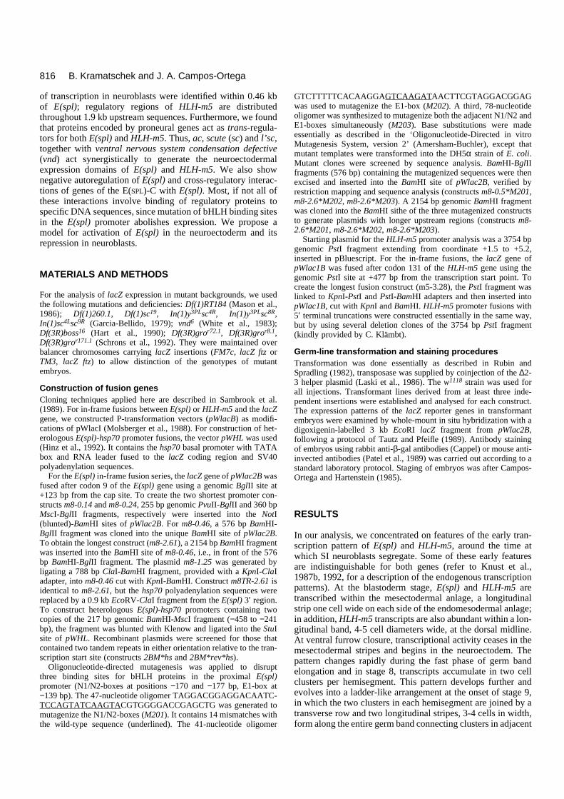
Germ-line transformation and staining proceduresTransformation was done essentially as described in Rubin andSpradling (1982), transposase was supplied by coinjection of the ∆2-3 helper plasmid (Laski et al., 1986). The w1118 strain was used forall injections. Transformant lines derived from at least three inde-pendent insertions were established and analysed for each construct.The expression patterns of the lacZ reporter genes in transformantembryos were examined by whole-mount in situ hybridization with adigoxigenin-labelled 3 kb EcoRI lacZ fragment from pWlac2B,following a protocol of Tautz and Pfeifle (1989). Antibody stainingof embryos using rabbit anti-β-gal antibodies (Cappel) or mouse anti-invected antibodies (Patel et al., 1989) was carried out according to astandard laboratory protocol. Staging of embryos was after Campos-Ortega and Hartenstein (1985).
RESULTS
In our analysis, we concentrated on features of the early tran-scription pattern of E(spl) and HLH-m5, around the time atwhich SI neuroblasts segregate. Some of these early featuresare indistinguishable for both genes (refer to Knust et al.,1987b, 1992, for a description of the endogenous transcriptionpatterns). At the blastoderm stage, E(spl) and HLH-m5 aretranscribed within the mesectodermal anlage, a longitudinalstrip one cell wide on each side of the endomesodermal anlage;in addition, HLH-m5 transcripts are also abundant within a lon-gitudinal band, 4-5 cell diameters wide, at the dorsal midline.At ventral furrow closure, transcriptional activity ceases in themesectodermal stripes and begins in the neuroectodem. Thepattern changes rapidly during the fast phase of germ bandelongation and in stage 8, transcripts accumulate in two cellclusters per hemisegment. This pattern develops further andevolves into a ladder-like arrangement at the onset of stage 9,in which the two clusters in each hemisegment are joined by atransverse row and two longitudinal stripes, 3-4 cells in width,form along the entire germ band connecting clusters in adjacent
B. Kramatschek and J. A. Campos-Ortega

817Regulation of
E(spl)
segments (see Fig. 5A,B). SI neuroblasts segregate at this timefrom the cells of the ladder and cease to express the RNAs,whereas the neuroectodermal cells continue to express bothgenes (not shown). Some of these neuroectodermal cells willeventually develop as epidermoblasts; the others will still haveto decide between fates one or more times (Hartenstein andCampos-Ortega, 1984; Doe, 1992).
0.46 kb of the E(spl) upstream region are sufficientto generate the normal pattern of embryonicexpression To determine how the E(spl) expression is regulated, we usedfusions of progressively smaller E(spl) promoter fragmentswith lacZ. The constructs m8-2.61, m8-1.25 and m8-0.46
include 2612, 1247 and 458 bp, respectively, of the E(spl)upstream region fused to lacZ (Fig. 1B). Their early expressionpatterns in transgenic embryos are nearly identical to that ofthe endogenous E(spl) gene summarized above. Therefore, cis-acting sequences necessary for normal transcription of E(spl)during early embryonic development are located within 460 bpupstream of the transcription start site. Only two minor differ-ences from the endogenous pattern were seen. During cellularblastoderm (stage 5), transcription of the reporter gene in themesectodermal anlage is modulated following a pair rule-likepattern rather than being continuous, and extends into themesodermal anlage rather than being restricted to the mesec-todermal anlage (Fig. 2A,B). Since these differences also applyto HLH-m5-lacZ constructs, to be considered later on, and were
Fig. 1. E(spl)-lacZ constructs. (A) A map of the genomic region of E(spl) and HLH-m7. Direction of transcription is indicated by arrows, RNAleaders and trailers are shown as filled bars, coding regions as open bars. Sequences encoding the bHLH motifs are hatched. E1 and E2 boxesare indicated by filled triangles, N-boxes are shown by squares. Restriction sites are indicated as B (BamHI), C (ClaI), G (BglII), M (MscI), U(PvuII), V (EcoRV). (B-D) Maps of the different E(spl)-lacZ constructs (refer to text). m8TR-2.61 is identical to m8-2.61 but contains a 0.9 kbgenomic fragment, encompassing the complete E(spl) RNA trailer and approximately 0.5 kb of the 3′ non-transcribed region instead of hsp70polyadenylation sequences. m8TR-2.61 generates a normal pattern of expression, similar to that of m8-2.61. However, in each of 10independent transformant lines examined, both E(spl)-lacZ RNA and β-galactosidase levels were strongly reduced, compared to m8-2.61.Sequence data revealed the presence of two AUUUA pentamers at positions +762 and +783 bp relative to the E(spl) transcription start sitewithin the 3′ non-translated region (Klämbt et al., 1989). Such elements have been shown to target several other messengers for rapid decay(Shaw and Kamen, 1986; Jones and Cole, 1987; Shyu et al., 1989) and their presence here suggests that the 3′ non-translated region controlsmRNA half-life. Therefore, the 3′ region of the constructs m8-1.25, m8-0.46, m8-0.24, and m8-0.14 carried hsp70 polyadenylation sequences toincrease mRNA stability. The ability to direct transcription of the fusion genes in the mesectodermal anlage (ME), in the neuroectoderm (NE)and neuroblasts (NB) is indicated.

818
found with different constructs after independent integrationevents, they are likely to be due to interactions between theupstream regions and vector sequences. During stage 8, E(spl)-lacZ-expressing neuroectodermal cells are arranged in twoclusters per hemisegment, similarly to the endogenous tran-script (Fig. 2C,D). Double-labelling demonstrates that theanterior borders of the clusters coincide with the anteriorborders of the expression domains of the gene invected (orengrailed) (Fig. 5A). The clusters are visible for only 10-15minutes; as SI neuroblasts segregate, other neuroectodermalcells start transcribing E(spl)-lacZ and the pattern progres-sively develops into a ladder-like arrangement (Fig. 2F). Thispattern is fully established by the onset of stage 9, by whichtime SI neuroblast segregation has ended.
The 458 bp promoter region was further analysed. The −458to −241 interval is necessary for E(spl) transcription in the
mesectodermal anlage and in the neuroectoderm, as shown byconstruct m8-0.24. Strikingly enough, from stage 10 onwards,both constructs drive transcription of lacZ in neuroblasts of thetrunk and the procephalon, the former more abundantly thanthe latter. This suggests that 5′ truncation of the E(spl)upstream region beyond −458 bp allows the activation of cis-regulatory functions located between positions −241 and −136bp and positions −136 and +96 bp, that direct abundant tran-scription in neuroblasts.
Two copies of the −458 to −241 sequence alone, when fusedin either orientation upstream of the hsp70 basal promoter(constructs 2BM*hs and 2BM*rev*hs, Fig. 1C), are not capableof directing lacZ expression in either mesectoderm or neu-roectoderm. Instead, both constructs drive lacZ transcription inmost neuroblasts from stage 9 onwards (not shown). This resultsuggests that transcription of E(spl) in mesectoderm and neu-
B. Kramatschek and J. A. Campos-Ortega
Fig. 2. Transcription pattern of the m8-2.61 construct. A, C and E show lateral, B, D and F ventral planes of focus. These, as well as all otherphotographs in Figs 4-6, show in situ hibridizations with a digoxigenin-labelled lacZ probe. (A,B) Reporter gene transcription starts at cellularblastoderm in cells of the mesectodermal anlage, projecting medially into the mesodermal anlage (ms). (C,D) Two clusters of E(spl)-lacZtranscribing cells per hemisegment appear in the ventral neuroectoderm (ne) during stage 8. (E,F) This pattern evolves into a ladder-likearrangement in stage 9, with two longitudinal rows on either side of the midline, one lateral and the other paramedial, and several cells in-between. Scale bar in A for all photographs 50 µm. as, amnioserosa; ne, neuroectoderm; pc, pole cells; pm, posterior midgut.

819Regulation of E(spl)
Fig. 4. Transcription pattern of the m5-3.28 construct. A, C and E are lateral, B and D dorsal and F ventral views of transgenic embryos. (A,B)During early gastrulation, the promoter construct is strongly transcribed in a dorsomedial band. Notice that RNA is also present in lowerconcentration within the mesectodermal anlage (arrow). (C,D) The dorsal expression domain during early germ band extension. (E,F) Ladder-like arrangement in an early stage 9 embryo. Scale bar in A for all photographs 50 µm. as, amnioserosa; cf, cephalic furrow; ne,neuroectoderm; pc, pole cells.
Fig. 3. HLH-m5-lacZ constructs. (A) A restriction map of the HLH-m5 region, including the transcription unit m6. Two different types of E-boxes (CANNTG and A/GCAGNTG) are indicated with open and filled triangles, respectively. For other symbols see Fig. 1. Restrictionenzyme sites are G (BglII), H (HindIII), O (XhoI), P (PstI), U (PvuII), V (EcoRV). (B) The six constructs used in this study. All constructs arefused after codon 131 of the HLH-m5 gene and, therefore, contain most of the coding region, including the HLH motif. Their ability to directtranscription of the fusion genes in the dorsal midline (AS) and mesectodermal anlage (ME), in the neuroectoderm (NE) and neuroblasts (NB)is indicated.

820
roectoderm is not entirely directed by sequences within the 217bp fragment, but requires more proximal sequences in theregion between −241 and +96 bp. However, cis-regulatoryfunctions exerted autonomously by the −458 to −241 fragment,i.e., the activation of transcription in neuroblasts, act indepen-dently of their orientation relative to the transcription start site.
Cis-acting elements directing HLH-m5 expressionare contained within 1.9 kb of upstream sequenceTo test whether the promoter of HLH-m5 is organized in asimilar manner, promoter-lacZ fusions were studied (Fig. 3B).The embryonic transcription patterns driven by m5-3.28 (Fig.4) and m5-1.92 which are 3277 and 1923 bp of the HLH-m5upstream region, respectively, are identical and reproduce theendogenous HLH-m5 transcription pattern (Knust et al., 1987b,1992); smaller constructs (Fig. 3) lack some of the features ofthe pattern. The distribution of HLH-m5-lacZ RNA and of β-galactosidase expressed by m5-3.28 and m5-1.92 is similar,although the protein is detected exclusively within the nuclei(not shown), whereas the transcripts are located in thecytoplasm (Fig. 4A-F). Since the lacZ gene was not providedwith a nuclear localization sequence, the observed nuclearaccumulation of the HLH-m5-β-galactosidase fusion proteinmust be due to the amino-terminal 131 amino acids of theHLH-m5 protein, present in the fusions (see Materials andMethods; Fig. 3B).
At early stages, m5-1.92 or longer constructs, are stronglytranscribed in the mesectodermal anlage and in the dorsalmidline (Fig. 4A,B). Transcription in the mesectodermalanlage is modulated according to a pair rule pattern rather thancontinuous, as in the endogenous pattern (Knust et al., 1987b,1992), and invades mesodermal territories (not shown). Thisalso applies to E(spl)-lacZ as mentioned above. The expressionof a dorsal transcription domain (Fig. 4A-D) is the only qual-itative difference between the expression pattern of the HLH-m5-lacZ and that of the E(spl)-lacZ fusions.
There is no transcription of m5-1.13, which is 1125 bp ofthe HLH-m5 upstream region, in the mesectodermal and dorsalexpression domains (not shown). Transcription of this contructstarts in stage 7-8 with the neuroectodermal clusters (as shownin Fig. 4E-F for m5-3.28) and subsequently exhibits all featuresof the endogenous pattern. This indicates that early mesecto-dermal and dorsomedial expression of HLH-m5 requiressequences between positions −1923 and −1125 bp upstream ofthe transcription start site. Further truncation of the upstreamregion to position −509 bp (construct m5-0.51) leads to ageneral reduction in transcriptional activity in all seven trans-formant lines studied. However, neuroectodermal clusters andladder-like expression can still be observed. Since the spatialexpression pattern of m5-0.51 remains the same as that of m5-1.13, the difference may be due either to a general activatorelement between positions −1125 and −509 bp, or to adispersed distribution of regulatory sites within this region.The embryonic expression of m5-0.35, which contains 348 bpof the HLH-m5 upstream region, is similar to that of m5-0.51.Finally, construct m5-0.13, encompassing 126 bp of the HLH-m5 upstream region, is transcribed exclusively, althoughweakly, in the optic lobes during stages 12-14 (not shown).These results indicate that cis-regulatory regions necessary forthe spatially restricted expression of HLH-m5 in neuroecto-derm are located in the interval −348 to −126 bp.
Neuroectodermal transcription of E(spl)-lacZ andHLH-m5-lacZ depends on proneural trans-activatorsThe neuroectodermal clusters and the ladder-like pattern ofE(spl) and HLH-m5 transcripts are reminiscent of theexpression patterns of the AS-C genes, suggesting regulatoryinteractions between these genes. To test this hypothesis, weanalysed the lacZ expression of m8-2.61 and m5-3.28, whichgenerate the normal spatial transcription pattern of E(spl) andHLH-m5, respectively, in the background of genetic variantsaffecting either AS-C genes, or vnd, or both (Garcia-Bellidoand Santamaria, 1978; Garcia-Bellido, 1979; Jiménez andCampos-Ortega, 1979, 1987, 1990; White, 1980). All thesevariants were balanced over a FM7c, lacZ ftz balancer chro-mosome to allow distinction of the genotypes of mutantembryos. Since the results obtained were similar for bothgenes, we restrict our description to E(spl).
In(1)y3PLsc8R lacks ac and is associated with a generaldecrease in the number of E(spl)-lacZ transcribing cells in theclusters, within the anterior half of the invected stripes andposteriorly to the stripes, whereas the ladder-like patternshows metameric interruptions within the invected stripes(Fig. 5C-D). In In(1)y3PLsc4R embryos, which lack both acand sc, neuroectodermal expression clusters are affectedmore severely than in ac− mutants, and only parts of the stepsof the ladder-like pattern remain (Fig. 5E-F). Loss of l’sc inIn(1)sc4Lsc9R embryos affects the clusters in the region imme-diately posterior to the invected stripes (compare Fig. 5A and5G). However, the ladder-like pattern shows broad metamericdisruptions 3-4 cells in width (Fig. 5H). In contrast to ac− andto ac−-sc− embryos, transcription in the intermediate zonesbetween medial and lateral clusters is nearly completelyabolished in l’sc− embryos (Fig. 5H). Clusters of E(spl)-lacZtranscribing cells are smaller in alternate metameres of bothac− and l’sc− embryos. Deletion of ac, sc and l’sc, as inDf(1)sc19, results in near total loss of expression in the neu-roectodermal clusters in stage 8 (Fig. 5K) and in an irregulararrangement of transcribing cells in stage 9 (Fig. 5L). Con-comitant deletion of asense (ase), that is to say, deletion ofthe entire AS-C, as in Df(1)260.1, does not modify theresidual neuroectodermal E(spl) transcription pattern seen inDf(1)sc19 embryos, suggesting that ase is not involved in gen-erating the cluster and ladder-like patterns (not shown). Inembryos homozygous for Df(1)RT184, which lack the AS-Ctogether with three proximal genes, silver, embryonic lethalabnormal visual system and vnd, and the entire chromosomalsubdivison 1A, neuroectodermal expression of m8-2.61 istotally abolished (not shown). Since, apart from the AS-C,vnd (White, 1980; White et al., 1983) is the only gene in thisregion known to affect neuroblast development (Jiménez andCampos-Ortega, 1990), we studied the expression of thetransgenes in vnd6 embryos. Here, the medial neuroectoder-mal clusters are lacking, whereas the lateral expressiondomain consists of a more or less continuous row of E(spl)-lacZ transcribing cells, 2-3 cells wide, instead of clusters(Fig. 5I). Medial transcription is also strongly affected in theladder-like stage, while expression in the intermediate andlateral zones of the neuroectoderm seems to be less affected(Fig. 5J).
To extend these observations on E(spl)-lacZ transcriptionalregulation to the endogenous gene, we performed a series ofin situ hybridizations with digoxigenin-labelled E(spl) probes
B. Kramatschek and J. A. Campos-Ortega

821Regulation of E(spl)
to AS-C mutant embryos (In(1)y3PLsc8R, In(1)y3PLsc4R,In(1)sc4Lsc9R, Df(1)sc19 and Df(1)RT184). The results (notshown) were indistinguishable from those described above forthe E(spl)-lacZ constructs.
bHLH-binding sites are essential for transcriptionalregulation of E(spl)Proteins encoded by proneural genes bind to the E-box sequenceCAGCTG at −139 bp (E1 box), whereas HLH-m5 and E(spl)bind to a related motif, CACNAG, called the N-box, two ofwhich are present as a tandem repeat at −170 and −177 bp(N1/N2 boxes) in the E(spl) promoter region (Tietze et al., 1992;N. Oellers, M. Dehio and E. Knust, personal communication).To test whether the E(spl) cis-regulatory elements describedabove require these binding sites, oligonucleotide-directedmutagenesis was used to disrupt either the N1/N2 boxes (m8-2.6*M201), or the E1 box (m8-2.6*M202), or both binding sites(m8-2.6*M203; see Materials and Methods). The mutagenizedconstructs (Fig. 1D) contained 2612 bp of the promoter region,i.e., sufficient genomic DNA to mimic the endogenousembryonic transcription pattern (see above). As all conservedbases together with the flanking nucleotides were exchanged,binding of bHLH proteins to these sites should be abolished(Murre et al., 1989). The patterns of expression of these con-structs exhibit differences to that of m8-2.61. The expression ofm8-2.6*M202, in which only the E1 box is mutated, is weakeras compared to the m8-2.6 pattern, but no qualitative differencescan be detected (Fig. 6C,D). The cluster and the ladder-likeexpression domains are very severely reduced in the transcrip-tion pattern of m8-2.6*M203, in which N1/N2 and E1 sites weremutated (Fig. 6E-H). Following disruption of the N1/N2 boxesalone, as in m8-2.6*M201, neuroectodermal expression isstrongly reduced (Fig. 6E) and most neuroblasts in the head (notshown) and trunk (Fig. 6E,F) contain E(spl)-lacZ RNA. Theresults demonstrate that the bHLH-binding sites N1/N2 areessential for neuroectodermal transcription of E(spl)-lacZ andfor its suppression in the neuroblasts.
Neuroectodermal transcription domains of E(spl)are expanded in E(SPL)-C− mutantsIn embryos homozygous for deficiencies of the E(SPL)-C(Schrons et al., 1992), the neuroectodermal transcriptionpattern of m8-2.61 is enhanced: more neuroectodermal cellsexpress the construct than in a wild-type background. Thiseffect differs in severity depending on the number of genes ofthe complex lacking in the deletions analysed. Thus, inDf(3R)boss16 homozygotes, lacking from HLH-mδ to HLH-m7, m8-2.61 is overexpressed in the neuroectoderm (Fig. 6I,J).In embryos homozygous for Df(3R)gror72.1, which lack HLH-m7 and E(spl), overexpression of m8-2.61 in the neuroecto-derm is much less pronounced; overexpression is even less pro-nounced in Df(3R)gror8.1 homozygotes, which lack only theE(spl) gene (not shown). As all the above variants have, inaddition, a partially defective gro gene, we examined m8-2.61transcription in embryos homozygous for Df(3R)gror171.1,which lack only gro. In these mutants, the E(spl) promoterconstruct shows normal neuroectodermal expression (notshown), thus excluding the possibility that gro itself is directlyinvolved in E(spl) regulation. Therefore, these results suggestnegative auto- and cross-regulatory effects of products of theE(SPL)-C on E(spl).
DISCUSSION
Prior to neuroblast segregation, six of the E(SPL)-C genes aretranscribed in zones of the neuroectoderm from which neu-roblasts segregate; after segregation, transcription of thesegenes ceases in neuroblasts and becomes restricted to theremaining cells of the proneural clusters and, eventually, tothe epidermoblasts (Knust et al., 1987b, 1992). A currenthypothesis proposes that lineage segregation is the conse-quence of interactions between the proneural genes and thegenes of the E(SPL)-C (Campos-Ortega, 1993). Thus, to under-stand how lineage segregation is regulated, it is important tounderstand how transcription of these genes is initiallyactivated in the neuroectoderm and subsequently repressed inthe neuroblasts.
Activating and repressing elements for E(spl)expression during segregation of SI neuroblastsOur results indicate that interaction of activating and repress-ing factors is required to regulate E(spl) transcription in theneuroectoderm and its derivatives (Fig. 7). We conclude thatthe N1/N2 and the E1 boxes, and other undefined sequenceelements within the interval −458 to −241 bp, are essential foractivating E(spl) in the neuroectoderm on the basis of thefollowing data. First, the disruption of all three boxes by invitro mutagenesis in the m8-2.6 construct leads to almostcomplete abolition of E(spl)-lacZ transcription in the neuroec-toderm. Second, disruption of the N1/N2 boxes alone resultsin severe reduction of neuroectodermal expression, in additionto activating transcription in the neuroblasts, as will bediscussed below. Third, disruption of the E1 box alone causesa slight reduction in the amount of RNA expressed. Two Eboxes within the promoter fragment of the m8-2.61 construct,E1 and E2, at −139 and −463 bp, respectively, bind proneuralproteins (Oellers et al., personal communication). However,only the E1 box was disrupted in our constructs. Functionalredundancy of the E boxes would explain why the disruptionof the E1 box only attenuates the activation response. Fourth,additional sequences necessary for neuroectodermalexpression of E(spl) are located in the −458 to −241 bp region,as shown by comparing the expression of m8-0.46 and m8-0.24. However, the −458 to −241 bp region alone is insuffi-cient in either orientation to drive transcription from an het-
Fig. 5. Transcription of the m8-2.61 construct in proneural mutants.The embryos shown in A,B,D,F,I,J,K and L were simultaneouslystained with an anti-invected antibody to mark the parasegmentalborders (brown colour). All photographs show ventral planes offocus (arrowheads point to the midline), those on the left are of stage8, those on the right are of stage 9 embryos. (A,B) The normal E(spl)neuroectodermal expression clusters (two per hemisegment, arrows)and ladder-like transcription pattern, respectively, of construct m8-2.61 in wild-type embryos. (C,D) Embryos homozygous forIn(1)y3PLsc8R, which eliminates ac. Additional deletion of sc, as inIn(1)y3PLsc4R embryos (E,F), leads to a more severe reduction ofneuroectodermal E(spl)-lacZ transcription. (G,H) In(1)sc4Lsc9R
embryos (l’sc−). The embryos in I and J are homozygous for vnd6,where paramedial domains of transcription are missing in stage 8 andreduced in stage 9. (K,L) Df(1)sc19 embryos (ac−, sc−, l’sc−).Transcription in the neuroectodermal clusters is totally abolished(K), the ladder-like expression strongly reduced (L). Scale bar in Afor all photographs 50 µm.

822 B. Kramatschek and J. A. Campos-Ortega
Fig. 5. See p. 821

823Regulation of E(spl)
Fig. 6. Neuroectodermal transcription of the constructs m8-2.61, m8-2.6*M201, m8-2.6*M202 and m8-2.6*M203. All photographs showventral planes of focus, those on the left are of stage 8, those on the right are of stage 9 embryos. (A,B) The normal E(spl) cluster and ladder-like transcription patterns by construct m8-2.61, respectively. (C,D) Disrupting the E1 site in construct m8-2.6*M202 causes a reducedneuroectodermal transcription, compared to construct m8-2.61. However, eliminating the N1/N2 boxes (construct m8-2.6*M201) leads to a lossof the neuroectodermal expression clusters (E) and an ectopic transcription in neuroblasts during later stages (F), whereas transcription inneuroectodermal cells or epidermoblasts is abolished. (G,H) In construct m8-2.6*M203, where both N1/N2 and E1 bHLH-binding sites aredisrupted, neuroectodermal transcription of the reporter gene is strongly reduced and there is no transcription in neuroblasts. I and J show theexpression of m8-2.61 in embryos homozygous for Df(3R)boss16, which deletes the E(SPL)-C genes HLH-mα to HLH-m7. This causes apronounced overexpression in the neuroectoderm. Scale bar in A for all photographs, 50 µm.

824
erologous hsp70 basal promoter, as shown by the results withconstruct 2BM*hs. Specific binding sites apparently exist in the−458 to −241 interval for regulatory proteins, which we assumeto interact with bHLH proteins bound to N1/N2 and E1; thisinteraction would activate transcription of E(spl) in the neu-roectoderm.
We were surprised to find regions that direct transcription ofE(spl) in neuroblasts (Fig. 7). Two regions with transcriptionalactivation sequences are present in the −241 to −136 (NBII)and −136 to +96 bp (NBI) intervals, respectively, as the con-structs m8-0.24 and m8-0.14 are transcribed at different levelsin the neuroblasts. In addition, the interval −458 to −241 bpappears to contain other sequences to activate E(spl) tran-scription in neuroblasts (NBIII), since this interval, in isolation,autonomously induces transcription from a hsp70 basalpromoter in most neuroblasts from stage 9 onwards (construct2BM*hs). Binding sites N1/N2 are necessary to repress tran-scription in neuroblasts. Since m8-0.46, which includes NBI,NBII and NBIII, is not expressed in neuroblasts, we postulatethat the −458 to −241 bp interval contributes an element thatoverrides the activating function of NBI, NBII and NBIII. Boththis element and N1/N2 are essential: the elimination of eitherone, as in m8-0.24 and m8-0.14, or in m8-2.6*M201 leads totranscription in neuroblasts; hence, neither alone is sufficientto repress neuroblast transcription.
In conclusion, the activation of transcription of E(spl) in theneuroectoderm and its repression in the neuroblasts may beexplained by assuming distinct interactions of proximalpromoter regions, containing the N1/N2 and E1 boxes, withthe −458 to −241 interval in each case. Distal truncations ofthe E(spl) upstream region that eliminate the −458 to −241interval, or disruption of the N1/N2 boxes, would abolish thisinteraction and thereby block neuroectodermal activation.Conversely, neuroblast-specific activation mediated byregions NBI, NBII and NBIII, must be prevented by the pos-tulated interaction between regulators acting on the −458 to −241 interval and bHLH proteins bound to the N1/N2 boxes.Disruption of the N1/N2 sites would thus allow neuroblast-specific transactivators to ectopically activate E(spl) in neu-roblasts. As discussed below, proteins encoded by proneural
genes are likely to be the postulated transactivators, acting onthe E1 box.
Transcripts of HLH-m5, as well as HLH-m7, HLH-mβ, HLH-mγ and HLH-mδ, exhibit the same distribution as E(spl) aroundthe period of SI neuroblast segregation (Knust et al., 1987b,1992). Thus, a mechanism that represses transcription in thosecells of the proneural clusters that develop as neuroblasts hasto operate on these genes as well. However, none of the HLH-m5 constructs studied here directed expression in neuroblasts,and elements that activate transcription in neuroblasts,analogous to the postulated NBI, NBII and NBIII, were notdetected in the HLH-m5 upstream region. This could be due toseveral reasons. For example, the repression elements may belocated proximal to −130 bp or, although improbable, evenwithin coding regions of HLH-m5. Another possibility wouldbe the poor expression driven by construct m5-0.51 and smaller,which makes the detection of transcripts in neuroblasts difficult.Finally, it is of course also possible that repression of tran-scription of HLH-m5 in the neuroblasts involves a mechanismcompletely different from that which controls E(spl).
Trans-acting factors for neuroectodermal E(spl) andHLH-m5 transcription Hinz et al. (1994) have recently shown that ectopic expressionof lethal of scute is capable of activating transcription of E(spl)in the wing imaginal disc. Our data indicate that proneuralproteins are involved in regulating the transcription of E(spl)and HLH-m5 in the embryo as well. ac, sc, l’sc and vnd actsynergistically to generate the neuroectodermal clusters andthe ladder-like pattern of transcription of both E(spl) and HLH-m5, whereas the vnd function appears to be dominant over thatof the AS-C genes in generating the medial pattern elements.The deletion of the entire subdivision 1B, including ac, sc, l’scand vnd, causes the disappearance of the ladder-like transcrip-tion pattern. In vnd6 embryos, transcription of E(spl) and HLH-m5 promoter constructs is selectively blocked in the medialzone of the neuroectoderm. Hence, vnd participates in the reg-ulation of E(spl) and HLH-m5 in the neuroectoderm. However,the effects of vnd and the AS-C genes are not simply additive.This becomes evident if we compare the remnants of theladder-like pattern in vnd+ embryos lacking the AS-C, withthat of vnd6 embryos. Besides the AS-C, vnd is the only geneof the subdivision 1B known to affect early neurogenesis(Jiménez and Campos-Ortega, 1987, 1990). Hence, this dis-crepancy may be indicative of synergistic interactions betweenvnd and the genes of the AS-C. The primary structure of thevnd product is still unknown. Therefore, the molecular natureof transcriptional interactions between vnd and the E(SPL)-Cgenes remains an open question.
Besides interactions with proneural genes, our resultssuggest cross-regulatory interactions among the E(SPL)-Cgenes. Since E(SPL)-C− mutants show increased numbers ofE(spl)-lacZ transcribing neuroectodermal cells, a negativefeedback seems to operate among the E(SPL)-C genes todelimit the final neuroectodermal transcription domains ofE(spl). However, this interaction may well be indirect, due tothe fact that, as in other neurogenic mutants, all neuroecto-dermal cells of the E(SPL)-C− mutants have acquired aproneural state and therefore could be a consequence of acti-vation of transcription of the E(spl)-lacZ construct byproneural proteins (see below).
B. Kramatschek and J. A. Campos-Ortega
Fig. 7. Summary diagram of regulatory regions and elementsidentified in our study. Transcript stability depends on sequences atthe 3′ end of the gene. Sequences required to activate transcription inneuroblasts are located in three regions, NBI, NBII and NBIII;repression of transcription in neuroblasts depends on sequences inthe interval −458 to −241 (R-NB) and the N1/N2 boxes. Finally, theN1/N2 and the E1 boxes, as well as sequences within the −458 to−241 interval (NE) are necessary for activation of transcription in theneuroectoderm.

825Regulation of E(spl)
On the function of E(SPL)-C genes during lineagedichotomyAccording to a current hypothesis (Campos-Ortega, 1993), thedecision to adopt the neural or the epidermal cell fate dependson reciprocal interactions involving transcription factorsencoded by the E(SPL)-C and the proneural genes. Neurogenicgenes, probably those of the E(SPL)-C, are thought to suppressthe genes of the AS-C thus allowing the cells of the proneuralclusters to develop as epidermoblasts (Brand and Campos-Ortega, 1988; Skeath and Carroll, 1992; Ruiz-Gómez andGhysen, 1993). Indeed, using cell transfection assays, Oellers etal. (unpublished data) have shown that the transcriptional acti-vation of the E(spl) promoter mediated by heterodimers com-prising lethal of scute and daughterless proteins is suppressed byadding E(spl) and/or HLH-m5 protein. The proteins of theE(SPL)-C may thus suppress the AS-C function in the epider-moblasts by binding to the N1/N2 boxes. After disruption of theN1/N2 boxes, we found that E(spl) is transcribed in neuroblasts.This can be explained as a result of the loss of the N1/N2mediated suppression, allowing continued transcription of E(spl)in the neuroblasts. The concomitant disruption of the E1 box andthe N1/N2 boxes leads to abolition of neuroblast transcription;hence, proneural proteins binding to E1 are probably the acti-vators of transcription of E(spl) in the neuroblasts. In a similarmanner, the observed increase of neuroectodermal transcriptionin E(SPL)-C− mutants can be interpreted as indicative of insuffi-cient binding of the proteins encoded by the E(SPL)-C to the Nboxes and consequent persistence of transcriptional activationby proneural proteins. Recall that the neuroectoderm of thesemutants is abnormal, in that the segregation of neural andepidermal lineages does not take place normally, and inDf(3R)boss16 all the neuroectodermal cells develop as neurob-lasts, whereas in Df(3R)gror72.1, Df(3R)gror8.1 neuralization ofthe neuroectoderm is weaker (Schrons et al., 1992).
As the transcription patterns of the AS-C and the E(SPL)-Cgenes overlap extensively around the time of SI neuroblast seg-regation, proneural and E(SPL)-C gene products are co-localized in neuroectodermal cells for some time before thedecision to adopt one of the developmental fates is made. It isprobable that lateral inhibitory signals conveyed by the otherneurogenic genes trigger the repressing action of the E(SPL)-Cproteins on the AS-C function in the prospective epider-moblasts. Conversely, when a cell initiates neuroblast devel-opment, the activating effect of the proneural proteins on tran-scription of E(SPL)-C genes must be inhibited to allow controlof the neural developmental pathway by the proneural genes.
We are grateful to Nipam Patel and Corey Goodman for the anti-invected antibody, to Beate Hoss for technical assistance, and to P.Hardy, U. Hinz, C. Klämbt, T. Klein and E. Knust for comments onthe manuscript and discussions. This work was supported by grantsfrom the Deutsche Forschungsgemeinschaft (DFG, SFB 243), and theFonds der Chemischen Industrie.
REFERENCES
Alonso, M. C. and Cabrera, C. V. (1988). The achaete-scute gene complex ofDrosophila melanogaster comprises four homologous genes. EMBO J. 7,2585-2591.
Brand, M. and Campos-Ortega, J. A. (1988). Two groups of interrelatedgenes regulate early neurogenesis in Drosophila melanogaster. Roux’s Arch.Dev. Biol. 197, 457-470.
Cabrera, C. V. (1990). Lateral inhibition and cell fate during neurogenesis inDrosophila, the interaction between scute, Notch, and Delta. Development109, 733-742.
Cabrera, C. V., Martinez Arias, A. and Bate, M. (1987). The expression ofthree members of the achaete-scute gene complex correlates with neuroblastsegregation in Drosophila. Cell 50, 425-433.
Campos-Ortega, J. A. (1993). Early neurogenesis in Drosophilamelanogaster. In Development of Drosophila. (eds Bate, C. M., MartinezArias, A.) Cold Spring Harbor, New York: Cold Spring Harbor LaboratoryPress.
Campos-Ortega, J. A. and Hartenstein, V. (1985). The EmbryonicDevelopment of Drosophila melanogaster. New York: Springer-Verlag.
Caudy, M., Vässin, H., Brand, M., Tuma, R., Jan, L. Y. and Jan, Y. N.(1988b). daughterless, a gene essential for both neurogenesis and sexdetermination in Drosophila, has sequence similarities to myc and theachaete-scute complex. Cell 55, 1061-1067.
de la Concha, A., Dietrich, U., Weigel, D. and Campos-Ortega, J. A. (1988).Functional interactions of neurogenic genes of Drosophila melanogaster.Genetics. 118, 499-508.
Delidakis, C. and Artavanis-Tsakonas, S. (1992). The Enhancer of split(E(spl)) locus of Drosophila encodes seven independent helix-loop-helixproteins. Proc. Natl. Acad. Sci. USA 89, 8731-8735.
Doe, C. Q. (1992). Molecular markers for identified neuroblasts and ganglionmother cells in the Drosophila central nervous system. Development 116,855-863.
Garcia-Bellido, A. (1979). Genetic analysis of the achaete-scute system ofDrosophila melanogaster. Genetics 91, 491-520.
Garcia-Bellido, A. and Santamaria, P. (1978). Developmental analysis of theachaete-scute system of Drosophila melanogaster. Genetics 88, 469-486.
Ghysen, A. and Dambly-Chaudière, C. (1989). Genesis of the Drosophilaperiheral nervous system. Trends Genet. 5, 251-255.
Ghysen, A., Dambly-Chaudière, C., Jan, L. Y. and Jan, Y.-N. (1993). Cellinteractions and gene interactions in peripheral neurogenesis. Genes Dev. 7,723-733.
González, F., Romani, S., Cubas, P., Modolell, J. and Campuzano, S.(1989). Molecular analysis of asense, a member of the achaete-scutecomplex of Drosophila melanogaster, and its novel role in optic lobedevelopment. EMBO J., 8, 3553-3562.
Hart, A. C., Krämer, H., Van Vactor, D. L. Jr., Paidhungat, M. andZipursky, S. L. (1990). Induction of cell fate in the Drosophila retina, thebride of sevenless protein is predicted to contain a large extracellular domainand seven transmembrane segments. Genes Dev. 4, 1835-1847.
Hartenstein, V. and Campos-Ortega, J. A. (1984). Early neurogenesis inwildtype Drosophila melanogaster. Roux’s Arch. Dev. Biol. 193, 308-325.
Hinz, U., Giebel, B. and Campos-Ortega, J. A. (1994). The basic-helix-loop-helix domain of the Drosophila lethal of scute protein is sufficient forproneural function and activates neurogenic genes. Cell 76, 77-87.
Hinz, U., Wolk, A. and Renkawitz-Pohl, R. (1992). Ultrabithorax is aregulator of β3 tubulin expression in the Drosophila visceral mesoderm.Development 116, 543-554.
Jan, Y. N. and Jan, L. Y. (1990). Genes required for specifying cell fates inDrosophila embryonic sensory nervous system. Trends Neurosci. 13, 493-498.
Jiménez, F. and Campos-Ortega, J. A. (1979). A region of the Drosophilagenome ncessary for CNS development. Nature 282, 310-312
Jiménez, F. and Campos-Ortega, J. A. (1987). Genes in subdivision 1B of theDrosophila melanogaster X-chromosome and their influence on neuronaldevelopment. J. Neurogenet. 4, 179-200.
Jiménez, F. and Campos-Ortega, J. A. (1990). Defective neuroblastcommitment in mutants of the achaete-scute complex and adjacent genes ofDrosophila melanogaster. Neuron 5, 81-89.
Jones, T. R. and Cole, M. D. (1987). Rapid cytoplasmic turnover of c-mycmRNA. Requirement of the 3′ untranslated sequences. Mol. Cell. Biol. 7,4513-4521.
Klämbt, C., Knust, E., Tietze, K. and Campos-Ortega, J. A. (1989). Closelyrelated transcripts encoded by the neurogenic gene complex Enhancer ofsplit of Drosophila melanogaster. EMBO J. 8, 203-210.
Knust, E., Bremer, K. A., Vässin, H., Ziemer, A., Tepass, U. and Campos-Ortega, J. A. (1987a). The Enhancer of split locus and neurogenesis inDrosophila melanogaster. Dev. Biol. 122, 262-273.
Knust, E., Tietze, K. and Campos-Ortega, J. A. (1987b). Molecular analysisof the neurogenic locus Enhancer of split of Drosophila melanogaster.EMBO J. 6, 4113-4123.
Knust, E., Schrons, H., Grawe, F. and Campos-Ortega, J. A. (1992). Seven

826
genes of the Enhancer of split complex of Drosophila melanogaster encodehelix-loop-helix proteins. Genetics 132, 505-518.
Laski, F. A., Rio, D. C. and Rubin, G. M. (1986). Tissue specificity ofDrosophila P element transposition is regulated at the level of mRNAsplicing. Cell 44, 7-19.
Lehmann, R., Jiménez, F., Dietrich, U. and Campos-Ortega, J. A. (1983).On the phenotype and development of mutants of early neurogenesis inDrosophila melanogaster. Roux’s Arch. Dev. Biol. 192, 62-74.
Martin-Bermudo, M. D., Martinez, C., Rodriguez, A. and Jiménez, F.(1991). Distribution and function of the lethal of scute gene product duringearly neurogenesis in Drosophila. Development 113, 445-454.
Mason, J. M., Voelker, R. A., Rosen, D., Campos, A. R., White, K. and Lim,J. K. (1986). Localization of terminal deficiency breakpoints on the Xchromosome. Drosophila Inform. Serv. 63, 164-165.
Molsberger, G., Schäfer, U. and Schäfer, M. (1988). A new set of lacZ fusionvectors, pUCPlac, for studying gene expression in Drosophila by P-mediatedtransformation. Gene 3, 147-151.
Murre, C., Schonleber McCaw, P. and Baltimore, D. (1989). Theamphipathic helix-loop-helix, a new DNA-binding and dimerization motif inimmunglobulin enhancer binding, daughterless, MyoD and myc proteins.Cell 56, 777-783.
Patel, N., Martin-Blanco, E., Coleman, K. G., Poole, S. J., Ellis, M. C.,Kornberg, T. B. and Goodman, C. S. (1989). Expression of engrailedproteins in arthropods, annelids and chordates. Cell 58, 955-968.
Romani, S., Campuzano, S. and Modolell, J. (1987). The achaete-scutecomplex is expressed in neurogenic regions of Drosophila embryos. EMBOJ. 6, 2085-2092.
Romani, S., Campuzano, S., Macagno, E. R. and Modolell, J. (1989).Expression of achaete and scute genes in Drosophila imaginal discs and theirfunction in sensory organ development. Genes Dev. 3, 997-100.
Rubin, G. M. and Spradling, A. C. (1982). Genetic transformation ofDrosophila with transposable element vectors. Science 218, 348-353.
Ruiz-Gómez, M. and Ghysen, A. (1993). The expression and role of aproneural gene, achaete, in the development of the larval nervous system ofDrosophila.. EMBO J. 12, 1121-1130.
Sambrook, J., Fritsch, E. F. and Maniatis, T. (1989). Molecular Cloning. A
Laboratory Manual. Second edition. Cold Spring Harbor, New York: ColdSpring Harbor Laboratory Press.
Schrons, H., Knust, E. and Campos-Ortega, J. A. (1992). The Enhancer ofsplit complex and adjacent genes in the 96 F region of Drosophilamelanogaster are required for segregation of neural and epidermal progenitorcells. Genetics 132, 481-503.
Shaw, G. and Kamen, R. (1986). A conserved AU sequence from the 3′untranslated region of GM-CSF mRNA mediates selective mRNAdegradation. Cell 46, 659-667.
Shyu, A.-B., Greenberg, M. E. and Belasco, J. G. (1989). The c-fos transcriptis targeted for rapid decay by two distinct mRNA degradation pathways.Genes Dev. 3, 60-72.
Simpson, P. (1990). Lateral inhibition and the development of the sensorybristles of the adult peripheral nervous system of Drosophila. Development109, 509-519.
Skeath, J. B. and Carroll, S. B. (1992). Regulation of proneural geneexpression and cell fate during neuroblast segregation in the Drosophilaembryo. Development 114, 939-946.
Tautz, D. and Pfeifle, C. (1989). A non radioactive in situ hybridizationmethod for the localization of specific RNAs in Drosophila embryos revealstranslational control of the segmentation gene hunchback. Chromosoma 98,81-85.
Tietze, K., Oellers, N. and Knust, E. (1992). Enhancer of splitD, a dominantmutation of Drosophila and its use in the study of functional domains of ahelix- loop-helix protein. Proc. Natl Acad. Sci. USA 89, 6152-6156.
Villares, R. and Cabrera, C. V. (1987). The achaete-scute gene complex ofDrosophila melanogaster: conserved domains in a subset of genes requiredfor neurogenesis and their homology to myc. Cell. 50, 415-424.
White, K. (1980). Defective neural development in Drosophila melanogasterembryos deficient for the tip of the X-chromosome. Dev. Biol. 80, 322-344
White, K., Decelles, N. L. and Enlow, T. C. (1983). Genetic anddevelopmental analysis of the locus vnd in Drosophila melanogaster.Genetics 104, 433-488.
(Accepted 6 January 1994)
B. Kramatschek and J. A. Campos-Ortega