vironostika hiv-1 plus o microelisa system - label · vironostika® hiv-1 plus o microelisa system...
TRANSCRIPT
Vironostika® HIV-1 Plus O Microelisa System Store between 2-8°C. For in vitro diagnostic use.
INTENDED USE The Vironostika® HIV-1 Plus O Microelisa System is an enzyme-linked immunosorbent assay (ELISA) for the qualitative detection of antibodies to Human Immunodeficiency Virus Type 1 (HIV-1), including Group O, in human specimens collected as serum, plasma, or dried blood spots. The Vironostika HIV-1 Plus O Microelisa System is intended for use as an aid in diagnosis of infection with HIV-1. It is not intended for use in screening blood donors.
SUMMARY AND EXPLANATION OF THE TEST Published data indicate a strong correlation between the acquired immunodeficiency syndrome (AIDS) and a retrovirus referred to as Human Immunodeficiency Virus (HIV).1,2 Currently, two HIV serotypes, designated as HIV-1 and HIV-2, have been identified based on the results of serologic and molecular studies. Both HIV serotypes have been isolated from patients with AIDS and AIDS-related complex (ARC), as well as from apparently healthy individuals at high risk for AIDS.2 Both viruses have the same morphology, lymphotropism,3 and modes of transmission.4 Since 1984, reports have indicated that HIV-1 can be isolated from a variety of tissues and body fluids of infected individuals.2,5 In 1990, a more divergent strain of HIV-1, which is known as Group O HIV-1, was isolated and characterized.6 Antibodies specific for group O HIV-1 are difficult to be detected with some HIV antibody detection systems unless these systems use whole viral lysate and/or contain group O specific epitopes as antigens.7,8 In addition, an increasing number of variants have been identified with HIV-1 serotype, some of which have arisen from genetic recombinations within a host having infection with multiple types of HIV-1. Such variants may be more difficult to be detected with ELISA systems that are not based on viral lysate.7,8 Worldwide distribution of HIV shows prevalence of the serotypes in different areas, with HIV-1 most widely distributed and Group O appearing primarily in West Central Africa.
Following infection with HIV, an individual rapidly (e.g. within 4 weeks) develops antibodies to viral proteins, a process known as seroconversion. After seroconversion, HIV specific antibodies can be readily detected in the blood specimen. A majority of patients who exhibit symptoms of AIDS or ARC have HIV specific antibodies in their blood. In addition, a significant proportion of apparently healthy individuals at increased risk for AIDS also contain HIV specific antibodies in their blood specimens. Due to close relation of HIV-1 and HIV-2, proteins of the two viruses, especially the core and polymerase proteins, result in serologic cross-reactivity.3
The Vironostika HIV-1 Plus O Microelisa System assay was designed to be highly sensitive for a spectrum of HIV-1 serotypes, including group O virus. As a result, nonspecific reactions may occasionally be seen in specimens from people who have prior pregnancy, blood transfusion, or exposure to human cells or media containing cultured HIV antigen.9 Because of these and other potential nonspecific reactions, specimens reactive with the Vironostika HIV-1 Plus O Microelisa System assay should be confirmed with a confirmatory test ,e.g., Western Blot testing.
Reactive specimens upon initial testing with the Vironostika HIV-1 Plus O Microelisa System assay should be re-tested in duplicate. Reactivity in either or both of the duplicate tests indicates a potential for the presence of HIV-specific antibody. In individuals at increased risk of infection, such as homosexual men, hemophiliacs, or intravenous drug users, repeatedly reactive specimens are usually found to contain antibodies to HIV by additional, more specific tests. However, when the ELISA is used to screen populations with a low prevalence of HIV infections, nonspecific reactions may be more common than specific reaction. Information about prevalence of HIV infections in persons in various categories of risk, as well as clinical and public health guidelines, are available in each CDC publication of Morbidity and Mortality Weekly Reports. Although information about the degree of risk for HIV-1 infection and the degree of reactivity of the serum are of value in interpreting the test, a diagnosis should not be based only on this information. Therefore, it is appropriate to investigate repeatedly reactive specimens by additional, more specific tests, such as Western Blot, immunofluorescence, radioimmunoprecipitation, viral antigen based immunoassays, peptide ELISAs, or nucleic acid amplification assays.
1

PRINCIPLE OF THE TEST This test uses HIV-1 antigens, which are coated onto the wells of microwell plates, for the detection of antibodies specific for HIV-1, including Group O. These antigens include inactivated, purified HIV-1 viral lysate proteins,10 purified viral envelope proteins, and a synthetic peptide with amino acid sequence corresponding to that of the transmembrane immunodominant domain of the HIV-1 Group O (ANT 70) isolate.6
Upon the addition of a diluted test specimen containing antibodies to HIV-1 or HIV-1 Group O to a microwell, immune complexes are formed through the interaction between anti-HIV in the specimen and HIV antigens coated on the microwell. Following incubation, the specimen is aspirated and the well is washed with buffer. Subsequently, anti-human immunoglobulin (goat) conjugated with horseradish peroxidase (HRP) is added which binds to the anti-HIV-antigen complex during a second incubation. Following a wash and incubation with ABTS (2,2'-azino-di-[3-ethylbenzthiazoline-6-sulfonate]) substrate, a green color is produced. The enzyme reaction is stopped by the addition of a fluoride solution.11 The amounts of antibodies to HIV present in the specimen are qualitatively proportional to color intensity.
REAGENTS For in vitro diagnostic use.
Components in each Vironostika HIV-1 Plus O Microelisa System
384 Tests 960 Tests 9,600 Tests Component Description
4 stripholders 10 stripholders 100 stripholders HIV-1 Plus O Microelisa Strips – Twelve per holder, each containing 8 HIV-1 lysate (inactivated) and synthetic HIV-1 Group O antigen-coated wells, contained in a foil pack (2 stripholders/pack) with silica gel desiccant.
1 bottle (120 ml)
2 bottles (120 ml)
20 bottles (120 ml)
Dilsim III – Liquid specimen diluent; contains bovine and caprine proteins, salt, surfactant, Chlorophenol Red as specimen addition indicator, and antimicrobial agents (contains 0.03% [w/v] bromonitrodioxane).
3 vials (0.5 ml)
6 vials (0.5 ml)
18 vials (0.5 ml)
Negative Calibrator Serum (Human) – Contains human serum and goat serum as stabilizer with 0.05% (w/v) bromonitrodioxane as preservative and Fast Green FCF as coloring agent; nonreactive to HBsAg, antibodies to HIV-1 or HCV.
1 vial (0.5 ml)
2 vials (0.5 ml)
6 vials (0.5 ml)
HIV-1 Positive Control Serum (Human) – Inactivated human serum containing protein stabilizers. Contains 0.05% (w/v) bromonitrodioxane as preservative and FD&C red dye no. 2 as coloring agent; reactive for antibodies to HIV-1 and nonreactive to HBsAg and antibodies to HCV.
1 vial (0.5 ml)
2 vials (0.5 ml)
6 vials (0.5 ml)
HIV-O Positive Control Serum (Human) – Inactivated human serum containing protein stabilizers. Contains 0.05% (w/v) bromonitrodioxane as preservative and Orange II as coloring agent; reactive for antibodies to HIV-1 Group O and nonreactive to HBsAg and antibodies to HCV.
2

2 vials 4 vials 38 vials Peroxidase Conjugated Goat Anti-human Immunoglobulins (EnzAbody) – Lyophilized with protein stabilizers and FD&C red dye no. 2.
1 bottle (120 ml)
2 bottles (120 ml)
16 bottles (120 ml)
Conjugate Diluent – Phosphate buffered saline containing protein stabilizers and 0.03% (w/v) bromonitrodioxane as preservative.
2 bottles (62 ml)
3 bottles (62 ml)
32 bottles (62 ml)
ABTS Substrate Solution – 2,2'-azino-di-[3-ethylbenzthiazoline-6-sulfonate], containing hydrogen peroxide.
1 bottle (120 ml)
2 bottles (120 ml)
16 bottles (120 ml)
Stop Solution – Contains 0.28% sodium fluoride and 0.03% (w/v) bromonitrodioxane as preservative.
10 sheets 40 sheets 400 sheets Plate sealers – Adhesive.
1 each 1 each 4 each Clamp and rod – Closure for foil pack.
Note: The Wash Concentrate and DBS Elution Medium are provided separately and individually as accessories to the kit:
Accessory Product Number
Quantity
Accessory Description
250622 4 bottles (500 ml)
Wash Concentrate 50X – Contains 2.5% surfactant in phosphate buffered medium containing 0.05% (w/v) bromonitrodioxane.
250626 4 bottles (120 ml)
DBS Elution Medium – Liquid specimen diluent; contains bovine and caprine proteins, salt, surfactant, and anti-microbial agents (contains 0.03% [w/v] bromonitrodioxane).
Note: Dilsim III and DBS Elution Medium contain Dilsim, a patented diluent.
WARNINGS AND PRECAUTIONS For in vitro diagnostic use.
This test kit is intended for use with serum, plasma, or dried blood spots.
1. Caution: Handle all Vironostika® HIV-1 Plus O Microelisa System biological materials as though capable of transmitting infectious agents. The antigen used to coat the microelisa wells and the positive control sera have been inactivated; nevertheless, both should be handled as though they contain potentially infectious agents. Other components prepared from human serum or plasma have been tested using FDA-licensed tests and found to be nonreactive for the presence of HIV-1 antibody, HTLV-I/II antibody, hepatitis B surface antigen (HBsAg) and HCV antibody. However, as no test method can offer complete assurance that infectious agents are absent, all materials of human origin should be handled as though they contain infectious agents.
2. Do not pipet any of the materials by mouth. Do not smoke, eat, or drink in areas where specimens or kit reagents are handled.
3. Do not perform the test in the presence of reactive vapors (e.g., from sodium hypochlorite, acids, alkalis, or aldehydes) or dust, because the enzymatic activity of the conjugate may be affected.
4. Use disposable gloves. Handle specimens and materials contacting specimens as potentially infectious biological materials in accordance with "Universal Precautions for Prevention of Transmission of Human Immunodeficiency Virus, Hepatitis B Virus, and Bloodborne Pathogens in Health-Care Setting" (CDC, MMWR, June 24, 1988). Consult a physician immediately in the event that contaminated materials are ingested or come in contact with open lacerations, lesions, or other breaks in the skin.
3
5. Immediately clean up any spillage of material potentially containing antigen or antibody with a 1:10 dilution of 5% sodium hypochlorite. Dispose of the cleaning material by an acceptable method.
6. Dispose of all specimens and materials used to perform the test according to local guidelines. For example:
a) Autoclave for 60 minutes at 121°C.
b) Incinerate disposable materials.
c) Mix liquid waste with 5% sodium hypochlorite solution so that the final concentration is approximately 0.5% sodium hypochlorite. Allow to stand at least 30 minutes before disposal.
7. The Stop Solution contains sodium fluoride. Avoid contact with skin. If contact is made, thoroughly wash area with water.
REAGENT PREPARATION Prepare all reagents before beginning assay procedure. All reagents and specimens should be at room temperature (15-30°C) before beginning the assay procedure and can remain at room temperature during testing.
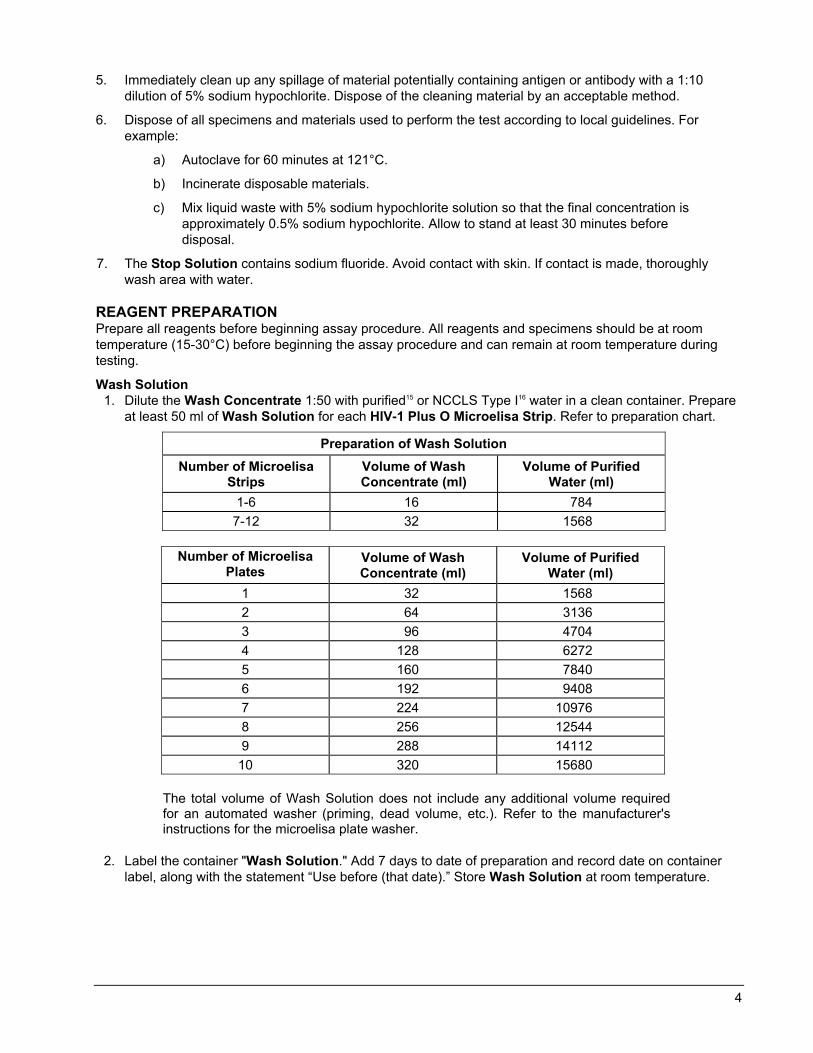
Wash Solution 1. Dilute the Wash Concentrate 1:50 with purified15 or NCCLS Type I16 water in a clean container. Prepare
at least 50 ml of Wash Solution for each HIV-1 Plus O Microelisa Strip. Refer to preparation chart.
Preparation of Wash Solution Number of Microelisa
Strips Volume of Wash Concentrate (ml)
Volume of Purified Water (ml)
1-6 16 784 7-12 32 1568
Number of Microelisa
Plates Volume of Wash Concentrate (ml)
Volume of Purified Water (ml)
1 32 1568 2 64 3136 3 96 4704 4 128 6272 5 160 7840 6 192 9408 7 224 10976 8 256 12544 9 288 14112 10 320 15680
The total volume of Wash Solution does not include any additional volume required for an automated washer (priming, dead volume, etc.). Refer to the manufacturer's instructions for the microelisa plate washer.
2. Label the container "Wash Solution." Add 7 days to date of preparation and record date on container label, along with the statement “Use before (that date).” Store Wash Solution at room temperature.
4
EnzAbody 1. Pipet 50 ml Conjugate Diluent into 1 vial of EnzAbody. Mix by inverting several times. Avoid excessive
foaming. Allow EnzAbody to rehydrate a minimum of 30 minutes before use. Mix again before using to ensure a homogeneous solution. All dye particles should be in solution prior to use.
2. Add 28 days to date of reconstitution and record date on vial label, along with the statement “Use before (that date).” Store prepared EnzAbody at 2-8°C.
KIT STORAGE INSTRUCTIONS Store kit reagents at 2-8°C when not in use. The expiration date printed on the kit indicates the date beyond which the product should not be used. Stability of kit reagents after reconstitution or dilution is listed in "REAGENT PREPARATION." Do not store frozen.
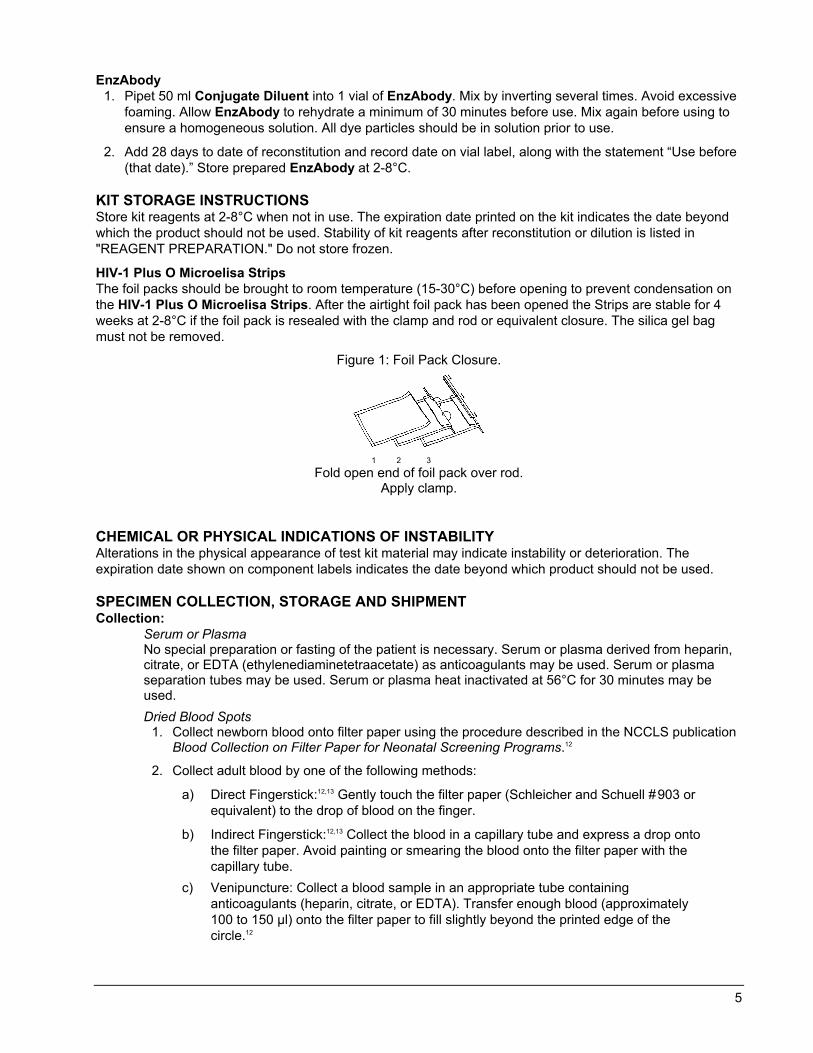
HIV-1 Plus O Microelisa Strips The foil packs should be brought to room temperature (15-30°C) before opening to prevent condensation on the HIV-1 Plus O Microelisa Strips. After the airtight foil pack has been opened the Strips are stable for 4 weeks at 2-8°C if the foil pack is resealed with the clamp and rod or equivalent closure. The silica gel bag must not be removed.
Figure 1: Foil Pack Closure.
1 2 3
Fold open end of foil pack over rod. Apply clamp.
CHEMICAL OR PHYSICAL INDICATIONS OF INSTABILITY Alterations in the physical appearance of test kit material may indicate instability or deterioration. The expiration date shown on component labels indicates the date beyond which product should not be used.
SPECIMEN COLLECTION, STORAGE AND SHIPMENT Collection:
Serum or Plasma No special preparation or fasting of the patient is necessary. Serum or plasma derived from heparin, citrate, or EDTA (ethylenediaminetetraacetate) as anticoagulants may be used. Serum or plasma separation tubes may be used. Serum or plasma heat inactivated at 56°C for 30 minutes may be used. Dried Blood Spots
1. Collect newborn blood onto filter paper using the procedure described in the NCCLS publication Blood Collection on Filter Paper for Neonatal Screening Programs.12
2. Collect adult blood by one of the following methods:
a) Direct Fingerstick:12,13 Gently touch the filter paper (Schleicher and Schuell #903 or equivalent) to the drop of blood on the finger.
b) Indirect Fingerstick:12,13 Collect the blood in a capillary tube and express a drop onto the filter paper. Avoid painting or smearing the blood onto the filter paper with the capillary tube.
c) Venipuncture: Collect a blood sample in an appropriate tube containing anticoagulants (heparin, citrate, or EDTA). Transfer enough blood (approximately 100 to 150 µl) onto the filter paper to fill slightly beyond the printed edge of the circle.12
5
Notes:
With fingersticks, the first drop should be wiped away before collection since the first drop is most likely to contain excess tissue fluid.12,13
The blood should soak completely into the filter paper. Prepare one or more spots for each patient. Blood should not be applied to both sides of the filter paper nor should clotted blood be smeared onto the filter paper.
Allow the specimens to dry in a horizontal position without touching any surface for at least three hours.12 The filter paper may be allowed to dry at room temperature overnight.
3. Do not test dried blood specimens having any of the following characteristics:
• Foreign substance contamination • Blood clots • Non-uniform saturation with blood
Storage: Serum or Plasma Specimens should be free of microbial contamination and can be stored at 2-8°C for up to 7 days. For long-term storage, specimens should be frozen at –20°C or colder. Specimens repeatedly frozen and thawed more than five (5) times or those containing particulate matter may give erroneous results.
Dried Blood Spots Dried blood spots may be stored refrigerated (2-8°C), or at room temperature (15-30°C) for 90 days as long as they are not exposed to elevated humidity (>50%). For long-term storage, dried blood spots may be frozen at –20°C or colder at <50% humidity. Although specimens exposed to humidity ≥50% and elevated temperature (37°C) for 14 days did not exhibit a detectable loss in reactivity, bioMérieux does not recommend routine storage of dried blood spots at elevated temperatures and humidity.
Shipment: Specimens to be shipped must be packaged in compliance with federal regulations governing the transport of etiologic agents.
Dried Blood Spots For shipment, dried blood spot specimens should be placed in a sealed container, such as a heavy-duty zippered bag with a desiccant. Exposure to greater than 50% relative humidity may adversely affect stability of the specimen.
VIRONOSTIKA® HIV-1 PLUS O MICROELISA TEST PROCEDURE Materials provided HIV-1 Plus O Microelisa Strips Dilsim III Negative Calibrator Serum (Human) HIV-1 Positive Control Serum (Human) HIV-O Positive Control Serum (Human) Peroxidase Conjugated Goat Anti-human Immunoglobulins (EnzAbody) Conjugate Diluent ABTS Substrate Solution Stop Solution Plate sealers
6
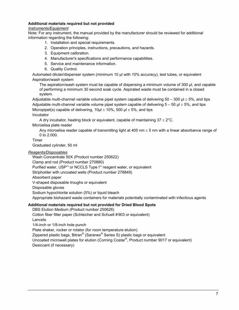
Additional materials required but not provided Instruments/Equipment Note: For any instrument, the manual provided by the manufacturer should be reviewed for additional information regarding the following:
1. Installation and special requirements. 2. Operation principles, instructions, precautions, and hazards. 3. Equipment calibration. 4. Manufacturer's specifications and performance capabilities. 5. Service and maintenance information. 6. Quality Control.
Automated diluter/dispenser system (minimum 10 µl with 10% accuracy), test tubes, or equivalent Aspiration/wash system The aspiration/wash system must be capable of dispensing a minimum volume of 300 µl, and capable
of performing a minimum 30 second soak cycle. Aspirated waste must be contained in a closed system.
Adjustable multi-channel variable volume pipet system capable of delivering 50 – 300 µl ± 5%, and tips Adjustable multi-channel variable volume pipet system capable of delivering 5 – 50 µl ± 5%, and tips Micropipet(s) capable of delivering, 10µl ± 10%, 500 µl ± 5%, and tips Incubator A dry incubator, heating block or equivalent, capable of maintaining 37 ± 2°C. Microelisa plate reader
Any microelisa reader capable of transmitting light at 405 nm ± 5 nm with a linear absorbance range of 0 to 2.000.
Timer Graduated cylinder, 50 ml
Reagents/Disposables Wash Concentrate 50X (Product number 250622) Clamp and rod (Product number 270880) Purified water, USP15 or NCCLS Type I16 reagent water, or equivalent Stripholder with uncoated wells (Product number 278849) Absorbent paper V-shaped disposable troughs or equivalent Disposable gloves Sodium hypochlorite solution (5%) or liquid bleach Appropriate biohazard waste containers for materials potentially contaminated with infectious agents
Additional materials required but not provided for Dried Blood Spots DBS Elution Medium (Product number 250626) Cotton fiber filter paper (Schleicher and Schuell #903 or equivalent) Lancets 1/4-inch or 1/8-inch hole punch Plate shaker, rocker or rotator (for room temperature elution) Zippered plastic bags, Bitran® (Saranex® Series S) plastic bags or equivalent Uncoated microwell plates for elution (Corning Costar®, Product number 9017 or equivalent) Desiccant (if necessary)
7

Materials available from bioMérieux Wash Concentrate 50X (Product number 250622) DBS Elution Medium (Product number 250626) Incubator Microelisa washer Microelisa reader Stripholder with uncoated wells (Product number 278849) 12-channel pipet and tips Micropipet and tips Microprocessor-controlled diluter/dispenser
Procedural notes 1. HIV-1 Plus O Microelisa Strips, EnzAbody, Negative Calibrator, and Positive Controls used in an
assay must be from the same master lot number. Materials should not be used after the expiration date shown on the package label. Components and test specimens should be at room temperature (15-30°C) before testing begins. Return the reagents to 2-8°C after use.
2. HIV-1 Plus O Microelisa Strips of the microelisa plate are removable. Remove Strips not needed and replace with uncoated Strips. Store unused Strips as described in "KIT STORAGE INSTRUCTIONS." Before testing begins, inspect the microelisa stripholder and ensure that all wells are secure. Stripholders should be handled with care to ensure that no Strip is dislodged during testing. Strips may be numbered to ensure re-insertion should Strips become dislodged.
3. HIV-1 Plus O Microelisa Strips and plate sealers may be used only once.
4. Do not touch the top or bottom of strips, or the edge of wells with fingers or pipet tips.
5. All reagents and specimens must be mixed well before use. The Positive Controls and Negative Calibrator may be vortexed before pipetting. One of each Positive Control and three Negative Calibrator replicates must be run on each plate (stripholder). If more than one stripholder is processed, ensure that all specified incubation times are met.
6. This assay refers to reagents used to calculate the cutoff for assay results as "Calibrators." Instrument printouts and associated literature used in conjunction with this assay may refer to these reagents as "Controls." This difference does not affect assay results.
7. The Vironostika HIV-1 Plus O Microelisa System utilizes a Negative Calibrator to calculate the cutoff value (refer to note below regarding use of an external negative control).
Note: CLIA regulations require control reagents to be used according to 42 CFR 493. If a bloodborne pathogen test kit uses any of its manufacturer supplied reagents to serve as a calibrator function, i.e., either or both of the test kit controls (negative or positive) are used to calculate the assay cutoff, then CLIA regulations require that (an) additional "control" reagent(s) be included in each run. Such reagents may be procured or developed in-house. In any case, prior to placing the additional controls in routine use, each lot of such reagents should have: 1) a known dating period, i.e., validated stability (supplied by a control reagent manufacturer or validated by the user on in-house developed control reagent); and 2) known performance parameters, i.e., specifications for acceptance. Prior to implementation, additional control reagents should be qualified to minimize possible incompatibilities that may exist with particular test kits.
8. Do not allow the microelisa wells to dry once the assay has begun. Fill the wells with the next required reagent immediately after washing; if not possible, fill the wells within 10 minutes. The assay should be repeated if the wells can not be filled within 10 minutes after washing.
9. Inspect wells after wash steps. Remove any extraneous material on the bottom of any well that could interfere with absorbance reading.
10. All pipetting steps should be performed with the utmost care and accuracy. Cross-contamination between reagents will invalidate test results. Use micropipets for quantitative delivery of specimens and reagents. For the manual pipetting of controls, calibrator, and specimens, use individual, disposable specimen tips to prevent carryover of specimens. Avoid microbial or any other contamination of reagents.
8
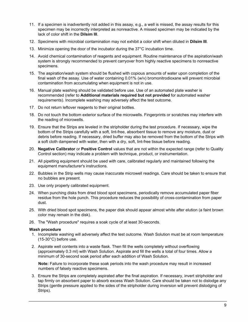
11. If a specimen is inadvertently not added in this assay, e.g., a well is missed, the assay results for this specimen may be incorrectly interpreted as nonreactive. A missed specimen may be indicated by the lack of color shift in the Dilsim III.
12. Specimens with microbial contamination may not exhibit a color shift when diluted in Dilsim III.
13. Minimize opening the door of the incubator during the 37°C incubation time.
14. Avoid chemical contamination of reagents and equipment. Routine maintenance of the aspiration/wash system is strongly recommended to prevent carryover from highly reactive specimens to nonreactive specimens.
15. The aspiration/wash system should be flushed with copious amounts of water upon completion of the final wash of the assay. Use of water containing 0.01% (w/v) bromonitrodioxane will prevent microbial contamination from accumulating when equipment is not in use.
16. Manual plate washing should be validated before use. Use of an automated plate washer is recommended (refer to Additional materials required but not provided for automated washer requirements). Incomplete washing may adversely affect the test outcome.
17. Do not return leftover reagents to their original bottles.
18. Do not touch the bottom exterior surface of the microwells. Fingerprints or scratches may interfere with the reading of microwells.
19. Ensure that the Strips are leveled in the stripholder during the test procedure. If necessary, wipe the bottom of the Strips carefully with a soft, lint-free, absorbent tissue to remove any moisture, dust or debris before reading. If necessary, dried buffer may also be removed from the bottom of the Strips with a soft cloth dampened with water, then with a dry, soft, lint-free tissue before reading.
20. Negative Calibrator or Positive Control values that are not within the expected range (refer to Quality Control section) may indicate a problem with technique, product, or instrumentation.
21. All pipetting equipment should be used with care, calibrated regularly and maintained following the equipment manufacturer's instructions.
22. Bubbles in the Strip wells may cause inaccurate microwell readings. Care should be taken to ensure that no bubbles are present.
23. Use only properly calibrated equipment.
24. When punching disks from dried blood spot specimens, periodically remove accumulated paper fiber residue from the hole punch. This procedure reduces the possibility of cross-contamination from paper dust.
25. With dried blood spot specimens, the paper disk should appear almost white after elution (a faint brown color may remain in the disk).
26. The "Wash procedure" requires a soak cycle of at least 30-seconds.
Wash procedure 1. Incomplete washing will adversely affect the test outcome. Wash Solution must be at room temperature
(15-30°C) before use.
2. Aspirate well contents into a waste flask. Then fill the wells completely without overflowing (approximately 0.3 ml) with Wash Solution. Aspirate and fill the wells a total of four times. Allow a minimum of 30-second soak period after each addition of Wash Solution.
Note: Failure to incorporate these soak periods into the wash procedure may result in increased numbers of falsely reactive specimens.
3. Ensure the Strips are completely aspirated after the final aspiration. If necessary, invert stripholder and tap firmly on absorbent paper to absorb excess Wash Solution. Care should be taken not to dislodge any Strips (gentle pressure applied to the sides of the stripholder during inversion will prevent dislodging of Strips).
9

Test procedure for serum or plasma specimens Note: For Dried Blood Spots, see Test procedure for Dried Blood Spot (DBS) specimens below.
1. Fit stripholder with the required number of HIV-1 Plus O Microelisa Strips. If less than twelve Strips are needed, use uncoated strips to complete the plate when using a 96-well washer.
2. Prepare a 1:21 dilution of each serum or plasma test specimen, Calibrator, and Controls. Include three wells of Negative Calibrator, and one well each of HIV-1 Positive Control and HIV-O Positive Control on each run.
Caution: Use a clean tip for each specimen. Do not pipet specimen into an empty well without Dilsim III. Do not allow the microelisa wells to dry once the assay has begun.
a) Automated diluter/dispenser : Pipet 10 µl of specimen, Calibrator, or Control with 200 µl Dilsim III into the designated microelisa well.
b) Premixed manual method : Pipet 15 µl specimen, Calibrator, or Control into a clean test tube containing 300 µl Dilsim III. Mix well. Pipet 210 µl of the diluted specimen into the designated microelisa well.
c) Direct manual method : Using a multichannel pipet, add 100 µl Dilsim III to each microelisa well. Pipet 10 µl specimen, Calibrator, or Control into the designated wells. Using a multichannel pipet and clean tips, add an additional 100 µl Dilsim III to each well and repeatedly aspirate and dispense to mix.
Note: The addition of a serum or plasma specimen to Dilsim III will cause the specimen addition indicator to turn the dilution to a lavender color.
3. Cover the Strips with adhesive plate sealers or equivalent. Within 60 minutes of specimen/control addition, incubate Strips at 37 ± 2°C for 60-70 minutes.
4. Wash each well four times with Wash Solution (refer to "Wash procedure") using a soak cycle of at least 30-seconds.
5. Pipet 150 µl of reconstituted EnzAbody working solution into each well.
Caution: Do not allow EnzAbody to contaminate ABTS Substrate Solution. If the same equipment is used to add both reagents, new disposable tips must be used.
6. Cover the Strips with adhesive plate sealers or equivalents. Incubate at 37 ± 2°C for 60 to 65 minutes.
7. Wash each well four times with Wash Solution (refer to "Wash procedure") using a soak cycle of at least 30-seconds.
8. Pipet 150 µl of ABTS Substrate Solution into each well. Do not mix or agitate. Do not cover the Strips.
9. Incubate at room temperature (15-30°C) for 10 to 13 minutes.
10. Stop the reaction by adding 150 µl of Stop Solution to each well (maintain the same sequence and time intervals used for ABTS Substrate Solution addition). Plates should be read within two hours.
11. Blank the microelisa reader on air (without stripholder and Strips) and read the absorbance of the solution in each well at 405 nm.
Test procedure for Dried Blood Spot (DBS) specimens 1. Prepare each dried blood spot specimen using one of the following methods. Ensure that the identity of
each specimen is maintained.
a) 1/4-inch punch : Punch one disk from each dried blood spot into a designated uncoated strip well. Pipet 150 µl of DBS Elution Medium into each well containing a punched dried blood spots and cover the Strips with a new adhesive plate sealer.
b) 1/8-inch punch : Punch four disks from each dried blood spot into a designated uncoated strip well. Pipet 150 µl of DBS Elution Medium into each well containing punched dried blood spots and cover the Strips with a new adhesive plate sealer.
10

Note: To minimize carryover when retesting, the punch may be purged between spots by punching 2-3 clean areas of the filter paper. Remove disks and fibers from the punch by tapping lightly onto an absorbent paper for disposal.
2. Incubate at room temperature (15-30°C) for 60-90 minutes with agitation (400-600 rpm) or overnight (14-22 hours) at 2-8°C.
3. After incubation, fit a stripholder with the required number of HIV-1 Plus O Microelisa Strips. If less than twelve Strips are needed, use uncoated strips to complete the plate when using a 96-well washer.
4. Pipet 125 µl Dilsim III into each well of the HIV-1 Plus O Microelisa Strip that will be used for a dried blood spot specimen.
5. Before addition of DBS eluates to the test plate, mix eluates thoroughly using a plate shaker, or by repeatedly aspirating and dispensing contents with a pipet using a clean tip for each specimen. Transfer 25 µl of each eluate into the HIV-1 Plus O Microelisa Strip well containing Dilsim III and mix again.
6. Cover the elution plate containing the remaining dried blood spot eluate with a new plate sealer. Dried blood spot elutions may be stored at 2-8°C for 5 days. For long term storage, eluates may be frozen in microtubes or equivalent at –20°C or colder.
7. Prepare and pipet Negative Calibrator, HIV-1 Positive Control and HIV-O Positive Control as described in Step 2 under "Test procedure for serum or plasma specimens."
8. Cover the Strips with adhesive plate sealers or equivalent. Within 60 minutes of specimen/control addition, incubate Strips at 37 ± 2°C for 120-130 minutes.
9. Wash each well four times with Wash Solution (refer to "Wash procedure") using a minimum 30-second soak cycle.
10. Pipet 150 µl of reconstituted EnzAbody working solution into each well.
Caution: Do not allow EnzAbody to contaminate ABTS Substrate Solution. If the same equipment is used to add both reagents, new disposable tips must be used.
11. Cover the Strips with adhesive plate sealers or equivalent. Incubate at 37 ± 2°C for 60 to 65 minutes.
12. Wash each well four times with Wash Solution (refer to "Wash procedure") using a soak cycle of at least 30-seconds.
13. Pipet 150 µl of ABTS Substrate Solution into each well. Do not mix or agitate. Do not cover the Strips.
14. Incubate at room temperature (15-30°C) for 10 to 13 minutes.
15. Stop reaction by adding 150 µl of Stop Solution to each well (maintain the same sequence and time intervals used for ABTS Substrate Solution addition). Plates should be read within two hours.
16. Blank the microelisa reader on air (without stripholder and Strips) and read the absorbance of the solution in each well at 405 nm.
QUALITY CONTROL Qualification of Negative Calibrator (NC) values: The absorbances of each NC must be greater than or equal to 0.080 and less than or equal to 0.400. Eliminate outliers and calculate the NC mean (NCX). Absorbance of NC must be less than or equal to 1.5 multiplied by NCX and greater than or equal to 0.5 multiplied by NCX. If two or more values are outside range, the run is invalid and must be repeated.
Qualification of HIV-1 (PC1) and HIV-O (PCO) Positive Control values: The absorbance values of the PC1 and PCO must be greater than or equal to 0.700. If either the PC1 or the PCO absorbance value is below this limit, the run is invalid and should be repeated.
11
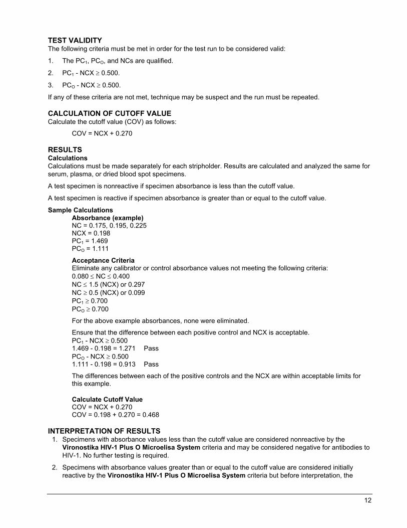
TEST VALIDITY The following criteria must be met in order for the test run to be considered valid:
1. The PC1, PCO, and NCs are qualified.
2. PC1 - NCX ≥ 0.500.
3. PCO - NCX ≥ 0.500.
If any of these criteria are not met, technique may be suspect and the run must be repeated.
CALCULATION OF CUTOFF VALUE Calculate the cutoff value (COV) as follows:
COV = NCX + 0.270
RESULTS Calculations Calculations must be made separately for each stripholder. Results are calculated and analyzed the same for serum, plasma, or dried blood spot specimens.
A test specimen is nonreactive if specimen absorbance is less than the cutoff value.
A test specimen is reactive if specimen absorbance is greater than or equal to the cutoff value.
Sample Calculations Absorbance (example) NC = 0.175, 0.195, 0.225 NCX = 0.198 PC1 = 1.469 PCO = 1.111
Acceptance Criteria Eliminate any calibrator or control absorbance values not meeting the following criteria: 0.080 ≤ NC ≤ 0.400 NC ≤ 1.5 (NCX) or 0.297 NC ≥ 0.5 (NCX) or 0.099 PC1 ≥ 0.700 PCO ≥ 0.700
For the above example absorbances, none were eliminated.
Ensure that the difference between each positive control and NCX is acceptable. PC1 - NCX ≥ 0.500 1.469 - 0.198 = 1.271 Pass PCO - NCX ≥ 0.500 1.111 - 0.198 = 0.913 Pass
The differences between each of the positive controls and the NCX are within acceptable limits for this example.
Calculate Cutoff Value COV = NCX + 0.270 COV = 0.198 + 0.270 = 0.468
INTERPRETATION OF RESULTS 1. Specimens with absorbance values less than the cutoff value are considered nonreactive by the
Vironostika HIV-1 Plus O Microelisa System criteria and may be considered negative for antibodies to HIV-1. No further testing is required.
2. Specimens with absorbance values greater than or equal to the cutoff value are considered initially reactive by the Vironostika HIV-1 Plus O Microelisa System criteria but before interpretation, the
12

specimen should be retested in duplicate. If either duplicate retest is reactive, the specimen is considered repeatedly reactive.
3. Initially reactive specimens that do not react in both of the duplicate repeat tests are considered negative for antibodies to HIV-1.
4. If the specimen is repeatedly reactive, the probability that antibodies to HIV are present is high, especially in specimens obtained from subjects at increased risk for HIV infection.14 In addition, persons who have participated in an HIV vaccine study may develop antibodies to the vaccine and may or may not be infected with HIV. In most settings it is appropriate to investigate repeatedly reactive specimens by additional, more specific tests. Specimens found repeatedly reactive by ELISA and positive by additional, more specific tests are considered positive for antibodies to HIV-1. Clinical correlation is indicated with appropriate counseling, medical evaluation and possibly additional testing to decide whether a diagnosis of HIV infection is accurate. Interpretation of results of specimens found repeatedly reactive by ELISA and negative by additional, more specific tests is unclear; further clarification may be obtained by testing another specimen obtained three to six months later.
PERFORMANCE CHARACTERISTICS OF THE TEST Reproducibility Replicates of HIV-1 antibody positive serum, plasma, and dried blood spot specimens with various degrees of reactivity, negative specimens, and kit controls were tested at multiple sites (n=3), using multiple kit lots (n=3) and multiple technicians (n=3) on multiple days (n=4). Total, inter-assay, and intra-assay precision is reported in table 1 below. The total coefficient of variation (CV) for specimens 1-4 ranged from 11.0 to 22.4%.
Table 1: Assay Reproducibility Total Inter-assay Intra-assay
Specimen Type
ID
N
Mean
SD
CV (%)
SD
CV (%)
SD
CV (%)
1 72 2.78 0.561 20.2 0.533 19.2 0.185 6.7 2 72 2.67 0.549 20.6 0.472 17.7 0.287 10.8 3 72 5.48 0.602 11.0 0.538 9.8 0.278 5.1 4 72 4.77 0.797 16.7 0.711 14.9 0.369 7.7
NC 108 0.33 0.031 9.4 PC 1 36 4.78 0.732 15.3
Serum or Plasma
PC O 36 4.04 0.883 21.8 1 72 1.57 0.272 17.3 0.240 15.3 0.131 8.4 2 72 1.86 0.416 22.4 0.347 18.7 0.232 12.5 3 72 3.70 0.782 21.1 0.678 18.3 0.398 10.8 4 72 3.60 0.676 18.8 0.611 16.9 0.299 8.3
NC 108 0.34 0.036 10.7 PC 1 36 5.38 0.502 9.3
Dried Blood Spots
PC O 36 4.25 0.674 15.9
ID Specimen Identification N Number of Replicates Mean Mean Signal to Cutoff Ratio (SCR) SD Standard Deviation of SCR CV Coefficient of Variation of SCR NC Negative Control PC 1 HIV-1 Positive Control PC O HIV-1 Group O Positive Control
13

Specificity The specificity of the Vironostika HIV-1 Plus O Microelisa System was assessed by testing 6,032 serum/plasma specimens and 3,031 dried blood spot specimens collected from three low risk populations, including voluntary blood donors, insurance applicants and Planned Parenthood clinic patients. The licensed Vironostika HIV-1 Microelisa System was used for comparison. All specimens that were repeatedly reactive with either test were further tested with an FDA licensed Western blot assay.
Of the 6,032 serum/plasma specimens tested, thirteen (13) were repeatedly reactive with the Vironostika HIV-1 Plus O Microelisa System and subsequently confirmed HIV-1 antibody positive with a Western blot assay. In contrast, only 12 of the 13 positive specimens were reactive with the comparative test. As summarized in table 2, the specificity for the Vironostika HIV-1 Plus O Microelisa System in this study was estimated to be 100% (6,019/6,019), with a 95% confidence interval of 99.94 - 100%.
Of the 3,031 dried blood spot specimens, thirteen (13) were repeatedly reactive with the Vironostika HIV-1 Plus O Microelisa System. These repeatedly reactive specimens matched those of serum specimens, which were confirmed positive with a Western blot assay. The comparative test again detected only 12 of the 13 positive specimens. The specificity for the Vironostika HIV-1 Plus O Microelisa System was estimated in this study to be 100% (3,018/3018), with a 95% confidence interval of 99.88 - 100% for dried blood spots.
Table 2: Estimated Specificity in Low-Risk Populations
Number tested
Non-
reactive
Initially
Reactive
Repeatedly
Reactive
Western Blot Positive
Population 1 1,500 1,500 0 0 N/A Population 2 3,012 3,012 0 0 N/A Population 3 1,520 1,506 14 13 13
Serum or Plasma
Total 6,032 6,018 14 13 13
Population 1 0 N/A N/A N/A N/A Population 2 1,511 1,511 0 0 N/A Population 3 1,520 1,506 14 13 13
Dried Blood Spot
Total 3,031 3,017 14 13 13 N/A Not applicable
The estimation of specificity is as follows:
(Number screened – Number repeatedly reactive) (Number screened – Number Western blot test positive)
X 100
14

Sensitivity The clinical sensitivity of the Vironostika HIV-1 Plus O Microelisa System was evaluated by testing matched serum/plasma specimens and dried blood spot specimens collected from 1,010 HIV-1 infected individuals with various CD4+ counts. These specimens were collected from six sites. Of the 1,010 dried blood spot specimens, 904 (90%) were collected directly as dried blood spots while 106 (10%) were collected with an indirect technique (100 µl of anticoagulated blood spotted onto filter paper).
As summarized in table 3, all serum/plasma specimens and dried blood spot specimens were repeatedly reactive with the Vironostika HIV-1 Plus O Microelisa System. Therefore, the sensitivity for both specimen types in this study was 100% (95% CI: 99.64-100%).
Table 3: Estimation of Clinical Sensitivity
Specimen
Type
CD4+
Stratum
Number of specimens
Number of Initially
Reactive
Number of Repeatedly
Reactive <200 250 250 250
200-499 385 385 385 >499 375 375 375
Serum or Plasma
Total 1,010 1,010 1,010 <200 250 250 250
200-499 385 385 385 >499 375 375 375
Dried Blood Spots Total 1,010 1,010 1,010
High-risk populations To assess the performance of the test with specimens collected from high-risk populations, fifteen hundred and fourteen (1,514) specimens were collected from four high-risk populations, which included prison inmates, STD (sexually transmitted diseases) clinic patients, inner city hospital emergency room patients, and HIV-1 outreach clinic patients. In addition, seven hundred and fifty (750) dried blood spot specimens were also collected from two of the study populations.
Serum/plasma specimens were tested with both the licensed Vironostika HIV-1 Microelisa System for comparison (comparative test) and the Vironostika HIV-1 Plus O Microelisa System. If repeatedly reactive with either or both tests, the specimens were further tested with a Western blot assay for confirmation. Dried blood spot specimens were tested only with the Vironostika HIV-1 Plus O. Table 4 lists the test results for the Vironostika HIV-1 Plus O Microelisa System.
Table 4: High-risk Populations
Specimen Type
Population
Number of specimens
Number of Initially
Reactive
Number of Repeatedly
Reactive
Western Blot
Positive 1 251 16 14 8 2 513 13 13 13 3 500 73 68 68 4 250 28 27 27
Serum or Plasma
Total 1,514 130 122 116* 1 0 N/A N/A N/A 2 0 N/A N/A N/A 3 500 68 68 68 4 250 27 27 27
Dried Blood
Spots
Total 750 95 95 95 *Three repeatedly reactive specimens in population 1 were indeterminate with the Western blot assay due to the presence of only a gp160, a gp120 or a 70 kDa band. The other 3 specimens were not tested by Western blot. N/A Not applicable
15

Table 5 shows a summary of the agreement of test results obtained with the Vironostika HIV-1 Plus O and the licensed test with specimens from high risk populations. The 6 specimens that were repeatedly reactive (RR) with the Vironostika HIV-1 Plus O assay but non-reactive (NR) with the licensed test were considered to be false positive on the Vironostika HIV-1 Plus O assay. Therefore, the specificity of the Vironostika HIV-1 Plus O assay in this study of high risk populations was calculated to be 1392/1398 = 99.57% (95% CI = 99.07% – 99.84%).
Table 5: Comparison of the Vironostika HIV-1 Plus O Assay with the licensed Vironostika HIV-1 Microelisa System in testing Specimens from High Risk Populations
Licensed Test
RR NR Total
RR 116 6* 122
NR 0 1392 1392
Vironostika HIV-1 Plus O
Total 116 1398 1514
*Western blot results were indeterminate for 3 of these specimens. The other 3 specimens were not tested by Western blot.
Seroconversion Panel Testing Twelve (12) seroconversion panels were tested in triplicate with the Vironostika HIV-1 Plus O Microelisa System and the licensed Vironostika HIV-1 Microelisa System. Due to insufficient amounts of samples, only 10 of the 12 panels were also tested as DBS specimens.
For serum specimens, the Vironostika HIV-1 Plus O Microelisa System detects the reactivity earlier than the comparative test in all 12 panels.
Table 6: Seroconversion Panel Testing
First Reactive Bleed (Days from the First Bleed) Serum DBS1
Panel ID
Sample Collection Days
Vironostika HIV-1 Plus O
System
Comparative
Test2
Vironostika HIV-1 Plus O
System
Comparative
Test2 924 8, 10, 26, 33, 35,
40 33 40 33 NR
927 0, 28, 33, 35, 40 33 40 35 40 931 9, 15, 28, 33, 35,
42 28 33 28 33
932 0, 3, 13, 27, 34, 50, 78, 163, 194
34 NR3 78 NR
940 0, 7, 11, 15, 18, 22, 25, 29
15 22 18 25
071 1, 3, 17, 22, 28 17 22 22 28 111 1, 2, 8, 16, 20,
22, 27 8 16 NT4 NT
241 1, 7, 9, 15, 17, 22, 24
15 22 17 24
321 1, 8, 12, 15, 21 15 21 15 21 341 1, 7, 10, 21, 23,
28 21 28 28 28
351 1, 8, 11, 15 15 NR NT NT 361 1, 3, 9, 11, 16, 18 18 NR NR NR
1Dried Blood Spot specimens 2The licensed Vironostika HIV-1 Microelisa System was used 3None of the specimens in this panel was reactive 4Not tested due to limited sample volumes
16
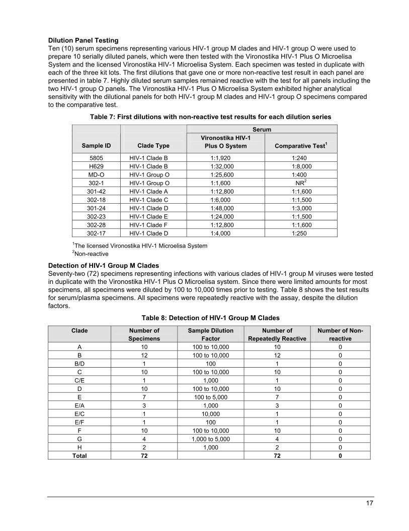
Dilution Panel Testing Ten (10) serum specimens representing various HIV-1 group M clades and HIV-1 group O were used to prepare 10 serially diluted panels, which were then tested with the Vironostika HIV-1 Plus O Microelisa System and the licensed Vironostika HIV-1 Microelisa System. Each specimen was tested in duplicate with each of the three kit lots. The first dilutions that gave one or more non-reactive test result in each panel are presented in table 7. Highly diluted serum samples remained reactive with the test for all panels including the two HIV-1 group O panels. The Vironostika HIV-1 Plus O Microelisa System exhibited higher analytical sensitivity with the dilutional panels for both HIV-1 group M clades and HIV-1 group O specimens compared to the comparative test.
Table 7: First dilutions with non-reactive test results for each dilution series
Serum
Sample ID
Clade Type Vironostika HIV-1
Plus O System
Comparative Test1
5805 HIV-1 Clade B 1:1,920 1:240 H629 HIV-1 Clade B 1:32,000 1:8,000 MD-O HIV-1 Group O 1:25,600 1:400 302-1 HIV-1 Group O 1:1,600 NR2
301-42 HIV-1 Clade A 1:12,800 1:1,600 302-18 HIV-1 Clade C 1:6,000 1:1,500 301-24 HIV-1 Clade D 1:48,000 1:3,000 302-23 HIV-1 Clade E 1:24,000 1:1,500 302-28 HIV-1 Clade F 1:12,800 1:1,600 302-17 HIV-1 Clade D 1:4,000 1:250
1The licensed Vironostika HIV-1 Microelisa System 2Non-reactive
Detection of HIV-1 Group M Clades Seventy-two (72) specimens representing infections with various clades of HIV-1 group M viruses were tested in duplicate with the Vironostika HIV-1 Plus O Microelisa system. Since there were limited amounts for most specimens, all specimens were diluted by 100 to 10,000 times prior to testing. Table 8 shows the test results for serum/plasma specimens. All specimens were repeatedly reactive with the assay, despite the dilution factors.
Table 8: Detection of HIV-1 Group M Clades
Clade Number of Specimens
Sample Dilution Factor
Number of Repeatedly Reactive
Number of Non-reactive
A 10 100 to 10,000 10 0 B 12 100 to 10,000 12 0
B/D 1 100 1 0 C 10 100 to 10,000 10 0
C/E 1 1,000 1 0 D 10 100 to 10,000 10 0 E 7 100 to 5,000 7 0
E/A 3 1,000 3 0 E/C 1 10,000 1 0 E/F 1 100 1 0 F 10 100 to 10,000 10 0 G 4 1,000 to 5,000 4 0 H 2 1,000 2 0
Total 72 72 0
17

Detection of HIV-1 Group O Specimens Archived serum specimens from eleven (11) HIV-1 Group O infected individuals were tested after dilution. These specimens were tested in duplicate with both the Vironostika HIV-1 Plus O Microelisa System and the licensed Vironostika HIV-1 Microelisa System. As shown in table 9, all eleven (11) specimens were repeatedly reactive with the Vironostika HIV-1 Plus O Microelisa System, while only 7 of the 11 were reactive with the comparative test. For all 11 specimens, the Signal to Cutoff Ratio (SCRs) were significantly higher with the Vironostika HIV-1 Plus O as compared to the licensed Vironostika HIV-1 based on the Wilcoxon Signed Rank test.
Table 9: Detection of HIV-1 Group O Specimens
Vironostika HIV-1 Plus O System
Comparative Test1
Specimen ID
Sample Dilution Mean SCR2 Mean SCR
1 1:100 6.1 2.0 2 1:50 4.0 0.4 3 1:50 7.4 1.6 4 1:50 5.6 1.1 5 1:50 7.7 2.0 6 1:50 7.7 7.2 7 1:50 5.3 0.5 8 1:50 7.6 2.2 9 1:50 5.9 0.4 10 1:50 3.9 0.4 11 1:50 7.6 4.5
Total Repeatedly Reactive 11/11 7/11 1The licensed Vironostika HIV-1 Microelisa System 2SCR = Signal to Cutoff Ratio
18

Detection of HIV-2 Positive Specimens Twenty (20) HIV-2 positive serum/plasma specimens were diluted and tested in duplicate with the Vironostika HIV-1 Plus O Microelisa System. All HIV-2 specimens were repeatedly reactive with the Vironostika HIV-1 Plus O Microelisa System. The test results are listed below in table 10.
Table 10: Detection of HIV-2 Specimens
Specimen ID Sample Dilution Mean SCR*
1 1:10 3.3 2 1:10 5.8 3 1:500 1.5 4 1:10 4.3 5 1:50 1.7 6 1:10 5.5 7 1:10 2.5 8 1:10 5.0 9 1:100 3.7 10 1:1,000 1.8 11 1:200 1.3 12 1:200 1.4 13 1:10 1.9 14 1:50 1.3 15 1:1,000 4.9 16 1:1,000 3.6 17 1:50 1.7 18 1:1,000 2.7 19 1:10 2.0 20 1:10 1.8
*Specimens with SCR equal to or greater than 1.0 are considered reactive with the test.
Reactivity with Potentially Interfering Substances or Medical Conditions Serum or plasma specimens (S/P) Samples were collected from individuals with medical conditions that may cause nonspecific assay reactivity. These specimens were tested using the Vironostika HIV-1 Plus O Microelisa System. As shown in table 11, all but one specimens (184/185) were non reactive with the test. The one specimen weakly reactive with the test was from an individual with elevated bilirubin. Further analysis of this specimen with a Western blot assay showed the presence of antibodies weakly reactive with gp160. Therefore, elevated bilirubin was unlikely the cause for the reactivity of the specimen. Rather, the presence of nonspecific antibody in the sample contributed to the weak reactivity.
In a second study, the 184 non-reactive samples were spiked with a small volume of HIV-1 positive sample and tested with the system. All specimens were reactive with the Vironostika HIV-1 Plus O Microelisa System. The medical conditions listed in table 11 did not affect the reactivity of specimen spiked with HIV-1 seropositive sample.
19

Dried blood spot specimens (DBS) A similar study was also performed using dried blood spot specimens. As shown in table 11, all 184 specimens were non-reactive with the Vironostika HIV-1 Plus O Microelisa System. When spiked with a small volume of HIV-1 seropositive sample, all 184 specimens were reactive with the system. No interference was observed with dried blood spot specimens.
Table 11: Reactivity of Specimens from Individuals with Potentially Interfering Substances or with Medical Conditions
Neat Specimens Spiked Specimens
Specimen type
Number of specimens
No. of Reactive
No. of non-reactive
No. of Reactive
No. of non-reactive
S/P DBS S/P DBS S/P DBS S/P DBS S/P DBS Antinuclear antibody positive 10 10 0 0 10 10 10 10 0 0 CMV antibody positive 10 10 0 0 10 10 10 10 0 0 EBV antibody positive 10 10 0 0 10 10 10 10 0 0 Rubella antibody positive 9 9 0 0 9 9 9 9 0 0 Elevated bilirubin1 10 10 1 0 9 10 9 9 0 0 Hemolysed specimens 10 10 0 0 10 10 10 10 0 0 HSV-1 or HSV-2 antibody positive
10 10 0 0 10 10 10 10 0 0
HTLV-I or HTLV-II antibody positive
10 10 0 0 10 10 10 10 0 0
Lipemic2 10 10 0 0 10 10 10 10 0 0 Multiple transfusion 10 10 0 0 10 10 10 10 0 0 Multiparous females 10 10 0 0 10 10 10 10 0 0 Rhematoid factor positive 10 10 0 0 10 10 10 10 0 0 SLE positive 10 10 0 0 10 10 10 10 0 0 Syphilis antibody positive 10 10 0 0 10 10 10 10 0 0 Toxaplasmosis gondii positive 7 7 0 0 7 7 7 7 0 0 HBV antigen positive 10 10 0 0 10 10 10 10 0 0 HCV antibody positive 10 10 0 0 10 10 10 10 0 0 Hypergammaglobulinemia 9 8 0 0 9 8 9 8 0 0 Influenza vaccinated 10 10 0 0 10 10 10 10 0 0
Total 185 184 1 0 184 184 184 184 0 0 1 >6.3 mg/dL 2 >980 mg/dL
20

REFERENCES 1. Hardy AM, Allen JR, Morgan WM, et al. The Incidence Rate of Acquired Immunodeficiency Syndrome in
Selected Populations. JAMA 1985;253(2):215-20.
2. Gallo RC, Salahuddin SZ, Popovic M, et al. Frequent Detection and Isolation of Cytopathic Retroviruses (HTLV-III) from Patients with AIDS and at Risk for AIDS. Science 1984;224:500-3.
3. Brun-Vezinet F, Katlama C, Roulot D, et al. Lymphadenopathy associated virus type 2 in AIDS and AIDS-related complex. Lancet 1987;1:128-32.
4. Quinn TC, Zacarias FRK, St. John RK, et al. AIDS in the Americas: an emerging public health crisis. New Engl. J. Med. 1989;320:1005-7.
5. Markham P, Salahuddin SZ, et al., Advances in the isolation of HTLV-III from patients with AIDS and AIDS-related complex and from donors at risk. Cancer Res. (Suppl.) 1985;45:4588-91.
6. De Leys R, Vanderborght B, Vanden HaeseVelde M, et al. Isolation and partial characterization of an unusual human immunodeficiency retrovirus from two persons of West-Central African origin. J. Virol. 1990;64:1207-16.
7. Loussert-Ajaka I, Ly TD, Chaix ML, et al. HIV-1/HIV-2 seronegativity in HIV-1 subtype O infected patients. The Lancet 1994;343:1393-4.
8. Schable C, Zekeng C, Pau CP, et al. Sensitivity of United States HIV Antibody tests for detection of HIV-1 Group O infections. The Lancet 1994;344:1333-4.
9. Kuhnl P, Seidl S, Holzberger G: HLA DR4 Antibodies Cause Positive HTLV-III Antibody ELISA Results. The Lancet 1985;1222-3.
10. Popovic M, Sarngadharan MG, Read E, et al. Detection, Isolation, and Continuous Production of Cytopathic Retroviruses (HTLV-III) from Patients with AIDS and Pre-AIDS. Science 1984;224:497-500.
11. Bartlett ML: Substrate Evaluation for the Horseradish Peroxidase Enzyme Immunoassay. Am Soc Micro 79th Annual Meeting 1979; Abstract C25.
12. National Committee for Clinical Laboratory Standards: Blood Collection on Filter Paper for Neonatal Screening Programs. Approved Standard – Third Edition. Villanova, PA, NCCLS, 1997;LA4-A3.
13. National Committee for Clinical Laboratory Standards: Procedures for the Collection of Diagnostic Blood Specimens by Skin Puncture. Approved Standard – Third Edition. Villanova, PA, NCCLS, 1991;H4-A3.
14. Curran JW, Morgan WM, Hardy AM, et al. The Epidemiology of AIDS: Current Status and Future Prospects. Science 1985;229:1352-7.
15. The U.S. Pharmacopeia 24/National Formulary 19: 1999; Purified Water : pg. 1753.
16. National Committee for Clinical Laboratory Standards: Preparation and Testing of Reagent Water in the Clinical Laboratory, Villanova, PA, NCCLS, 1997;C3-A3.
21

AVAILABILITY bioMérieux Vironostika® HIV-1 Plus O Microelisa System
384 Test Kit Product number 259859 960 Test Kit Product number 259860 9600 Test Kit Product number 259861
For technical assistance in the U.S.A., contact bioMérieux Customer Service at 1-800-682-2666. Outside the U.S.A., contact your local bioMérieux Representative.
Dilsim is a trademark of bioMérieux, Inc. Vironostika and EnzAbody are registered trademarks of bioMérieux in the USA and other countries. Bitran is a registered trademark of Com-pac International, Inc. Costar is a registered trademark of Corning, Inc. Saranex is a registered trademark of Dow Chemical Company.
Dilsim is a patented diluent shown to inactivate HIV-1 upon contact; US Patent No. 4,839,298.
bioMérieux, Inc. Box 15969 Durham, NC 27704-0969 USA
bioMérieux, S.A. 69280 Marcy-l'Etoile France www.biomerieux.com
©BIOMÉRIEUX 2000 June 2003 4PIHIV1PlusO.doc Revised 06-05-03
22