vision: what is abnormal vision? fovea centralis for 20/20 vision. photoreceptors specialized cells...
TRANSCRIPT

Friday February 3, 2017Honors Anatomy & Physiology
Lesson Objective:
• The student will understand the relationship between abnormal structure of the eye and abnormal vision.
• The student will understand and describe the differences between myopia, hyperopia, and emmetropia.

Lesson Outcome:
Students can describe the shapeof the eye related to the vision test performed yesterday.
Students will understand the functions of the structures of the eye related to visual acuity.
Students will evaluate the impact of corrective lenses and their ability to provide better vision/accommodation.

Vision: What is Abnormal Vision?
Susan Chabot
Honors A&P
Lemon Bay High School

Review• Light must pass through several structures as
it enters the eye to focus on the retina.
• As it passes through these structures, light rays are bent, or REFRACTED.

Checkpoint Question
• What 4 structures/substances must light pass through in order to reach the sensory portion of the eye?
• FIRST = Cornea
• SECOND = Aqueous Humor
• THIRD = Lens
• FOURTH = Vitreous Humor

Lens
The lens has the ability to change shape depending on the distance that light has traveled BEFORE it enters the eye.
Ciliary bodies are the muscles responsible for changing the shape of the lens.
Accommodation is the ability
of the eye to focus specifically
on close objects.
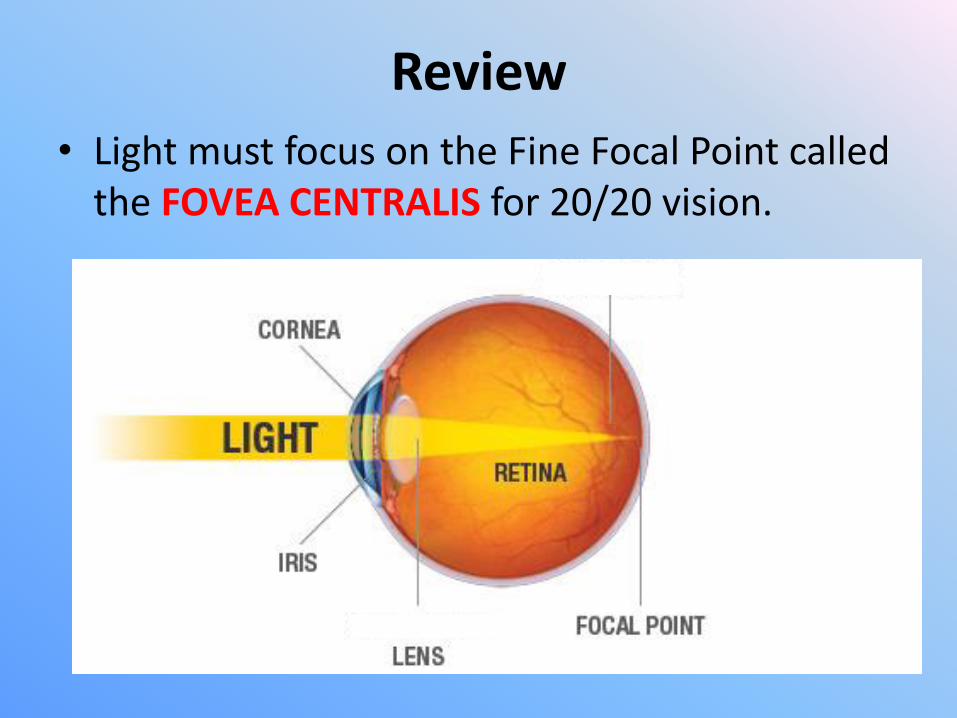
Review
• Light must focus on the Fine Focal Point called the FOVEA CENTRALIS for 20/20 vision.
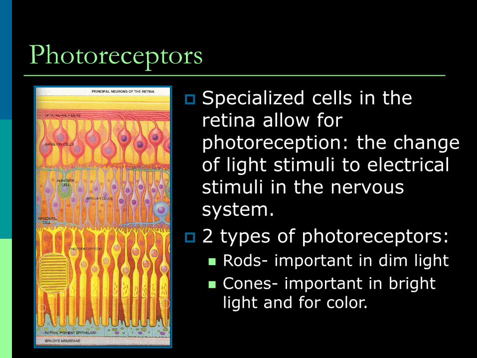
Photoreceptors
Specialized cells in the retina allow for photoreception: the change of light stimuli to electrical stimuli in the nervous system.
2 types of photoreceptors:
Rods- important in dim light
Cones- important in bright light and for color.

Optic Chiasm
The retinal fibers from the medial side of each eye cross over to the opposite side of the brain at the optic chiasm.
This crossing-over allows the visual fields to overlap, permitting binocular vision.

Vocabulary Introduction
• - opia = vision
• - oma = swelling
• - ism = condition of
• a/stigma- = without point/focus
• emme (a)- = harmony
• my- = close/limited
• hyper- = exaggerated/large
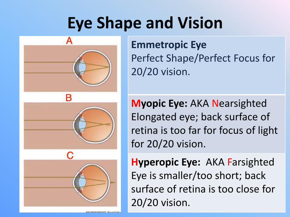
Eye Shape and VisionEmmetropic EyePerfect Shape/Perfect Focus for 20/20 vision.
Myopic Eye: AKA NearsightedElongated eye; back surface of retina is too far for focus of light for 20/20 vision.
Hyperopic Eye: AKA FarsightedEye is smaller/too short; back surface of retina is too close for 20/20 vision.

Vision Correction/LensesEmmetropia: No correction needed
Myopia:Concave lens to expand light rays since they have further to travel.
Hyperopia:Convex lens to push light rays closer together since they don’t have to travel as far.

Surgical CorrectionThe list of possible vision correction procedures is quite long. Here is ONE example of the many.

Other Vision AbnormalitiesStructural Changes
• Astigmatism:– “condition of no focus”
– Abnormality of cornea.
– Bends light AWAY FROM fovea rather than TOWARD fovea.
Degenerative Changes
• Presbyopia:– Aging eyes
– Loss of lens elasticity
• Cataracts:– Lens becomes cloudy
– Must be surgically repaired.
• Glaucoma:– Increases intraocular
pressure.
– Can lead to blindness

Diseases of the Eye Conjunctivitis: AKA pink-eye;
bacterial infection of the lining of the inner eyelid; use antibiotic to cure; highly infectious.
Night blindness: improper rod function; can be helped by vitamin A supplement.
Color blindness: improper cone function;
Cataracts: lens becomes increasingly hard and opaque; vision becomes hazy and eventually causes blindness in the affected eye. Treatment includes surgical removal of the lens and replacement with an artificial lens.
Replacement lens