vmii and icalteches.library.caltech.edu/176/01/brain.pdf · has been doing, and will also serve ......
TRANSCRIPT
Roger I f ". Sperry, Hixon professor of psychobiology at Calkch.
HE VERTEBRATE BRAIN, with an organizations complexity far surpassing that of any other natura
or man-made system, and possessing in certain of its parts the puzzling property of conscious awareness, will probably continue to remain a challenge to man's under- standing for many decades to come. At the present time, the cerebral events underlying even the simplest forms of mental activity remain quite obscure. Although it
IH should someday be possib e to start correlating subjec- tive experience with the corresponding brain process- perhaps even to comprehend the basis and derivation of the "mental" propert ieewe have to be satisfied, for the present, to work at mafty removes from this ultimate goal.
by R. W. SPERRY ow far removed can be judged from the following

senes of experi~nental observations that VMII serve to illustrate some of the things our ~)sychobiologj group has been doing, and will also serve to indicate the gen- eral status of some of the current problems in brain organization.
I will start with some early work dealing with nerve growth and regeneration. the results of 'which have been interpreted broadly to mea.n that brain function in the vertebrates generally is predetermined by inheritance to a much greater degree than formerly had been $up- posed. The findings also give iis some ideas about how the inherent patterning of the brain circuits is achieved iri embryonic development.
Our information on the de\eIopn~ental patterning of brain pathwajs has been obtained mainly from fishes and amphibians because the early developmental stages in these lower vertebrates are accessible to surgery, and because the central nervous sjstem, in the larval and adult stages, retains a capacity for regrowth after surgi- cal intervention that is almost entirely lacking in higher forms.
As shown in the diagram below. it is possible in these animals to cut the nerves of the eye where they cross, and to reunite them surgically in such manner that, when they regenerate, the eyes become connected to the wrong sides of the brain. Under these conditions. the animals respond thereafter as if everything seen through one eye were being viewed through the opposite eye. For ex- ample, when a fly moves within the field of view of a frog's left eye, the frog will strike out at a correspond- ing point in the right field of view. This right-left re- versal of visual reactions persists indefinitely, with no evidence of correction by re-education.
The sensory surface or retina of the eye in all verte- brates is projected through the optic nerve fibers onto the brain in an orderly, topographic, or map-like fash- ion. In the foregoing experiment the behavioral teats (and other evidence) indicate that this orderly topo-
graphic projection is restored w i h systematic precision in the regeneration process-despite extensive intertang- ling of the regenerating fillers. In the diagram below, for instance, the relationships of X t o Y, and of X to any and all oiher poiuts wihin the same visual field are restored to their normal patterns. The fact that this occurb, despite the maladaptive functional effect pro- duwd by crossing the optic nmesi. means that learuhig, or any other kind of functional readjustment. is not responsible for the orderly topographic patterning of the central hook-ups.
Fhe fact, albo, that this orderly restoration occcurs in the face of random iiiterrnixing and intertangling of the regenerated fibers- -particularly in the region of the nerve transection--has forced us to conclude that the optic fibers must differ from one another in quality.
In the lower vertebrates the optic fibers number around 25,000 (there are over a million in the optic nerve of man) , and we have to infer that these indi- vidual fibers differ from one another in their biochemical constitution according to the particular points of the retinal field from which they arise. The further infer- ence here is that the ingrowing fibers, on entering the brain, establish their central hook-ups in a selective, discriminative manner, governed by specific chemical affinities between the different types of ingrowing fibers and the central cells on which they terminate. This in- ference requires the corollary conclusion that a similar topical specificity exists among the nerve cells of the optic centers.
There is good reason to think that the qualitative spe- cificity of the optic fibers is achieved in development through a polarized chemical differentiation of the retina. First, a naso-temporal, or front-back gradient of differentiation is laid down, and later-superimposed at right angles on top of this-an up-down gradient. This would mark each retinal locus with a latitude and longi- tude, so to speak, expressed as a unique ratio of chemical
Connectiizg of eyes to wrong side of brain results in an illusory right-left rez~ersal of vi,s~~,al field. Howe~jer, the rela- t io11~/1{ps of - 4 to Y, and of X to any and all other points within the same visual field are restored to normal patterns.
25
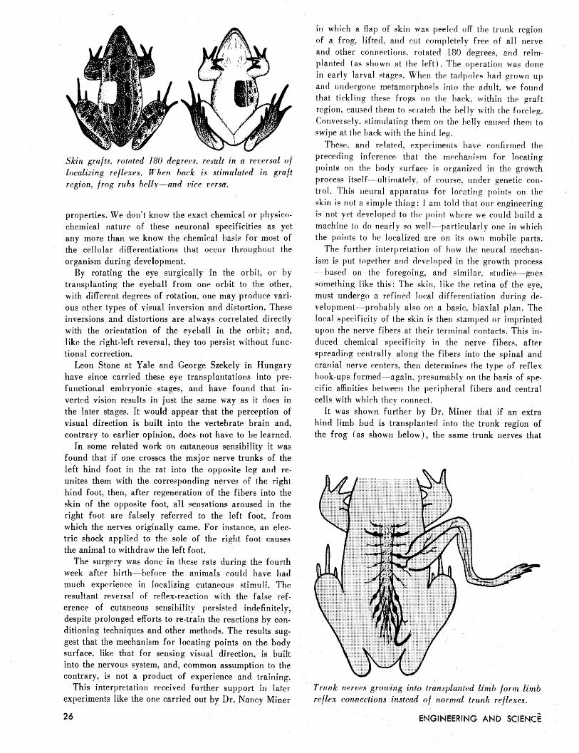
Skin grafts, rotnted 180 dqrees. result in a reversal o f localizing reflexes. When back is stimulated in graft region, frog rubs bellyÑan vice versa.
properties. We don't know the exact chemical or physico- chemical nature of these neuronal specificities as yet any more than we know the chemical basis for most of the cellular differentiations that occur throughout the organism during development.
By rotating the eye surgically in the orbit, or by transplanting the eyeball from one orbit to the other, with different degrees of rotation. one may produce vari- ous other types of visual inversion and distortion. These inversions and distortions are always correlated directly with the orientation of the eyeball in the orbit; and, like the right-left reversal, they too persist without func- tional correction.
Leon Stone at Yale and George Szekely in Hungary have since carried these eye transplantations into pre- functional embryonic stages, and have found that in- verted vision results in just the same way as it does in the later stages. It would appear that the perception of visual direction is built into the vertebrate brain and, contrary to earlier opinion, does not have to be learned.
In some related work on cutaneous sensibility it was found that if one crosses the major nerve trunks of the left hind foot in the rat into the opposite leg and re- unites them with the corresponding nerves of the right hind foot, then, after regeneration of the fibers into the skin of the opposite foot, all sensations aroused in the right foot are falsely referred to the left foot, from which the nerves originally came. For instance, an elec- tric shock applied to the sole of the right foot causes the animal to withdraw the left foot.
The surgery was done in these rats during the fourth week after birth--before the animals could have had much experience in localizing cutaneous stimuli. The resultant reversal of reflex-reaction with the false ref- erence of cutaneous sensibility persist? despite prolonged efforts to re-train the reactions by con' ditioning techniques and other methods. The results sug- gest that the mechanism for locating points on the body surface, like that for sensing visual direction, is built into the nervous system, and, common assum contrary, is not a product of experience a
This interpretation received further support in later experiments like the one carried out by Dr. Nancy Miner
in which a flap of skin was peeled off the trunk region of a frog, lifted, and cut completely free of all nerve and other connections. rotated 180 degrees, and reim- planted (as shown at the left). The operation was done in early larval stages, When the tadpoles had grown up and undergone metamorphosis into the adult, we found that tickling these frogs on the hack. within the graft region, caused them to scratch the belly with the foreleg. Conversely, stimulating them on the belly caused them to swipe at the back with the hind leg.
These. and related, experirnen t s have ronfirmed the preceding inference that the mechanism for locating points on the body curface is organized in the growth process itself---ultimately. of course, under genetic eon- trol. This neural apparatus for locating points on the skin is not a simple thing: I am told that our engineering is not yet developed to the point where we could build a machine to do nearly so well---particularly one in which the points to be localized are on its own mobile parts.
The further interpretation of how the neural mechan- ism is put together and developed in the growth process -based on the foregoing, and similar. studies-goes something like this: The skin. like the retina of the eye, must undergo a refined local differentiation during de- velopment--probably also on a basic. biaxial plan. The local specificity of the skin i s then stamped or imprinted upon the nerve fibers at their terminal contacts. This iri- duced chemical specificity in the nerve fibers. after spreading centrally along the fibers into the spinal and cranial nerve renters. then determines the type of reflex hook-ups formed--again. presumably on the basis of spe- cific affinities between the peripheral fibers and central cells with which they connect.
It was shown further by Dr. Miner that if an extra hind limb bud is transplanted into the trunk region of the frog (as shown below), the same trunk nerves that
Trunk nerves grozuirzg into transplanted limb form limb reflex connections instead u f normal trunk reflexes.

A-ray picture shou'me, tantalum u ire inserts f i l l ing visual urea of cortex. These, are designed t o short-circuit electric brain currents d u r i n g visual pattern pt'rcfption.
were i m olved in the preceding graft experiment-and which normally form belly; Lack, and side-niping reflex patterns-now fonn entirely different patterns of central connections, suited in each case 1 0 the particular areas of the transplanted limb with which the fibers connect. By stimulating different points in the extra limb we get knee-wiping, thigh-wiping. and various iypes of kicking reactions. This means that cutaneous nerve fibers des- tined normally to form central hook-tips appropriate for the belly, flank, and hack skin of the trunk, formed in- stead connections appropriate for the digits. heel, and knee of the limb.
All these responses, i~icidentaJJy, are made by the normal limb on the same side as the transplant; the transplant itself has no motor func~ion. The important point is that here again the local quality of the skin with which the fibers connect in the periphery, and not the functional effects for the organism, determines the patterns of synaptic hook-ups fonntd in the central nervous system.
It used to he thought h a t the nervous system was first laid out in emhr>oriic development pretty much as a random equipotential network that was gradually chan- nelized through experience and training. The training ef- fects were presumed to start way back in the early move- ments of the fetus in utero. Now our picture is quite dif- ferent. We think that the great bulk of the neural circuits are laid down in precise, predetermined patterns in the growth mechanisin itself. The effects? of learning are presiimahly confnied to [he highpat association centers, particularly the cerebral cortex, and are so minute a part of tlie total central nenous striicture that they have thus
of perception that developed out of the Ce-talt school of psychology, and is perhaps most commonly referred to as the "electriral field theory of cerebral integration." Proponents of field theory have ascribed a primary role in brain function to grijes electric currents that sprrad through the cortex en n m s e ; thai is. ihrough tlie cortical tissue as a volume conductor. Mo3t a 3 p a of percvptio~~ appear to be more readily correlated with these gross electric currents in the brain than &it11 the more ortho- dox type of nerve impulses that travel in scattered discon- tinuous patterns along discrete fiber pathways.
In an experiment aimed at testing this electrical field theory, the visual area of the cortex in ihe cat was filled with metallic insertb of tantalum wire ( a s shown at the le f t ) . The aim here was to short-circuit, and hence to dis- tort, the normal patterning of DC current-flow in the cortex during visual form perception. These numerous metallic inserts, which are biologically inert and re- mained in the brain for months without any deleterious effects, proved to have no measurable effect on any visual reactions-incl uding previous] y-trained high-l eve1 pat- tern discriminations.
In another experiment aimed at testing the electrical field theory the approach was just the opposite. Die- lectric or insulating plates of mica were inserted vertical- ly into the cortex, in the patterns shown below, in an attempt to distort-this time by blocking instead of by shorting-the postulated patterns of DC flow in the visual area during pattern perception. Although some functional impairment was found in this series, it was shown in controls to he correlated with the tissue dam- age produced by the inserts rather than with their die- lectric effects, and the conclusion was the same as in the previous experiment.
The outcome of there two studies has rather discour- aged any inclination, on our part at least, toforsake the traditional fiber conduction doctrine of brain function in favor of the newer electrical field hypothesis.
far eluded any d i ~ e c t inurphologiral demonstration. Dielectric mica plates inserted in visual cortex t o distort
Another of our work [leal3 bitli a brain theory the patterning of brain currents du,ring perception.
MAY, 1957
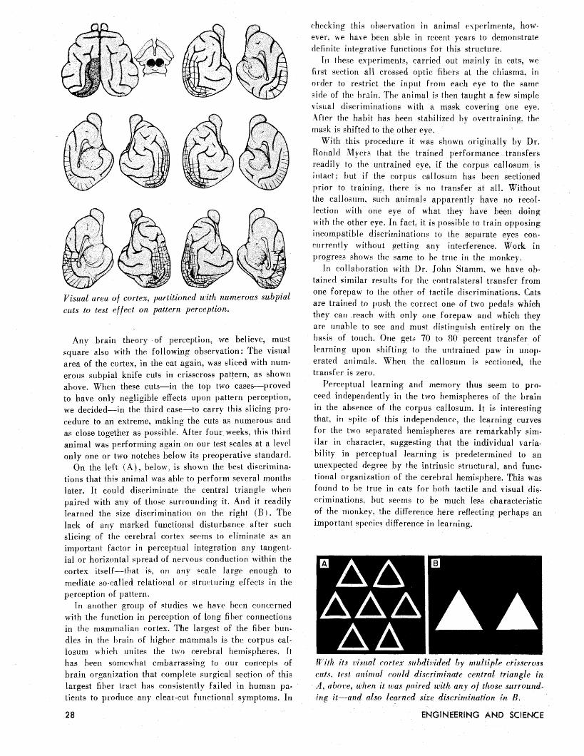
Visual area o f cortex, partitioned with numerous suhpial cu& to test effect on pattern perception.
Any brain theory of perception, we believe, must square also with the following observation: The visual area of the cortex, in the cat again, was sliced with nurn- erous subpial knife cuts in crisscross pattern, as shown above. When these cuts-in the top two cases-proved to have only negligible effects upon pattern perception, we decided-in the third case-to carry this slicing pro- cedure to an extreme, making the cuts as numerous and as close together as possible. After four weeks. this third animal was performing again on our test scales at a level only one or two notches below its preoperative standard.
On the left (A) , below, is shown the best discrimina- tions that this animal was able to perform several months later. It could discriminate the central triangle when paired with any of those surrounding it. And it readily learned the size discrimination on the right ( R >. The lack of any marked functional disturbance after such slicing of the cerebral cortex seems to eliminate as an important factor in perceptual integration any tangent- ial or horizontal spread of nervous conduction within the cortex itself-that is, on any scale large enough to mediate so-called relational or structuring effects in the perception of pattern.
In another group of qtudies we have been roncerried with the function in perception of long fiber connections in the mammalian cortex. The largest of the fiber hurl- dies in the brain of higher mammals is the corpus cal- losum which unites the two cerebral hemispheres. It has been somewhat embarrassing to our concepts of brain organization that complete surgical section of this largest fiber tract has consistently failed in human pa- tients to produce any clear-cut functional symptoms. In
checking this okser~ation in animal experiments, how- ever. we have been able in recent years to demonstrate definite integrative functions for this 'structure.
In these experiments. carried out mainly in cats, we first section all crossed optic fibers at the chiasrna. in order to restrict the input from each eye to the same side of the brain. The animal is then taught a few simple visual discriminations with a mask covering one eye. 4fter the habit has been gtabilized by overtraining, the rriask is shifted to the other eye.
With this procedure it was shown originally by Ronald Myers that the trained performance transfers readily to the untrained eye. if the corpus callosum is intact: but if the corpus callosum has been sectioned prior to training. there is no transfer at all. Without the callosum, such animal'-! apparently have no recol- lection with one eye of what they have been doing with the other eye. In fact, it is possible to train opposing incompatible discriminations to the separate eyes con- current1 y without getting any interference. Work i n progress shows the same to he true in the monkey.
In collaboration with Dr. John Starnni. we have ob- tained similar results for the contralateral transfer from one forepaw to the other of tactile discriminations. Cats are trained to push the correct one of two pedals which they can reach with only one forepaw and which they are unable to see and must distinguish entirely on the basis of touch. One gets 70 to 80 percent transfer of earning upon shifting to the untrained paw in unop- erated animals. When the callosurn is sectioned, the transfer is zero.
Perceptual learning and memory thus seem to pro- ceed independently in the two hemispheres of the brain in the absence of the corpus callosurn. It is interesting that. in spite of this independence, the learning curves for the two separated hemispheres are remarkably sim- ilar in character, suggesting that the individual \aria- bility in perceptual learning is predetermined to an unexpected degree by the intrinsic structural, and func- tional organization of the cerebral hemisphere. This was found to he true in cats for hoth tactile and visual dis- criminations. but seems to be much less characteristic of the monkey, h e difference here reflecting perhaps an important species difference in learning.
With its 1?i,sz1a/ cortex subdivided by multiple crisscross cuts, test animal could discriminate central triangle in A, above, when it was paired with any of those surround- ing b a n d also learned size discrimination i n B.

4lttw-1~1ts to localize in the brain the memory traces for particular habit5 have generally failed. The memory traces. or aigrams, appear to be extremely elusive and diffuse and so far have not been specificallj localized or demonstrated. In the case of the memory traces in- grained for the visual discriminations in the foregoing ex- periments. it was j~o>sible to shon that they are not con- fined to the directly trained hemisphere. One can remove the visual and the neighboring association cortex on the trained side in these animals before switching the mask. and still get the transfer to the untrained eye through the caHosum. Further, one can still get this transfer even if the entire calJosum is sectioned after the completion of training, but before testing for the transfer. Some kind of mnemonic; carryover into ihe oppos-ite hemisphere is fi idently effected via the corpub callosum.
At the present time we are invcstigatiiig the functionaJ capacities of small islands of cerebral cortex. In these studies ti/e put to use the above-mentioned functional independence of the two hemispheres in what we have come to call the "split-brain preparation." This is an animal in which the brain has been split down the mid- die by sectioning the corpus; callosuni. hippocampal corn- misf-ure and the optic chiasnia and. frequently aLo, sowe of the lower-level connertirig systems. To casual exam- ination, these split-brain animals after recovery are indistinguishable from normal in their general cage behavior,
In such animals the brain-le3ion analyses can be car- ried out in one hemisphere alone; the other hemisphere being kept intact to maintain generalized background functions. In the test hemisphere, instead of the custom- ary small lesions in the critical area. it becomes possible in f-uch preparations to use the opposite approach - that is. to remove the greater part of the cortex and to leave intact only the small critical area, the functions of
Removal o f non-visual cortex with preservation o/ visual area aboIi~h(>~*>Â visual functions for reasons still unde- termiiied.
Small island of intact cortex retains capacity to reinem- her and to learn new tactile discriminations almost us well as the whole heniispher~.
which one wishes to investigate. F o what extent would visual perception be possible,
for example, if all parts of the cerebral cortex were removed excepting just the visual area itself? W e have found that vision is practically absent on the test side when the visual area is isolated in cats to the degree shown in the drawing below.
If the non-vibual parts are removed in two or three beparate operations, starting with the cortex immedi- ately surrounding the sector to be preserved, it is not until the final removal of frontal or temporal lobes, as the case may he, that we get the really severe visual impairment.
Similar isolation of the cortical area for touch per- ception, as shown above, has yielded quite different results. In this case the cats, after operation, are still able to perform, at a high level, previously-trained tac- tile discriminatio~is. They- also are able to Jean) JICW
discriminations with the isolated area almost as well as with the whole hemisphere. If circumscribed lesions are subsequently placed in the forepaw tactile area in the opposite, intact hemisphere, it is possible to abolish all discrimination with the affected paw without significantly impairing the performance of the pa^\ that is controlled through the isolated remnant of cortex.
It would appear that the processes of cortical integra- tion and reintegration involvpd in the learning and memory of these tactile discriminations are localized within the intact cortical island. Pnder normal condi- tions it is entirely possible that the integrative processes are much more ^vide-spread through the cerebral hemi- sphere, but it is important to know at least that these unknown cerebral mechanisms are of such a naliire that they can be handled with a rather small. isolated sector of the cortex.
This is about where we stand on these projects at the moment. A s can he seen, we are still a very long way from being able to blueprint the circuit diagrams for perpetual integration, learning or memory. &or have we the vaguest notion of the general type of circuits needed. for example, to build into a machine so simple a thing as pain sensation. We don't know enough to say in theory even that it can-or e \e r could-he done.