vol 465 13 may 2010 doi:10.1038/nature08993 letters · products function to couple radical...
TRANSCRIPT
LETTERS
Stepwise [FeFe]-hydrogenase H-cluster assemblyrevealed in the structure of HydADEFG
David W. Mulder1,2, Eric S. Boyd1,2, Ranjana Sarma1,2, Rachel K. Lange1,2, James A. Endrizzi1,2, Joan B. Broderick1,2
& John W. Peters1,2
Complex enzymes containing Fe–S clusters are ubiquitous in nature,where they are involved in a number of fundamental processesincluding carbon dioxide fixation, nitrogen fixation and hydrogenmetabolism1,2. Hydrogen metabolism is facilitated by the activity ofthree evolutionarily and structurally unrelated enzymes: the [NiFe]-hydrogenases, [FeFe]-hydrogenases and [Fe]-hydrogenases3,4 (Hmd).The catalytic core of the [FeFe]-hydrogenase (HydA), termed theH-cluster, exists as a [4Fe–4S] subcluster linked by a cysteine thiolateto a modified 2Fe subcluster with unique non-protein ligands5,6. The2Fe subcluster and non-protein ligands are synthesized by the hydro-genase maturation enzymes HydE, HydF and HydG; however, themechanism, synthesis and means of insertion of H-cluster compo-nents remain unclear7–10. Here we show the structure of HydADEFG
(HydA expressed in a genetic background devoid of the active siteH-cluster biosynthetic genes hydE, hydF and hydG) revealing thepresence of a [4Fe–4S] cluster and an open pocket for the 2Fe sub-cluster. The structure indicates that H-cluster synthesis occurs in astepwise manner, first with synthesis and insertion of the [4Fe–4S]subcluster by generalized host-cell machinery11,12 and then withsynthesis and insertion of the 2Fe subcluster by specialized hydE-,hydF- and hydG-encoded maturation machinery7–10. Insertion of the2Fe subcluster presumably occurs through a cationically chargedchannel that collapses following incorporation, as a result of confor-mational changes in two conserved loop regions. The structure,together with phylogenetic analysis, indicates that HydA emergedwithin bacteria most likely from a Nar1-like ancestor lacking the2Fe subcluster, and that this was followed by acquisition in severalunicellular eukaryotes.
The biosynthesis and assembly of active-site metallo-cofactorsrequires multiple enzymes, scaffolds and carriers2,11,13. For [FeFe]-hydrogenases, the gene products HydE, HydF and HydG are requiredfor the maturation of the active-site H-cluster14 (Fig. 1). These geneproducts function to couple radical S-adenosyl-L-methionine (SAM)chemistry and nucleotide binding and hydrolysis to ligand synthesis,cluster assembly and insertion, and, ultimately, [FeFe]-hydrogenasematuration7–9,15,16. Although several plausible schemes have been pro-posed for the generation of the carbon monoxide, cyanide and dithio-late ligands at the Fe site, including radical SAM-mediated sulphurinsertion coupled to the decomposition or condensation of aminoacids7–9,17,18, the precise mechanism by which the various enzymes, scaf-folds and carriers coordinate H-cluster maturation is unknown. Owingto their high catalytic rates of hydrogen production, much interestsurrounds [FeFe]-hydrogenases as alternative biological catalysts tothose containing precious metals such as platinum in hydrogen-fuel-cell technology. Advancements in understanding how the H-cluster issynthesized by HydE, HydF and HydG could contribute significantly toboth the genetic engineering of hydrogen-producing microorganismsand the synthesis of biomimetic hydrogen-production catalysts.
HydF has been shown to transfer a cluster precursor to HydA inthe final stage of [FeFe]-hydrogenase maturation and may act as ascaffold on which an H-cluster precursor is assembled8. Our mostrecent results indicate that the H-cluster maturation machinery (theactivities of HydE, HydF and HydG) is directed at the synthesis ofonly the 2Fe unit of the 6Fe cluster, and that the [4Fe–4S] subclustercan be synthesized independently10. To better understand the syn-thesis and insertion of the individual [4Fe–4S]-subcluster and 2Fe-subcluster components of the H-cluster during [FeFe]-hydrogenasematuration, we determined the X-ray crystal structure of HydADEFG.
We determined the structure of HydADEFG from Chlamydomonasreinhardtii, heterologously expressed in Escherichia coli, by molecularreplacement, using the structure of the [FeFe]-hydrogenase fromClostridium pasteurianum5 (CpI) as a search model, and refined itto a resolution of 1.97 A (Fig. 2a). We focused the study on the[FeFe]-hydrogenase from C. reinhardtii because our complementarybiochemical and spectroscopic analyses examining maturation wereconducted using this enzyme10. In addition, C. reinhardtii HydA is ofbiotechnological interest and no structural information about it yetexists.
The overall structure of C. reinhardtii HydADEFG (Fig. 2a) is similarto that observed for active-site domains of the previously characterized[FeFe]-hydrogenases from C. pasteurianum5 (Fig. 2c) and Desulfovibrio
1Astrobiology Biogeocatalysis Research Center, 2Department of Chemistry & Biochemistry, Montana State University, Bozeman, Montana 59717, USA.
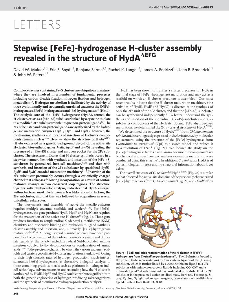
Figure 1 | Ball-and-stick representation of the H-cluster in [FeFe]-hydrogenase from Clostridium pasteurianum29. The H-cluster is bound tothe protein (tube representation) by four cysteine ligands of the [4Fe–4S]subcluster, which is further linked by a cysteine thiolate ligand to a 2Fesubcluster with unique non-protein ligands including CO, CN– and adithiolate ligand30. A water molecule is coordinated to the distal Fe of the 2Fesubcluster in the presumed-active, oxidized state. Dark red, Fe; orange, S;grey, C; blue, N; light red, oxygen; magenta, central atom of the dithiolateligand. Protein Data Bank ID, 3C8Y.
Vol 465 | 13 May 2010 | doi:10.1038/nature08993
248Macmillan Publishers Limited. All rights reserved©2010

desulfuricans6 (DdH) purified from the native hosts. The primary dif-ference in [FeFe]-hydrogenase expressed in a background devoid of thematuration machinery (that is, in HydADEFG) is the absence of the 2Fesubcluster (Figs 2b, d and 3), which leaves an open cavity adjacent to the[4Fe–4S] cluster that is linked to the protein surface through an open,cationically charged channel. The channel is formed by significantstructural rearrangement in two loop regions (loop 1, residues240–255; loop 2, residues 279–286) in HydADEFG, as revealed by thesuperimposition of HydADEFG and CpI (Supplementary Fig. 1). Thesestructural differences indicate that the two loop regions adopt analternative conformation upon insertion of the 2Fe subcluster, effec-tively closing the channel and shielding the active site from surfaceexposure. A sequence alignment of HydA from a diversity of organismsindicates that loop 1 is highly conserved and that loop 2 is partlyconserved in HydA from bacteria and several unicellular eukaryotesincluding C. reinhardtii, but not in eukaryotic Nar1 homologues (Sup-plementary Figs 2 and 3). Nar1 homologues contain only the [4Fe–4S]cluster and are present in the genomes of nearly all eukaryotes, wherethey function in cytosolic and nuclear Fe–S-cluster maturation19.
Although it is clear that an intact 2Fe subcluster is not present atthe active site of HydADEFG (Fig. 3 and Supplementary Fig. 4), ana-lysis of Fo–Fc electron density maps reveals some residual densityadjacent to the [4Fe–4S] cluster where the 2Fe subcluster would beexpected to reside (Fig. 3a and Supplementary Figs 4a and 5a). Toverify the absence of Fe in the residual density, we performed Fe-edgeanomalous-difference Fourier analysis, which confirmed that theonly anomalous scatters at the Fe absorption edge were within the[4Fe–4S] cluster (Fig. 3a and Supplementary Fig. 4a and 5b). Anacetate molecule and a chloride ion, both of which were present inthe crystallization buffer, could be modelled and refined reasonablywell into the unknown density. Thus, the active site seems to exist as a[4Fe–4S] cluster adjoined to an open binding cavity for insertion ofthe 2Fe subcluster.
The overall surface representations of C. reinhardtii HydADEFG
reveal a positively charged channel leading to the active-site area(Fig. 4a–c), a feature that is not present in the intact active hydrogenase,presumably owing to conformational change following insertion of the2Fe subcluster (Fig. 4d). The channel for 2Fe-subcluster insertion issolvent accessible, 8–15 A in width and ,25 A deep, with positively
charged residues (Arg 275, Lys 288 and Lys 409) lining the channelentrance. Lys 188, whose equivalent residue (Lys 358) hydrogen-bondsto a cyanide ligand of the H-cluster in the intact CpI hydrogenase5, liesat the end of the channel at the active cavity and may have a role inorienting the 2Fe subcluster during insertion. Importantly, thebridging cysteine thiolate ligand (Cys 381) between the [4Fe–4S] clus-ter and 2Fe subcluster is located ,19 A into the channel, and its sul-phur side chain is exposed on the surface of the channel, providing thesite for covalent attachment of the 2Fe subcluster following insertion.
The structure of HydADEFG presented here provides strong sup-port for a stepwise mechanism for H-cluster biosynthesis in which[4Fe–4S]-subcluster insertion precedes 2Fe-subcluster insertion.Previously we showed that the [4Fe–4S]-subcluster synthesis and 2Fe-subcluster synthesis occur independently10. Here we show that the[4Fe–4S] subcluster makes up part of a binding cavity that is linkedto a channel leading to the surface of the protein. If the [4Fe–4S] sub-cluster were absent, the binding cavity would not exist. Furthermore,because the [4Fe–4S] subcluster is at the base of the channel, it would beimpossible to insert it after insertion of the 2Fe subcluster as the path-way would effectively be blocked. In addition, if the process were con-served through all organisms, alternative paths for insertion of the[4Fe–4S] cluster would be precluded in the majority of organisms thatexpress [FeFe]-hydrogenases with accessory Fe–S-cluster domainsadjacent to the [4Fe-4S] subcluster of the H-cluster. Therefore, thestructural observations from HydADEFG indicate that the [4Fe–4S]subcluster must be synthesized and inserted first, by Fe–S-clustergeneralized host-cell machinery11,12, and that this is followed by thesynthesis and insertion of the 2Fe subcluster by Hyd-protein matura-tion machinery7–10.
The implied structural rearrangement that occurs during 2Fe-subcluster insertion reveals an interesting parallel to the maturationof the nitrogenase complex. Molybdenum nitrogenase comprises aniron protein (NifH) and a MoFe-protein (NifDK), with the lattercontaining P-clusters (8Fe–7S) and FeMo-cofactors (Mo–7Fe–9S–X–homocitrate, where X 5 C, O, N)20,21. Structural determinationof a form of Azotobacter vinelandii molybdenum nitrogenase (Av1)NifDK deficient in FeMo-cofactor (Av1DnifB) reveals major structuralrearrangement in the domain involved in FeMo-cofactor binding,resulting in channel formation to the FeMo-cofactor site in Av1DnifB
a b
c d
Figure 2 | X-ray crystal structure ofC. reinhardtii HydADEFG determined to aresolution of 1.97 A, compared with HydA fromCpI. a, Ribbon diagram of the overall HydADEFG
structure, with a space-filling representation ofthe associated [4Fe–4S] cluster. The twoconserved loop regions thought to undergomajor conformational rearrangement arecoloured green. b, HydADEFG active-site area,where a 2Fe subcluster recipient cavity is adjacentto the [4Fe–4S] cluster. c, Ribbon diagram of theoverall CpI structure in the same orientation asHydADEFG, with a space-filling representation ofthe intact H-cluster. The regions of CpIcorresponding to the loop regions of HydADEFG
shown in a are coloured green. d, CpI active-siteregion, with ball-and-stick representation of theH-cluster. Protein representations are colouredaccording to secondary structure (light blue,a-helices and loops; violet, b-sheets). The atomiccolouring scheme is the same as in Fig. 1.
NATURE | Vol 465 | 13 May 2010 LETTERS
249Macmillan Publishers Limited. All rights reserved©2010

(ref. 22; Fig. 4e–g). Superimposition of Av1 NifDK (containingFeMo-cofactor; ref. 20) and Av1DnifB NifDK (lacking FeMo-cofactor;ref. 22) indicates that the FeMo-cofactor site is at the end of a cationicchannel (Fig. 4f, g) that is absent from the structure of Av1 NifDK(Fig. 4h). The structural rearrangements resulting in the formation ofa cationic channel in both HydA and NifDK indicate that the processfor complex Fe–S-cluster insertion into apoproteins may be con-served and that the evolution of functional HydA may have occurredstepwise, a feature that is consistent with the evolution of nitrogen-ase23 and possibly Fe–S enzymes in general.
Phylogenetic analysis indicates that HydA and Nar1 homologuesform two separate stem lineages (Supplementary Figs 2 and 3), one ofwhich comprises Nar1 lineages from eukaryotic genomes that lack hydE,hydF and hydG, and the other of which comprises well-supportedlineages of HydA from bacteria and several eukaryotic genomes thatgenerally contain hydE, hydF and hydG. The earliest-branching HydAlineages are from bacteria with HydA from C. reinhardtii andTrichomonas vaginalis nested among bacterial sequences, suggesting
that these HydA derive from lateral gene transfer from a bacteriumand/or endosymbiosis of a bacterium24. Collectively, these results indi-cate that the origin of the 2Fe subcluster containing HydA post-dates thedivergence of bacteria and archaea, a proposal that is consistent with theabsence of HydA from archaea. Consistent with previous results24,25, thisset of observations implies that Nar1 was acquired in eukarya by meansof endosymbiosis of, or lateral gene transfer with, a bacterium24. Thisevent is likely to have occurred before the recruitment of hydE, hydF and
a
A376
C377
M375 A54P53
C381
C130
C185
C129
C499
b
A498
M497
S232
G418
P231
C503
C300
C355
K358
Q325
S323
C299M353
K188
M183
S153
Q155
P154
P324
F417
Figure 3 | Active-site comparison between C. reinhardtii HydADEFG andHydA from CpI. a, The [4Fe–4S]-cluster active-site environment inHydADEFG. The anomalous-difference Fourier map (blue) is showncontoured at 4.5s, indicating the positions of the Fe atoms localized to the[4Fe–4S] cluster. An acetate molecule and a chloride ion are modelled into theFo–Fc map (magenta, contoured at 3.5s) of the HydADEFG cavity. Residues arelabelled according to single-letter amino-acid abbreviations and sequencenumber. The side chain of Cys 129 has increased conformational freedom andcan be refined in three conformations (one shown). b, H-cluster active-siteenvironment in CpI, shown in the same orientation as HydADEFG in a. Theatomic colouring scheme is the same as in Fig. 1.
c
e
g h
f
a
d
b
Figure 4 | Channels for insertion into hydrogenase and nitrogenase duringcomplex Fe–S-cluster assembly. a, Surface representation of HydADEFG (grey)superimposed on native CpI (green ribbon and ball and stick representations).b, HydADEFG channel leading to the active-site 2Fe-subcluster recipient cavity.c, d, Electrostatic surface representations of HydADEFG (c) and the catalyticdomain of CpI (d), calculated using the PYMOL plug-in APBS. Red, negative(210kBT/e); blue, (10kBT/e); kB, Boltzmann’s constant; e, elementary charge.e, Surface representation of the FeMo-cofactor-deficient form of NifDK(Av1DnifB) (grey; PDB ID, 1L5H; ref. 22) superimposed on NifDK from nativenitrogenase MoFe-protein (Av1) (PDB ID, 3MIN; ref. 20) (green ribbon andball and stick representations). f, Av1DnifB NifDK channel leading to the active-site FeMo-cofactor recipient cavity. g, h, Electrostatic surface representationsof Av1DnifB NifDK (g) and Av1 NifDK (h) calculated with identical APBSparameters as in c and d and represented using the same colouring scheme. Theatomic colouring scheme is the same as in Fig. 1 and the unknown atom (X) ofthe FeMo-cofactor in f is blue.
LETTERS NATURE | Vol 465 | 13 May 2010
250Macmillan Publishers Limited. All rights reserved©2010

hydG and the development of the ability to generate the 2Fe subcluster, ahypothesis that is supported by the lack both of conservation in Nar1loop regions and of hydE, hydF and hydG in eukaryotes with Nar1 (seeSupplementary Information for further discussion of this).
The structure of HydADEFG presented here reveals the stepwiseassembly of the H-cluster in [FeFe]-hydrogenases and the structuralpathway for 2Fe-subcluster insertion in the final step of [FeFe]-hydrogenase maturation. By providing significant insights intoH-cluster biosynthesis, the results provide a foundation for enhancingmechanisms of biological hydrogen production in genetically engi-neered hydrogenases, for the synthesis of biomimetic catalysts and,thus, ultimately for developing hydrogen as a renewable fuel. In addi-tion, this structure reveals several novel and unifying themes for Fe–S-enzyme biosynthesis and evolution. Both the FeMo-cofactors ofNifDK and the 2Fe subcluster of the H-cluster of HydA are synthesizedon specialized scaffold proteins and are inserted into the respectiveenzymes through a conserved, cationically charged channel, resultingin catalytically active proteins7–9,13,26. Recent evolutionary studies ofproteins involved in synthesizing the FeMo-cofactor of nitrogenaseindicate that, like the 2Fe subcluster of HydA, the FeMo-cofactor inthe active site of NifDK was not present in the last universal commonancestor and is thus a more recent innovation23. Therefore, the step-wise evolution of complex Fe–S metalloenzymes may be pervasive inbiology, resulting in protein complexes with improved specificity and/or enhanced catalytic efficiency.
METHODS SUMMARYHydADEFG from C. reinhardtii was heterologously expressed in E. coli and purified
as described previously10. Crystal screens were set up in a nitrogen-atmosphere
glovebox (Unilab, MBRAUN), using the anaerobic microcapillary batch diffusion
method27. Favourable conditions for crystal growth at room temperature (298 K)
were found using 25.5% polyethylene glycol 8000 as a precipitate, 0.085 M sodium
cacodylate (pH 6.5), 0.17 M sodium acetate trihydrate and 1 mM dithionite. The
data were collected from a single flash-cooled crystal on beamline 9-1 (wavelength,
0.954 A; resolution, 1.97 A) and beamline 9-2 (wavelength, 1.74 A; resolution,
3.00 A) at the Stanford Synchrotron Radiation Lightsource (SSRL; Supplemen-
tary Table 1). The structure of HydADEFG was solved using molecular replacement
with CpI as a search model (PDB ID, 1FEH; ref. 5), and the final model was refined
using data at 1.97 A to Rfactor 5 17.0% and Rfree 5 21.6%. Homologues of
HydADEFG were compiled using BLASTP, and phylogenetic analyses were con-ducted using Bayesian inference and maximum-likelihood approaches. Figures
were prepared using PYMOL28.
Full Methods and any associated references are available in the online version ofthe paper at www.nature.com/nature.
Received 21 October 2009; accepted 5 March 2010.Published online 25 April 2010.
1. Drennan, C. L. & Peters, J. W. Surprising cofactors in metalloenzymes. Curr. Opin.Struct. Biol. 13, 220–226 (2003).
2. Fontecilla-Camps, J. C., Amara, P., Cavazza, C., Nicolet, Y. & Volbeda, A.Structure-function relationships of anaerobic gas-processing metalloenzymes.Nature 460, 814–822 (2009).
3. Vignais, P. M. & Billoud, B. Occurrence, classification, and biological function ofhydrogenases: an overview. Chem. Rev. 107, 4206–4272 (2007).
4. Shima, S. & Thauer, R. K. A third type of hydrogenase catalyzing H2 activation.Chem. Rec. 7, 37–46 (2007).
5. Peters, J. W., Lanzilotta, W. N., Lemon, B. J. & Seefeldt, L. C. X-ray crystal structureof the Fe-only hydrogenase (Cpl) from Clostridium pasteurianum to 1.8 angstromresolution. Science 282, 1853–1858 (1998).
6. Nicolet, Y., Piras, C., Legrand, P., Hatchikian, C. E. & Fontecilla-Camps, J. C.Desulfovibrio desulfuricans iron hydrogenase: the structure shows unusualcoordination to an active site Fe binuclear center. Structure 7, 13–23 (1999).
7. Nicolet, Y. et al. X-ray structure of the [FeFe]-hydrogenase maturase HydE fromThermotoga maritima. J. Biol. Chem. 283, 18861–18872 (2008).
8. McGlynn, S. E. et al. HydF as a scaffold protein in [FeFe] hydrogenase H-clusterbiosynthesis. FEBS Lett. 582, 2183–2187 (2008).
9. Pilet, E. et al. The role of the maturase HydG in [FeFe]-hydrogenase active sitesynthesis and assembly. FEBS Lett. 583, 506–511 (2009).
10. Mulder, D. W. et al. Activation of HydADEFG requires a preformed [4Fe-4S]cluster. Biochemistry 48, 6240–6248 (2009).
11. Lill, R. Function and biogenesis of iron-sulphur proteins. Nature 460, 831–838 (2009).12. Johnson, D. C., Dean, D. R., Smith, A. D. & Johnson, M. K. Structure, function, and
formation of biological iron-sulfur clusters. Annu. Rev. Biochem. 74, 247–281 (2005).13. Schwarz, G., Mendel, R. R. & Ribbe, M. W. Molybdenum cofactors, enzymes and
pathways. Nature 460, 839–847 (2009).14. Posewitz, M. C. et al. Discovery of two novel radical S-adenosylmethionine
proteins required for the assembly of an active [Fe] hydrogenase. J. Biol. Chem.279, 25711–25720 (2004).
15. Rubach, J. K., Brazzolotto, X., Gaillard, J. & Fontecave, M. Biochemicalcharacterization of the HydE and HydG iron-only hydrogenase maturationenzymes from Thermatoga maritima. FEBS Lett. 579, 5055–5060 (2005).
16. Brazzolotto, X. et al. The [Fe-Fe]-hydrogenase maturation protein HydF fromThermotoga maritima is a GTPase with an iron-sulfur cluster. J. Biol. Chem. 281,769–774 (2006).
17. McGlynn, S. E., Mulder, D. W., Shepard, E. M., Broderick, J. B. & Peters, J. W.Hydrogenase cluster biosynthesis: organometallic chemistry nature’s way. DaltonTrans. 22, 4274–4285 (2009).
18. Peters, J. W., Szilagyi, R. K., Naumov, A. & Douglas, T. A radical solution for thebiosynthesis of the H-cluster of hydrogenase. FEBS Lett. 580, 363–367 (2006).
19. Balk, J., Pierik, A. J., Netz, D. J., Muhlenhoff, U. & Lill, R. The hydrogenase-likeNar1p is essential for maturation of cytosolic and nuclear iron-sulphur proteins.EMBO J. 23, 2105–2115 (2004).
20. Peters, J. W. et al. Redox-dependent structural changes in the nitrogenaseP-cluster. Biochemistry 36, 1181–1187 (1997).
21. Einsle, O. et al. Nitrogenase MoFe-protein at 1.16 A resolution: a central ligand inthe FeMo-cofactor. Science 297, 1696–1700 (2002).
22. Schmid, B. et al. Structure of a cofactor-deficient nitrogenase MoFe protein.Science 296, 352–356 (2002).
23. Fani, R., Gallo, R. & Lio, P. Molecular evolution of nitrogen fixation: the evolutionaryhistory of the nifD, nifK, nifE, and nifN genes. J. Mol. Evol. 51, 1–11 (2000).
24. Meyer, J. [FeFe] hydrogenases and their evolution: a genomic perspective. Cell.Mol. Life Sci. 64, 1063–1084 (2007).
25. Hug, L. A., Stechmann, A. & Roger, A. J. Phylogenetic distributions and histories ofproteins involved in anaerobic pyruvate metabolism in eukaryotes. Mol. Biol. Evol.27, 311–324 (2010).
26. Rubio, L. M. & Ludden, P. W. Biosynthesis of the iron-molybdenum cofactor ofnitrogenase. Annu. Rev. Microbiol. 62, 93–111 (2008).
27. Georgiadis, M. M. et al. Crystallographic structure of the nitrogenase iron proteinfrom Azotobacter vinelandii. Science 257, 1653–1659 (1992).
28. DeLano, W. L. PyMOL Molecular Viewer Æhttp://www.pymol.orgæ (2002).29. Pandey, A. S., Harris, T. V., Giles, L. J., Peters, J. W. & Szilagyi, R. K.
Dithiomethylether as a ligand in the hydrogenase H-cluster. J. Am. Chem. Soc. 130,4533–4540 (2008).
30. Silakov, A., Wenk, B., Reijerse, E. & Lubitz, W. 14N HYSCORE investigation of theH-cluster of [FeFe] hydrogenase: evidence for a nitrogen in the dithiol bridge.Phys. Chem. Chem. Phys. 11, 6592–6599 (2009).
Supplementary Information is linked to the online version of the paper atwww.nature.com/nature.
Acknowledgments This work was supported by a US Air Force Office of ScientificResearch Multidisciplinary University Research Initiative Award(FA9550-05-01-0365, J.W.P.) and the NASA Astrobiology Institute (NAI)-fundedAstrobiology Biogeocatalysis Research Center (NNA08C-N85A, J.B.B. andJ.W.P.). E.S.B. was supported by a NAI postdoctoral fellowship. Portions of thisresearch were carried out at the Stanford Synchrotron Radiation Lightsource(SSRL), a national user facility operated by Stanford University on behalf of the USDepartment of Energy, Office of Basic Energy Sciences. The SSRL StructuralMolecular Biology programme is supported by the US Department of Energy,Office of Biological and Environmental Research, the US National Institutes ofHealth, National Center for Research Resources, Biomedical Technologyprogramme, and the US National Institute of General Medical Sciences.
Author Contributions The structural work was conducted by D.W.M. withcontributions from R.S. and J.A.E. E.S.B. led the phylogenetic work withcontributions from R.K.L. J.W.P. supervised the work with assistance from J.B.B.D.W.M., E.S.B. and J.W.P. led the manuscript preparation with contributions fromJ.B.B., R.S., R.K.L. and J.A.E.
Author Information Coordinates and structure factors of C. reinhardtii HydADEFG
have been deposited in the Protein Data Bank under the accession code 3LX4.Reprints and permissions information is available at www.nature.com/reprints.The authors declare no competing financial interests. Correspondence andrequests for materials should be addressed to J.W.P.([email protected]).
NATURE | Vol 465 | 13 May 2010 LETTERS
251Macmillan Publishers Limited. All rights reserved©2010
METHODSStructure determination and refinement. HydADEFG from C. reinhardtii was
heterologously expressed in E. coli and purified under strict anaerobic conditions
as described previously10. Before crystallization, purified HydADEFG was diluted
over a Sephadex G-25 column (GE Healthcare) in 50 mM Tris buffer (pH 7.8)
with 300 mM NaCl, 20% glycerol and 1 mM dithionite, to a final concentration
of 28 mg ml21. Crystals were obtained at room temperature (298 K) by means of
the anaerobic microcapillary batch diffusion method27 in a nitrogen-atmosphere
glovebox (Unilab, MBRAUN), using 25.5% polyethylene glycol 8000 as precipi-
tate and 0.085 M sodium cacodylate (pH 6.5) with 0.17 M sodium acetate trihy-drate and 1 mM dithionite.
The initial data were collected from a single flash-cooled crystal on beamline
9-1 at the SSRL, with a continuous flow of liquid nitrogen at 100 K, and a single-
wavelength data set (wavelength, 0.954 A) was collected up to a resolution of
1.97 A (Supplementary Table 1). An additional single anomalous data set at the
Fe edge (wavelength, 1.74 A) was collected at a later time using the same crystal
on beamline 9-2 at the SSRL, up to a resolution of 3.00 A (Supplementary Table
1). The data at 1.97 A were processed and scaled using DENZO and SCALPACK
of the HKL-2000 software package (version 1.98.7)31.
The structure was solved by molecular replacement using AutoMR of the
CCP4 suite of programs (version 6.0)32 with CpI (PDB ID, 1FEH; ref. 5) as a
search model. The solution was subjected to rigid-body refinement in REFMAC5
(version 5.2.0019)33 and further improved using ARP/wARP (version 7.0)34. The
maps obtained were solvent-flattened and histogram-averaged using RESOLVE
(version 2.10)35. Model-building was subsequently completed manually using
COOT (version 0.3.3)36 with subsequent refinement (REFMAC5) using NCS
and B-factor restraints. This resulted in the determined structure. The final
model was refined on the data at 1.97 A to Rfactor 5 17.0% and Rfree 5 21.6%(Supplementary Table 1). We could not determine the positions of residues
1–24, 116–120, 201–203, 312–322 and 451–457, owing to disorder. Also, the
side-chain sulphur atom of Cys 129 could be modelled and refined in three
different conformational states. In the final model, 98.4% of all residues were
in favoured regions and 100% of all residues were in allowed regions of the
Ramachandran plot (calculated with MOLPROBITY (version 3.17)37).
Because of undetermined density adjacent to the [4Fe–4S]-cluster site, an
additional single anomalous data set at the Fe edge (wavelength, 1.74 A), at a
resolution of 3.00 A, was used to confirm the Fe positions in the determined
structure. The data were processed as describe above and Fe positions deter-
mined using SOLVE (version 2.13)38. The native and anomalous reflection data
was merged using CAD in CCP4 and a difference Fourier synthesis between the
native model phases and anomalous scattering was calculated using FFT39 in
CCP4.
Phylogenetic analysis. BLASTP was used to compile all HydA, HydE, HydF,
HydG and HydA-homologue deduced amino-acid sequences from genomic
sequences using the DOE-IMG and the NCBI Genome Blast servers
(Supplementary Table 2). A total of 435 HydA and HydA-homologue sequenceswere compiled and these were aligned using CLUSTALX (version 2.0.8) with the
Gonnet 250 protein matrix and default gap extension and opening penalties40.
The alignment was scrutinized and manually aligned using known catalytic
residues24. A single maximum-likelihood phylogenetic tree was computed
with PHYML (version 3.0)41 using the LG substitution matrix42 (Sup-
plementary Fig. 2). This tree was used to empirically select 90 HydA sequences
that represented the primary lineages. Sequences were trimmed to contain only
the H-cluster domain present in HydADEFG in C. reinhardtii24. The trimmed
sequences were realigned using CLUSTALX as described above.
PROTTEST (version 2.0)43 was used to select the WAG1I1G as the best-fit
protein evolutionary model. The phylogeny of each locus was evaluated using
PHYML with the WAG evolutionary model with a proportion of invariable sites
and gamma-distributed rate variation (I1G). A composite phylogram was con-
structed from 100 bootstrap replicate phylograms and the tree was projected
using FIGTREE (version 1.2.2) (http://tree.bio.ed.ac.uk/software/figtree)
(Supplementary Fig. 3). Similarly, the phylogeny of each locus was evaluated
using MRBAYES (version 3.1.2)44,45. A composite phylogram was constructed
using the WAG evolutionary model with gamma-distributed rate variation with
a proportion of invariable sites (I1G). Using MRBAYES, we sampled tree topo-
logies at likelihood stationary during two separate runs every 500 generations
over 3.7 3 106 generations, with a ‘burnin’ parameter of 2.0 3 106 (standard
deviation of split trees was ,0.07).
31. Otwinowski, Z. & Minor, W. Processing of X-ray diffraction data collected inoscillation mode. Methods Enzymol. 276, 307–326 (1997).
32. Collaborative. Computation Project, Number 4. The CCP4 suite: programs forprotein crystallography. Acta Crystallogr. D 50, 760–763 (1994).
33. Murshudov, G. N., Vagin, A. A. & Dodson, E. J. Refinement of macromolecularstructures by the maximum-likelihood method. Acta Crystallogr. D 53, 240–255(1997).
34. Perrakis, A., Morris, R. & Lamzin, V. S. Automated protein model buildingcombined with iterative structure refinement. Nature Struct. Biol. 6, 458–463(1999).
35. Terwilliger, T. C. SOLVE and RESOLVE: automated structure solution and densitymodification. Methods Enzymol. 374, 22–37 (2003).
36. Emsley, P. & Cowtan, K. Coot: model-building tools for molecular graphics. ActaCrystallogr. D 60, 2126–2132 (2004).
37. Davis, I. W. et al. MolProbity: all-atom contacts and structure validation forproteins and nucleic acids. Nucleic Acids Res. 35, W375–W383 (2007).
38. Terwilliger, T. C. & Berendzen, J. Automated MAD and MIR structure solution.Acta Crystallogr. D 55, 849–861 (1999).
39. Ten Eyck, L. F. Crystallographic fast Fourier transforms. Acta Crystallogr. A 29,183–191 (1973).
40. Larkin, M. A. et al. Clustal W and Clustal X version 2.0. Bioinformatics 23,2947–2948 (2007).
41. Guindon, S. & Gascuel, O. A simple, fast, and accurate algorithm to estimate largephylogenies by maximum likelihood. Syst. Biol. 52, 696–704 (2003).
42. Le, S. Q. & Gascuel, O. An improved general amino acid replacement matrix. Mol.Biol. Evol. 25, 1307–1320 (2008).
43. Abascal, F., Zardoya, R. & Posada, D. ProtTest: selection of best-fit models ofprotein evolution. Bioinformatics 21, 2104–2105 (2005).
44. Ronquist, F. & Huelsenbeck, J. P. MrBayes 3: Bayesian phylogenetic inferenceunder mixed models. Bioinformatics 19, 1572–1574 (2003).
45. Huelsenbeck, J. P. & Ronquist, F. MRBAYES: Bayesian inference of phylogenetictrees. Bioinformatics 17, 754–755 (2001).
doi:10.1038/nature08993
Macmillan Publishers Limited. All rights reserved©2010