vps factors are required for efficient transcription - genetics
TRANSCRIPT
1
Vps factors are required for efficient transcription elongation in budding
yeast
Naseem A. Gaur1,4, Jiri Hasek2, Donna Garvey Brickner3, Hongfang Qiu1, Fan Zhang1, Chi-Ming
Wong1,5, Ivana Malcova2, Pavla Vasicova2, Jason H. Brickner3, and Alan G. Hinnebusch1*
1Laboratory of Gene Regulation and Development, Eunice K. Shriver National Institute of Child
Health and Human Development, National Institutes of Health, Bethesda, Maryland 20892;
2Laboratory of Cell Reproduction, Institute of Microbiology, Academy of Sciences of the Czech
Republic, The Czech Republic
3Department of Molecular Biosciences, Northwestern University, Evanston, IL 60208
4Current address: Synthetic Biology and Biofuel Group, International Centre For Genetic Engineering and Biotechnology (ICGEB),
Aruna Asaf Ali Marg, New Delhi-110067
5Current address: Department of Medicine, The University of Hong Kong, Hong Kong
Genetics: Early Online, published on January 18, 2013 as 10.1534/genetics.112.146308
Copyright 2013.

2
There is increasing evidence that certain Vps proteins, factors that mediate vesicular
protein trafficking, have additional roles in regulating transcription factors at the
endosome. We found that yeast mutants lacking the PI(3)P kinase Vps34 or its associated
protein kinase Vps15 display multiple phenotypes indicating impaired transcription
elongation. These phenotypes include reduced mRNA production from long or G+C-rich
coding sequences (CDS) without affecting the associated GAL1 promoter activity, and a
reduced rate of RNA polymerase II (Pol II) progression through lacZ CDS in vivo.
Consistent with reported genetic interactions with mutations affecting the histone
acetyltransferase complex NuA4, vps15Δ and vps34Δ mutations reduce NuA4 occupancy in
certain transcribed CDS. vps15Δ and vps34Δ mutants also exhibit impaired localization of
the induced GAL1 gene to the nuclear periphery. We found unexpectedly that, similar to
known transcription elongation factors, these and several other Vps factors can be cross-
linked to the CDS of genes induced by Gcn4 or Gal4 in a manner dependent on
transcriptional induction and stimulated by Cdk7/Kin28-dependent phosphorylation of the
Pol II CTD. We also observed co-localization of a fraction of Vps15-GFP and Vps34-GFP
with nuclear pores at nucleus-vacuole (NV) junctions in live cells. These findings suggest
that Vps factors enhance the efficiency of transcription elongation in a manner involving
their physical proximity to nuclear pores and transcribed chromatin.
3
Newly synthesized proteins that are transported from the Golgi to the lysosome/vacuole
traverse the endosome, as do ubiquitinated proteins that are removed from the plasma membrane
by endocytosis en route to the vacuole for degradation. Ubiquitinated cargo proteins progress
through early and late endosomes, are concentrated at the outer membranes of multivesicular
bodies (MVB), and are then sequestered in intralumenal vesicles (ILVs) of the MVB. Fusion of
the MVB with the vacuole delivers cargo proteins to the vacuole lumen for degradation by
vacuolar hydrolases. “Class C and D” Vps factors participate in vesicle fusion at the endosome,
while cargo sorting and delivery to the ILVs at the MVB involves class E Vps proteins,
including the components of the soluble ESCRT (endosomal sorting complex required for
transport) complexes ESCRT-0, -I, -II, and –III (BOWERS and STEVENS 2005; HURLEY and EMR
2006; RAIBORG and STENMARK 2009).
It is thought that ESCRT-0 is recruited from the cytoplasm to the endosomal outer
membrane by interaction with the phosphoinositide PI(3)P where it acts to recruit and
concentrate ubiquitinated cargo proteins and transfer them to the ESCRT-I complex. ESCRT-I
activates the ESCRT-II heterotrimer that, in turn, recruits the ESCRT-III components, which are
believed to assemble filaments instrumental in invagination of the MVB membrane. The AAA-
ATPase Vps4, recruited by ESCRT-III subunits, functions to pinch off the membrane
invaginations to produce ILVs containing cargo proteins and to recycle the ESCRT factors back
to the cytoplasm (RAIBORG and STENMARK 2009).
There is increasing evidence that certain Vps proteins have additional functions in
cytoplasmic signaling pathways that regulate transcription in the nucleus. In budding yeast,
ESCRT-III factor Snf7/Vps32 and the subunits of ESCRT-II were first identified genetically by

4
their requirements for robust accumulation of SUC2 mRNA (KAMURA et al. 2001; TU et al.
1993; YEGHIAYAN et al. 1995). The transcription factor Rim101 is proteolytically activated on
recruitment to the MVB outer membrane via ESCRT-III factor Snf7/Vps32 to permit expression
of pH-responsive genes (BOYSEN and MITCHELL 2006). The Gα subunit (Gpa1) of a
heterotrimeric G-protein activates the PI 3-kinase Vps34 (a class D Vps factor) at the endosomal
membrane to promote the transcriptional response to mating factors (SLESSAREVA et al. 2006).
Activation of genes for utilization of alternative nitrogen sources by Gln3 is enhanced by Vps
factors, and it appears that Gln3 must traffic in vesicles containing Vps10 between Golgi and
endosome for subsequent nuclear entry (PURIA et al. 2008). Recently, evidence was presented
that the phosphoinositide PI(3,5)P2, produced at the late endosome promotes assembly of a
transcriptional cofactor complex that enhances galactose induction of GAL gene transcription in
the nucleus (HAN and EMR 2011).
We found previously that robust activation of amino acid biosynthetic genes by yeast
transcription factor Gcn4 requires a subset of Vps factors that function at the MVB. The defects
in activation of Gcn4 target genes were most pronounced in mutants lacking certain Vps C or -D
factors, with lesser but still significant defects observed in vps mutants lacking particular ESCRT
proteins, including ESCRT-II factors (Snf8/Vps22, Vps25, or Vps36) and ESCRT-III subunits
Snf7/Vps32 and Vps20. Gcn4 synthesis is induced at the translational level in response to
starvation for any single amino acid (HINNEBUSCH 2005). In the vps mutants, Gcn4 synthesis was
induced properly and Gcn4 could enter the nucleus and bind to upstream activation sequences
(UAS), but did not efficiently stimulate preinitiation complex (PIC) assembly at the promoter
(ZHANG et al. 2008). We hypothesized that a signal transduction pathway operates to dampen the
transcriptional response to amino acid starvation by Gcn4 in response to endosome dysfunction

5
(ZHANG et al. 2008). More recently, evidence was provided that sterol limitation also down-
regulates Gcn4 function in the nucleus, in a manner involving sterol binding protein Kes1 and its
ability to inhibit PI(4)P-directed vesicular protein trafficking with an attendant increase in
cellular sphingolipids. It appears that elevated sphingolipids provoke a reduction in Gcn4
function in a manner involving the CDK8 module of the transcriptional coactivator Mediator
complex (MOUSLEY et al. 2012).
In our previous study, some of the strongest defects in transcriptional activation by Gcn4
were observed in mutants lacking Vps34 and Vps15. Vps34 is the sole kinase in budding yeast
that synthesizes PI(3)P in endomembranes, and Vps15 is a protein kinase associated with Vps34
required for its function (BOWERS and STEVENS 2005). Galactose induction of a GAL1-lacZ
reporter was impaired in vps15Δ and vps34Δ mutants, but not as dramatically as seen for Gcn4-
dependent reporters, suggesting that the function of Gal4, and possibly other transcriptional
activators besides Gcn4, is also down-regulated to a lesser extent in response to endosome
dysfunction. However, as shown below, we found subsequently that PIC assembly and
transcription initiation occurs normally at the GAL1 promoter in vps15Δ and vps34Δ mutants.
Furthermore, vps15Δ and vps34Δ mutations evoked much stronger reductions in expression of
lacZ reporters driven by Gcn4 compared to mRNA transcripts of authentic Gcn4 target genes
(ZHANG et al. 2008). These last observations led us to suspect that the greatly reduced
expression of lacZ reporters observed in vps mutants involved a defect in transcription
elongation in addition to the defective PIC assembly observed specficially for Gcn4 target genes.
Indeed, it is well established by Aguilera and colleagues that efficient elongation through lacZ
coding sequences (CDS) requires a full complement of transcription elongation factors in yeast

6
cells (CHAVEZ et al. 2001;MORILLO-HUESCA et al. 2006)—a fact which has been exploited to
identify novel factors involved in transcription elongation (TOUS et al. 2011).
Accordingly, we set out to investigate whether Vps15, Vps34, and various other soluble Vps
factors are required for efficient transcription elongation in yeast cells. The results presented
below support this possibility, including evidence that the rate of elongation by Pol II through
lacZ coding sequences is reduced in vps15Δ and vps34Δ cells. Our findings further suggest that
reduced co-transcriptional recruitment of the histone acetyltransferase complex NuA4 to CDSs
could be one factor underlying the elongation defect in these mutants. Interestingly, we also
obtained evidence that elimination of Vps15 or Vps34 impairs localization of the GAL1 and
INO1 genes to the nuclear periphery during transcriptional activation. Unexpectedly, using
chromatin immunoprecipitation analysis, we detected cross-linking of these and several other
Vps proteins to the CDSs of various genes in vivo in a manner dependent on transcriptional
activation. Consistent with this, we detected dynamic association of a fraction of GFP-tagged
Vps15 and Vps34 with nuclear pores in live cells under various culture conditions. Together, our
results indicate that inactivation of various Vps proteins reduces the efficiency of transcription
elongation in vivo and raise the possibility that at least a subset of these proteins might stimulate
transcription elongation in a manner involving their physical association with, or proximity to,
transcribed chromatin at the nuclear periphery.
MATERIALS AND METHODS
Yeast strain and plasmid constructions. All strains and plasmids used in this study are listed in
Tables 1 and 2, respectively. Wild-type (WT) strain BY4741 and deletion derivatives thereof

7
were described previously (GIAEVER et al. 2002) and purchased from Research Genetics. The
presence of the reported deletion alleles was confirmed by PCR amplification of genomic DNA
(SWANSON et al. 2003). Strains harboring myc13 epitope tags were generated as described
previously (SWANSON et al. 2003) and verified by PCR analysis of chromosomal DNA and
Western blot analysis with anti-myc antibodies. Strains harboring trp1∆::hisG were generated
using TRP1 knock-out construct pNK1009 as described previously (ALANI et al. 1987).
Strains CRY1605 and CRY1606 were generated by transformation of strains
CRY1541and CRY1581 with plasmid pNSP1-RFP. Strains HQY1584 and HQY1586 were
constructed by transforming strains CRY1541 and CRY1581, respectively, with NurI-digested
pHQ2061, harboring the SPT4-mCherry::hphMX4 cassette, selecting for growth on YPD
containing hygromycin. Replacement of SPT4 with SPT4-mCherry::hphMX4 was confirmed by
PCR analysis of chromosomal DNA using primers 972 and 1167 (Table 3), and by Western blot
analysis using antibodies against mCherry.
NSP1 was tagged at its C-terminus with mTagBFP in strains HQY1584 and HQY1587 to
yield strains CRY1718 and CRY1719, respectively, using a newly constructed tagging plasmid
(I. Malcova, manuscript in preparation). The latter was produced by inserting a XhoI-BamHI
fragment containing the gene encoding yeast enhanced mTagBFP (ytBFP, a kind gift of Dr.
Kaern's laboratory at the University of Ottawa) into pFA6a-natNT2. The ytBFP::natNT2 cassette
was PCR-amplified from the resulting tagging plasmid with primers Nsp1mbfp5 and Nsp1C2
using Phusion DNA polymerase (NEB) and used to transform HQY1584 and HQY1587 by
selecting for growth on YPD medium containing nourseothricin (100 µg/ml, cloNat, Werner
Bioagents, Jena). Correct integration of the cassette was verified by PCR analysis using primers

8
Nsp1diaC and mTAGBFPrev, and the size of the protein fusion was checked by Western blot
analysis using anti-tRFP antibody (Evrogen).
Plasmid pHQ2061 was constructed by PCR amplification from genomic DNA of a
fragment containing the WT SPT4 CDS flanked by HindIII/NruI and BamHI sites using primers
1853/1854 and inserted between the HindIII and BamHI sites of pBS35 (fusing SPT4 CDS in-
frame to mCherry CDS); subsequently a fragment containing the SPT4 3'UTR flanked by SacI-
NruI and SpeI sites was PCR-amplified from genomic DNA using primers 1855/1856 and
inserted between the SacI and SpeI sites of pBS35 to produce pHQ2061. Plasmid pNG18 was
constructed by PCR amplification of a ~0.97 kb URA3 fragment from genomic DNA of WT
strain BY4741by using primer N381/N382. The PCR amplified URA3 fragment was digested
with BamH1/ClaI and cloned into BamH1/ClaI-digested pCM184LAUR.
Strains H1486 (wild type), NGY11 (vps34∆) and NGY12 (vps15∆) were transformed with GFP-
Lac repressor plasmid pAFS144 digested with NheI (Straight et al., 1996). The resulting strains
were transformed with either p6LacO128-GAL1 digested with NruI (Brickner et al., 2007)
or p6LacO128-INO1 digested with StuI (Brickner & Walter, 2004), giving rise to the following
six strains: DBY375 (GAL1:LacO), DBY376 (vps34∆ GAL1:LacO), DBY377 (vps15∆
GAL1:LacO), DBY452 (INO1:LacO), DBY455 (vps34∆ INO1:LacO) and DBY457 (vps15∆
INO1:LacO).
Gene length-dependent accumulation of mRNA (GLAM) assays and Northern analysis.
Measurement of Pho5 enzymatic activity for GLAM ratio determinations was carried out as
described previously using transformants of the appropriate strains harboring plasmids YCplac33
(empty URA3 vector), pSCH202 (PGAL1-PHO5, URA3) and either pSCH209 (PGAL1-PHO5-
LAC4, URA3) or pSCH212 (PGAL1-PHO5-lacZ, URA3) (MORILLO-HUESCA et al. 2006).

9
Isolation of total RNA from yeast and Northern blot analysis were carried out as described
previously (GINSBURG et al. 2009). DNA probes used were a 0.9 kb EcoRV digested PHO5
internal fragment isolated from plasmid pSCH202, or the following PCR fragments amplified
from the genomic DNA of BY4741 using primers listed in Table 3: 0.45 kb of YAT1 CDS, 0.57
kb of LYS2 CDS, 0.4 kb of IMD2 CDS and 0.9 kb of the SCR1 gene. Each DNA fragment was
radiolabeled with [α32P]-dCTP by using the Megaprime DNA labeling system (Amersham).
Analysis of GAL1 positioning in yeast nuclei. Chromatin localization experiments were
performed as described ((BRICKNER et al. 2010) using strains expressing GFP fused to Lac
repressor and the GAL1 gene marked with an array of Lac repressor binding sites. Cells were
stained with antibodies against GFP and Nsp1.
Chromatin immunoprecipitation (ChIP) analysis. Yeast cell cultures (100 ml) at A600 of 0.5
to 0.6 were mixed with 11 ml of formaldehyde solution (50 mM HEPES-KOH [pH 7.5], 1 mM
EDTA, 100 mM NaCl and 11% formaldehyde) and cross-linked for 20 min at room temperature
with intermittent shaking and then quenched with 15 ml 2.5 M glycine. Cells were collected by
centrifugation and washed twice with 100 ml ice cold Tris-buffered saline. The cells were broken
by vortexing with glass beads in 500 µl of FA-lysis buffer (50 mM HEPES-KOH [pH 7.5], 1
mM EDTA, 140 mM NaCl, 1% Triton X-100, 0.1% sodium deoxycholate, and protease
inhibitors). Glass beads were removed from the lysates and washed with 500 µl of FA lysis
buffer, and the resulting 1ml lysates were sonicated to yield DNA fragments of 300–500 bp.
Supernatants containing soluble chromatin were obtained by centrifugation at 13000g and stored
at −80°C. Fifty microliters of chromatin were used for immunoprecipitations, and an identical
10
aliquot was reserved as the “Input” sample. Chromatin was immunoprecipitated using
dynabeads® Pan Mouse IgG (invitrogen) coupled with antibodies described below for 2 h at
4°C, recovered immune complexes were washed once with phosphate-buffered saline (PBS)
containing BSA (5 mg/ml), twice each with FA-lysis buffer, wash-buffer I (50 mM HEPES-
KOH [pH 7.5], 1 mM EDTA, 500 mM NaCl, 1% Triton X-100 and 0.1% Na-deoxycholate) and
wash-buffer II (10 mM Tris-HCl [pH 8.5], 250 mM LiCl, 1 mM EDTA, 0.5% NP-40 and 0.5%
sodium deoxycholate), and once with TE (10 mM Tris-HCl [pH 8.0] and 1 mM EDTA). The
immunoprecipitated complexes were eluted at 65°C for 15 min with 100 µl elution buffer (50
mM Tris-HCl [pH 8.0], 10 mM EDTA and 1% SDS) and for 10 min with 150 µl of elution wash
buffer (50 mM Tris-HCl [pH 8.0], 1 mM EDTA and 0.67% SDS), and the eluates were
combined (IP sample). The matched input and IP samples were incubated overnight at 65°C to
reverse the cross-links. The samples were then treated with proteinase K (Ambion), at 100
µg/250 µl of chromatin, for 2 h, and DNA was extracted twice with phenol:chloroform:isoamyl
alcohol (25:24:1) and once with chloroform:isoamyl alcohol (24:1), ethanol precipitated, and
resuspended in 30 µl TE containing RNase (10 µg/ml). Two microliters of resuspended DNA
from IP and Input samples were used for each PCR reaction in the presence of [33P]-dATP with
the appropriate primers (listed in Table 4). The radiolabeled amplified fragments were resolved
by PAGE and quantified with a phosphorimager. For each primer set employed, we optimized
the PCR conditions to ensure that the amounts of amplified 33P-labeled products being generated
are proportional to the amounts of input DNA over the range of DNA concentrations present in
the IP or Input samples. For the ChIP analysis of Snf7-myc and Snf8-myc in Fig. 7 G-I, and of
Rpb3 in Fig. 3A, POLI CDS were amplified as a negative control in addition to the specific
ARG1 or GAL1 sequences of interest. An intergenic sequence from chromosome V (ChrV-1) was

11
amplified as a negative control for all other ChIP experiments using primers 948/949, with the
exception of the Rpb3 ChIP in Fig. 4 where primers ExtChrV1/ExtChrV2 (ChrV-2) were used
instead. Two Immunoprecipitations were conducted on at least two chromatin samples isolated
from independent cultures and the PCR analysis of IP and Input DNA samples was carried out in
duplicate or triplicate. Ratios of the amounts of PCR fragments for specific to control DNA
sequences generated from IP samples were normalized to the corresponding ratios for Input
samples to yield occupancy values, and mean occupancies were calculated from replicate
experiments.
Antibodies. Unless stated otherwise, 1 µl of the following antibodies was used for each ChIP
assay: Mouse monoclonal anti-Rpb3 (Neoclone), mouse monoclonal anti-myc (Roche) and
mouse anti-Ser5P-Rpb1 (H14 from Covance). For Western blot analysis of myc13-tagged
proteins in whole cell extracts prepared under denaturing conditions (REID and SCHATZ 1982),
the anti-myc antibody was used at the dilution recommended by the vendor.
Live-cell imaging by fluorescence microscopy. Distributions of fusion proteins in living cells
were analyzed with an oil immersion 100x/1.4 objective using the Olympus Cell RTM detection
and analyzing system based on the motorized Olympus IX-81 inverted microscope,
Hammamatsu Orca/ER digital camera and the following highly specific mirror units: (i) EGFP
filter block U-MGFPHQ, excitation (exc.) max. 488 nm, emission (em.) max. 507 nm; (ii) RFP
filter block U-MFRFPHQ, exc. max. 558 nm, em. max. 583 nm; (iii) BFP filter block U-
MFBFPHQ, exc. max. 390 nm, em. max. 460 nm). The Cell RTM system enables us to obtain
12
several optical sections through the cell. Images were processed, merged and analyzed using
Olympus Cell RTM , ImarisTM , NIH ImageJ and Adobe CS5 software. Images from selected
optical layers were presented.
RESULTS
Elimination of Vps15 or Vps34 confers sensitivity to 6-AU and MPA and dampens IMD2
induction by these inhibitors. The strong reductions in lacZ reporter expression observed in
vps15Δ and vps34Δ mutants (ZHANG et al. 2008) led us to suspect that the efficiency of
transcription elongation was impaired in cells lacking these Vps proteins. To gain additional
evidence for a transcription elongation defect, we examined sensitivity of vps15Δ and vps34Δ
cells to the drugs 6-azauracil (6-AU) and mycophenolic acid (MPA). These drugs lower GTP
pools by inhibiting IMP dehydrogenase (IMPDH) encoded by IMD3 and IMD4, which catalyzes
the rate-limiting step in de novo guanine nucleotide biosynthesis. Numerous mutations affecting
transcription elongation factors confer sensitivity to both 6-AU and MPA ((EXINGER and
LACROUTE 1992; JENKS and REINES 2005) and references therein). Interestingly, vps15Δ and
vps34Δ cells display sensitivity to these drugs comparable in degree to that exhibited by mutant
cells lacking the Spt4 subunit of elongation factor DSIF (HARTZOG et al. 1998), and these 6-AUS
and MPAS phenotypes were complemented by the corresponding wild-type (WT) VPS alleles on
plasmids (Fig.1A). Significant, albeit reduced, sensitivity to MPA and 6-AU was also evident in
the pep7Δ/vps19Δ, pep12Δ/vps6Δ, and vps45∆ mutants, and sensitivity to an even smaller degree
was evident in the vps4Δ and snf7Δ/vps32 strains as well (Fig. 1B-C). Like vps15Δ and vps34Δ,
these other five vps mutants are also defective for vesicular protein transport from the Golgi to
the vacuole (BOWERS and STEVENS 2005). Vps10 is an integral membrane protein that functions

13
as the receptor for carboxypeptidase Y to mediate its trafficking from the late Golgi to late
endosome (BOWERS and STEVENS 2005); and it was recently implicated in promoting nuclear
entry of Gln3 (PURIA et al. 2008); however, eliminating the VPS10 gene conferred no sensitivity
to 6-AU or MPA (Fig. 1C).
Sensitivity to 6-AU and MPA does not necessarily indicate a transcription elongation
defect; however, mutations in bona fide elongation factors have been found to reduce
transcriptional induction of IMD2, encoding an MPA-resistant form of IMPDH (MCPHILLIPS et
al. 2004), in response to 6-AU or MPA treatment (RILES et al. 2004). It is significant, therefore,
that the vps34Δ mutation reduces IMD2 mRNA abundance in the presence of 6-AU or MPA to
an extent similar to that given by mutations that eliminate Spt4 or the Thp1 subunit of the
elongation/mRNA export complex TREX-2 (Fig. 1D-E & F-G). Furthermore, complementation
of the IMD2 expression defect in vps34Δ cells by episomal VPS34 was impaired by a mutation
(N736K) that inactivates its PI 3-kinase activity (SLESSAREVA et al. 2006) (Fig. 1D-G). These
findings are consistent with the possibility that inactivation of the PI 3-kinase activity of Vps34
compromises the efficiency of transcription elongation in vivo.
Elimination of Vps15 or Vps34 impairs expression of lacZ reporters. We determined that
vps34Δ and vps15Δ cells display another prominent phenotype of yeast elongation mutants, of
inefficient elongation through the GC-rich or long CDS of bacterial lacZ and fungal LAC4
(CHAVEZ et al. 2001; MORILLO-HUESCA et al. 2006). First, we employed an in vivo reporter
described by Aguilera and colleagues in which a lacZ-URA3 translational fusion is expressed
from the Ptet promoter in ura3 auxotrophic strains. Mutations in known elongation factors
impair expression of the URA3 portion of the reporter and confer poor growth on (-Ura) medium

14
lacking uracil (JIMENO et al. 2002). To control for possible effects of mutations on Ptet promoter
activity, we constructed a matched Ptet-URA3 reporter missing the lacZ CDS and compared
growth of vps mutants harboring the Ptet-lacZ-URA3 versus Ptet-URA3 reporters on –Ura
medium. In the otherwise wild-type ura3 strain, the Ptet-lacZ-URA3 transformants grew more
slowly, and exhibited a lower plating efficiency, on SC-Ura compared to the corresponding Ptet-
URA3 transformants (Fig. 2A, WT, lacZ versus control [C]), indicating reduced expression of
URA3 CDS from the Ptet-lacZ-URA3 reporter. The reduction in plating efficiency of Ptet-lacZ-
URA3 versus Ptet-URA3 transformants was clearly exacerbated in the vps15Δ,
vps34Δ, pep7Δ and pep12Δ mutants, and possibly also in the snf7Δ strain, but not in the
vps4Δ mutant (Fig. 2A, SC-Ura, cf. lacZ and C transformants). These results, combined with the
sensitivity of the vps mutants to 6-AU and MPA shown above, suggest that eliminating particular
Vps proteins reduces the efficiency of transcription elongation, with relatively stronger defects
conferred by the absence of Vps15 or Vps34 compared to other Vps factors, eg. Vps4, that are
equally critical for vesicular protein transport to the vacuole.
To provide additional evidence for elongation defects in these vps mutants, we employed
the “GLAM” (Gene-length-dependent accumulation of mRNA) assay developed by Chavez et al.
involving a PGAL1-PHO5-LAC4 construct harboring LAC4 CDS inserted into the 3’UTR and a
matched PGAL1-PHO5 reporter with the same promoter but without LAC4 CDS. The LAC4
sequences add ~3 kb to the transcript length but are not translated, such that both reporter
transcripts produce the same Pho5 protein. A variety of mutants lacking known elongation
factors express reduced steady-state amounts of the long reporter mRNA and, hence, decreased
“gene-length dependent accumulation of mRNA” (GLAM) ratios of Pho5 enzyme activity
produced from the long versus short construct (MORILLO-HUESCA et al. 2006). In agreement

15
with previous results for these reporters, the spt4Δ mutant exhibits a GLAM ratio that is only
~20% of the WT value (Fig. 2B). By comparison, the GLAM ratios in the vps15Δ and
vps34Δ mutants were 50% and 40% of WT, and these defects were largely (vps15Δ) or
completely (vps34Δ) complemented by the cognate WT alleles (Fig. 2B).
We also conducted Northern analysis to quantify the GLAM ratios, and comparing
mRNA expression from the PGAL1-PHO5-LAC4 and PGAL-PHO5 constructs revealed that
deletions eliminating Spt4 or the Thp2 subunit of the THO elongation complex (CHAVEZ et al.
2000) confer strong reductions in the long/short mRNA ratio (Fig. 2C-D), as expected from
previous results (MORILLO-HUESCA et al. 2006). Northern analysis of the vps15Δ and
vps34Δ mutants confirmed the occurrence of ~60% reductions in the ratio of long to short
mRNAs produced by these two reporters, and further revealed that complementation of these
defects by the cognate plasmid-borne alleles was abolished by mutations that impair the protein
kinase activity of Vps15 or PI-3 kinase activity of Vps34 (SLESSAREVA et al. 2006) (Fig. 2C-D).
Similar results were obtained using a PGAL1-PHO5-lacZ reporter that contains lacZ instead of
LAC4 CDS in the 3’UTR (MORILLO-HUESCA et al. 2006) (Fig. 2E-F). These findings support the
conclusion that the efficiency of elongation is reduced in vps15Δ and vps34Δ cells, albeit not to
the same extent observed in the known elongation mutants spt4Δ and thp2Δ.
The analyses of mRNA expression from the PGAL1-PHO5 constructs in the vps15Δ and
vps34Δ mutants shown above suggested that these mutations do not significantly affect PGAL1
promoter activity (Fig. 2C & E, PGAL1-PHO5 blots), such that the reductions in expression of the
long PGAL1-PHO5-LAC4 and PGAL1-PHO5-lacZ reporters in these mutants likely involve defects
in transcription elongation. To confirm that these vps mutations do not affect PIC assembly at the
PGAL1 promoter, we conducted chromatin immunoprecipitation (ChIP) analysis of the Pol II

16
subunit Rpb3 at the native GAL1 promoter. As expected, elimination of the transcriptional
activator Gal4 in the gal4Δ mutant reduces Rpb3 occupancy at the GAL1 TATA box and CDS
under galactose induction to the low, background levels observed in WT cells under non-
inducing conditions (Fig. 3A). By contrast, the vps15Δ and vps34Δ mutants exhibit essentially
WT levels of Rpb3 occupancy in the promoter region on galactose induction, confirming that
PIC assembly is unaffected by elimination of the Vps15 and Vps34 proteins. A moderate
reduction in Rpb3 occupancy in the CDS was observed in the vps15Δ strain, which would be
consistent with a transcription elongation defect.
It has been shown that the long CDS of the native gene LYS2 and the GC-rich CDS of
YAT1 exhibit enhanced requirements for elongation factors for efficient mRNA production
(CHAVEZ et al. 2001). To determine if the vps15Δ and vps34Δ mutations also reduce the
efficiency of transcription elongation through these native CDS, we examined mRNA expression
from plasmid-borne PGAL1-YAT1 and PGAL1-LYS2 constructs harboring the YAT1 or LYS2 CDS.
On induction with galactose, these constructs produce transcript levels far in excess of the
endogenous YAT1 or LYS2 transcripts, and in a manner strongly dependent on the Hpr1 subunit
of the THO elongation complex (CHAVEZ et al. 2001) and Spt4 (RONDON et al. 2003a). As
shown by the Northern analyses in Figs. 3B-C, the vps15Δ and vps34Δ mutations reduce mRNA
production from the PGAL1-YAT1 and PGAL1-LYS2 constructs by 40-60%, comparable to that given
by the spt4Δ and thp2Δ mutations for the PGAL1-LYS2 construct, but much less than that
observed for the PGAL1-YAT1 construct in spt4Δ cells.
As shown above, deletions of various VPS genes besides VPS15 and VPS34 conferred
sensitivity to 6-AU and MPA (Fig. 1A-C) and also appeared to reduce transcription elongation
through the Ptet-lacZ-URA3 reporter (Fig. 2A), albeit to lesser extents than observed in vps15Δ

17
and vps34Δ cells. Hence, we characterized additional vps mutants in the GLAM assay using the
PGAL1-PHO5-LAC4 and PGAL1-PHO5 constructs described above. With the exception of the
vps10Δ and snf7Δ deletions, all of the VPS deletions we tested reduce the GLAM ratio
significantly, but to different extents. Among the defective mutants, the snf8Δ/vps22Δ strain
exhibits the smallest effect, reducing the ratio by only 21%, whereas vps4Δ and pep7Δ decrease
the ratio by ~40%. Consistent with its lack of 6-AU and MPA sensitivity (Fig. 1C), the vps10∆
mutant displays a WT GLAM ratio (Fig.3D). Although the vps15Δ and vps34Δ mutants display
the strongest reductions in the GLAM ratio among the vps mutants tested, a defect in elongation
is exhibited to different extents by other vps mutants using LAC4 (Fig. 3D) and lacZ reporters
(Fig. 2A). However, because Snf7, Snf8, and Vps4 are required for vesicular protein trafficking
from the MVB to vacuole, it appears that disruption of this process per se is not sufficient to
confer the relatively stronger elongation defects displayed by vps15Δ and vps34Δ mutants.
Vps15 and Vps34 stimulate the rate of Pol II elongation through lacZ coding sequences in
vivo. To gain further insight into the nature of the elongation defect displayed by the vps15Δ and
vps34Δ mutants, we explored whether eliminating Vps15 or Vps34 reduces the rate of
transcription elongation in vivo. To this end, we analyzed the kinetics of Pol II elongation
through the lacZ coding sequences of the PGAL1-PHO5-lacZ reporter. On glucose addition to cells
growing with galactose, Pol II recruitment to the GAL1 promoter is blocked and pre-existing
elongating Pol II molecules finish transcribing the CDS. The kinetics of Pol II run-off during this
last wave of elongation can be determined by ChIP analysis of Rpb3 occupancy at various times
after adding glucose, providing a measure of the elongation rate in vivo (MASON and STRUHL
2005).

18
As expected, after addition of glucose to WT cells, Pol II vacated the promoter and 5’ end
of the PGAL1-PHO5-lacZ CDS more rapidly than from the 3’ end of the CDS. Thus, after 2min,
there was a large decline in Rpb3 occupancy at the promoter, progressively smaller reductions at
locations 1.52kb and 2.87kb downstream from the promoter, and no reduction 4.06kb from the
promoter; whereas by 3 to 4min, most of the Pol II had cleared the entire lacZ CDS (Fig. 4B).
Interestingly, the rate of Pol II run-off was significantly lower in the vps15Δ and vps34Δ
mutants. This point is appreciated by noting that the decreases in Rpb3 occupancies at the 2.87kb
and 4.06kb locations between 2min and 3min in glucose in the mutant cells (Fig. 4C-D) were
considerably smaller than the reductions observed between 2min and 3min at the corresponding
locations in WT cells (Fig. 4B). Even after 5 min, the Pol II run-off was incomplete in both vps
mutants (Fig. 4C-D). These findings suggest that elimination of Vps15 or Vps34 evokes a
reduced rate of elongation by Pol II through the GC-rich lacZ CDS. The fact that in vps15∆ cells
the Rpb3 occupancies were actually higher at the 3’ end of the CDS after 2 min in glucose than
in galactose medium (Fig. 4C) might indicate that the elongation defect in this mutant is
exacerbated by the switch from galactose to glucose, leading to a modest build-up of Pol II
towards the 3’ end of the CDS. Finally, ChIP analysis of Rpb3 occupancies across the lacZ CDS
under steady-state inducing conditions (exponential growth in galactose medium), revealed no
reductions in Pol II occupancy at the 3’ end relative to the 5’ end of the CDS in the vps15Δ and
vps34Δ mutants compared to the Pol II occupancies seen at the corresponding locations in WT
cells (data not shown). Thus, elimination of these Vps factors does not seem to provoke
dissociation of Pol II from the template DNA despite a reduction in the elongation rate during
transcription of the lacZ CDS.

19
Co-transcriptional recruitment of NuA4 to CDS in vivo. A genome-wide study of genes
directly involved in histone acetylation or deacetylation revealed an unexpected enrichment of
genetic interactions with genes involved in endosome/vacuole functions. In particular vps15 and
vps34 mutations were found to be synthetically lethal with a mutation in Epl1, a subunit of the
histone acetyltransferase (HAT) complex NuA4, and a snf8 mutation was synthetically lethal
with mutations in the Yng2 and Esa1 subunits of NuA4 (LIN et al. 2008). We recently presented
evidence that NuA4 is co-transcriptionally recruited to CDS and that a conditional mutation
affecting Esa1, the HAT catalytic subunit of NuA4, reduces the rate of Pol II elongation in vivo
(GINSBURG et al. 2009). These findings led us to consider whether eliminating the Vps15 or
Vps34 proteins from cells would reduce NuA4 recruitment to CDS as one aspect of the
impairment of transcription elongation in vps15Δ and vps34Δ cells. To this end, we conducted
ChIP analysis of a functional Myc-tagged version of the NuA4 subunit Epl1 at the GAL1 gene.
In agreement with previous results (GINSBURG et al. 2009), induction of GAL1 with galactose
leads to increased Myc-Epl1 occupancy of both the 5’ and 3’ ends of the GAL1 CDS (Fig. 5B, cf.
WT uninduced [UI] vs. WT induced [IN]). Interestingly, the Myc-Epl1 occupancy under
inducing conditions is reduced at both locations in the GAL1 CDS in vps34Δ cells to nearly the
same levels seen in WT cells under non-inducing conditions. In addition, the vps15Δ mutation
reduces the galactose induction of Myc-Epl1 at the 3’ end of the GAL1 CDS. Highly similar
results were observed in ChIP analysis of Myc-Yng2, another subunit of NuA4 (Fig. 5C). As
shown above in Fig. 3A, Rpb3 occupancy in the 3’ end of the GAL1 CDS was reduced somewhat
in these strains. However, the small reduction in Pol II occupancy clearly cannot account for the
strong decrease in NuA4 occupancy seen in vps34Δ cells, and the reduction for NuA4 exceeds
that of Pol II even in vps15Δ cells. Thus, we conclude that co-transcriptional recruitment of

20
NuA4 to GAL1 CDS is greatly compromised in cells lacking Vps34 and is also significantly
impaired in the absence of Vps15.
Localization of activated GAL1 and INO1 at the nuclear periphery requires Vps15 and
Vps34. The GAL1 gene, when transcriptionally active, localizes at the nuclear periphery in
association with the nuclear pore complex (CASOLARI et al. 2004; CABAL et al. 2006;BRICKNER
et al. 2007). Having found that Vps15 and Vps34 are required for efficient transcription
elongation through various CDS driven by the GAL1 promoter, we next asked if these proteins
are also required for targeting of active GAL1 to the nuclear periphery. An array of Lac
repressor binding sites was integrated downstream of the GAL1 gene in wild type, vps15∆ and
vps34∆ strains expressing a GFP fusion to Lac repressor (BRICKNER et al. 2007; STRAIGHT et al.
1996). Cells were shifted to galactose medium for 2h and the fraction of cells in the population in
which GAL1 colocalized with the nuclear periphery (marked with the nuclear pore protein Nsp1;
Fig. 6A) was scored (BRICKNER et al. 2010). A random distribution results in ~25-30%
colocalization (BRICKNER and WALTER 2004). In agreement with previous findings (BRICKNER
et al. 2007), in the WT strain, GAL1 localized at the nuclear periphery in 35 ± 2% of cells
cultured in glucose and 65 ± 5% of cells grown with galactose. In the vps15∆ and vps34∆
mutant, we observed a strong defect in the targeting of GAL1 to the nuclear periphery in
galactose (32 ± 2% and 40 ± 4%, respectively) (Fig. 6B). The INO1 gene also is targeted to the
nuclear periphery in WT cells cultured without inositol, which derepresses INO1 transcription
(BRICKNER and WALTER 2004). Using a set of strains with Lac repressor binding sites integrated
at INO1, we found that both vps15∆ and vps34∆ evoke marked reductions in INO1 localization

21
under derepressing conditions (Fig. 6C). Thus, Vps15 and Vps34 are required for normal
targeting of the activated GAL1 and INO1 genes to the nuclear periphery.
Vps15 and Vps34 are co-transcriptionally cross-linked to the CDS of various yeast genes.
Having found that vps15Δ and vps34Δ mutations reduce the efficiency of transcription
elongation, co-transcriptional recruitment of NuA4, and targeting of several activated genes to
the nuclear periphery, we wondered whether Vps15, Vps34, and other Vps factors could be
physically associated with sites of transcription in yeast cells. This possibility was stimulated by
reports (discussed below) that Vps factors from other species can be found in the nucleus in
association with chromatin, or co-purified with transcription elongation factors. To test this
possibility in yeast, we first conducted ChIP analysis of functional myc-tagged versions of
Vps15, Vps34, Snf7, and Snf8 at the Gcn4 target genes ARG1 and ARG4 in response to
induction of Gcn4 by treatment with sulfometuron (SM), which inhibits biosynthesis of
isoleucine and valine (Ilv). Our previous work has shown that Ilv starvation with SM evokes a
rapid increase in Gcn4 occupancy of UAS elements, followed by recruitment of coactivators
Mediator and SWI/SNF to the UAS and the HAT complexes SAGA and NuA4 to both the UAS
and CDS of various Gcn4 target genes (GINSBURG et al. 2009; GOVIND et al. 2005; GOVIND et
al. 2007; QIU et al. 2005).
Interestingly, we found that Vps15, Vps34, Snf7 and Snf8 can all be detected in
association with both ARG1 and ARG4 on transcriptional induction by Gcn4, and the
occupancies of all four Vps factors is consistently higher in the 3’ end of the CDS versus the
UAS or promoter (TATA) regions (Fig. 7A-H, cf. IN vs. UN for WT strains). In the case of
Vps15 and Vps34, we established that their increased occupancies at ARG1 and ARG4 on

22
treatment with SM were completely dependent on Gcn4, being abrogated in isogenic gcn4Δ
strains (Fig. 7C-F, cf. WT IN vs. gcn4∆ IN). We observed that Snf7, Vps15, and Vps34 are also
associated with the GAL1gene, strictly during its induction by Gal4 in galactose medium, and
again exhibiting higher occupancies in the CDS versus UAS or promoter regions (Fig. 8A-D, cf.
Gal vs. Raf). Furthermore, Vps15 and Vps34 were detected in association with the ADH1 and
PMA1 CDS, two genes transcribed constitutively in amino acid and glucose-containing medium
(Fig. 8E-G).
The fact that occupancies of Vps factors are consistently higher in CDS versus UAS or
promoter, suggests that their recruitment requires elongating Pol II and not merely activator
binding at the UAS. Supporting this idea, deleting the TATA element at ARG1, which reduces
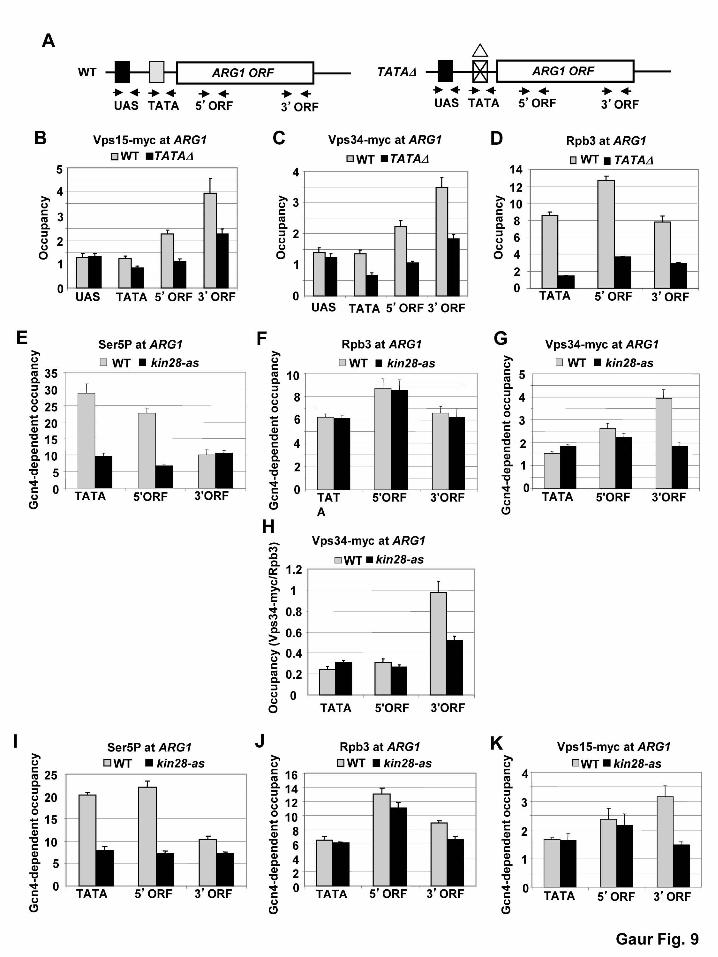
Pol II in the CDS, likewise reduces Vps15 and Vps34 occupancies at ARG1 (Fig. 9A-D, cf. WT
vs. TATA∆). We showed previously that this arg1-TATAΔ mutation does not reduce the UAS
occupancy of Gcn4 itself (QIU et al. 2006).
The recruitment of histone modifying enzymes, elongation and termination factors, and
mRNA processing factors that function during transcription elongation is stimulated by the
heptad repeats (Tyr1Ser2Pro3Thr4Ser5Pro6Ser7) in the C-terminal domain of Pol II subunit Rpb1
(Pol II CTD), dependent on phosphorylation of the CTD repeats on Ser2, Ser5, or Ser7 by various
cyclin-dependent kinases (PHATNANI and GREENLEAF 2006;BURATOWSKI 2009). Cdk7 (Kin28 in
yeast) is the enzyme responsible for the majority of Ser5 and Ser7 CTD phosphorylation in yeast
cells (BATAILLE et al. 2012; TIETJEN et al. 2010). As expected, we observed that the occupancy
of Rpb1 harboring Ser5-phosphorylated CTD repeats (S5P) is higher at the promoter and 5’ end
versus the 3’ end of the CDS at ARG1, and inhibiting an analog-sensitive (as) version of Kin28
with an ATP analog reduced the occupancy of the S5P form of Rpb1 (Fig. 9E), while having

23
little effect on levels of total Pol II (Rpb3) at ARG1 (Fig. 9F). Interestingly, the occupancy of
Vps34 was reduced at the 3’ end of ARG1 on inhibition of Kin28-as (Fig. 9G), leading to a
reduced Vps34:Rpb3 ratio at this location (Fig. 9H). Similar results were obtained for Vps15 on
inhibition of kin28-as (Fig. 9I-K). This finding suggests that S5P or S7P enhances, directly or
indirectly, the co-transcriptional association of these Vps factors at promoter-distal locations. We
conclude that association of the Vps15 and Vps34 with CDS is coupled to transcription
elongation and stimulated by CTD phosphorylation by Kin28.
Interestingly, we found that vps4Δ cells display reduced association of ESCRT-II factor
Snf8 and ESCRT-III component Snf7 with the ARG1 CDS (Fig. 7I). It is known that cells
lacking the AAA-ATPase Vps4 fail to produce ILVs and recycle ESCRT factors to the
cytoplasm, causing accumulation of ESCRT-III component Snf7 at the MVB (BABST et al.
1998). The fact that association of Snf7 and Snf8 with the ARG1 CDS is diminished in vps4Δ
cells is consistent with the idea that these ESCRT factors are partitioned between endosome- and
nucleus-associated pools and that their impaired recycling from the MVB to the cytoplasm in
vps4Δ cells decreases their association with chromatin.
We went on to examine association of Myc-tagged versions of several other Vps factors
with ARG1 or GAL1 CDS and observed induction-dependent association of Vps4, Vps27, and
Vps10 at ARG1 and Vps45, Vps10 and Pep7 at GAL1 at levels comparable to those described
above for Vps15, Vps34, Snf7 and Snf8 (Fig. 10A-D). In parallel, we analyzed two different
functional myc-tagged protein synthesis initiation factors, eIF4E/Cdc33 and eIF3a/Tif32, that we
expected to be primarily, if not exclusively cytoplasmic. We observed 2-fold or less increases in
association of Cdc33-myc and Tif32-myc occupancy on transcriptional induction at ARG1 (Fig.
10C), and also at ARG4 and GAL1 (data not shown). Thus, although induction-dependent

24
chromatin association of the translation initiation factors is not negligible, it is significantly
lower in magnitude than that seen for the Vps factors.
Fractions of Vps15 and Vps34 co-localize with nucleoporin Nsp1 at edges of nucleus-
vacuole junctions.
To examine further the possibility that fractions of Vps15 and Vps34 are physically
associated with transcribed genes in the nucleus, we visualized GFP fusions to these proteins in
living cells that also contain RFP or BFP fusions to the nucleoporin Nsp1 (AITCHISON and ROUT
2012) or an mCherry fusion to transcription elongation factor Spt4. Consistent with previous
results (HUH et al. 2003; OBARA et al. 2006), both Vps15-GFP and Vps34-GFP were found
primarily in cytoplasmic punctae reflecting their association with endosomes and the outer
membrane of the vacuole. Nsp1-RFP was found in punctae localized at the nuclear rim in the
manner expected for a nucleoporin, as observed previously (MACKINNON et al. 2009). Under
conditions where the Vps proteins were found cross-linked to transcribed chromatin, including
30 min cultivation in SC-galactose medium, we did not observe any obvious accumulation of
Vps34 or Vps15 inside the nucleus. However, a fraction of the Vps-GFP punctae co-localized
with Nsp1-RFP at the edges of junctions formed between the nucleus and vacuole, from which
Nsp1 is largely excluded. This pattern was observed in essentially all cells we examined in
cultures prepared in SC with glucose, raffinose or galactose as carbon source (Figs. S1-S2 in
Supporting Information). It is known that nucleus-vacuole (NV) junctions are mediated by
interactions between vacuolar membrane protein Vac8 and outer nuclear-membrane protein
Nvj1, and are devoid of nuclear pores (PAN et al. 2000). Our time-lapse microscopic
observations suggest that Vps34-GFP or Vps15-GFP punctae at the vacuolar membrane are

25
dynamic and transiently associate with nuclear pores (Nsp1-RFP) at the edges of NV-junctions
(Movies S1-6 in Supporting Information). Interestingly, when cells were cultivated overnight on
rich glucose (YPD) agar medium and then investigated under agarose strips cast in minimal
galactose medium, more than 50% of them displayed obvious overlapping signals of Vps34-GFP
or Vps15-GFP with Nsp1-RFP (Fig. S3).
Spt4-mCherry, examined as a nuclear marker, showed relatively homogenous nuclear
localization in the manner expected for a transcription elongation factor distributed throughout
the nucleoplasm in exponentially growing cells (HUH et al. 2003). We found that in the cells
shifted from YPD plates to minimal galactose medium before microscopy, Spt4-mCherry usually
did not occupy the entire nuclear space defined by the Nsp1-BFP nuclear rim. In rare instances a
portion of Vps15 appeared to be located inside the nucleus of these cells (Fig. S4A), and analysis
of optical sections revealed that the Vps15-GFP signal was surrounded by Spt4-mCherry
material within the nuclear rim defined by Nsp1-BFP (see layer 2). Similarly, as shown in
Figure S4B, the Vps15-GFP signal could be observed as an inclusion inside the Spt4-mCherry
domain (layer 2). Together, our microscopic findings indicate that a fraction of Vps15 and Vps34
are in proximity to nuclear pores at the edges of NV junctions, and that rarely, the Vps15-GFP
fusion protein can be found within the nucleus under certain culture conditions.
DISCUSSION
We demonstrated previously that eliminating certain VPS genes whose products are
involved in Golgi-to-vacuole vesicular protein trafficking reduced the ability of transcriptional
activator Gcn4 to stimulate PIC assembly and transcription initiation in vivo. Based on the
results of assaying a GAL1- lacZ reporter gene, it appeared that the function of Gal4 was also

26
impaired by the vps15Δ and vps34Δ mutations, which reduced GAL1-lacZ expression by 60%
and 80%, respectively (ZHANG et al. 2008). We showed here, however, that Gal4 function in PIC
assembly appears to be normal in vps15Δ and vps34Δ cells, and presented several lines of
evidence that the reductions in Gal4-dependent lacZ reporter expression observed in these
mutants is engendered by a defect at the elongation stage of transcription. Indeed, Aguilera and
colleagues have established that reduced mRNA production from lacZ reporters and certain
native genes with long or GC-rich CDS are hallmarks of mutations in various elongation factors
in yeast (CHAVEZ et al. 2001; MORILLO-HUESCA et al. 2006; RONDON et al. 2004; RONDON et al.
2003a; TOUS et al. 2011). We consistently observed decreased mRNA expression from several
such reporters, all driven by the GAL1 promoter, in vps15Δ and vps34Δ cells. Employing a ChIP
analysis of the kinetics of Pol II elongation in vivo (MASON and STRUHL 2005), modified to
include lacZ CDS, we also obtained evidence that elimination of Vps15 or Vps34 reduces the
rate of Pol II elongation through lacZ CDS in vivo.
We further identified defects in co-transcriptional recruitment of the HAT complex NuA4
to the GAL1 CDS in vps15Δ and vps34Δ cells. Considering that conditional inactivation of the
HAT subunit of NuA4 (Esa1) also impairs the rate of Pol II elongation in vivo (GINSBURG et al.
2009), the reduced NuA4 occupancy in CDS we observed in vps15Δ and vps34Δ mutants could
contribute to their transcription elongation defects. We additionally demonstrated that vps15Δ
and vps34Δ cells are defective for localization of activated GAL1 and INO1 to the nuclear
periphery (gene positioning), a process that requires a number of nuclear pore complex (NPC)
proteins or associated factors and likely facilitates coordination of transcription and mRNP
biogenesis with mRNA export at nuclear pores (EGECIOGLU and BRICKNER 2011). Indeed, the
nucleoporin Nup2 is required for targeting of both GAL1 and INO1 to the nuclear periphery

27
(BRICKNER et al. 2007), and the TREX-2/THSC complex is located at nuclear pores and is
required for gene positioning of GAL1 (CABAL et al. 2006) and efficient mRNA export (FISCHER
et al. 2002; LEI et al. 2003).
Our ability to demonstrate an elongation defect for particular genes with long or GC-rich
CDS, such as lacZ, does not rule out a general requirement for Vps factors for efficient
elongation, as transcription of most genes in yeast is unaffected in mutants lacking only a single
elongation factor. The elongation process appears to be overdetermined (MASON and STRUHL
2005), such that only a few specialized genes require a full complement of cofactors for efficient
transcription elongation in vivo (CHAVEZ et al. 2001). Whereas the elongation defect described
here likely applies broadly to many genes, the impairment of transcription initiation we observed
previously in vps mutants (ZHANG et al. 2008) is restricted to a subset of activators that includes
Gcn4, but not Gal4, and seems to involve a signaling pathway that responds to sterol limitation
(MOUSLEY et al. 2012). Hence, we now interpret the dramatic reductions in expression of Gcn4-
dependent lacZ reporters we observed previously in vps15Δ and vps34Δ mutants (ZHANG et al.
2008) to be the compound effect of reduced PIC assembly by Gcn4 and impaired transcription
elongation through lacZ CDS.
Given their well-established functions in vesicular protein trafficking, we anticipated that
Vps15 and Vps34 would promote transcription elongation by an indirect mechanism. Hence, we
were surprised to obtain evidence by ChIP assays for physical association of these and several
other functionally related Vps proteins with transcribed chromatin. The Vps factors consistently
displayed higher occupancies of the CDS versus UAS or promoter regions of the examined
genes, consistent with a widespread role in transcription elongation. The CDS-occupancies of
the Vps proteins were lower than we observed previously for canonical elongation factors,

28
including Spt4, Spt5, Bur2, and the Paf1C complex (QIU et al. 2009; QIU et al. 2006), but they
were comparable to the occupancies of subunits of SAGA (GOVIND et al. 2007), NuA4
(GINSBURG et al. 2009), and histone deacetylase complexes (GOVIND et al. 2010), and they were
significantly higher than the ≤2-fold enrichment of two different protein synthesis factors we
analyzed at Gcn4 and Gal4 target genes. Importantly, as observed for conventional elongation
factors, the association of Vps proteins with ARG1 or GAL1 CDS was strongly dependent on
target gene transcription, occurring at high levels only under conditions where the relevant
transcriptional activators are induced (Gcn4) or functional (Gal4). In addition, the TATA
promoter element is required for high-level Vps15 and Vps34 occupancies at ARG1 under
inducing conditions, and Vps15 and Vps34 occupancies of ARG1 CDS were stimulated by the
Pol II CTD kinase Cdk7/Kin28—a characteristic of numerous factors involved in co-
transcriptional histone-modifications, mRNA processing or nuclear export, and the elongation or
termination phases of transcription (GINSBURG et al. 2009; GOVIND et al. 2010; GOVIND et al.
2007; PASCUAL-GARCIA et al. 2008; PHATNANI and GREENLEAF 2006; QIU et al. 2012).
The specific association of Vps factors with transcribed coding sequences raises the
possibility that they function in association with the chromatin to promote transcription
elongation rather than influencing the process indirectly at the endosome. In this view, certain
Vps factors would be partitioned between cytoplasm and nucleus and have dual functions in
protein trafficking and transcription in these two compartments. Further evidence for this
partitioning is provided by our finding that association of Snf7/Vps32 and Snf8/Vps22 with
ARG1 chromatin is diminished in vps4∆ cells, in which Snf7 accumulates on the MVB outer
membrane owing to a defect in ILV formation and recycling of ESCRT-III factors back to the
cytoplasm (BABST et al. 1998). By fluorescence microscopy of living cells, we also observed

29
association of a fraction of Vps15-GFP and Vps34-GFP fusions with an RFP fusion to
nucleoporin Nsp1 at the edges of NV junctions. This pattern of vacuole/nucleus interaction was
observed frequently under various growth conditions. Our time-lapse microscopy revealed that
association of Vps punctae with the edges of NV junctions is highly dynamic, such that
Vps15/Vps34 colocalization with nuclear pores is transient and observed only in particular
optical layers. These findings raise the possibility of a dynamic interaction of Vps15/Vps34
proteins with actively transcribed genes at nuclear pores, which could be consistent with their
requirement for efficient gene localization to the nuclear periphery. It is unclear, however,
whether an association of Vps15/Vps34 with nuclear pores that is restricted to the borders of NV
junctions could account for their widespread cross-linking to transcribed genes and their
apparently general effect on transcription elongation. It is worth noting that the Vps34 homolog
in C. elegans is concentrated in a perinuclear location, although this cellular location has been
connected so far only with its role in vesicle budding from the outer nuclear membrane directed
towards the cell periphery (ROGGO et al. 2002).
Despite our evidence for physical association of a fraction of Vps15 and Vps34 with
transcribed chromatin and nuclear pores, we have been unable to observe a defect in Pol II
elongation in whole cell extracts of vps34Δ cells using a DNA template containing two G-less
cassettes flanking lacZ CDS (data not shown). This assay was employed previously to provide
evidence for direct roles of various factors in transcription elongation, including Spt4 and
subunits of the THO complex (RONDON et al. 2004; RONDON et al. 2003a; RONDON et al.
2003b). Accordingly, the elongation defect we observed in vps15Δ and vps34Δ cells presumably
involves an aspect of nuclear structure or nucleus-cytoplasm interaction that cannot be
recapitulated in homogenized cell extracts. However, we cannot eliminate the possibility that the

30
elongation defect we documented in vps15Δ and vps34Δ mutants represents an indirect
consequence of a defect in vesicular protein trafficking in the cytoplasm that is incidental to the
association of Vps15 and Vps34 with transcribed chromatin and nuclear pores.
Notwithstanding this last reservation, one interesting hypothesis would be that
Vps15/Vps34 function at the nuclear periphery to promote transcription elongation of activated
genes localized to nuclear pores. Vps34 generates PI(3)P at endosomal membranes, which
recruits ESCRT-0 factor Vps27 via its FYVE domain (RAIBORG and STENMARK 2009). To our
knowledge, there is no prior evidence that Vps27 or any other known yeast FYVE protein (Pep7,
Fab1, Pib1, and Pib2) functions in the nucleus. (Although the FYVE factor Fab1 regulates
assembly of the Cti6/Cyc8/Tup1 transcriptional cofactor complex, it performs this function at
endosomes (HAN and EMR 2011). Interestingly, however, plant homeodomain (PHD) fingers
can bind various PIPs, including PI(3)P (GOZANI et al. 2003), and are found in many chromatin
and transcription-related proteins, including the Yng2 subunit of NuA4 (http://smart.embl.de/).
Given our finding that transcription-coupled recruitment of NuA4 to the GAL1 CDS is
diminished in vps15Δ and vps34Δ cells, it could be proposed that PI(3)P produced by
Vps15/Vps34 on the nuclear membrane, possibly at nuclear pores, stimulates recruitment of
NuA4 and other PHD-containing transcription cofactors to enhance elongation by Pol II. A
precedent for such a mechanism is provided by evidence indicating that mammalian Ing2, an
HDAC component, is recruited to chromatin by a nuclear pool of PI(5)P via its PHD finger,
where it regulates p53 acetylation and apoptosis in response to DNA damage (GOZANI et al.
2003). Indeed, there is increasing evidence for phosphoinositides in the inner nuclear periphery
and for isoforms of phosphoinositide kinases being located inside the nucleus (BARLOW et al.
2010).

31
If PI(3)P is produced by Vps15/Vps34 on the nuclear membrane in the manner just
proposed, this might account for our ability to detect transcription-dependent chromatin
association of the FYVE-containing proteins Vps27 and Pep7 by ChIP assays. Moreover, the
similar ChIP results observed for Vps factors Snf7, Snf8, Vps4, Vps45, and possibly Vps10,
might reflect their association, direct or indirect, with Vps27 or Pep7. However, it seems more
difficult to account for our evidence that these other Vps proteins also contribute to efficient
transcription elongation, albeit to a lesser extent than do Vps15 and Vps34. One possibility is
that impairing vesicular protein trafficking in the cytoplasm by deletions of various Vps factors
reduces the nucleus-associated pools of Vps15 and Vps34 and thereby impairs transcription
elongation indirectly.
There are other indications of nuclear functions for certain Vps proteins, including the
fact that mammalian ESCRT-II was first purified in association with Pol II elongation factor
ELL (SHILATIFARD 1998), and a report that plant Vps34 co-localizes with sites of transcription in
plant cell nuclei (BUNNEY et al. 2000). In addition, the mammalian homolog of the ESCRT-III-
related protein Vps46/Did2, when overexpressed, was found to enter the nucleus and locally
condense chromatin, and to produce a gene-silencing phenotype in Xenopus embryos (STAUFFER
et al. 2001). Clearly more work is required to determine at the molecular level exactly how Vps
factors promote transcription elongation in yeast cells and whether the mechanisms involved
depend on their physical interaction with chromatin and nuclear pores.
ACKNOWLEDGEMENTS
We thank Sebastián Chávez, Andrés Aguilera, Henrik Dohlman, Steven Hahn, Kristine
Willis, Roger Tsien, and the Yeast Resource Center at the University of Washington for gifts of
32
plasmids, and Thomas Dever for useful suggestions. This work was supported in part by the
Intramural Research Program of the NIH. J.H. was supported by P305/12/0480 and
RVO61388971, and D.G.B. and J.H.B. were supported by a W.M. Keck Young Scholars in
Medical Research Award.
REFERENCES
AITCHISON, J. D., and M. P. ROUT, 2012 The yeast nuclear pore complex and transport through it. Genetics 190: 855-883.
ALANI, E., L. CAO and N. KLECKNER, 1987 A method for gene disruption that allows repeated use of URA3 selection in the construction of multiply disrupted yeast strains. Genetics 116: 541-545.
BABST, M., B. WENDLAND, E. J. ESTEPA and S. D. EMR, 1998 The Vps4p AAA ATPase regulates membrane association of a Vps protein complex required for normal endosome function. Embo J 17: 2982-2993.
BARLOW, C. A., R. S. LAISHRAM and R. A. ANDERSON, 2010 Nuclear phosphoinositides: a signaling enigma wrapped in a compartmental conundrum. Trends Cell Biol 20: 25-35.
BATAILLE, A. R., C. JERONIMO, P. E. JACQUES, L. LARAMEE, M. E. FORTIN et al., 2012 A Universal RNA Polymerase II CTD Cycle Is Orchestrated by Complex Interplays between Kinase, Phosphatase, and Isomerase Enzymes along Genes. Mol Cell 45: 158-170.
BOWERS, K., and T. H. STEVENS, 2005 Protein transport from the late Golgi to the vacuole in the yeast Saccharomyces cerevisiae. Biochim Biophys Acta 1744: 438-454.
BOYSEN, J. H., and A. P. MITCHELL, 2006 Control of Bro1-domain protein Rim20 localization by external pH, ESCRT machinery, and the Saccharomyces cerevisiae Rim101 pathway. Mol Biol Cell 17: 1344-1353.
BRICKNER, D. G., I. CAJIGAS, Y. FONDUFE-MITTENDORF, S. AHMED, P. C. LEE et al., 2007 H2A.Z-mediated localization of genes at the nuclear periphery confers epigenetic memory of previous transcriptional state. PLoS Biol 5: e81.
BRICKNER, D. G., W. LIGHT and J. H. BRICKNER, 2010 Quantitative localization of chromosomal loci by immunofluorescence. Methods Enzymol 470: 569-580.
BRICKNER, J. H., and P. WALTER, 2004 Gene recruitment of the activated INO1 locus to the nuclear membrane. PLoS Biol 2: e342.
BRYANT, G. O., and M. PTASHNE, 2003 Independent recruitment in vivo by Gal4 of two complexes required for transcription. Mol Cell 11: 1301-1319.
BUNNEY, T. D., P. A. WATKINS, A. F. BEVEN, P. J. SHAW, L. E. HERNANDEZ et al., 2000 Association of phosphatidylinositol 3-kinase with nuclear transcription sites in higher plants. Plant Cell 12: 1679-1688.
BURATOWSKI, S., 2009 Progression through the RNA polymerase II CTD cycle. Mol Cell 36: 541-546.

33
CABAL, G. G., A. GENOVESIO, S. RODRIGUEZ-NAVARRO, C. ZIMMER, O. GADAL et al., 2006 SAGA interacting factors confine sub-diffusion of transcribed genes to the nuclear envelope. Nature 441: 770-773.
CASOLARI, J. M., C. R. BROWN, S. KOMILI, J. WEST, H. HIERONYMUS et al., 2004 Genome-wide localization of the nuclear transport machinery couples transcriptional status and nuclear organization. Cell 117: 427-439.
CHAVEZ, S., T. BEILHARZ, A. G. RONDON, H. ERDJUMENT-BROMAGE, P. TEMPST et al., 2000 A protein complex containing Tho2, Hpr1, Mft1 and a novel protein, Thp2, connects transcription elongation with mitotic recombination in Saccharomyces cerevisiae. EMBO J. 19: 5824-5834.
CHAVEZ, S., M. GARCIA-RUBIO, F. PRADO and A. AGUILERA, 2001 Hpr1 is preferentially required for transcription of either long or G+C-rich DNA sequences in Saccharomyces cerevisiae. Mol Cell Biol 21: 7054-7064.
EGECIOGLU, D., and J. H. BRICKNER, 2011 Gene positioning and expression. Curr Opin Cell Biol.
EXINGER, F., and F. LACROUTE, 1992 6-Azauracil inhibition of GTP biosynthesis in Saccharomyces cerevisiae. Curr Genet 22: 9-11.
FISCHER, T., K. STRASSER, A. RACZ, S. RODRIGUEZ-NAVARRO, M. OPPIZZI et al., 2002 The mRNA export machinery requires the novel Sac3p-Thp1p complex to dock at the nucleoplasmic entrance of the nuclear pores. EMBO J 21: 5843-5852.
GIAEVER, G., A. M. CHU, L. NI, C. CONNELLY, L. RILES et al., 2002 Functional profiling of the Saccharomyces cerevisiae genome. Nature 418: 387-391.
GIETZ, R. D., and A. SUGINO, 1988 New yeast-Escherichia coli shuttle vectors constructed with in vitro mutagenized yeast genes lacking six-base pair restriction sites. Gene 74: 527-534.
GINSBURG, D. S., C. K. GOVIND and A. G. HINNEBUSCH, 2009 The NuA4 lysine acetyltransferase Esa1 is targeted to coding regions and stimulates transcription elongation with Gcn5. Mol Cell Biol.
GOVIND, C. K., H. QIU, D. S. GINSBURG, C. RUAN, K. HOFMEYER et al., 2010 Phosphorylated Pol II CTD recruits multiple HDACs, including Rpd3C(S), for methylation-dependent deacetylation of ORF nucleosomes. Mol Cell 39: 234-246.
GOVIND, C. K., S. YOON, H. QIU, S. GOVIND and A. G. HINNEBUSCH, 2005 Simultaneous recruitment of coactivators by Gcn4p stimulates multiple steps of transcription in vivo. Mol Cell Biol 25: 5626-5638.
GOVIND, C. K., F. ZHANG, H. QIU, K. HOFMEYER and A. G. HINNEBUSCH, 2007 Gcn5 promotes acetylation, eviction, and methylation of nucleosomes in transcribed coding regions. Mol Cell 25: 31-42.
GOZANI, O., P. KARUMAN, D. R. JONES, D. IVANOV, J. CHA et al., 2003 The PHD finger of the chromatin-associated protein ING2 functions as a nuclear phosphoinositide receptor. Cell 114: 99-111.
HAN, B. K., and S. D. EMR, 2011 Phosphoinositide [PI(3,5)P2] lipid-dependent regulation of the general transcriptional regulator Tup1. Genes Dev 25: 984-995.
HARTZOG, G. A., T. WADA, H. HANDA and F. WINSTON, 1998 Evidence that Spt4, Spt5, and Spt6 control transcription elongation by RNA polymerase II in Saccharomyces cerevisiae. Genes Dev 12: 357-369.
HINNEBUSCH, A. G., 2005 Translational regulation of GCN4 and the general amino acid control of yeast. Annu Rev Microbiol 59: 407-450.

34
HUH, W. K., J. V. FALVO, L. C. GERKE, A. S. CARROLL, R. W. HOWSON et al., 2003 Global analysis of protein localization in budding yeast. Nature 425: 686-691.
HURLEY, J. H., and S. D. EMR, 2006 The ESCRT complexes: structure and mechanism of a membrane-trafficking network. Annu Rev Biophys Biomol Struct 35: 277-298.
JANKE, C., M. M. MAGIERA, N. RATHFELDER, C. TAXIS, S. REBER et al., 2004 A versatile toolbox for PCR-based tagging of yeast genes: new fluorescent proteins, more markers and promoter substitution cassettes. Yeast 21: 947-962.
JENKS, M. H., and D. REINES, 2005 Dissection of the molecular basis of mycophenolate resistance in Saccharomyces cerevisiae. Yeast 22: 1181-1190.
JIMENO, S., A. G. RONDON, R. LUNA and A. AGUILERA, 2002 The yeast THO complex and mRNA export factors link RNA metabolism with transcription and genome instability. EMBO J 21: 3526-3535.
KAMURA, T., D. BURIAN, H. KHALILI, S. L. SCHMIDT, S. SATO et al., 2001 Cloning and characterization of ELL-associated proteins EAP45 and EAP20. a role for yeast EAP-like proteins in regulation of gene expression by glucose. J Biol Chem 276: 16528-16533.
KVAM, E., and D. S. GOLDFARB, 2007 Nucleus-vacuole junctions and piecemeal microautophagy of the nucleus in S. cerevisiae. Autophagy 3: 85-92.
LEI, E. P., C. A. STERN, B. FAHRENKROG, H. KREBBER, T. I. MOY et al., 2003 Sac3 is an mRNA export factor that localizes to cytoplasmic fibrils of nuclear pore complex. Mol Biol Cell 14: 836-847.
LIN, Y. Y., Y. QI, J. Y. LU, X. PAN, D. S. YUAN et al., 2008 A comprehensive synthetic genetic interaction network governing yeast histone acetylation and deacetylation. Genes Dev 22: 2062-2074.
LIU, Y., C. KUNG, J. FISHBURN, A. Z. ANSARI, K. M. SHOKAT et al., 2004 Two cyclin-dependent kinases promote RNA polymerase II transcription and formation of the scaffold complex. Mol Cell Biol 24: 1721-1735.
MACKINNON, M. A., A. J. CURWIN, G. J. GASPARD, A. B. SURACI, J. P. FERNANDEZ-MURRAY et al., 2009 The Kap60-Kap95 karyopherin complex directly regulates phosphatidylcholine synthesis. J Biol Chem 284: 7376-7384.
MASON, P. B., and K. STRUHL, 2005 Distinction and relationship between elongation rate and processivity of RNA polymerase II in vivo. Mol Cell 17: 831-840.
MCPHILLIPS, C. C., J. W. HYLE and D. REINES, 2004 Detection of the mycophenolate-inhibited form of IMP dehydrogenase in vivo. Proc Natl Acad Sci U S A 101: 12171-12176.
MORILLO-HUESCA, M., M. VANTI and S. CHAVEZ, 2006 A simple in vivo assay for measuring the efficiency of gene length-dependent processes in yeast mRNA biogenesis. Febs J 273: 756-769.
MOUSLEY, C. J., P. YUAN, N. A. GAUR, K. D. TRETTIN, A. H. NILE et al., 2012 A sterol-binding protein integrates endosomal lipid metabolism with TOR signaling and nitrogen sensing. Cell 148: 702-715.
OBARA, K., T. SEKITO and Y. OHSUMI, 2006 Assortment of phosphatidylinositol 3-kinase complexes--Atg14p directs association of complex I to the pre-autophagosomal structure in Saccharomyces cerevisiae. Mol Biol Cell 17: 1527-1539.
PAN, X., P. ROBERTS, Y. CHEN, E. KVAM, N. SHULGA et al., 2000 Nucleus-vacuole junctions in Saccharomyces cerevisiae are formed through the direct interaction of Vac8p with Nvj1p. Mol Biol Cell 11: 2445-2457.

35
PASCUAL-GARCIA, P., C. K. GOVIND, E. QUERALT, B. CUENCA-BONO, A. LLOPIS et al., 2008 Sus1 is recruited to coding regions and functions during transcription elongation in association with SAGA and TREX2. Genes Dev 22: 2811-2822.
PHATNANI, H. P., and A. L. GREENLEAF, 2006 Phosphorylation and functions of the RNA polymerase II CTD. Genes Dev 20: 2922-2936.
PURIA, R., S. A. ZURITA-MARTINEZ and M. E. CARDENAS, 2008 Nuclear translocation of Gln3 in response to nutrient signals requires Golgi-to-endosome trafficking in Saccharomyces cerevisiae. Proc Natl Acad Sci U S A 105: 7194-7199.
QIU, H., C. HU, N. A. GAUR and A. G. HINNEBUSCH, 2012 Pol II CTD kinases Bur1 and Kin28 promote Spt5 CTR-independent recruitment of Paf1 complex. EMBO J.
QIU, H., C. HU and A. G. HINNEBUSCH, 2009 Phosphorylation of the Pol II CTD by KIN28 enhances BUR1/BUR2 recruitment and Ser2 CTD phosphorylation near promoters. Mol Cell 33: 752-762.
QIU, H., C. HU, C. M. WONG and A. G. HINNEBUSCH, 2006 The Spt4p subunit of yeast DSIF stimulates association of the Paf1 complex with elongating RNA polymerase II. Mol Cell Biol 26: 3135-3148.
QIU, H., C. HU, S. YOON, K. NATARAJAN, M. J. SWANSON et al., 2004 An array of coactivators is required for optimal recruitment of TATA binding protein and RNA polymerase II by promoter-bound Gcn4p. Mol Cell Biol 24: 4104-4117.
QIU, H., C. HU, F. ZHANG, G. J. HWANG, M. J. SWANSON et al., 2005 Interdependent recruitment of SAGA and Srb mediator by transcriptional activator Gcn4p. Mol Cell Biol 25: 3461-3474.
RAIBORG, C., and H. STENMARK, 2009 The ESCRT machinery in endosomal sorting of ubiquitylated membrane proteins. Nature 458: 445-452.
REID, G. A., and G. SCHATZ, 1982 Import of Proteins into Mitochondria. J Biol Chem 257: 13062-13067.
RILES, L., R. J. SHAW, M. JOHNSTON and D. REINES, 2004 Large-scale screening of yeast mutants for sensitivity to the IMP dehydrogenase inhibitor 6-azauracil. Yeast 21: 241-248.
ROBERTS, P., S. MOSHITCH-MOSHKOVITZ, E. KVAM, E. O'TOOLE, M. WINEY et al., 2003 Piecemeal microautophagy of nucleus in Saccharomyces cerevisiae. Mol Biol Cell 14: 129-141.
ROGGO, L., V. BERNARD, A. L. KOVACS, A. M. ROSE, F. SAVOY et al., 2002 Membrane transport in Caenorhabditis elegans: an essential role for VPS34 at the nuclear membrane. EMBO J 21: 1673-1683.
RONDON, A. G., M. GALLARDO, M. GARCIA-RUBIO and A. AGUILERA, 2004 Molecular evidence indicating that the yeast PAF complex is required for transcription elongation. EMBO Rep 5: 47-53.
RONDON, A. G., M. GARCIA-RUBIO, S. GONZALEZ-BARRERA and A. AGUILERA, 2003a Molecular evidence for a positive role of Spt4 in transcription elongation. EMBO J 22: 612-620.
RONDON, A. G., S. JIMENO, M. GARCIA-RUBIO and A. AGUILERA, 2003b Molecular evidence that the eukaryotic THO/TREX complex is required for efficient transcription elongation. J Biol Chem 278: 39037-39043.
SHILATIFARD, A., 1998 Identification and purification of the Holo-ELL complex. Evidence for the presence of ELL-associated proteins that suppress the transcriptional inhibitory activity of ELL. J Biol Chem 273: 11212-11217.

36
SLESSAREVA, J. E., S. M. ROUTT, B. TEMPLE, V. A. BANKAITIS and H. G. DOHLMAN, 2006 Activation of the phosphatidylinositol 3-kinase Vps34 by a G protein alpha subunit at the endosome. Cell 126: 191-203.
STAUFFER, D. R., T. L. HOWARD, T. NYUN and S. M. HOLLENBERG, 2001 CHMP1 is a novel nuclear matrix protein affecting chromatin structure and cell-cycle progression. J Cell Sci 114: 2383-2393.
STRAIGHT, A. F., A. S. BELMONT, C. C. ROBINETT and A. W. MURRAY, 1996 GFP tagging of budding yeast chromosomes reveals that protein-protein interactions can mediate sister chromatid cohesion. Curr Biol 6: 1599-1608.
SWANSON, M. J., H. QIU, L. SUMIBCAY, A. KRUEGER, S.-J. KIM et al., 2003 A Multiplicity of coactivators is required by Gcn4p at individual promoters in vivo. Mol.Cell.Biol. 23: 2800-2820.
TIETJEN, J. R., D. W. ZHANG, J. B. RODRIGUEZ-MOLINA, B. E. WHITE, M. S. AKHTAR et al., 2010 Chemical-genomic dissection of the CTD code. Nat Struct Mol Biol 17: 1154-1161.
TOUS, C., A. G. RONDON, M. GARCIA-RUBIO, C. GONZALEZ-AGUILERA, R. LUNA et al., 2011 A novel assay identifies transcript elongation roles for the Nup84 complex and RNA processing factors. Embo J 30: 1953-1964.
TU, J., L. G. VALLIER and M. CARLSON, 1993 Molecular and genetic analysis of the SNF7 gene in Saccharomyces cerevisiae. Genetics 135: 17-23.
WEK, R. C., J. F. CANNON, T. E. DEVER and A. G. HINNEBUSCH, 1992 Truncated protein phosphatase GLC7 restores translational activation of GCN4 expression in yeast mutants defective for the eIF-2α kinase GCN2. Mol Cell Biol 12: 5700-5710.
WINZELER, E. A., D. D. SHOEMAKER, A. ASTROMOFF, H. LIANG, K. ANDERSON et al., 1999 Functional Characterization of the S. cerevisiae Genome by Gene deletion and Parallel Analysis. Science 285: 901-906.
YEGHIAYAN, P., J. TU, L. G. VALLIER and M. CARLSON, 1995 Molecular analysis of the SNF8 gene of Saccharomyces cerevisiae. Yeast 11: 219-224.
ZHANG, F., N. A. GAUR, J. HASEK, S. J. KIM, H. QIU et al., 2008 Disrupting vesicular trafficking at the endosome attenuates transcriptional activation by Gcn4. Mol Cell Biol 28: 6796-6818.
FIGURE LEGENDS
Fig. 1. Vps factors are required for WT resistance to 6-AU and MPA and for robust
induction of IMD2 transcription by these elongation inhibitors. (A) Serial 10-fold dilutions
of transformants of the indicated genotypes harboring an empty URA3 vector (YCplac33), or
URA3 plasmids containing VPS15 (pVPS15; row 4) or VPS34 (pVPS34; row 6) spotted on SC-
37
Ura or SC-Ura containing 15 µM MPA or 100 µg/ml 6-AU and incubated for 3da (SC) or 5 da
(MPA and 6-AU) at 30oC. Transformants of WT (BY4741), spt4Δ (6986), vps15Δ (3236) and
vps34Δ (5149) strains were analyzed. (B-C) Conducted as in (A) using transformants of WT
(BY4741), spt4Δ (6986), vps15Δ (3236), vps34Δ (5149), snf7Δ (1580), vps4Δ (5588), pep7Δ
(3682), pep12Δ (1812), vps10Δ (3043) and vps45Δ (4462) strains except that cells were
incubated for 6 da on plates containing 75 µg/ml 6-AU. (D-E) Northern analysis of IMD2 and
SCR1 transcripts in strains of the indicated genotypes cultured to mid-exponential phase in SC-
Ura at 30oC and incubated with 6-AU at 100 µg/ml for 2h. Total RNA was extracted and
subjected to blot-hybridization analysis with probes for IMD2 and SCR1 transcripts. A
representative blot is shown in (D), in which successive lanes for each strain contain amounts of
the same RNA samples differing by a factor of 2 (indicated by the ramps), and the mean ratios of
IMD2 to SCR1 intensities quantified by phosphorimaging analysis of multiple experiments are
plotted in (E) with the S.E.M.s shown as error bars. Empty vector transformants of WT
(BY4741, lanes 1-2), spt4Δ (6986, lane 3-4), thp1Δ (1764, lane 5-6), and vps34Δ (5149, lane 7-8)
strains, a pVPS34 transformant of strain 5149 (lanes 9-10), and a pVPS34N736K transformant of
strain 5149 (lanes 11-12) were analyzed. (F-G) Same as (D-E) except, using 15 µM MPA
instead of 6-AU.
Fig. 2. Vps factors are required for efficient transcription elongation through long and GC-
rich coding sequences.
(A) Serial 10-fold dilutions of transformants of the indicated genotypes harboring pCM184-
LAUR (lacZ) containing Ptet-lacZ-URA3 or control plasmid pNG18 (C) containing Ptet-URA3
spotted on SC-Leu or SC-Leu-Ura and incubated for 5 da at 30oC. The Ptet-lacZ-URA3 and Ptet-

38
URA3 constructs are depicted schematically at the top. Transformants of WT (NGY1), snf7Δ
(NGY3), pep7Δ (NGY4), pep12Δ (NGY5), vps15Δ (NGY6), vps34Δ (NGY7) and vps4Δ
(FZY810) strains were analyzed. (B) Ratios of Pho5 enzymatic specific activity in transformants
of the indicated genotypes harboring plasmids containing PGAL1::PHO5-LAC4 (pSCH209) or
PGAL1-PHO5 (pSCH202) (both shown schematically above), ie. GLAM ratios, were measured.
Strains also contained either empty LEU2 vector YCplac111 (lane 1-3 and 5) or plasmids
containing VPS15 (pNG9, lane 4) or VPS34 (pNG11, lane 6). The appropriate transformants of
WT (BY4741), spt4Δ (6986), vps15Δ (3236) or vps34Δ (5149) strains were grown to mid
exponential phase in SC-Ura-Leu with 2% galactose as carbon source at 30oC before measuring
Pho5 activity in whole cell extracts (WCE). (C-D) Northern analysis of transcripts from PGAL1-
PHO5 (top two panels) and PGAL1-PHO5-LAC4 (bottom two panels) in strains of the indicated
genotype grown to mid-exponential phase in SC-Ura-Leu containing 2% galactose as carbon
source at 30oC. Total RNA was extracted and subjected to blot-hybridization analysis with
probes for the PHO5 CDS or SCR1. A representative blot is shown in (C) and the mean ratios of
PHO5-LAC4 (Long) to PHO5 (Short) mRNA levels, each normalized to SCR1, was quantified
by phosphorimaging analysis of multiple experiments and plotted in (D) with the S.E.M.s shown
as error bars. Empty vector transformants of WT (BY4741), spt4Δ (6986), thp2Δ (2861), vps15Δ
(3236) and vps34Δ (5149) strains (lanes 1-4, &7), a pNG9 transformant of strain 3236 (lane 5), a
pNG10 transformant of strain 3236 (lane 6), a pNG11 transformant of strain 5149 (lane 8), and a
pNG12 transformant of strain 5149 (lane 9) were analyzed. (E-F) same as in panels C-D except
that PGAL1-PHO5-lacZ (pSCH212) was analyzed instead of PGAL1-PHO5-LAC4.

39
Fig. 3. Vps factors do not affect PIC assembly at GAL1 but are required for efficient
transcription of several PGAL1-driven CDS.
(A) Occupancies of Rpb3 at the TATA region in the promoter and 3’ORF of chromosomal GAL1
were measured by ChIP analysis of strains of the indicated genotype cultured in SC medium
containing 2% raffinose, and either induced by adding galactose to 2% for 30 min (IN) or kept
untreated (UI). Cross-linked chromatin was immunoprecipitated with anti-Rpb3 antibodies and
DNA from immunoprecipitated (IP) and input samples was subjected to PCR (using primers
listed in Table 4) in the presence of [33P] dATP to amplify radiolabeled fragments of the GAL1
TATA region, 3’ORF or POL1 CDS (Table 4) analyzed as a control, and the PCR amplicons
were quantified by phosphorimaging analysis. Ratios of GAL1 TATA and 3’ORF to POL1 CDS
signals for IP samples were normalized to the corresponding ratios for input samples to yield
occupancy values. Immunoprecipitations were conducted in duplicate on at least two chromatin
samples prepared from two replicate cultures and the PCR analysis of precipitated DNA
sequences was conducted in duplicate for each immunoprecipitated sample. Error bars represent
the standard error of the mean (S.E.M.). WT (BY4741), gal4Δ (1044), vps15Δ (3236) and
vps34Δ (5149) strains were analyzed. (B) Northern analysis of transcripts expressed from
PGAL1-YAT1 (shown schematically and contained on pSCH247) in strains of the indicated
genotypes grown at 30oC to mid-exponential phase in SC-Ura-Leu containing 2% raffinose and
induced with 2% galactose for 2h. Total RNA was extracted and subjected to blot-hybridization
analysis with probes to the YAT1 CDS or SCR1. A representative blot is shown on the left and
the mean ratios of YAT1 mRNA to SCR1 RNA levels were quantified by phosphorimaging
analysis of multiple experiments and plotted here with the S.E.M.s shown as error bars. Empty
vector transformants of WT (BY4741), spt4Δ (6986), thp2Δ (2861), vps15Δ (3236), and vps34Δ
40
(5149) strains (lanes 1-4 & 6), a pNG9 transformant of strain 3236 (lane 5), a pNG11
transformant of strain 5149 (lane 7), and a pNG12 transformant of strain 5149 (lane 8), all
harboring pSCH247, were analyzed. (C) Northern analysis of transcripts from PGAL1-LYS2 in
strains of the indicated genotype conducted as in (B) except that the strains harbored pSCH227
versus pSCH247, a pNG10 transformant of strain 3236 (lane 6) was analyzed, and probes of
LYS2 CDS and SCR1 were employed. (D) GLAM ratios were measured as in Fig. 2B using the
appropriate transformants of WT (BY4741), spt4Δ (6986), vps15Δ (3236), vps34Δ (5149), snf7Δ
(1580), snf8Δ (2826), vps4Δ (5588), pep7Δ (3682) and vps10Δ (3043) strains, harboring
pSCH202 or pSCH209, and normalized to the GLAM ratio of the WT strain.
Fig. 4. vps15Δ and vps34Δ mutations reduce the transcription elongation rate through lacZ
CDS in vivo. (A-D) ChIP analysis of Pol II (Rpb3) occupancies across the PGAL1-PHO5-lacZ
construct. pSCH212 transformants of WT (BY4741) (B), vps15Δ (3236) (C), or vps34Δ (5149)
(D) strains were grown in SC medium containing 2% raffinose at 30°C, induced by adding
galactose to 2% for 1h, and repressed by adding glucose to 4% for the indicated times (min)
before cross-linking. ChIP was performed with anti-Rpb3 antibodies and PCR amplification in
the presence of [α33P]dATP using the primers shown schematically in (A), and listed in Table 4,
located in the PGAL1 promoter or in lacZ CDS at the indicated distances downstream of the
transcription start site, and the control ChrV-2 primers (that amplify an intergenic region on
chromosome V). Rpb3 occupancies were calculated as described in Fig. 3A, and the
occupancies in glucose (2-5 min) were normalized to those in galactose (0 min) and set to unity
for time zero. Mean relative occupancies and S.E.M.s (error bars) were calculated from
immunoprecipitations conducted in duplicate on at least two chromatin samples prepared from
41
two replicate cultures and the PCR analysis of precipitated DNA sequences was conducted in
duplicate for each immunoprecipitated sample.
Fig. 5. vps15Δ and vps34Δ mutations reduce co-transcriptional recruitment of NuA4. (A-
C) ChIP analysis of myc-tagged NuA4 subunits Epl1 (B) and Yng2 (C) at GAL1 CDS using
primers depicted in (A). In panel B, WT (NGY117), vps15Δ (NGY102) and vps34Δ (NGY103)
strains, harboring EPL1-myc13, were grown in SC medium containing 2% raffinose at 30°C (UI),
and induced with 2% galactose (IN) for 30 min before cross-linking. ChIP analysis was
performed as described in Fig. 3A except using anti-myc antibodies and the GAL1 CDS primers
depicted in (A) and control ChrV-1 primers (that amplify an intergenic region on chromosome
V) listed in Table 4. In panel C, ChIP was performed as in (B) except using the following
YNG2-myc13 strains: WT (NGY118), vps15Δ (NGY119) and vps34Δ (NGY120).
Immunoprecipitations were conducted in duplicate on at least two chromatin samples prepared
from two replicate cultures and the PCR analysis of precipitated DNA sequences was conducted
in duplicate for each immunoprecipitated sample. Error bars represent the standard error of the
mean (S.E.M.)
Fig. 6. Localization of activated GAL1 at the nuclear periphery requires Vps15 and Vps34.
Strains DBY375 (WT), DBY376 (vps34∆), and DBY377 (vps15∆), containing an array of Lac
repressor binding sites integrated at GAL1 and expressing a GFP fusion to Lac repressor, were
grown overnight in glucose-containing medium and then shifted to galactose medium for 2h
before fixing in methanol and processing for immunofluorescence using antibodies against GFP-
Lac repressor and Nsp1. (A) Representative cells stained for GFP (green) and Nsp1 (red) that
42
were scored as nucleoplasmic or peripheral. (B) Average peripheral localization of GAL1 from
three biological replicates. For each replicate, ≥ 30 cells were counted. The mean percentage of
cells in which the Lac repressor dot colocalized with the nuclear envelope (Nsp1) is displayed ±
S.E.M. The dynamic range of this assay is from 20%-80%, and the hatched line represents the
distribution of the URA3 gene with respect to the nuclear envelope. (C) Strains DBY452 (WT),
DBY455 (vps34∆), and DBY457 (vps15∆), harboring an array of Lac repressor binding sites
integrated at INO1 and expressing GFP-tagged Lac repressor were cultured overnight in SC
lacking inositol either containing or lacking 100µM myo-inositol and analyzed as in (B).
Fig.7. Transcription-dependent recruitment of Vps factors at Gcn4 target genes ARG1 and
ARG4.
(A-D) ChIP analysis of myc-tagged Vps15 in VPS15-myc13strains NGY26 (WT) and NGY28
(gcn4Δ) at ARG1 (A & C) and ARG4 (B & D), using the primers depicted schematically in
panels A and B, respectively, and control primer ChrV-1listed in Table 4. (E-F) ChIP analysis of
Vps34-myc in VPS34-myc13 strains NGY27 (WT) and NGY29 (gcn4Δ) at ARG1 (E) and ARG4
(F) using the primers depicted in panels A-B and control primer ChrV-1 listed in Table 4. (G-I)
ChIP analysis of Snf7-myc (G & I) at ARG1 in SNF7-myc13 strain FZY610 and vps4Δ SNF7-
myc13 strain FZY642, and of Snf8-myc in SNF8-myc13 strain FZY644 and vps4Δ SNF8-myc13
strain FZY657( listed in Table 1) using the primers depicted in (A) and the control primers
POL1CDS listed in Table 4. For all experiments, cells were cultured in SC-Ilv and either treated
with 0.5µM sulfometuron methyl (SM) for 30min to induce Gcn4 (IN) or left untreated (UI).
Cross-linked chromatin was immunoprecipitated with anti-myc antibodies and analyzed as in
Fig. 3A Immunoprecipitations were conducted in duplicate on at least two chromatin samples
43
prepared from two replicate cultures and the PCR analysis of precipitated DNA sequences was
conducted in duplicate for each immunoprecipitated sample. Error bars represent the standard
error of the mean (S.E.M.)
Fig.8. Recruitment of Vps factors to the CDS at GAL1, ADH1 and PMA1.
(A-D) ChIP analysis of Vps15-myc in VPS15-myc13 strain NGY26 (B), Vps34-myc in VPS34-
myc13 strain NGY27 (C), and Snf7-myc in SNF7-myc13 strain FZY610 (D) conducted for GAL1
using the primers depicted in (A) and control primer ChrV-1 for B and C, POL1 CDS for C
listed in Table 4. Cells were cultured and ChIP analysis was conducted as described in Fig. 3A.
(F-G) ChIP analysis of Vps15-myc in NGY26 and Vps34-myc in NGY27 conducted for ADH1
and PMA1 using the primers depicted in (E) and ChrV-1 as control listed in Table 4. Cells were
grown to mid log phase in SC-Ilv and induced with 0.5µm of SM for 30min. ChIP analysis was
performed as in Fig. 3A. Immunoprecipitations were conducted in duplicate on at least two
chromatin samples prepared from two replicate cultures and the PCR analysis of precipitated
DNA sequences was conducted in duplicate for each immunoprecipitated sample. Error bars
represent the standard error of the mean (S.E.M.)
Fig. 9. Co-transcriptional recruitment of Vps15-myc and Vps34-myc is stimulated by the
Pol II CTD kinase Cdk7/Kin28.
(A-D) ChIP analysis at ARG1 conducted for Vps15-myc in WT (NGY26) and arg1-TATAΔ
(NGY56) strains harboring VPS15-myc13 (B), and for Vps34-myc in WT (NGY27) and arg1-
TATAΔ (NGY57) strains harboring VPS34-myc13 (C) and for Rpb3 in WT (NGY27) and arg1-
TATAΔ (NGY57) strains (D), using primers depicted in (A-B) and control primer ChrV-1 listed

44
in Table 4. Cells were grown to mid log phase in SC-Ilv and induced with 0.5µm of SM for
30min. Occupancies of Vps34-myc (C) and Rbp3 (D) were measured in the same chromatin
extracts. (E-H) ChIP analysis of Rpb1 phosphorylated on Ser5 of its CTD (Ser5P, panel E), Rpb3
(F) and Vps34-myc (G-H) was conducted on the same chromatin extracts from KIN28 VPS34-
myc13 strain NGY69 (WT), kin28-as VPS34-myc13 strain NGY71 (kin28-as), and gcn4Δ VPS34-
myc13 strain NGY29. Cells were grown in SC -Ilv and treated with NAPP1 at 12 µM for 42 min
and SM 0.5µm for the last 30 min of the NAPP1 treatment. ChIP analysis was conducted as in
Fig. 3A and values obtained for the KIN28 and kin28-as strains were normalized to those
obtained for the gcn4Δ strain to produce the plotted Gcn4-dependent occupancies in panels E-G.
Panel H presents the ratios of occupancies in panels G and F. (I-K) ChIP analysis conducted as
in (E-G) except using KIN28VPS15-myc13 strain NGY68, kin28-as VPS15-myc13 strain NGY70
and gcn4Δ VPS15-myc13 strain NGY28. For all ChIP experiments, Immunoprecipitations were
conducted in duplicate on at least two chromatin samples prepared from two replicate cultures
and the PCR analysis of precipitated DNA sequences was conducted in duplicate for each
immunoprecipitated sample. Error bars represent the standard error of the mean (S.E.M.)
Fig.10. Vps factors exhibit higher occupancies of transcribed ARG1 CDS than do
translation factors eIF4E and eIF3a.
(A & C) ChIP analysis of Vps27-myc and Vps4-myc in strains NGY98 and NGY99,
respectively (A), and Vps34-myc, Vps10-myc, eIF4E/Cdc33-myc, and eIF3a/Tif32-myc in
strains NGY27, NGY116, H3881, and H3879, respectively (C), all conducted for ARG1 as
described in Fig. 7. Except control primer for Cdc-33my and Tif32-myc was POL1CDS (B)
ChIP analysis of Pep7-myc, Vps45-myc, and Vps10-myc in NGY100, NGY101, and NGY116,

45
respectively, conducted for GAL1 as described in Fig. 3A. Immunoprecipitations were conducted
in duplicate on at least two chromatin samples prepared from two replicate cultures and the PCR
analysis of precipitated DNA sequences was conducted in duplicate for each immunoprecipitated
sample. Error bars represent the standard error of the mean (S.E.M.)

46
Table 1. Yeast strains used in this study Name Parent Relevant genotypeb Reference BY4741a NAd MATa his3∆1 leu2∆0 met15∆0 ura3∆ Research Genetics 249 a BY4741 a gcn4∆::kanMX4 Research Genetics 1044a BY4741 gal4∆::kanMX4 Research Genetics 3236 a BY4741 a vps15∆::kanMX4 Research Genetics 5149 a BY4741 a vps34∆::kanMX4 Research Genetics 1580a BY4741 a snf7∆::kanMX4 Research Genetics 2826 a BY4741 a snf8∆::kanMX4 Research Genetics 5381a BY4741 a vps27∆::kanMX4 Research Genetics 5588 a BY4741 a vps4∆::kanMX4 Research Genetics 6986a BY4741 a spt4∆::kanMX4 Research Genetics 1764a BY4741 a thp1∆::kanMX4 Research Genetics 2861a BY4741 a thp2∆::kanMX4 Research Genetics 3682a
1812a
3043a
4462a
BY4741a
BY4741a
BY4741a
BY4741a
pep7∆::kanMX4 pep12∆::kanMX4 vps10∆::kanMX4 vps45∆::kanMX4
Research Genetics Research Genetics Research Genetics Research Genetics
FZY610 BY4741a SNF7-myc13::HIS3* This study FZY642 5588a SNF7-myc13::HIS3* vps4∆::kanMX4 This study FZY644 BY4741a SNF8-myc13::HIS3* This study FZY657 5588a SNF7-myc13::HIS3* vps4∆::kanMX4 This study NGY26 BY4741a VPS15-myc13::HIS3* This study NGY27 BY4741a VPS34-myc13::HIS3* This study NGY28 249a VPS15-myc13::HIS3* gcn4∆::kanMX4 This study NGY29 249a VPS34-myc13::HIS3* gcn4∆::kanMX4 This study H3834/ HQY700
BY4741a arg1-ΔTATA (QIU et al. 2006)
NGY56 H3834 arg1-ΔTATA VPS15-myc13::HIS3* This study NGY57 H3834 arg1-ΔTATA VPS34-myc13::HIS3* This study BY4705
NAd MATα ade2Δ::hisGc his3-200Δ leu2Δ0 ura3Δ0 trp1-63Δ lys2Δ0met15Δ0
(LIU et al. 2004)
SHY508A BY4705
kin28:kin28-as(L83G) pSH579[kin28-as(L83G) URA3 CEN]
(LIU et al. 2004)
NGY68 BY4705 VPS15-myc13::HIS3* This study NGY69 NGY102 NGY103 NGY117 NGY118 NGY119 NGY120 NGY98 NGY99 NGY100 NGY101 NGY116 H3881 H3879
BY4705 3236a 5149a BY4741a BY4741a 3236a 5149a
5381a
5588a
3682a
4462a
3043a
BY4741 a
BY4741 a
VPS34-myc13::HIS3* EPL1-myc13::HIS3* EPL1-myc13::HIS3* EPL1-myc13::HIS3* YNG2-myc13::HIS3* YNG2-myc13::HIS3* YNG2-myc13::HIS3* VPS27-myc13::HIS3* VPS4-myc13::HIS3* PEP7-myc13::HIS3* VPS45-myc13::HIS3* VPS10-myc13::HIS3* CDC33-myc13::HIS3* TIF32-myc13::HIS3*
This study This study This study This study This study This study This study This study This study This study This study This study This study This study

47
Table 1. Yeast strains used in this study NGY70 SHY508A kin28:kin28-as(L83G) VPS15-myc13::HIS3*
pSH579[kin28-as(L83G) URA3 CEN] This study
NGY71 SHY508A kin28:kin28-as(L83G) VPS34-myc13::HIS3* pSH579[kin28-as(L83G) URA3 CEN]
This study
NGY1 BY4741 a trp1∆::hisGc (ZHANG et al. 2008) NGY3 1580a snf7∆::kanMX4 trp1∆::hisGc (ZHANG et al. 2008) NGY4 3682a pep7∆::kanMX4 trp1∆::hisGc (ZHANG et al. 2008) NGY5 1812 a pep12∆::kanMX4 trp1∆::hisGc (ZHANG et al. 2008) NGY6 3236 a vps15∆::kanMX4 trp1∆::hisGc (ZHANG et al. 2008) NGY7 5149 a vps34∆::kanMX4 trp1∆::hisGc (ZHANG et al. 2008) FZY810 5588a vps4∆::kanMX4 trp1∆::hisGc (ZHANG et al. 2008) CRY1541 CRY1581 CRY1605 CRY1606 HQY1584 HQY1587 CRY1718 CRY1719 H1486 NGY11 NGY12 DBY375 DBY376 DBY377 DBY452 DBY455 DBY457
BY4741 BY4741 CRY1541 CRY1581 CRY1541 CRY1581 HQY1584 HQY1587 NAd H1486 H1486 H1486 NGY11 NGY12 H1486 NGY11 NGY12
MATα his3∆1 leu2∆0 met15∆0 ura3∆0 VPS34-GFP::HIS3MX6 MATα his3∆1 leu2∆0 met15∆0 ura3∆0 VPS15-GFP::HIS3MX6 VPS34-GFP::HIS3MX6 pNSP1-RFP [NSP1-RFP, URA3, CEN] VPS15-GFP::HIS3MX6 pNSP1-RFP [NSP1-RFP, URA3, CEN] SPT4-mCherry::hphMX4, VPS34-GFP::HIS3MX SPT4-mCherry::hphMX4, VPS15-GFP::HIS3MX SPT4::mCherry::hphMX4 VPS34-GFP::HIS3MX NSP1-BFP::natNT2MX, MATa SPT4-mCherry::hphMX4 VPS15-GFP::HIS3MX NSP1-BFP::natNT2MX MATα his1-29 leu2-3, -112 ura3-52 ino1(?) <HIS4-lacZ, ura3-52> vps34Δ::kanMX4 vps15Δ::kanMX4 GFP13-lacI-I12, lacO::GAL1 GFP13-lacI-I12, lacO::GAL1 vps34Δ::kanMX4 GFP13-lacI-I12, lacO::GAL1 vps15Δ::kanMX4 GFP13-lacI-I12, lacO::INO1 GFP13-lacI-I12, lacO::INO1 vps34Δ::kanMX4 GFP13-lacI-I12, lacO::INO1 vps15Δ::kanMX4
(HUH et al. 2003) (HUH et al. 2003) This study This study This study This study This study This study This study (WEK et al. 1992) (ZHANG et al. 2008) (ZHANG et al. 2008) This study This study This study This study This study This study
_____________________________________________________________________________________________________________________________________
aPurchased from Research Genetics; bHIS3* designates the HIS3 allele from S. kluyveri. chisG(S.t.): hisG sequences from S. typhimurium dNA, not applicable
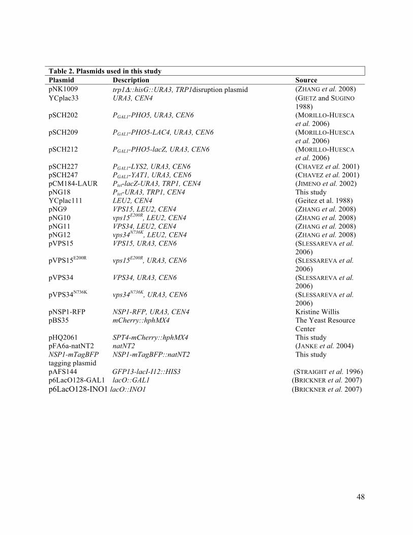
48
Table 2. Plasmids used in this study Plasmid Description Source pNK1009 trp1Δ::hisG::URA3, TRP1disruption plasmid (ZHANG et al. 2008) YCplac33 URA3, CEN4 (GIETZ and SUGINO
1988) pSCH202 PGAL1-PHO5, URA3, CEN6 (MORILLO-HUESCA
et al. 2006) pSCH209 PGAL1-PHO5-LAC4, URA3, CEN6 (MORILLO-HUESCA
et al. 2006) pSCH212 PGAL1-PHO5-lacZ, URA3, CEN6 (MORILLO-HUESCA
et al. 2006) pSCH227 PGAL1-LYS2, URA3, CEN6 (CHAVEZ et al. 2001) pSCH247 PGAL1-YAT1, URA3, CEN6 (CHAVEZ et al. 2001) pCM184-LAUR Ptet-lacZ-URA3, TRP1, CEN4 (JIMENO et al. 2002) pNG18 YCplac111
Ptet-URA3, TRP1, CEN4 LEU2, CEN4
This study (Geitez et al. 1988)
pNG9 VPS15, LEU2, CEN4 (ZHANG et al. 2008) pNG10 vps15E200R, LEU2, CEN4 (ZHANG et al. 2008) pNG11 VPS34, LEU2, CEN4 (ZHANG et al. 2008) pNG12 vps34N736K, LEU2, CEN4 (ZHANG et al. 2008) pVPS15 VPS15, URA3, CEN6 (SLESSAREVA et al.
2006) pVPS15E200R vps15E200R, URA3, CEN6 (SLESSAREVA et al.
2006) pVPS34 VPS34, URA3, CEN6 (SLESSAREVA et al.
2006) pVPS34N736K vps34N736K, URA3, CEN6 (SLESSAREVA et al.
2006) pNSP1-RFP pBS35
NSP1-RFP, URA3, CEN4 mCherry::hphMX4
Kristine Willis The Yeast Resource Center
pHQ2061 SPT4-mCherry::hphMX4 This study pFA6a-natNT2 natNT2 (JANKE et al. 2004) NSP1-mTagBFP tagging plasmid
NSP1-mTagBFP::natNT2 This study
pAFS144 GFP13-lacI-I12::HIS3 (STRAIGHT et al. 1996) p6LacO128-GAL1 lacO::GAL1 (BRICKNER et al. 2007) p6LacO128-INO1 lacO::INO1 (BRICKNER et al. 2007)
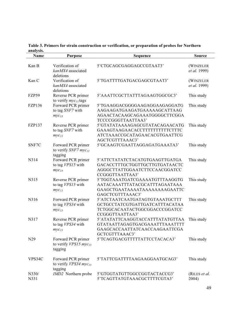
49
Table 3. Primers for strain construction or verification, or preparation of probes for Northern analysis.
Name Purpose Sequence Source Kan B Verification of
kanMX4 associated deletions
5’CTGCAGCGAGGAGCCGTAAT3’ (WINZELER et al. 1999)
Kan C Verification of kanMX4 associated deletions
5’TGATTTTGATGACGAGCGTAAT3’ (WINZELER et al. 1999)
FZP59 Reverse PCR primer to verify myc13 tags
5’AAATTCGCTTATTTAGAAGTGGCGC3’ This study
FZP136 Forward PCR primer to tag SNF7 with myc13
5’TGAAGGACGGGGAAGAGGAAGAGGATGAAGAAGATGAAGATGAAAAAGCATTAAGAGAACTACAAGCAGAAATGGGGCTTCGGATCCCCGGGTTAATTAA3’
This study
FZP137 Reverse PCR primer to tag SNF7 with myc13
5’GTATATAAAAGAGCGTATACAGAACATGGAAAGTAAGAACACCTTTTTTTTTTCTTTCATCTAAACCGCATAGAACACGTGAATTCGAGCTCGTTTAAAC3’
This study
SNF7C Forward PCR primer to verify SNF7 myc13 tagging
5’GCAAGTCGAATTAGGAGATGAAATA3’ This study
N314
Forward PCR primer to tag VPS15 with myc13
5’ATTCTATATCTACATGTGAAGTTGATGAGACACCTTTGCTGGTTGCTTGTGATAACTCAGGGCTTATTGGAATCTTCCAACGGATCCCCGGGTTAATTAA3’
This study
N315 Reverse PCR primer to tag VPS15 with myc13
5’TGGTAAATGATCGAAAATGTTTAAGGTGAATACAAATTTATACGCATTTAGAATAAAGAAGCTGAATAAAATAAAAAAAAGAATTCGAGCTCGTTTAAAC3’
This study
N316 Forward PCR primer to tag VPS34 with myc13
5’ATCTAATCAATGATAGTGTAAATGCTTTGCTGCCTATCGTGATTGATCATTTACATAATCTGGCACAATACTGGCGGACCCGGATCCCCGGGTTAATTAA3’
This study
N317 Reverse PCR primer to tag VPS34 with myc13
5’ATATATTCAAGGTACCATTTATATGTTAAGTATAATTAGAGTGACGAAATTTAAATTTTGAAGCACCAATTATCAACCAAGAATTCGAGCTCGTTTAAAC3’
This study
N29 Forward PCR primer to verify VPS15 myc13 tagging
5’TCAGTGACGTTTTTATTCCTACACA3’ This study
VPS34C
Forward PCR primer to verify VPS34 myc13 tagging
5’TATTCGATTTTAAGAAGGAATGCAG3’
This study
N330/ N331
IMD2 Northern probe 5’GTGGTATGTTGGCCGGTACTACCG3’ 5’TCAGTTATGTAAACGCTTTTCGTA3’
(RILES et al. 2004)

50
Table 3. Primers for strain construction or verification, or preparation of probes for Northern analysis. N332/ N333
LYS2 Northern probe 5’TCAGTCCAATTAGGTAAGATTC3’ 5’ AAGCTGCTGCGGAGCTTCCAC3’
This study
N334/ N335
YAT1 Northern probe 5’AATTCAGCTCTTGCACGACGC3’ 5’ CACGCTCACGTCGAAGTAATC3’
This study
SCR698/ SCR1699
SCR1 Northern probe 5’CGCGGTACCTGATCAACTTAGCCAGGACATCC3’ 5’TGCTCTAGAGTTCTAAGTATTCTCATTTTATCC3’
(QIU et al. 2004)
N381/ N382
URA3 cloning for pNG18 construction
5’GATCGGATCCATGTCGAAAGCTACATATAAGGA3’ 5’GATCATCGATTTAGTTTTGCTGGCCGCATCTT3’
This study
N394 Forward PCR primer to tag YNG2 with myc13
5’AAGAACCTCCGAAAGGAACATGGTACTGTCCCGAATGTAAAATTGAGATGGAAAAAAACAAACTGAAAAGAAAACGTAACCGGATCCCCGGGTTAATTAA3’
This study
N395 Reverse PCR primer to tag YNG2 with myc13
5’AGAATAGCAATCACCGTTTCTGGCTTATGGAATATTATTAGTGTAAATGAGGTCATTCAGTCTCAAAAAGGTATTTTTGTGAATTCGAGCTCGTTTAAAC3’
This study
N396 Forward PCR primer to verify YNG2 myc13 tagging
5’AATGCGTTCTGGCGAACACTGC3’ This study
N362 Forward PCR primer to tag VPS27 with myc13
5’CACTTCCTGCAAGTTCCTCTGAAATTCCGATCAAAGAGGAAAGGCCGCCTAGTCCTCAAGAGGAACTGCTAATAGAGCTTCGGATCCCCGGGTTAATTAA3’
This study
N363 Reverse PCR primer to tag VPS27 with myc13
5’CAAGCAATTATATATATATGTATGTATATATTTATAAGCGCTAGGTTTCTTTTTACAAATACATAGAAAAGGCTACAATAGAATTCGAGCTCGTTTAAAC3’
This study
N106 Forward PCR primer to verify VPS27 myc13 tagging
5’TCCAGCAACTATCATCTCATCAATA3’
This study
N364 Forward PCR primer to tag VPS4 with myc13
5’AATCGACGAGACCTACCGTGAATGAGGATGACTTGCTGAAGCAAGAACAGTTCACTAGAGATTTTGGTCAAGAAGGTAACCGGATCCCCGGGTTAATTAA3’
This study
N365 Reverse PCR primer to tag VPS4 with myc13
5’TAATTATTCTGTTTTTTTTTTTTTTTTATTTTTTATTTTCATGTACACAAGAAATCTACATTAGCACGTTAATCAATTGAGAATTCGAGCTCGTTTAAAC3’
This study
N118 Forward PCR primer to verify VPS4 myc13 tagging
5’GCCAGGATAAAATGACTCAAAGTAA3’
This study
N377 Forward PCR primer to tag PEP7 with myc13
5’TCACACTTAATAAGAACTTAGAAGAATTGCACTCAAGAATACATACCGTTCAATCGAAGCTGGGTGACCATGGGTTTAATCGGATCCCCGGGTTAATTAA3’
This study

51
Table 3. Primers for strain construction or verification, or preparation of probes for Northern analysis. N378 Reverse PCR primer
to tag PEP7 with myc13
5’TACCCTTAAATAGATCTGATTGGCTCCGCGGACCAATTAGCGTGGAGATTCCGCGAATGATCGAATAATAAAGATATGTCGAATTCGAGCTCGTTTAAAC3’
This study
N308 Forward PCR primer to verify PEP7 myc13 tagging
5’TTGCTAAATATGATAGCATGCAAAA3’
This study
N379 Forward PCR primer to tag VPS45 with myc13
5’ATAACAGAATGAGGGTGGTTTTAGGAGGCACCTCTATACTTTCAACTAAAGAATATATGGATTCTATTAGATCTGCAAAACGGATCCCCGGGTTAATTAA3’
This study
N380 Reverse PCR primer to tag VPS45 with myc13
5’ATGTTTTATCTGCATCGAAAAAGTTATATAGATTTATGCCTCATATATAAAATAGAATTTTAGAATAAGATAATCCTTATGAATTCGAGCTCGTTTAAAC3’
This study
N118 Forward PCR primer to verify PEP7 myc13 tagging
5’ GCCAGGATAAAATGACTCAAAGTAA3’
This study
N386 Forward PCR primer to tag VPS10 with myc13
5’ATGTTCCAGATACATTACCAGAAGGAAATGAGGAAAACATCGACAGGCCTGATTCTACAGCGCCATCTAACGAAAACCAGCGGATCCCCGGGTTAATTAA3’
This study
N387 Reverse PCR primer to tag VPS10 with myc13
5’TGATATTCTGTCTAAAGGATCTGCTCGTTTTTGTTTATGAAAAGTATATGGAATTATCTACTCTATGTAAAGTAATCTCTGAATTCGAGCTCGTTTAAAC3’
This study
N114 Forward PCR primer to verify VPS10 myc13 tagging
5’AGGAAAAGAAAAAGAATTCAAGGAA3’
This study
FZP177 Forward PCR primer to tag SNF8 with myc13
5'CTAACGGTTTATTGTGGATTGATTATCAAGGTGGTGCAGAGGCGTTGTATTGGGATCCCTCTTGGATTACGAGGCAACTGCGGATCCCCGGGTTAATTAA3’
This study
FZP178 Reverse PCR primer to tag SNF8 with myc13
5'CGGTAAGAACTGTGCTCAATAAAATACAACATCGATAAATACTTAACGCTCATCCACCATTCCTGTTCAAGTCAACTATCGAATTCGAGCTCGTTTAAAC3’
This study
SNF8C Forward PCR primer to verify SNF8 myc13 tagging
5'AGTAAATCTGCTCTTGATGAAATGG3’ This study
1853/1854 PCR amplification of the SPT4 CDS for construction of pHQ2061
5’CGTACGAAGCTTTCGCGACGAGGTACAGTGTAA GAGATG3'/ 5'CCCGGGGATCCGCTCAACTTGACTGCCATCCCT3'
This study
1855/1856 PCR amplification of the SPT4 3’UTR for construction of pHQ2061
5'GAAAACGAGCTCTAAAACCTTCCGTTCTGATATC3'/ 5'TAGGCCACTAGTTCGCGACTGAGCTGCACGAGAAT3'
This study
972/1167 Verification of SPT4- 5' CGTTCTATGAGTGAAATCGAG3' This study

52
Table 3. Primers for strain construction or verification, or preparation of probes for Northern analysis.
mCherry::hphMX integration
5' CAATTCAACGCGTCTGTGAG3’
Nsp1mbfp5/ Nsp1C2
ytBFP cassette amplification
5‘GATAACTCCACCTCTCTGGAAAAACAAATCAACTCGATAAAGAAAGGTGCAGGTGCTGGAGCTGGT3’/ 5’CTATGTCAAATAAGTGTAGAATAGAGGGAATTTTTTCTTTTAGACACTATAGGGAGACCGGCAG3’
This study
Nsp1diaC/mTAGBFPrev
Verification of ytBFP cassette integration
5‘GGTACTAGTGAAAGGAGGTG3’/ 5’CTACCTTCTAAACCACCGTC3’
This study
Epl1CMyc+
Forward PCR primer to tag EPL1 with myc13
5’GATTCGTTAATCAATTCAGAAGCGAAACAGAACTCTTCTATAACCCAGAAAAATTCATCACGGATCCCCGGGTTAATTAA3’
This study
Epl1CMyc- Reverse PCR primer to tag EPL1 with myc13
5’AGGTTTGTTAAATGCCAATGCGTAGAAAGATGTTCGAGAGGTACAAAGTTGTGCTGCTCAGAATTCGAGCTCGTTTAAAC3’
This study
N385 Forward PCR primer to verify EPL1 myc13 tagging
5’AGATGAACCGGCAAGACTTAAC3’
This study
mycCDC33F2 mycCDC33R1 mycTIF32F1 mycTIF32R1
Forward PCR primer to tag CDC33 with myc13
Reverse PCR primer to tag CDC33 with myc13
Forward PCR primer to tag TIF32 with myc13
Reverse PCR primer to tag TIF32 with myc13
5’ATTAACCGATGACGGGCATTTGGAATTCTTTCCACATTCCAGTGCCAATGGTAGACACCCTCAACCATCAATCACCTTGCGGATCCCCGGGTTAATTAA3’ 5’GTATATATATTATATTATGATTATATATATAATTTTGATTAAAATAACAATTATCTTAAGAAAAATTCAGACTATCGAATTCGAGCTCGTTTAAAC3’ 5’CCCCAACTCCATCTTCTGGTCCAAAGAAAATGACTATGGCTGAAAAGTTGAGAGCCAAGAGATTGGCCAAGGGGGGCAGGCGGATCCCCGGGTTAATTAA3’
AATTGTTATTATGTACACATACTCTTATACGTATAAAAAACGGTATAAATTAAGTAGATCATTTTGCGTTGTCTGGAATTCGAGCTCGTTTAAAC3’
This study This study This study This study

53
Table 4. Primers for ChIP assays. Name Gene & location Sequence Source 273/274 POL1 CDS 5’GACAAAATGAAGAAAATGCTGATGCACC-3’
5’TAATAACCTTGGTAAAACACCCTG-3’ (SWANSON et al. 2003)
605/ 607 ARG1 UAS 5’ACGGCTCTCCAGTCATTTAT-3’ 5’GCAGTCATCAATCTGATCCA-3’
(SWANSON et al. 2003)
265/266 ARG1 TATA 5’TAATCTGAGCAGTTGCGAGA-3’ 5’ATGTTCCTTATCGCTGCACA-3’
(QIU et al. 2004)
876/877 ARG1 5’ORF 5' TGGCTTATTCTGGTGGTTTAG- 3' 5' ATCCACACAAACGAACTTGCA- 3'
(QIU et al. 2004)
870/871 ARG1 3’ORF 5’CAGATCTATGATCCAACCATC-3’ 5’CTCATCCATAGAGGATTCTGT-3’
(QIU et al. 2004)
UAS+/UAS- GAL1 UAS 5’CGCTTAACTGCTCATTGCTATATTG-3’ 5’TTGTTCGGAGCAGTGCGGCGC-3’
(GINSBURG et al. 2009)
TATA+/TATA- GAL1 TATA 5′AGTAACCTGGCCCCACAAAC-3′ 5′AAAGTGGTTATGCAGCTTTTCC-3
(GINSBURG et al. 2009)
5’ORF+/5’ORF- GAL1 5’ORF 5’GCTGGGGTGGTTGTACTGTT-3′ 5′ATAGACAGCTGCCCAATGCT-3′
(GINSBURG et al. 2009)
mORF+/mORF- GAL1 mORF 5’TGCTCGATCCTTCTTTTCCA-3’ 5’TTGCGAACACCCTTGTTGTA-3’
(GOVIND et al. 2010)
3’ORF+/3’ORF- GAL1 3’ORF 5’CGTTCATCAAGGCACCAAAT-3 5’TCAGAGGGCTAAGCATGTGT-3’
(BRYANT and PTASHNE 2003)
UAS1/ UAS2 ARG4 UAS 5′ GTTCTTGTGGTGGTTACTCA-3′ 5′ CCCTAGCTAAAGAAAGGTAG-3′
(QIU et al. 2004)
5’ORF1/5’ORF2 ARG4 5’ORF 5’AGATTCACTGGTGAAACCGAT-3’ 5’ACCTTCATGGATCTTTGCCAA-3’
(QIU et al. 2004)
3’ORF1/3’ORF2 ARG4 3’ORF 5’CCTTGGAGCAGTATCAAAAGA-3 5’CGTCGCCATCTGCTAATTTAA-3’
(QIU et al. 2004)
UAS1/ UAS2 ADH1 UAS 5’ATTCACGCACACTACTCTCTA-3’ 5’AACAAGGAAGAAAGGGAACGA-3’
(GOVIND et al. 2010)
3’ORF1/3’ORF2 ADH1 3’ORF 5’CTTACGTCGGTAACAGAGCTGA-3’ 5’ACCAACGATTTGACCCTTTTC-3’
(GOVIND et al. 2010)
5’ORF1/5’ORF2 ADH1 5’ORF 5’CCCACGGTMGTTGGAATA-3’ 5’TGACCACCGACTAATGGT-3’
This study
TATA1/ TATA2 PMA1 TATA 5'CGATTATATAAAAAGGCCAAATAT-3' 5'TATCAACG AGGTTGATAGAAAAA-3'
(QIU et al. 2004)
5’ORF1/5’ORF2 PMA1 5’ORF 5'CGACGACGAAGACAGTG ATA-3' 5'ATTCTTTTTCGTCAGCCATTTG-3'
(QIU et al. 2004)
3’ORF1/3’ORF2 PMA1 3’ORF 5’TGTTTTGGGTGGTTTCTACTACG-3’ 5’- TTAGGTTTCCTTTTCGTGTTGAG-3’
(QIU et al. 2004)
948/949 Intergenic ChrV (ChrV-1)
5' CCGATTTGTGAGATTCTTCCT-3' 5' GAACAAGGTTACAAATCC TGAT-3'
(QIU et al. 2004)
N376/ N352 PGAL1-PHO5-lacZ -Promoter
5′CTGGGGTAATTAATCAGCGAAGCGATG-3’ 5’CGGCTAAAATTGAATAAACAACAG-3’
This study
N355/N356 PGAL1-PHO5- 5’AAGAGGCCCGCACCGATCG-3’ This study

54
lacZ - lacZ1 5’GGATAGGTTACGTTGGTGTAG-3’ N357/N358 PGAL1-PHO5-
lacZ – lacZ2 5’GACACCACGGCCACCGATAT-3’ 5’CGCCAAGACTGTTACCCATC-3’
This study
N359/N360 PGAL1-PHO5-lacZ – lacZ3
5’CAGCTGGCGCAGGTAGCAG-3’ 5’CCGTTTTCGCTCGGGAAGAC-3’
This study
ExtChrV1/Ext ChrV2
Intergenic ChrV-2
5’CATTTGCAATTTGCAGTTCAAC-3’ 5’TCAAATTGGGTGGAAAATGAAC-3’
This study