· web viewyou will see the pons, the medulla, and the cerebellum. ***** interrupted analysis...
TRANSCRIPT

NERVOUS SYSTEM
Sheep Brain DissectionIntroductionThis guide is intended to lead you through the anatomy of the sheep brain dissection and also to make connections to the functional significance of the structures that you will locate – and why brain injury might cause functional and behavioral changes. While we are using the sheep brain for dissection, this guide will highlight the similarities between the sheep and human brain, so that this dissection will give you valuable insight into the structure of the human brain. Being able to locate structures in the sheep brain will help you to understand the anatomy of the human brain as well.
The brains are stored in a preservative solution. Before beginning, rinse the brain under a slow stream of running water. When not in use, the brains should be stored in the preservative solution in tightly sealed containers. As with all procedures involution animal specimens, exercise proper precautions and handle brains with care.
ProcedureAnatomical Directions
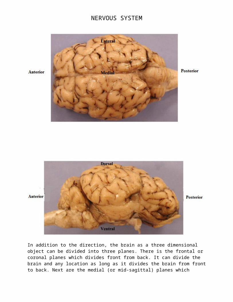
Before beginning the dissection of the sheep brain you will need to know the terms used to specify the location and relative location of various brain structures. Below are illustrations of directions:
NERVOUS SYSTEM
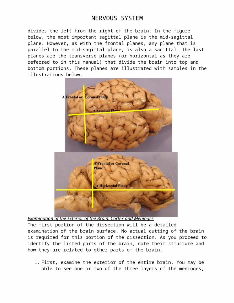
In addition to the direction, the brain as a three dimensional object can be divided into three planes. There is the frontal or coronal planes which divides front from back. It can divide the brain and any location as long as it divides the brain from front to back. Next are the medial (or mid-sagittal) planes which divides the left from the right of the brain. In the figure below, the most important sagittal plane is the mid-sagittal plane. However, as with the frontal planes, any plane that is parallel to the mid-sagittal plane, is also a sagittal. The last planes are the transverse planes (or horizontal as they are referred to in this manual) that divide the brain into top and bottom portions. These planes are illustrated with samples in the illustrations below.
NERVOUS SYSTEM
Examination of the Exterior of the Brain: Cortex and MeningesThe first portion of the dissection will be a detailed examination of the brain surface. No actual cutting of the brain is required for this portion of the dissection. As you proceed to identify the listed parts of the brain, note their structure and how they are related to other parts of the brain.
1. First, examine the exterior of the entire brain. You may be able to see one or two of the three layers of the meninges, the dura mater, the arachnoid layer, and the pia mater. The meninges are the protective coverings, which enclose the brain and spinal cord. The dura mater, the tough outer layer, will have been mostly removed when the brains were prepared for the dissection; however, some of the dura mater may remain near the base of the brain. The arachnoid layer, the middle layer, and pia mater, the inner layer, are still likely to cover the brain. The pia mater follows the gyri and sulci and most likely is still on your specimen and may be indistinguishable from the brain. Blood vessels are between the arachnoid layer and the pia mater. These vessels and the arachnoid layer will obscure your view of the sulci making the identifications below difficult and confusing. Before proceeding with the identification of structures on the surface of the brain you will need to remove the arachnoid layer and the blood vessels. Use your forceps and be very careful because the brain is soft and easily damaged.
2. Make a sketch of the brain from the lateral view as illustrated in the previous diagram. Make sure your sketch has a heading. In your sketch label the:
- Occipital lobe- Temporal lobe- Frontal lobe- Parietal lobe- Cerebellum- Brain stem
3. Below your sketch write a description about the external anatomy of your brain. How did it look, feel, was it smaller than you thought, what did the layers of the meninges feel like?
******************************************************************************Interrupted Analysis Questions
1. A patient comes to the emergency room with impaired motor ability on the right side of her body and impaired speech. The physician suspects that the patient suffered from a stroke, which causes the loss of blood flow to certain regions of the brain.
a. Which hemisphere of the brain did the stroke most likely occur in?b. What characteristics of the patient’s symptoms indicate that this is the region of
the stroke? (how can you tell that it is the left or right?)

NERVOUS SYSTEM
2. An 8 year old boy comes into the emergency room with is parents. He hit his head while playing baseball and, though he says he feels ok, his parents are still worried. The physician requests a brain scan that shows a crescent-shaped pattern of blood accumulation.
a. What kind of hematoma has this boy suffered from? How do you know?b. Should the physician be concerned, or send the child home since he seems fine?
Explain your response.

NERVOUS SYSTEM
3. Do you think the sheep brain has the Broca and/or Wernicke’s area(s)? Why or why not?
******************************************************************************Examination of the Exterior of the Brain: Brain Stem and Cerebellum
4. Once the protective layers of the meninges are removed, look at the ventral sides of the brain. You will see the pons, the medulla, and the cerebellum.

NERVOUS SYSTEM
******************************************************************************Interrupted Analysis Questions
4. “Pons” is the Latin word for “bridge.” Why might this structure be called bridge or pons?
5. “Medulla” refers to the “middle.” What is the medulla in the middle of?
****************************************************************************** Examination of the Internal Anatomy of the Brain: Medial CutDifferent cuts allow different views of brain structures. Structures that are not on the external surface of the brain are easier to visualize with coronal or sagittal sections. Together with your exploration of the brain surface anatomy, making a series of cuts will allow you to become more familiar with the three-dimensional structure of the brain.
5. Before making your first cut, place the brain so that you are looking at the dorsal surface. Using your hands (yes really) not the scalpel, gently pull the two hemispheres of the brain away from the medline. You don’t want to rip anything, but you should notice that the two hemispheres can be pulled slightly apart. You will see a structure that connects the two hemispheres: the corpus callosum. The corpus callosum contains bundles of fibers (axons) that connect neurons in the two hemispheres.
6. Make a medial cut. Holding the brain flat and level cut along the longitudinal fissure all the way through the brain. In this cut you can see the ventricles, which are open spaces in the brain that normally contain the cerebralspinal fluid. This fluid carries nutrients throughout the brain and regulate pressure. The ventricles ensure that the fluid has access to all areas of the brain, not only the external surfaces. As you cut, you may notice a thin layer or cells lining the ventricles (looks like pinkish “gunk”) that are responsible for producing the cerebralspinal fluid.

NERVOUS SYSTEM
7. Recall that you could identify the spinal cord, the medulla, and the pons on the exterior ventral surface of the brain. Identify these structures in the medial section you just made. You can also see the thalamus and the hypothalamus. The thalamus integrates information and relays it to the appropriate region of the cerebral cortex for processing. The hypothalamus is involved in many autonomic functions of the body including breathing and temperature regulation.

NERVOUS SYSTEM
8. Make a sketch of the median section of your brain. Make sure to give your sketch a heading. Label the:- Spinal cord- Pons- Medulla- Cerebellum- Hypothalamus- Thalamus - Pituitary gland- Corpus callosum- Ventricles
9. Finally, look at the gray and white matter of the medial section of the cerebellum. It resembles a tree and is called the arbor vitae, the “tree of life.” The white matter appears white because of the myelin sheaths that cover the axons. The gray matter appears gray because of the neural cell bodies.
******************************************************************************Interrupted Analysis Questions
6. Is the cerebral cortext composed of gray matter or white matter? Does it contain mostly axons or mostly cell bodies?
7. The word “cerebellum” is Latin for “little brain” How does the cerebellum resemble a little brain? Be specific.
******************************************************************************
ConclusionMake sure to include a conclusion in your lab write up.