welcomeback anatomic - mri and pain management centre
TRANSCRIPT

J. Neuroradiol., 2004, 31, 163-80© Masson, Paris, 2004Original article
THE ANATOMIC AND PHYSIOLOGIC BASIS OF LOCAL, REFERRED AND RADIATING LUMBOSACRAL PAIN SYNDROMES RELATED TO DISEASE OF THE SPINE
J. RANDY JINKINS
Department of Radiology, Downstate Medical Center, State University of New York, 450 Clarkson Avenue, Brooklyn, NY 11203,USA.
SUMMARY
Conscious perception and unconscious effects originating from the vertebral column and its neural structures, althoughcomplex, have definite pathways represented in a network of peripheral and central nervous system (CNS) ramifications.These neural relationships consequently result in superimposed focal and diffuse, local and remote conscious perceptionsand unconscious effects. Any one or combination of somatic and autonomic signs and symptoms may potentially be observedin a particular patient. This variety and inconsistency may mislead or confuse both the patient and the physician. A clearunderstanding of the basic anatomic and physiologic concepts underlying this complexity should accompany clinical consid-erations of the potential significance of spondylogenic and neurogenic syndromes in any disease process affecting the spine.
Key words: anatomy, pain, physiology, spine.
RÉSUMÉ
Bases anatomiques et physiologiques des syndromes douloureux d’origine rachidienneLa perception consciente et les effets inconscients provenant du rachis et des structures spinales empruntent un réseau complexe
appartenant aux systèmes nerveux central et périphérique. Ces rapports nerveux ont pour conséquence des effets inconscientset des sensations focaux et diffus, locaux et à distance. Signes et symptômes végétatifs et somatiques peuvent être observés chezun patient donné de façon isolée ou combinée. Cette expression variable et incohérente des signes peut tromper patient etclinicien. La compréhension des données anatomiques et physiologiques qui sous tendent cette complexité, est nécessaire pourcomprendre les syndromes vertébraux et nerveux observés en cas de lésion rachidienne
Mots-clés : anatomie, physiologie, douleur, rachis.
INTRODUCTION
The clinical state accompanying nonspecific injuryto the spinal column and perispinal soft tissues maybe manifest in a complex combination of somatic andautonomic syndromes. The overall combined symp-tom complex includes: local somatic spinal pain, radi-ating radicular pain, radiating/radicular paresthesias,radiating/referred skeletal muscle spasm/dysfunction,radiating/referred autonomic dysfunction, referredpain, and referred generalized alterations in visceros-omatic tone. In practice these clinical manifestationsare typically superimposed upon one another and areof varying individual expression.
The anatomic basis for the origin and mediation ofclinical signs and symptoms originating within the lum-bosacral spine is related to direct spinal innervation,the spinal nerve roots and nerves, and the lumbosacralsympathetic plexus. Specifically, the relevant neuralstructures include: afferent and efferent somaticneural branches emanating from the ventral and dor-sal rami of the lumbosacral spinal nerves, neural
branches projecting to and originating from theparavertebral autonomic (sympathetic) neural plexusand the spinal nerves/lumbosacral plexi themselves.Neural fibers from these structures originate and ter-minate in the spinal column and related nonneuraltissues (e.g., bone, periosteum, meninges, spinal liga-ments, perispinal musculature, spinal column andperispinal blood vessels) in the spinal neural tissueitself (e.g., spinal rami, nerves), and in the distantperipheral tissues within the somatic and visceraldistribution of these nerves (e.g., spinal column,perispinal soft tissues, buttocks, lower extremities,pelvis). Finally, the intimate connections of theseneural structures with the central nervous system,including the spinal cord and the higher cortical andnoncortical centers of the cerebrum, are ultimatelyresponsible for the manifestations of clinical syn-dromes in the patient with relevant spinal disease[10, 18, 35].
Thus, conscious perception and unconsciouseffects originating from the vertebral column, itsneural structures and the surrounding tissues,although complex, have definite pathways repre-sented in this network of peripheral and centralnervous system (CNS) ramifications. Although the
Reprint request: J. RANDY JINKINS, address above. e-mail: [email protected]
164 J. RANDY JINKINS
model for this discussion will center on the lum-bosacral spine, the particulars apply to all levels ofthe spine, after allowing for regional modifications.
ANATOMY OF LOCAL SPINAL SYNDROMES
Somatic Innervation of Ventral Spinal Elements
Innervation of the ventral spinal tissues restspartially with afferent somatic fibers originatingfrom the recurrent meningeal nerve (sinuvertebralnerve of von Luschka) supplying the posterior lon-gitudinal ligament, the meninges of the anterioraspect of the thecal sac, the regional anterior epi-dural blood vessels (arteries and veins), the poste-rior aspect of the outermost fibers of the annulusfibrosis, the anterior and posterior longitudinal lig-aments and the posterior portion of the periosteumof the vertebral body and related tissues over aninconstant range. In addition, irregular, unnamedafferent branches directly emanating from the ven-tral rami of the somatic spinal nerves themselvesalso contribute to direct spinal and adjacentperispinal soft tissue innervation laterally. Thus,these well-defined somatic neural networks formthe anatomic basis for discogenic (intervertebraldisc), spondylogenic (spinal bony structures), mus-culogenic (intrinsic spinal and external perispinalmuscles), ligamentogenic (intrinsic spinal liga-ments) normal sensation and pathologic pain [23,36, 45, 48, 74, 90, 91].
Any insult of these neural and non-neural tissuesmay incite well-circumscribed local somatic painbecause of this characteristic somatosensory inner-vation pattern (figure 1), and because of the directsegmental nature of the afferent inflow from the seg-ment of origin into the CNS via the somatic spinalnerves (figure 2a) [6, 9, 20, 23, 29, 38, 52, 63, 87, 90,91]. This direct somatosensory afferent inflow seems
to embryologically insure a relatively accurate CNSsomatotopic spatial registration of impulses incominginto the CNS with regard to stimulus origin, and thuslocal spine pain.
Somatic Innervation of Dorsal Spinal Elements
The dorsal rami of the spinal nerves give rise tomedial and lateral main branches. These neuralstructures innervate the posterior spinal facet (zyga-pophyseal) joints (bone, periosteum, articular struc-tures including the joint capsule), the lateral andposterior vertebral bony elements (laminae, trans-verse and spinous processes), as well as the sur-rounding posterior (dorsal) intrinsic spinal andperispinal muscular (multifidus, interspinalis mus-cles), and ligamentous tissues (interspinous liga-ment, supraspinous ligament).
In total there are potentially five or more mainbranches innervating these structures that are ofsomewhat irregular origin. These branches includeneural fibers arising directly from the main trunk ofthe dorsal ramus of the spinal nerve, from the medialbranch of the dorsal ramus, from the lateral branchof the dorsal ramus, and from the combined spinalnerve itself before its bifurcation into the dorsal andventral rami (figure 3) [2, 5, 8].
On careful anatomic study, the dorsal elements ofthe spinal column and surrounding tissues have beendemonstrated to have remarkably variable fields ofinnervation that are not confined to strict segmentalpatterns. This innervation shows bilateral asymmetrywith intra- and interindividual variation in the cran-iocaudal extent of the neural supply.
Nevertheless, injury to these neural and non-neu-ral spinal/perispinal tissues would in part, beexpected to result in well localized somatic painbecause of the direct afferent somatosensory inflowinto the CNS via the retrospective somatic spinalnerves. Thus, in general this neural handling of painseems to occur in a manner similar to that outlined
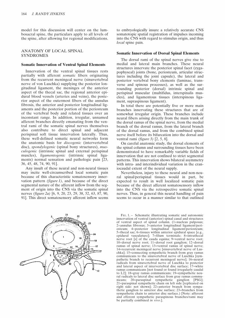
FIG. 1. – Schematic illustrating somatic and autonomicinnervation of ventral (anterior) spinal canal and structuresof ventral aspect of spinal column. (1=nucleus pulposus;2=annulus fibrosus; 3=anterior longitudinal ligament/peri-osteum; 4=posterior longitudinal ligament/periosteum;5=thecal sac; 6=tissues within anterior epidural space [e.g.,epidural vasculature]; 7=filum terminale; 8=intrathecalnerve root [s] of the cauda equina; 9=ventral nerve root;10=dorsal nerve root; 11=dorsal root ganglion; 12=dorsalramus of spinal nerve; 13=ventral ramus of spinal nerve;14=recurrent meningeal nerve [sinuvertebral nerve of Lus-chka]; 15=connecting sympathetic branch from gray ramuscommunicans to the sinuvertebral nerve of Luschka [sym-pathetic branch to recurrent meningeal nerve]; 16=neuralradicals from sinuvertebral nerve of Luschka to posteriorand lateral aspect of intervertebral disc surface; 17=whiteramus communicans [not found or found irregularly caudalto L2]; 18=gray ramus communicans; 19=sympathetic neu-ral radicals to lateral disc surface from gray ramus commu-nicans; 20=paraspinal sympathetic ganglion [PSG];21=paraspinal sympathetic chain on left side [replicated onright side: not shown]; 22=anterior branch from sympa-thetic ganglion to anterior disc surface; 23=branches fromsympathetic chain to anterior disc surface.) [Note: afferentand efferent sympathetic paraspinous branches/rami maybe partially combined in vivo.].

PAIN SYNROMES RELATED TO DISEASE OF THE SPINE 165
above for the ventral spinal elements (figure 2a). Inother words, these neural structures contribute todirect posterior (dorsal) spinal and perispinal softtissue innervation, and thus somatic spondylogenic,(posterior bony spinal tissues), articulogenic (poste-rior spinal facet joints), ligamentogenic (ligamentaflava, interspinous ligament, supraspinous ligament)and musculogenic (multifidus, interspinalis muscles)normal sensation and local spinal pain [1, 10, 36, 45,48, 74, 91].
Additional Considerations in the Innervation of Spinal Elements
Local innervations at the level of the dorsal andventral roots, spinal nerves, recurrent meningealnerves and other epidural structures at the pointof common expression of pathology (e.g., interver-
tebral disc herniation, spinal stenosis) is an impor-tant consideration regarding the understanding ofthe manifestations of the lumbosacral syndromes.In addition to peripheral and local somatic affer-ent sensory and efferent motor nerves traversingthis region (figure 4a-c), there are nerve fibersinnervating the nerves themselves, the nervi ner-vorum [21].
These nervi nervorum are theoretically of threetypes. First, there are afferent somatic sensory fibers tothe main nerve radicles in, traversing and around thespinal column (figure 4d). These are responsible forlocal sensation and even pain when the nerve itself isperturbed or nonspecifically injured. Second, there arelocal tissue and intrinsic radicular sympathetic afferentfibers. Upon leaving the spine, these fibers enter theparaspinal sympathetic chain via the gray rami com-municantes and return to the CNS via the white rami
FIG. 3. – Schematic illustrating innervation of structures of dorsal (posterior)aspect of spinal column. (1=main trunk of spinal nerve; 2=ventral ramus of spinalnerve; 3=lateral branch of dorsal ramus of spinal nerve; 4=neural fibers to posteriordorsal ramus of spinal nerve; 5=dorsal ramus of spinal nerve; 6=dorsal nerve rootand ganglion; 7=ventral nerve root; 8=gray ramus communicans; 9=white ramuscommunicans; 10=intervertebral disc; 11=articular cartilage of posterior spinalfacet [zygapophyseal] joint; 12=neural fibers from main trunk of spinal nerve;13=neural fibers to posterior facet joint from ventral ramus of spinal nerve;14=neural fibers to posterior facet joint from dorsal ramus; 15=medial branch ofdorsal ramus; 16=central spinal canal; 17=superior articular facet process; 18=infe-rior articular facet process; 19=zygapophyseal joint space and capsule; 20=spinousprocess; 21=interspinous ligament; 22=medial neural branches ramifying withinposterior spinal facet joint, the lamina, spinous process, interspinous ligament, andsupraspinous ligament; 23=branch of dorsal ramus ramifying within posterior peris-pinous tissues, 24=transverse process; 25=lamina; 26=supraspinous ligament;27=ligamenta flavum; 28=median retrothecal fat pad).
a b
FIG. 2. – Schematics illustrating lumbar afferent sen-sory patterns. a) Direct somatic afferent inflow into cen-tral nervous system (CNS) from branches of somaticspinal nerves at all levels. b) Ascending autonomic(sympathetic) afferent inflow diversion into CNS oflumbosacral sympathetic fibers. This inflow pattern isinconstant due to the absence or irregular distributionof white rami communicantes occurring between the L2and S2 vertebral levels. (ASC=ascending sympatheticchain; large diameter solid lines=afferent sympatheticnetwork; short arrows=afferent sympathetic inflow fromventral spinal elements; dashed lines=afferent sympa-thetic inflow from dorsal spinal elements).

166 J. RANDY JINKINS
FIG. 4. – Schematics of somatic and autonomic (sympathetic) innervation of spinal column and related structures. a) Peripheralsomatic afferent sensory neural fibers from ventral and dorsal tissues carried within the dorsal and ventral rami joining the spinalnerve proper. b) Peripheral somatic efferent motor neural fibers to ventral and dorsal tissues carried within the dorsal and ventralrami joining the spinal nerve proper. c) Local somatic afferent sensory neural fibers (including those within the recurrentmeningeal nerve) from ventral, intra- and perispinal tissues (e.g., peripheral disc, epidural tissues, dura, etc.), and dorsal spinal,perispinal tissues (e.g., facet joints, posterior spinal ligaments, posteromedial perispinal musculature, etc.). d) Local/radicularafferent nervi nervorum. e) Local/radicular sympathetic afferent neural fibers (including those within the recurrent meningealnerve) from ventral spinal, intra- and perispinal tissues (e.g., peripheral disc, epidural tissues, dura, etc.). f) Local/radicular sym-pathetic efferent neural fibers (including those within the recurrent meningeal nerve) to ventral spinal, intra- and perispinal tissues(e.g., peripheral disc, epidural tissues, dura, etc.). g) Local sympathetic afferent neural fibers from dorsal spinal and perispinaltissues (e.g., facet joints, posterior spinal ligaments, posteromedial perispinal musculature, etc.). h) Local sympathetic efferentneural fibers to dorsal spinal and perispinal tissues (e.g., facet joints, posterior spinal ligaments, posteromedial perispinal muscu-lature, etc.). (1=ventral nerve root; 2=dorsal nerve root; 3=dorsal root ganglion; 4=combined spinal nerve; 5=dorsal ramus of spi-nal nerve; 6=ventral ramus of spinal nerve; 7=white ramus communicans [not found or irregularly found caudal to L-1,2]; 8=grayramus communicans; 9=branch to recurrent meningeal nerve from spinal nerve; 10=recurrent meningeal nerve [sinuvertebralnerve of Luschka]; 1=autonomic [sympathetic] branch to recurrent meningeal nerve from gray ramus communicans; 12=annulusfibrous; 13=nucleus pulposis; 14=epidural vasculature; 15=leptomeninges; 16=intrathecal lumbosacral nerve root; 17=filum termi-nale).
a bc d
e fg h

PAIN SYNROMES RELATED TO DISEASE OF THE SPINE 167
communicantes (figure 4e). These fibers relay afferentinformation from the spinal roots, nerves and sur-rounding tissues to the somatic and sympatheticnervous systems. Third, there are local/radicular sym-pathetic efferent fibers which carry out sympatheticactions (e.g., vasoactive functions) upon the spinalroots, nerves and surrounding tissues (figure 4f).
This general format of spinal innervation is likelyreplicated in its essential points in the spinal andperispinal tissues, the autonomic fibers being initiallytransmitted via the dorsal roots of the spinal nerves,and later traveling in company with the blood vesselssupplying these tissues (figure 4g-h) [2, 7, 16].
With this level of neuroanatomic complexity, it isnot surprising that pathology affecting this particularregion would be expected to potentially be somewhatconfusing in its clinical manifestations. All possiblesomatoautonomic expressions (e.g., local pain,referred pain, radicular pain, autonomic dysfunction[see below]) could possibly emanate from this net-work of afferent and efferent fibers that traverse thisarea as well as originate and terminate here. At thesame time, injury to somatic efferent motor fiberscontained in the ventral/dorsal rami of the spinalnerve or the spinal nerve itself might yield muscularweakness, spasm and muscle reflex dysfunction. This
surely must be one of the more neurologically labyrin-thine regions in the entire peripheral nervous system.Why each patient might be expected to have a uniqueand compound-complex array of signs and symptomscan be easily appreciated if these intimately relatedanatomic neural ramifications are taken into account.
ANATOMY OF CENTRIPETALLY/CENTRIFUGALLY REFERRED SPINAL SYNDROMES
As noted above, many afferent fibers from the spi-nal column project immediately to the paraspinal sym-pathetic ganglia [29]. Afferent polymodal pain fiberstraversing the sympathetic ganglia have been identifiedin all of the vertebral structures except the central areasof the annulus fibrosus and the nucleus pulposus. Thetissues innervated by sympathetic fibers include thelongitudinal ligaments, the most peripheral laminae ofthe annulus fibrosus, the periosteum of the vertebralbody, the vertebral body itself, the posterior bony ele-ments (posterior spinal facet joints, laminae, spinousprocesses) and the spinal and perispinal soft tissues(blood vessels, muscles, ligaments) [16, 20, 36, 38, 63,77, 78].

168 J. RANDY JINKINS
A major autonomic branch also extends on bothsides posteriorly from either the sympathetic gan-glion directly or indirectly from the gray ramuscommunicans to make up the bulk of the recurrentmeningeal nerve (figure 4e) [6, 42, 46]. Thus, afferentsympathetic fibers supply the whole of the discperiphery, and indeed the entire vertebral column[1]. This extensive network, known as the paraverte-bral autonomic neural plexus, was initially detailedby Stilwell in 1956 (figure 1) [77].
Depending on the vertebral level, after traversingthe sympathetic ganglia many of these primary affer-ent fibers subsequently enter the ventral ramus ofthe somatic spinal nerve via the white ramuscommunicans [80]. These axons then pass into thedorsal root ganglion, where their cell bodies lie(figure 4e). Afterward, the dorsal nerve root carriesthe fibers until they penetrate the dorsolateralaspect of the spinal cord within the tract of Lissauer,adjacent to the dorsal-horn gray matter.
The anatomic path and embryologic origin ofthese neural elements within the autonomic ner-vous system contribute in part to the imperfectconscious perception and somatic localization ofmany pain stimuli [19]. Conscious somatotopiclocalization of pain is normally registered embryo-logically largely by means of the point of spatialentry of afferent fibers, and therefore neuralimpulses, into the CNS. Another way of saying thisis the means by which the cerebrum understands(i.e., imagines) the point of a painful stimulus inthe adult organism is dependent upon the somite ofimpulse origin must be isolevel with the somite ofthe spinal nerve serving that particular somite inorder to obtain accurate somatotopic localizationof the impulse origin within the CNS. For example,a somatic axon impulse entering the CNS (i.e., spi-nal cord) at the L5 level will be perceived mentallyat higher CNS centers in the L5 somatotopic sen-sory region.
Some pain related impulses entering afferent sym-pathetic fibers may result in appropriately localizedsymptomatology such as pain [19], while other axonswill be involved with important or even inappropri-ate autonomic reflex functions [4]. Impulses withinyet different (or perhaps the same) afferent fiber,however, will result in the conscious mental pictureof remote pain (i.e., perceived in the groin, pelvis,gluteal area, proximal posterolateral thigh). Suchreferred pain is mentally projected to the region cor-responding generally to the somatic distribution ofthe afferent fibers of the spinal nerve with which theafferent sympathetic fibers entered the spinal canalen route to the CNS, as was noted above for thesomatic nervous system.
A somatome is in part defined as a field ofsomatic and autonomic innervation that is basedon the embryologic segmental origin of thesomatic tissues [37]. The complete somatome iscomposed of three basic elements: the cutaneousstructures (dermatome), the skeletal musculature(myotome), and the bones, joints, and ligaments(sclerotome). The term “somatic” indicates thatthese tissues originate embryonically from the pre-cursor somites [64]. Tissues originating from thesame somite, therefore, will have a common neural
circuitry and thus a common pathway of neuralreferral. Thus, distant pain referral is mentally“projected” to these distant fields of innervation,within the lumbosacral somatomes. The conscioussomatic registration (or perhaps mental illusion) ofreferred pain is perceived peripherally by the brainwithin what have come to be known as the lum-bosacral zones of Head, named after Henry Headwho originally described these areas in 1893 [31].Unfortunately these peripheral regions of painreferral are found in the same physical distributionas is the radiating pain seen in true neurogenic sci-atica. It is for this reason that referred pain issometimes termed “pseudoradicular” pain. Never-theless, these zones of Head (as compared to thecutaneous dermatomes) are irregular, constricted,bilaterally asymmetric, and partially superimposedupon one another [24, 43, 45, 47, 53, 54]. Moreover,they are somewhat inconsistent from person toperson (figure 5) [55]. Proof that the referredpain’s origin is a process intimately involving anafferent limb of the peripheral nervous system,and that the illusory mental perception of distantreferred pain is a mechanism of the CNS, is con-firmed by the experimental finding that local anes-thesia of the actual region of impulse origin (i.e.,spinal tissues) abolishes the pain referral, however,anesthesia of the site of referral (i.e., the zone ofHead) does not consistently ablate this referredpain [17, 28].
The referred nature and poor definition of thepain are potentially further complicated by the dis-tribution patterns of the sympathetic afferent fibersof the spine, which overlap craniocaudally as wellas across the midline. In other words, there is nei-ther true anatomic midline nor accurate segmental
FIG. 5. – Schematics of right unilateral composite of lum-bosacral zones of Head representing regions of pain referraland proposed reflex autonomic dysfunction referral from seg-mental spinal levels (L1-S1, 2). Note the constricted, superim-posed, and skipped regions. a) Anterior aspect. b) Posterioraspect.
PAIN SYNROMES RELATED TO DISEASE OF THE SPINE 169
nature of the lumbosacral paravertebral autonomic(sympathetic) nervous system. In addition, afterafferent sympathetic fibers enter the paraspinalsympathetic ganglia, they cannot always exitdirectly into the nearby somatic ventral or dorsalrami of the spinal root. These fibers instead mayhave to ascend to a more craniad level beforeentering the spine. The reason for this is becausethe afferent fibers can only join the spinal nervesand subsequently enter the CNS via the white ramicommunicantes. An important anatomic patternand peculiarity of the lumbosacral spine illustratesthat there are no, few or irregularly distributedwhite rami communicantes below the L2 vertebrallevel and above the S2 level [27]. Any sympatheticafferent fibers from the lower lumbar and uppersacral region (i.e., between the L2 to S2 levels)therefore must ascend within the paravertebralsympathetic chain before they are able to enter thespine at a level that has a white ramus communi-cans (figure 2). As a result of this autonomic inflowdiversion [26, 56, 58, 59, 78-82], sympathetic painimpulses emanating from lumbosacral regions thatdo not have white rami communicantes (L3 to S1levels) will be referred to the somatome corre-sponding to the final spinal entry level of the affer-ent fiber (L2 or above). Thus, the consciousperception of sympathetically mediated pain will bemisregistered in the CNS (sensory cortex of cere-brum), and pain referral will thereby occur to asomatome different from that which its originwould have indicated. This may also possibly resultin summing of pain sensation due to the superim-position of afferent fiber input from several differ-ent levels [28]. These observations might explainthe partial segmental superimposition and irregularcontracted nature of the zones of Head in the lum-bosacral region, as depicted in figure 5 [24]. Itshould also be noted that the overlapping areas ofmost common centrifugal pain referral from alllumbar levels in fact fall largely within the area ofthe cutaneous dermatomes of the upper lumbar spi-nal nerves (i.e., along the inflow pathway of theascending sympathetic afferent diversion into theL2 spinal nerves and perhaps above).
These unusual lumbosacral innervation patternsmay also engender local referred pain to the spineitself and its surrounding tissues [29]. Conscious painreferral originating in the spinal column and spinalneural tissue and subsequently projected to the lum-bosacral zones of Head is linked with spinal nervesthat coincidentally have afferent somatic projectionfields within spinal and perispinal structures. Inother words, an integral component of thesomatomes of spinal nerves (e.g., myotomes, sclero-tomes) includes the spinal elements themselves.Thus, although the local referred pain is not per-ceived at the precise point of origin in the spine, it isstill consciously imagined diffusely in the region ofthe low back [24, 37, 47, 54].
A combination of local referred, distant referredand local somatic pain when combined with thesometimes concurrent radiating radicular neurogenicpain (see next section), seems to partly explain theparallel systems operating in the spinal column
responsible for the complex and often superimposedsyndromes of spinal pain [16, 28].
Further inspection of figure 5 reveals the sparseareas of nonsuperimposed pain referral extendingdistally into the lower extremities. This pattern maybe explained by the fact that there is direct sympa-thetic afferent inflow into the S2-S4 pelvic somaticnerve roots, and also by the observation that thesympathetic innervation of spinal structures mayoriginate from as few as three and as many as fivedifferent adjacent spinal levels [90]. Hence, direct,multisegmental sacral nerve inflow, and thereforedirect pain referral may occur over relatively wideareas [24, 71, 90]. These general anatomic conceptshelp to clarify some of the mechanisms within theperipheral nervous system responsible for therather nebulous fields that are characteristic of thezones of Head.
As the foregoing indicates, the majority if not theentire peripheral network resulting in the perceptionof referred pain could potentially be mediatedwithin the sympathetic nervous system. The periph-eral neurologic system follows two patterns duringembryologic development: somatic and autonomic.The somatic nervous system has one distribution,which ramifies solely within the somatic tissues.However, the autonomic (sympathetic) nervous sys-tem develops along two distinct different pathways:1) one within visceral tissues sometimes referred toas the visceral tissue autonomic nervous system, and2) one within the somatic tissues in a distributionsimilar to that of the peripheral somatic nerves thatmight be called the somatic tissue autonomic nervoussystem (carrying afferent and efferent sympatheticfibers from and to the somatic tissues). Logicallythere must be such parallel sympathetic afferentlinks to the CNS in order to complete somatic tissueautonomic reflex arcs (figure 6a). Normal autonomicfunctions depend upon this (e.g., vasomotor, sudo-motor and piloerector functions). In fact, the pres-ence of these peripheral autonomic fibers withinsomatic tissues have been clearly demonstrated [1,19, 36, 71, 78, 80]. Because both visceral and somatictissues are innervated by the sympathetic nervoussystem, and assuming that both tissues are served byafferent limbs, the CNS may then perceive animpulse origin within either tissue (visceral, somatic)or both on the basis of a central embryologically pre-determined linkage. In actuality, however, the CNS(sensory cortex) may not be able to accurately dis-criminate spatially between the visceral and thesomatic origin of a stimulus in certain circumstances.Thus, a visceral sympathetic afferent stimulus mayerroneously be consciously perceived as arisingwithin the somatic sector of the sympathetic afferentsensory projection field, and by definition is thusreferred mentally to this location. The converse ofthis phenomenon might also be true, although per-haps more rarely perceived [4].
As an example, this explanation concisely fits theobservation of referral of visceral sympathetic stim-uli (e.g., cardiac injury and related pain) to thesomatic sympathetic afferent projection field (e.g.,left shoulder), thereby defining the zones of Headpredominantly or entirely as a phenomenon of adevelopmentally dichotomous sympathetic nervous

170 J. RANDY JINKINS
a b
FIG. 6. – Schematics of proposed configuration of peripheral nervous system and its central terminations, illustrating some ofthe principles of CNS neural convergence of peripheral neural input, and explaining in part the mechanism of referred pain. a)Efferent pathways (solid arrows), afferent pathways (dashed arrows), visceral tissue sympathetic afferent fibers (open square),somatic tissue somatic afferent fibers (open circle), and hypothesized somatic tissue sympathetic afferent fibers (solid diamond).Note the use of the same symbols and meanings in figure b.
(VSNS=visceral sympathetic nervous system; SNS=somatic nervous system; SSNS=somatic sympathetic nervous system).b) Points of termination of visceral and somatic afferent fibers on cord neurons within dorsal and ventral gray matter of right spinalhemicord gray matter (ventral: top; dorsal: bottom; right hemicord to reader’s left). Note overlapping regions covered by squaresand diamonds that theoretically result in a central nervous system convergence of afferent fibers from divergent origins (i.e., periph-eral visceral and somatic neural fiber CNS convergence). (I-V=laminae of dorsal horn gray matter of spinal cord; termination ofsomatic [somatic tissue somatic] afferent fibers on somatic cord neurons [open circles]; termination s of visceral [visceral tissue sym-pathetic] afferent fibers on visceral cord neurons [open squares]; terminations of somatic [proposed somatic tissue sympathetic] affer-ent fibers [alternate proposal: somatic tissue somatic fibers] on viscerosomatic cord neurons [solid diamonds])
FIG. 7. – Schematic of proposed general organization ofperipheral afferent somatic and sympathetic nervous systemsillustrating CNS convergence of neural input from peripheralsomatic and visceral tissues. Theoretically, referred pain mayin part be a result of either of two possible mechanisms:convergence in spinal cord of separate fibers from visceraland somatic sources (1), or single convergence in spinal cordof bifurcating axons having diverging limbs within the visceraland somatic tissues (2). (1=dual afferent axon configuration;2=bifurcating afferent axon configuration; visceral sympa-thetic afferent fibers [solid axons]; somatic sympathetic affer-ent fibers [dashed axons]; VSNS=visceral sympatheticnervous system; SSNA=somatic sympathetic nervous system;arrowhead=point of bifurcation).
FIG. 8. – Schematic illustrating aberrant autonomic reflex arc.(manifestation: peripheral autonomic [sympathetic] dysfunc-tion). The afferent limb (dashed lines) of the autonomic reflexarc is theoretically carried within ascending paraspinal sympa-thetic chain (open ovals). After synapse in spinal cord, the effer-ent limb is carried within peripheral ramifications (e.g., spine,pelvis, extremities) of somatic and/or sympathetic components ofsomatic spinal nerves (long multiheaded curved arrow).

PAIN SYNROMES RELATED TO DISEASE OF THE SPINE 171
a b
FIG. 9. – Schematics illustrating hypothetical mechanism of normal sodium ion channel function across axon membrane. a) When asodium ion channel is closed, the passive influx of sodium ions into the cell intra-axonal space does not occur, the neuroelectrical voltagepotential is maintained and no neuroelectrical activity takes place along or across the axon membrane. b) When a sodium ion channelis open, as in normal longitudinal neuroelectrical axon conduction (neuroelectrical impulse transmission), the passive influx of sodiumions into the intra-axonal space is facilitated resulting in depolarization across the axon membrane (NA+=sodium ions).
a b c
FIG. 10. – Schematics illustrating hypothetical mechanism of normal function and dysfunction in axon membrane resulting inaxonal autodepolarization and ectopic neuroelectrical axon impulse generation (i.e., radiating radiculopathy). a) During normalneuroelectrical impulse transmission, sodium ion flow occurs into the axonal space, and potassium ions flow externally. Subse-quently, after normal impulse transmission subsides, the active energy-requiring sodium-potassium pump, that maintains the neu-roelectrical voltage potential across the axon membrane during inactivity, restores this ion voltage potential as the axon reaches aresting state. (NA+=sodium ions; K+=potassium ions; arrows in conduits=direction of flux in ion channels; C=central; P=periph-eral). b) Unbalanced relationship between the sodium ion channels and the potassium pump. In this circumstance, the functionalsodium ion channel spatial density in the axon membrane has increased without a consonant increase in the potential of thesodium-potassium pump to balance this phenomenon [compare with A)]. This abnormal relationship theoretically overwhelmsthe sodium-potassium pump’s ability to maintain the neuroelectrical voltage potential across the axon membrane resulting inautodepolarization (i.e., ectopic neuroelectrical axon impulse generation). (NA+=sodium ions; K+=potassium ions; arrows inconduits=direction of flux in ion channels; C=central; P=peripheral). c) The sodium-potassium pump itself may also be dysfunc-tional (oblique bar). Such a functional defect in the axon membrane constitutes an ectopic source (star burst) of neuroelectricalactivity (i.e., within the intra-axonal space). Because the resulting aberrant neuroelectrical action impulse is transmitted in bothdirections (centrally and peripherally) along the axon (bold arrows), this phenomenon is theoretically partly responsible for patho-logic efferent outflow peripheral nervous system (efferent somatic and autonomic fibers) involuntary expression (e.g., musclespasm, autonomic dysfunction) and afferent inflow central nervous system (afferent somatic and autonomic fibers) conscious per-ception (e.g., radicular pain, paresthesias). (NA+=sodium ions; K+=potassium ions; arrows in conduits=direction of flux in ionchannels; C=central; P=peripheral).
172 J. RANDY JINKINS
system dually ramifying within the visceral and thesomatic tissues [73]. Understood in this way,referred actions and conscious perceptions are anexpected if aberrant capacity of the autonomic(sympathetic) nervous system. Thus, while theascending afferent lumbar autonomic inflow diver-sion accounts for extrasegmental CNS misregistra-tion and patterns of mismapped and superimposedpain within the lumbosacral zones of Head, theactual primary referral seems to result from themediation of the painful stimulus within the auto-nomic nervous system [10].
However, only so much can be understood withinthe framework of the peripheral nervous system, andthereafter CNS mechanisms of pain referral must beconsidered [18, 35]. Anatomic data suggest thatsomatic and visceral autonomic afferents may havethe same or some of the same central connections atthe level of the spinal cord, thalamus, and sensorycortex [13, 71, 88-90]. The convergence theory for theoccurrence of referred pain states that because someof the same central pathways are shared by theconverging visceral and somatic afferent autonomicsystems, the CNS cannot precisely distinguishbetween the two origins of sensory input [72]. Anancillary hypothesis indicates that since the somatictissues are normally continually consciously relayingstimuli, as opposed to the viscera, through a processof pattern recognition, the CNS attributes most of thesegmental afferent inflow to somatic origins regard-less of the true site of the stimulus [71, 82, 88]. Thereis little doubt, however, that some degree of modula-tion of afferent input from any peripheral sourceoccurs at the level of the spinal cord and above [30,90].
Thus some of the mechanisms for somatotopicreferral of pain seem to lie at the level of the spinalcord and brain. As a further clarification, there seemsto be a definite somatotopic organization of the spinalcord with regard to entering afferent fibers. Somaticafferent fibers largely terminate on neurons (i.e.,somatic spinal cord neurons) within laminae II, III,and IV of the dorsal horn gray matter, while visceralafferent fibers terminate on neurons (i.e., visceralspinal cord neurons) in laminae I and V andwithin the ventral horn gray substance (figure 6b).However, there is a third population of cellslocated intermediately between these two groups(visceral and somatic) upon which some afferentsterminate known as viscerosomatic spinal cordneurons. The rationale for this terminology is thatthese latter neurons are driven by afferent stimulifrom both the somatic as well as the visceraltissues [71].
A complimentary theory to the foregoing forreferred pain considers the possibility of bifurcatingperipheral sympathetic afferent fibers, with onedistal limb entering the visceral tissues while theother ramifies within the somatic tissues (figure 7)[3, 13, 15, 66]. Nevertheless, the important conceptis still that of convergence of afferent impulsesfrom different regions upon the same visceroso-matotopic registration area (or neuron) of theCNS, either primarily or via connecting interneu-rons. This may cause a false mental image of thelocalization of a sensory event. In context there-
fore, pain referred to the peripheral tissues (e.g.,zones of Head) from a primary stimulus sourcesuch as the spine, does not have its stimulus originin the area of conscious perception (i.e., zones ofHead). This definition of central pain perceptionindicates that referred pain fields are thus wronglyimagined by the higher cognitive centers of theCNS in part because of afferent CNS sensoryimpulse convergence.
Autonomic nervous system function, however, isnot confined to the conscious perception of painfulstimuli. This network also has a major role in themediation of unconscious normal autonomic func-tion via autonomic reflex arcs occurring at the levelof the spinal cord, which in turn are influenced byhigher CNS levels [13, 39, 41, 88, 90]. Just as the con-scious perception of pain may be spatially misregis-tered, so too may be various autonomic functions.Current understanding suggests that somatic as wellas autonomic fibers both excite or otherwise sharethe same interneurons within the spinal cord [13].Aberrant autonomic reflex arcs resulting in referredautonomic dysfunction of spinal column origin mightbe represented in the form of aberrant centrifugalvasomotor, pilomotor, and sudomotor activity per-haps within the zones of Head [71]. In addition tothese positive sympathetic effects, reverse or para-doxical effects might be observed, presumably dueto pre- and/or postsynaptic efferent inhibition bypolysynaptic, polyaxonal afferent spinal cord input[88, 90].
However, these findings are seemingly minor, andare overshadowed by the manifestations of referredpain. Apparently, then, such autonomic dysfunctionis often disregarded clinically. These phenomena
a
b
FIG. 11. – Schematics illustrating spontaneous ectopic neuroelectrical activity resulting in central (C) and peripheral (Paberrant neuroelectrical impulse propagation in injured axon(wavy contoured channels). These phenomena could hypothetically occur in both spinal afferent axons (a) and efferenaxons (b), and explain in part radiating pain, muscular anreflex dysfunction, and autonomic dysfunction within the ditribution of the particular axon. a) Spontaneous ectopic neuroelectrical impulse (asterisk) originating in injured afferenaxon (open channel: somatic or autonomic). The aberranimpulse (solid arrows) propagates away from the site of spontaneous depolarization, both centrally as well as peripherall(central manifestations: well localized pain perceptioreferred signs and symptoms) (peripheral manifestation: sympathetic dystrophy [?]). b) Spontaneous ectopic neuroelectrcal impulses (asterisk) originating in injured efferent axo(shaded channel: somatic or autonomic). The aberranimpulse (open arrows) propagates away from the site of spontaneous depolarization, both peripherally as well as centrall(peripheral manifestations: skeletal muscle spasm; autonomeffector dysfunction).

PAIN SYNROMES RELATED TO DISEASE OF THE SPINE 173
may be more common than realized, and could per-haps be elicited with greater frequency and meaningif subjects were carefully scrutinized for these mani-festations at the time of clinical examination.
Somatic muscle spasm can also possibly be associ-ated with autonomic function/dysfunction [13, 22, 32,71, 90]. Skeletal muscle spasm, which may become apainful process in and of itself, is in part theoreticallyaccomplished by an aberrant reflex arc, similar tothat of the autonomic reflex dysfunction discussedearlier. Thus, referred reflex somatic muscle spasmin the lumbosacral myotome, known as a visceroso-matic reflex, may account for clinically significantsymptomatology [39]. The spasm itself could be pro-duced by an arrest of the usual negative feedbackmechanisms that ordinarily affect muscular contrac-tion, because the stimulus does not originate withinthe area of the effect (i.e., the lumbosacral zone of
Head), but instead from a distant referral source(i.e., the spine). Alternately, unopposed positivefeedback mechanisms may be responsible for themuscle spasm for similar reasons [88].
In all such autonomic reflex dysfunction, theafferent neural limb eventually enters the paraspinalsympathetic plexus. As previously discussed, largelybecause of the ascending autonomic inflow diversion(often entering at or above the L-2 segmental level)and because of the peculiarities of the autonomic(sympathetic) nervous system (both in its centraland peripheral ramifications), there may be a spatialmismapping of otherwise normal autonomic affer-ent/efferent function, causing the efferent effectorlimb of the arc to occur in the peripheral somatome(figure 8). This autonomic dysfunction, an aberrantsomatosympathetic (or sympathosomatic) reflex,might include any one or combination of dermal
a b
FIG. 12. – Schematics illustrating spontaneous ectopic neuroelectrical activity resulting in ephaptic axo-axonal transmission-stimulation between adjacent injured axons (wavy contoured channels). These phenomena could hypothetically occur betweenafferent axons (a), efferent axons (b), and between afferent and efferent axons (c, d). (manifestations: interaxonal ephapticrecruitment with multiplied peripheral and central effects and signs/symptoms). a) Spontaneous depolarization (single asterisk)originating in injured afferent axon (upper open channel: somatic or autonomic) results in a neuroelectrical impulse (single aster-isk) that propagates away from site of ectopic origin (upper solid arrows). At the same time, ephaptic transmission (open ser-pentine arrow) axo-axonally results in neuroelectrical stimulation (double asterisk) of adjacent injured afferent axon (lower openchannel: somatic or autonomic) acting as an ectopic receptor and effecting central (C) and peripheral (P) aberrant afferent axonimpulse propagation (lower solid arrows). b) Spontaneous depolarization (single asterisk) originating in injured afferent axon(open channel: somatic or autonomic) results in a neuroelectrical impulse (single asterisk) that propagates away from site ofectopic origin (open straight arrows). At the same time, ephaptic transmission (open serpentine arrow) of axo-axonally resultsin neuroelectrical (direct neuroelectric exchange or ion-imbalance driven) stimulation (double asterisk) of adjacent injured effer-ent axon (shaded channel: somatic or autonomic) acting as an ectopic receptor and effecting central (C) and peripheral (P) aber-rant efferent axon impulse propagation (open straight arrows). c) Spontaneous depolarization (single asterisk) originating ininjured afferent axon (upper open channel: somatic or autonomic) results in a neuroelectrical impulse (single asterisk) that prop-agates away from site of ectopic origin (upper solid straight arrows). At the same time ephaptic transmission (open serpentinearrow) axo-axonally results in neuroelectrical (direct neuroelectric exchange or ion-imbalance driven) stimulation (double aster-isk) of adjacent injured efferent axon (lower shaded channel: somatic or autonomic) acting as ectopic receptor and effectingcentral (C) and peripheral (P) aberrant afferent axon impulse propagation (lower open straight arrows). d) Spontaneous depo-larization (single asterisk) originating in injured efferent axon (upper shaded channel: somatic or autonomic) results in ectopicneuroelectrical impulse (single asterisk) that propagates away from site of ectopic origin (open straight arrows). At the sametime, ephaptic transmission (open serpentine arrow) axo-axonally results in a neuroelectrical (direct neuroelectric exchange orion-imbalance driven) stimulation (double asterisk) of adjacent injured afferent axon (lower open channel: somatic or auto-nomic) acting as ectopic receptor and effecting central (C) and peripheral (P) aberrant afferent axon impulse propagation (solidstraight arrows).
c d
174 J. RANDY JINKINS
blushing, pallor, piloerection, diaphoresis, or somaticmuscle spasm, reflecting genuine peripheral signsand symptoms within the lumbosacral zones of Head[71].
An additional possible referred phenomenon isthe conscious perception of paresthesias of thesomatic tissues within the zones of Head [22, 71]. Themechanism for this is presumably located at the levelof the spinal cord and/or above, which unpredictablyfacilitates (hyperasthesia) or blocks (hypoesthesia)somatic afferent activity within the somatome inresponse to elevated paraspinal sympathetic chainafferent inflow [75].
Finally, signs and symptoms of general sympa-thetic outflow may occasionally play a role in theoverall clinical complex during certain phases of spi-nal disease. For example, a large scale general sym-pathetic outflow is occasionally seen clinically andexperimentally in conjunction with acute traumaticstimulation of vertebral elements. This results in var-ied viscerosomatic reactions, including a change inblood pressure, heart rate and respiratory rate, aswell as elevations in alertness accompanied by nau-sea, all of which are not necessarily proportional tothe severity and extent of the induced pain perceivedby the patient [13, 22, 65].
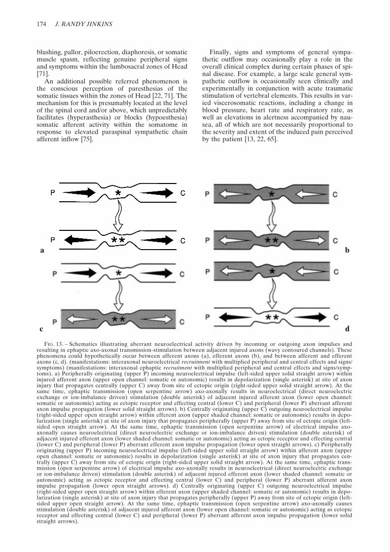
FIG. 13. – Schematics illustrating aberrant neuroelectrical activity driven by incoming or outgoing axon impulses andresulting in ephaptic axo-axonal transmission-stimulation between adjacent injured axons (wavy contoured channels). Thesephenomena could hypothetically occur between afferent axons (a), efferent axons (b), and between afferent and efferentaxons (c, d). (manifestations: interaxonal neuroelectrical recruitment with multiplied peripheral and central effects and signs/symptoms) (manifestations: interaxonal ephaptic recruitment with multiplied peripheral and central effects and signs/symp-toms). a) Peripherally originating (upper P) incoming neuroelectrical impulse (left-sided upper solid straight arrow) withininjured afferent axon (upper open channel: somatic or autonomic) results in depolarization (single asterisk) at site of axoninjury that propagates centrally (upper C) away from site of ectopic origin (right-sided upper solid straight arrow). At thesame time, ephaptic transmission (open serpentine arrow) axo-axonally results in neuroelectrical (direct neuroelectricexchange or ion-imbalance driven) stimulation (double asterisk) of adjacent injured afferent axon (lower open channel:somatic or autonomic) acting as ectopic receptor and effecting central (lower C) and peripheral (lower P) aberrant afferentaxon impulse propagation (lower solid straight arrows). b) Centrally originating (upper C) outgoing neuroelectrical impulse(right-sided upper open straight arrow) within efferent axon (upper shaded channel: somatic or autonomic) results in depo-larization (single asterisk) at site of axon injury that propagates peripherally (upper P) away from site of ectopic origin (left-sided open straight arrow). At the same time, ephaptic transmission (open serpentine arrow) of electrical impulse axo-axonally causes neuroelectrical (direct neuroelectric exchange or ion-imbalance driven) stimulation (double asterisk) ofadjacent injured efferent axon (lower shaded channel: somatic or autonomic) acting as ectopic receptor and effecting central(lower C) and peripheral (lower P) aberrant efferent axon impulse propagation (lower open straight arrows). c) Peripherallyoriginating (upper P) incoming neuroelectrical impulse (left-sided upper solid straight arrow) within afferent axon (upperopen channel: somatic or autonomic) results in depolarization (single asterisk) at site of axon injury that propagates cen-trally (upper C) away from site of ectopic origin (right-sided upper solid straight arrow). At the same time, ephaptic trans-mission (open serpentine arrow) of electrical impulse axo-axonally results in neuroelectrical (direct neuroelectric exchangeor ion-imbalance driven) stimulation (double asterisk) of adjacent injured efferent axon (lower shaded channel: somatic orautonomic) acting as ectopic receptor and effecting central (lower C) and peripheral (lower P) aberrant afferent axonimpulse propagation (lower open straight arrows). d) Centrally originating (upper C) outgoing neuroelectrical impulse(right-sided upper open straight arrow) within efferent axon (upper shaded channel: somatic or autonomic) results in depo-larization (single asterisk) at site of axon injury that propagates peripherally (upper P) away from site of ectopic origin (left-sided upper open straight arrow). At the same time, ephaptic transmission (open serpentine arrow) axo-axonally causesstimulation (double asterisk) of adjacent injured afferent axon (lower open channel: somatic or autonomic) acting as ectopicreceptor and effecting central (lower C) and peripheral (lower P) aberrant afferent axon impulse propagation (lower solidstraight arrows).
a b
c d

PAIN SYNROMES RELATED TO DISEASE OF THE SPINE 175
ANATOMY OF CENTRIPETALLY/CEN-TRIFUGALLY RADIATING SPINAL SYN-DROMES
On a yet more elemental level, if because of anonspecific pathologic influence upon a spinalnerve root or nerve [14, 40, 52, 60], the geometry ofthe cell membrane of the axon and its sodium ionchannels changes so that the functional channeldensity increases, the otherwise normal ion voltagepotential equilibrium across the membrane may bedisrupted [17]. The flow of normal ionic currentsand the maintenance of the normal ion voltagepotential across the axon membrane are in partdependent upon the functional sodium channeldensity within a given axon membrane area (itselfa manifestation in part of the ion channel stability
of the membrane) (figure 9). Theoretically, if thefunctional sodium ion channel density per squarearea escalates (i.e., too many overfunctioningsodium ion channels per square area), the influx ofsodium ions into the cell cannot be offset, as it nor-mally is by the sodium-potassium pump mechanism[17]. The passive influx (by the sodium ion chan-nels) out-paces the active efflux (by the sodium-potassium pump); for this reason the cell mem-brane in effect autodepolarizes producing anectopic bidirectional action potential within theinjured axon radiating away from the site of originof the initial membrane depolarization (i.e., neuralinsult). The CNS (cerebral sensory cortex) inter-prets this neuroelectrical signal (incoming withinappropriate afferent fibers) as pain and paresthe-
FIG. 14. – Schematics illustrating spontaneous ectopic neuroelectrical activity resulting in ephaptic transmission between injuredaxons (wavy contoured channels) and afferent nervi nervorum (open bifid channels). These phenomena could hypothetically occurin spinal dorsal roots (a), ventral roots (b), or in combined spinal nerves (a and b). (manifestations: magnified local pain in the dis-tribution of the somatic nervi nervorum; magnified autonomic afferent-efferent reflex phenomena in the distribution of the sympa-thetic nervi nervorum). a) Spontaneous depolarization (single asterisk) originating in injured afferent axon (lower open channel:somatic or autonomic) results in neuroelectrical impulse (solid straight arrows) that propagates centrally (C) and peripherally (P)away from site of ectopic origin. At the same time, ephaptic transmission (open serpentine arrow) axo-axonally causes stimulationof normal or injured (sensitized) receptor (open bifid channel: somatic or autonomic) of afferent nervi nervorum effecting central(C) aberrant afferent axon impulse propagation (stippled arrow). b) Spontaneous depolarization (single asterisk) originating ininjured efferent axon (lower shaded channel: somatic or autonomic) results in neuroelectrical impulse (open straight arrows) thatpropagates centrally (C) and peripherally (P) away from site of ectopic origin. At the same time, ephaptic transmission (open ser-pentine arrow) axo-axonally causes stimulation of normal or injured (sensitized) receptor (open bifid channel: somatic or autonomic)of afferent nervi nervorum effecting central (C) aberrant afferent axon impulse propagation (stippled arrows).
a b
a bFIG. 15. – Schematics illustrating ectopic neurogenic activity driven by incoming or outgoing somatic or autonomic axon impulses and
resulting in ephaptic axo-axonal transmission-stimulation between injured axon (wavy contoured channels) and afferent somatic or auto-nomic nervi nervorum (open bifid channels). These phenomena could hypothetically occur in spinal dorsal roots (a), ventral roots (b),or in combined spinal nerves (a and b). (manifestations: magnified local pain in the distribution of the somatic nervi nervorum; magnifiedautonomic afferent-efferent reflex phenomena in the distribution of the sympathetic nervi nervorum). a) Incoming neuroelectricalimpulse (solid straight arrows) within afferent axon (open channel: somatic or autonomic) results in depolarization (asterisk) at site ofinjured axon. At the same time, ephaptic transmission (open serpentine arrow) axo-axonally causes stimulation or normal or injuredreceptor (open bifid channel: somatic or autonomic) of afferent nervi nervorum and effects central (C) aberrant afferent axon impulsepropagation (stippled arrow). b) Outgoing neuroelectrical impulse (open straight arrows) within efferent axon (open channel: somaticor autonomic) results in depolarization (asterisk) at site of injured axon. At the same time, ephaptic transmission (open serpentinearrow) axo-axonally causes stimulation of normal or receptor (open bifid channel: somatic or autonomic) of afferent nervi nervorumand effects central (C) aberrant afferent axon impulse propagation (stippled arrow).

176 J. RANDY JINKINS
sias, a sign of involuntary muscular activity, or as asignal of/for autonomic action. In this manneraxons in and of themselves may become mechano-and chemosensitive ectopic sources of pathologicneuroelectrical activity resulting in abnormal clini-cal expression and conscious perception. This isbelieved to be the pathophysiologic basis for theso-called radiating radiculopathy.
Because of the presence of this ectopic axonalpacemaker, the transmission of normal incomingor outgoing neuroelectrical impulses occurring in,and adjacent to, fibers with pathologically alteredaxon membranes may also theoretically result inpain, paresthesias and somatic-autonomic dysfunc-tion that similarly originate at the level of theectopic source of activity. This aberrant impulsecoupling is believed to take place because ofabnormal interaxonal cross-stimulation based onchemical, neurochemical and/or ephaptic (directneuroelectrical exchange) factors [17, 25, 68]. The-oretically this cross-stimulation is responsible forabnormal links within and between the somaticand autonomic fibers within these two systems. Inthis way the resultant proximal and distant propa-gation may occur within axons that are anatomi-cally unrelated to the origin of the initial electricaltransmission. In addition, a recruitment phenome-non may take place in this circumstance whereby asingle ectopic neuroelectrical event may be respon-sible for firing within a large group of regionalaxons.
Experiments support the concept that focal neu-ral injury can act as both an ectopic intrinsic stim-ulus as well as an ectopic receptor/transmitter [11,12, 17, 25, 33, 50, 67-70, 75, 83, 85, 86]. Several pos-sibilities are suggested by this condition. First, achronically, repetitively injured afferent or efferentaxon may act as a primary intrinsic ectopic pace-maker spontaneously discharging and initiatingbidirectional intrinsic axonal impulse transmission(figure 11). Second, injured afferent or efferentaxons acting as ectopic pacemakers may ephapti-cally (neuroelectrically) transmit neuroelectricalimpulses extrinsically to ectopic axonal receptors inadjacent injured afferent and/or efferent axons(figure 12). This ephaptic transmission representsthe type of interaxonal neuroelectrical cross-stimu-lation referred to above [17, 25, 67, 68, 75]. Third,because of chronic injury, afferent or efferentaxons acting as primary ectopic neuroelectricalreceptors may be driven by incoming peripheralafferent or outgoing central efferent somatic orautonomic axonal impulses; and, they may thensecondarily ephaptically transmit neuroelectricalimpulses to ectopic receptors in adjacent injuredafferent and/or efferent somatic or autonomicaxons (figure 13). Thus, as an example, efferentautonomic fibers neuroelectrically driving afferentsomatic pain fibers in this way constitutes a rela-tively new theory of referred pain and dysfunction[17]. Fourth, because neural injury may result inpart in increased receptor sensitivity, afferent orefferent axons acting as ectopic pacemakers may
a b
FIG. 16. – Schematics illustrating aberrant neuroelectrical activity driven by outgoing autonomic [sympathetic] efferentimpulse resulting in ephaptic stimulation and transmission respectively between autonomic efferent nervi nervorum (shaded bifidchannels), an injured adjacent axon (wavy contoured channels) and the afferent somatic or autonomic nervi nervorum (openbifid channels). These phenomena could hypothetically occur in spinal dorsal roots (a), ventral roots (b), or in combined spinalnerves (a and b). (manifestations: magnified local pain in the distribution of the somatic nervi nervorum, driven by the autonomicnervous system; magnified autonomic afferent-efferent reflex phenomena in the distribution of the sympathetic nervi nervorum,driven by the autonomic nervous system [autonomic dysfunction, pain, muscle spasm {?}]; central and peripheral signs and symp-toms driven by the autonomic nervous system [pain, muscle spasm, autonomic dysfunction]). a) Incoming neuroelectrical impulse(hatched arrow) within efferent autonomic [sympathetic] axon (shaded bifid channel) of the nervi nervorum results in ephapticstimulation (lower open serpentine arrow) axo-axonally and depolarization (asterisk) at site of injured afferent axon (wavy con-tour, open channel: somatic or autonomic) acting as ectopic receptor and effecting central (C) and peripheral (P), aberrant affer-ent axon impulse propagation (solid straight arrows). At the same time, ephaptic transmission (upper open serpentine arrow)axo-axonally causes stimulation of normal or injured (sensitized) receptor (open bifid channel: somatic or autonomic) of afferentnervi nervorum and effects central (C) aberrant afferent axon impulse propagation (stippled arrow). b) Incoming neuroelectricalimpulse (hatched arrow) within normal or injured efferent autonomic [sympathetic] axon (shaded bifid channel) of the nervinervorum results in ephaptic stimulation (lower open serpentine arrow) axo-axonally and depolarization (asterisk) at site ofinjured efferent axon (wavy contour, shaded channel: somatic or autonomic) acting as ectopic receptor and effecting central (C)and peripheral (P) aberrant efferent axon impulse propagation (open straight arrows). At the same time, ephaptic transmission(upper open serpentine arrow) axo-axonally causes stimulation of normal or injured (sensitized receptor (open bifid channel:somatic or autonomic) of afferent nervi nervorum and effects central (C) aberrant afferent axon impulse propagation (stippledarrow).
PAIN SYNROMES RELATED TO DISEASE OF THE SPINE 177
ephaptically drive injured afferent fiber termina-tions on nerves, the afferent nervi nervorum(figure 14) [17]. Fifth, afferent or efferent axonsacting as ectopic transmitters may be driven byincoming peripheral afferent or outgoing centralefferent axonal impulses that may secondarilytransmit neuroelectrical impulses ephaptically toafferent somatic and autonomic fiber terminationson nerves, once again, the afferent nervi nervorum(figure 15). Sixth, autonomic efferent fiber termina-tions on nerves, the efferent sympathetic nervi ner-vorum, may drive afferent or efferent axons actingas ectopic receptors, and these may in turn transmitneuroelectrical impulse activity ephaptically to theafferent nervi nervorum (figure 16). This latterobservation is yet another possible mode of localpain, referred pain, radicular pain, muscular dys-
function, and aberrant autonomic activity. It is bythese mechanisms that pathologically peripherallydriven, centrally driven, spontaneously generated,mechanically and chemically stimulated and ephap-tically transmitted neuroelectric activity can hypo-thetically occur at and near the site of neural injury.This thereby causes ectopic single fiber neuroelec-trical phenomena and/or multifiber somatic-somatic, somatic-autonomic, autonomic-somatic orautonomic-autonomic aberrant neuroelectrical cou-pling [17]. The expression of this abnormal neuro-genic activity may potentially result in bizarrecombinations of subjective symptoms (e.g., pain,paresthesias) and objective signs (e.g., skeletal mus-cle spasm, sympathetic dysfunction).
Thus, otherwise normal peripheral sensory activ-ity, centrally initiated voluntary muscle activity,
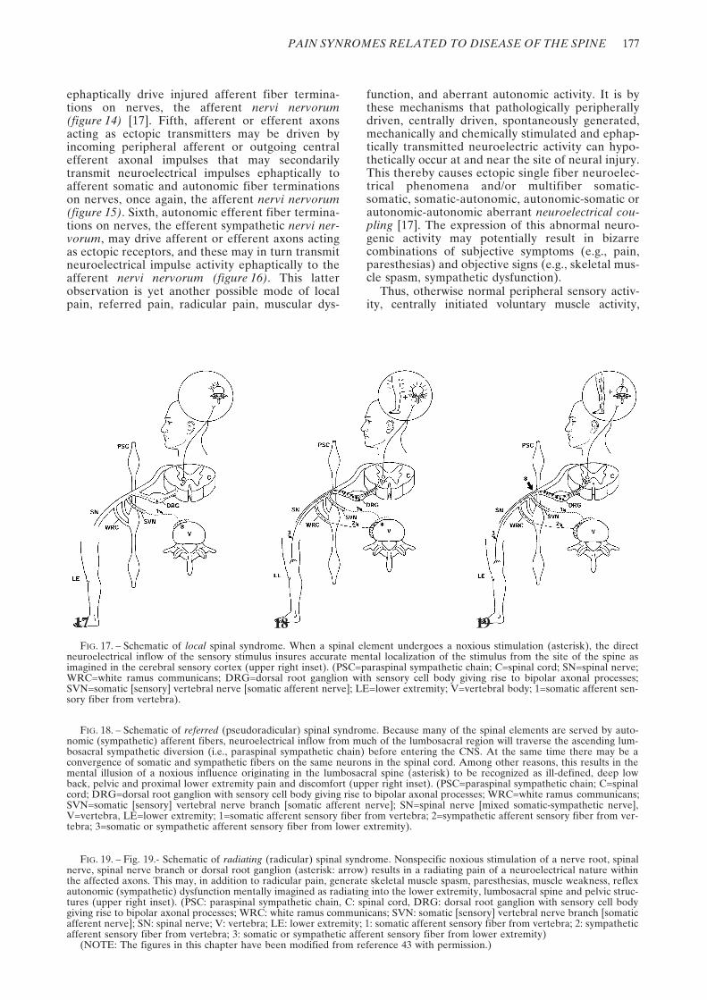
17 18 19FIG. 17. – Schematic of local spinal syndrome. When a spinal element undergoes a noxious stimulation (asterisk), the direct
neuroelectrical inflow of the sensory stimulus insures accurate mental localization of the stimulus from the site of the spine asimagined in the cerebral sensory cortex (upper right inset). (PSC=paraspinal sympathetic chain; C=spinal cord; SN=spinal nerve;WRC=white ramus communicans; DRG=dorsal root ganglion with sensory cell body giving rise to bipolar axonal processes;SVN=somatic [sensory] vertebral nerve [somatic afferent nerve]; LE=lower extremity; V=vertebral body; 1=somatic afferent sen-sory fiber from vertebra).
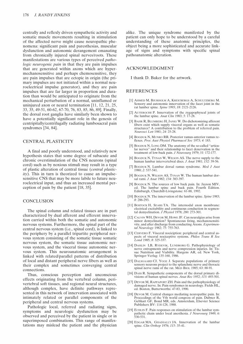
FIG. 18. – Schematic of referred (pseudoradicular) spinal syndrome. Because many of the spinal elements are served by auto-nomic (sympathetic) afferent fibers, neuroelectrical inflow from much of the lumbosacral region will traverse the ascending lum-bosacral sympathetic diversion (i.e., paraspinal sympathetic chain) before entering the CNS. At the same time there may be aconvergence of somatic and sympathetic fibers on the same neurons in the spinal cord. Among other reasons, this results in themental illusion of a noxious influence originating in the lumbosacral spine (asterisk) to be recognized as ill-defined, deep lowback, pelvic and proximal lower extremity pain and discomfort (upper right inset). (PSC=paraspinal sympathetic chain; C=spinalcord; DRG=dorsal root ganglion with sensory cell body giving rise to bipolar axonal processes; WRC=white ramus communicans;SVN=somatic [sensory] vertebral nerve branch [somatic afferent nerve]; SN=spinal nerve [mixed somatic-sympathetic nerve],V=vertebra, LE=lower extremity; 1=somatic afferent sensory fiber from vertebra; 2=sympathetic afferent sensory fiber from ver-tebra; 3=somatic or sympathetic afferent sensory fiber from lower extremity).
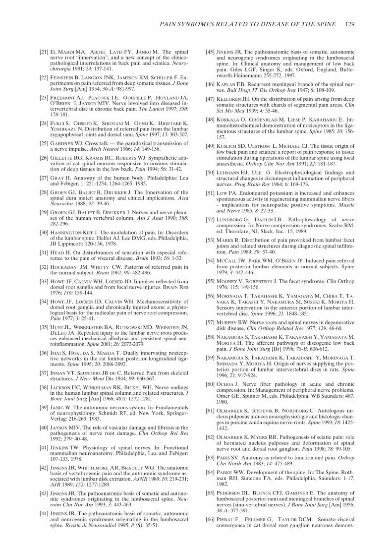
FIG. 19. – Fig. 19.- Schematic of radiating (radicular) spinal syndrome. Nonspecific noxious stimulation of a nerve root, spinalnerve, spinal nerve branch or dorsal root ganglion (asterisk: arrow) results in a radiating pain of a neuroelectrical nature withinthe affected axons. This may, in addition to radicular pain, generate skeletal muscle spasm, paresthesias, muscle weakness, reflexautonomic (sympathetic) dysfunction mentally imagined as radiating into the lower extremity, lumbosacral spine and pelvic struc-tures (upper right inset). (PSC: paraspinal sympathetic chain, C: spinal cord, DRG: dorsal root ganglion with sensory cell bodygiving rise to bipolar axonal processes; WRC: white ramus communicans; SVN: somatic [sensory] vertebral nerve branch [somaticafferent nerve]; SN: spinal nerve; V: vertebra; LE: lower extremity; 1: somatic afferent sensory fiber from vertebra; 2: sympatheticafferent sensory fiber from vertebra; 3: somatic or sympathetic afferent sensory fiber from lower extremity)
(NOTE: The figures in this chapter have been modified from reference 43 with permission.)
178 J. RANDY JINKINS
centrally and reflexly driven sympathetic activity andsomatic muscle movements resulting in stimulationof the affected nerve might cause neuropathic phe-nomena: significant pain and paresthesias, musculardysfunction and autonomic derangement emanatingfrom chronically injured spinal nerves/roots. Thesemanifestations are various types of perceived patho-logic neurogenic pain in that they are pain impulsesthat are generated within axons which are hyper-mechanosensitive and perhaps chemosensitive, theyare pain impulses that are ectopic in origin (the pri-mary impulses are not initiated within a normal neu-roelectrical impulse generator), and they are painimpulses that are far larger in proportion and dura-tion than would be anticipated to originate from themechanical perturbation of a normal, uninflamed oruninjured axon or neural termination [11, 12, 21, 25,33, 35, 49-51, 60-62, 67-70, 76, 83, 85, 86, 89]. Finally,the dorsal root ganglia have similarly been shown tohave a potentially significant role in the genesis ofcentripitally/centrifugally radiating lumbosacral painsyndromes [34, 84].
CENTRAL PLASTICITY
A final and poorly understood, and relatively newhypothesis states that some degree of subacute andchronic overstimulation of the CNS neurons (spinalcord) such as by noxious stimuli may result in a typeof plastic alteration of central tissue (central plastic-ity). This in turn is theorized to cause an impulse-sensitive CNS that may be more labile to future neu-roelectrical input, and thus an increased mental per-ception of pain by the patient [18, 35].
CONCLUSION
The spinal column and related tissues are in partcharacterized by dual afferent and efferent innerva-tion carried within both the somatic and autonomicnervous systems. Furthermore, the spinal part of thecentral nervous system (i.e., spinal cord), is linked tothe periphery by a parallel tripartite peripheral ner-vous system consisting of the somatic tissue somaticnervous system, the somatic tissue autonomic ner-vous system, and the visceral tissue autonomic ner-vous system. This neuroanatomic arrangement islinked with related/parallel patterns of distributionof local and distant peripheral nerve fibers as well astheir complex and sometimes converging centralconnections.
Thus, conscious perception and unconsciouseffects originating from the vertebral column, peri-vertebral soft tissues, and regional neural structures,although complex, have definite pathways repre-sented in this network of innervation associated withintimately related or parallel components of theperipheral and central nervous systems.
Pathologic local, referred and radiating signs,symptoms and neurologic dysfunction may beobserved and perceived by the patient in single or insuperimposed combinations. This range of manifes-tations may mislead the patient and the physician
alike. The unique syndrome manifested by thepatient can only hope to be understood by a carefulunderstanding of these anatomic principles, theobject being a more sophisticated and accurate link-age of signs and symptoms with specific spinalpathoanatomic alteration.
ACKNOWLEDGMENT
I thank D. Baker for the artwork.
REFERENCES
[1] AHMED M, BJURHOLM A, KREICBERGS A, SCHULTZBERG M.Sensory and autonomic innervation of the facet joint in therat lumber spine. Spine 1993; 18: 2121-2126.
[2] AUTEROCHE P. Innervation of the zygapophyseal joints ofthe lumbar spine. Anat Clin 1983; 5: 17-28.
[3] BAHR R, BLUMBERG H, JANIG W. Do dichotomizing afferentfibers exist which supply visceral organs as well as somaticstructures? A contribution to the problem of referred pain.Neurosci Lett 1981; 24: 25-28.
[4] BOGDUK N, MUNRO RR. Posterior ramus-anterior ramus re-flexes. Proc Aust Physiol Pharmacol Soc 1973; 4: 183.
[5] BOGDUK N, LONG DM. The anatomy of the so-called “articu-lar nerves” and their relationship to facet denervation in thetreatment of low-back pain. J Neurosurg 1979; 51: 172-177.
[6] BOGDUK N, TYNAN W, WILSON AS. The nerve supply to thehuman lumbar intervertebral discs. J Anat 1981; 132: 39-56.
[7] BOGDUK N. Lumbar dorsal ramus syndrome. Med J Aust1980; 2: 537-541.
[8] BOGDUK N, WILSON AS, TYNAN W. The human lumbar dor-sal rami. J Anat 1982; 134: 383-397.
[9] BOGDUK N. The sources of low back pain. In: Jayson MIV,ed. The lumbar spine and back pain. Fourth Edition.Edinburgh, Churchill-Livingstone: 61-88, 1992.
[10] BOGDUK N. The innervation of the lumbar spine. Spine 1983;8: 286-293.
[11] BOSTOCK H, SEARS TA. The internodal axon membrane:electrical excitability and continuous conduction in segmen-tal demyelination. J Physiol 1978; 280: 273-301.
[12] CALVIN WH, DEVOR M, HOWE JF. Can neuralgias arise fromminor demyelination? Spontaneous firing, mechanosensiti-vity, and after discharge from conducting Axons. Experimen-tal Neurology 1982; 75: 755-763.
[13] CERVERO F. Visceral nociciption: peripheral and central as-pects of visceral nociceptive systems. Philos Trans R SocLond 1985; 8: 325-337.
[14] DAHLIN LB, RYDEVIK, LUNDBORG G. Pathophysiology ofnerve entrapments and nerve compression injuries. In: Tis-sue Nutrition and Viability. Hargens AR, ed. New York,Springer-Verlag: 135-160, 1986.
[15] DALSGAARD CJ, YGGE J. Separate populations of primarysensory neurons project to the splanchnic nerve and thoracicspinal nerve rami of the rat. MED BIOL 1985; 63: 88-91.
[16] DASS R. Sympathetic components of the dorsal primary di-visions of human spinal nerves. Anat Rec 1952; 113: 493-501.
[17] DEVOR M, RAPPAPORT ZH. Pain and the pathophysiology ofdamaged nerve. In: Pain syndromes in neurology. Fields HL,ed. Boston, Butterworths: 47-83, 1990.
[18] DEVOR M. Central changes mediating neuropathic pain. In:Proceedings of the Vth world congress of pain. Dubner R,Gebhart GF, Bond MR, eds. Amsterdam, Elsevier SciencePublishers BV: 114-128, 1988.
[19] ECHLIN F. Pain responses on stimulation of the lumbar sym-pathetic chain under local anesthesia. J Neurosurg 1949; 6:530-531.
[20] EDGAR MA, GHADIALLY JA. Innervation of the lumbarspine. Clin Orthop 1976; 115: 35-41.
PAIN SYNROMES RELATED TO DISEASE OF THE SPINE 179
[21] EL MAHDI MA, ABDEL LATIF FY, JANKO M. The spinalnerve root “innervation”, and a new concept of the clinico-pathological interrelations in back pain and sciatica. Neuro-chirurgia 1981; 24: 137-141.
[22] FEINSTEIN B, LANGION JNK, JAMESON RM, SCHILLER F. Ex-periments on pain referred from deep somatic tissues. J BoneJoint Surg [Am] 1954; 36-A: 981-997.
[23] FREEMONT AJ, PEACOCK TE, GOUPILLE P, HOYLAND JA,O’BRIEN J, JAYSON MIV. Nerve involved into diseased in-tervertebral disc in chronic back pain. The Lancet 1997; 350:178-181.
[24] FUKUI S, OHSETO K, SHIOTANI M, OHNO K, HIDETAKE K,YOSHIKAZU N. Distribution of referred pain from the lumbarzygapophyseal joints and dorsal rami. Spine 1997; 13: 303-307.
[25] GARDNER WJ. Cross talk — the paradoxical transmission ofa nerve impulse. Arch Neurol 1966; 14: 149-156.
[26] GILLETTE RG, KRAMIS RC, ROBERTS WJ. Sympathetic acti-vation of cat spinal neurons responsive to noxious stimula-tion of deep tissues in the low back. Pain 1994; 56: 31-42.
[27] GRAY H. Anatomy of the human body. Philadelphia: Leaand Febiger, 1: 251-1254, 1264-1265, 1985.
[28] GROEN GJ, BALJET B, DRUKKER J. The Innervation of thespinal dura mater: anatomy and clinical implications. ActaNeurochir 1988; 92: 39-46.
[29] GROEN GJ, BALJET B, DRUKKER J. Nerves and nerve plexu-ses of the human vertebral column. Am J Anat 1990; 188:282-296.
[30] HANNINGTON-KIFF J. The modulation of pain. In: Disordersof the lumbar spine. Helfet AJ, Lee DMG, eds. Philadelphia,JB Lippincott: 120-136, 1978.
[31] HEAD H. On disturbrances of sensation with especial refe-rence to the pain of visceral disease. Brain 1893; 16: 1-32.
[32] HOCKADAY JM, WHITTY CW. Patterns of referred pain inthe normal subject. Brain 1967; 90: 482-496.
[33] HOWE JF, CALVIN WH, LOESER JD. Impulses reflected fromdorsal root ganglia and from focal nerve injuries. BRAIN RES1976; 116: 139-144.
[34] HOWE JF, LOESER JD, CALVIN WH. Mechanosensitivity ofdorsal root ganglia and chronically injured axons: a physio-logical basis for the radicular pain of nerve root compression.Pain 1977; 3: 25-41.
[35] HUNT JL, WINKELSTEIN BA, RUTKOWSKI MD, WEINSTEIN JN,DELEO JA. Repeated injury to the lumbar nerve roots produ-ces enhanced mechanical allodynia and persistent spinal neu-roinflammation. Spine 2001; 26: 2073-2079.
[36] IMAI S, HUKUDA S, MAEDA T. Dually innervating nocicep-tive networks in the rat lumbar posterior longitudinal liga-ments. Spine 1995; 20: 2086-2092.
[37] INMAN VT, SaUNDERS JB DE C. Referred Pain from skeletalstructures. J Nerv Ment Dis 1944; 99: 660-667.
[38] JACKSON HC, WINKELMAN RK, BICKEL WH. Nerve endingsin the human lumbar spinal column and related structures. JBone Joint Surg [Am] 1966; 48A: 1272-1281.
[39] JANIG W. The autonomic nervous system. In: Fundamentalsof neurophysiology. Schmidt RF, ed. New York, Springer-Verlag: 216-269, 1985.
[40] JAYSON MIV. The role of vascular damage and fibrosis in thepathogenesis of nerve root damage. Clin Orthop Rel Res1992; 279: 40-48.
[41] JENKINS TW. Physiology of spinal nerves. In: Functionalmammalian neuroanatomy. Philadelphia, Lea and Febiger:107-133, 1978.
[42] JINKINS JR, WHITTEMORE AR, BRADLEY WG. The anatomicbasis of vertebrogenic pain and the autonomic syndrome as-sociated with lumbar disk extrusion. AJNR 1989; 10: 219-231;AJR 1989; 152: 1277-1289.
[43] JINKINS JR. The pathoanatomic basis of somatic and autono-mic syndromes originating in the lumbosacral spine. Neu-roim Clin Nor Am 1993; 3: 443-463.
[44] JINKINS JR. The pathoanatomic basis of somatic, autonomicand neurogenic syndromes originating in the lumbosacralspine. Rivista di Neuroradiol 1995; 8 (S): 35-51.
[45] JINKINS JR. The pathoanatomic basis of somatic, autonomicand neurogenic syndromes originating in the lumbosacralspine. In: Clinical anatomy and management of low backpain. Giles LGF, Singer K, eds. Oxford, England, Butte-rworth-Heinemann: 255-272, 1997.
[46] KAPLAN EB. Recurrent meningeal branch of the spinal ner-ves. Bull Hosp JT Dis Orthop Inst 1947; 8: 108-109.
[47] KELLGREN JH. On the distribution of pain arising from deepsomatic structures with chards of segmental pain areas. ClinSci Mo Med 1939; 4: 35-46.
[48] KORKALA O, GRÖENBLAD M, LIESE P, KARAHARJU E. Im-munohistochemical demonstration of nociceptors in the liga-mentous structures of the lumbar spine. Spine 1985; 10: 156-157.
[49] KUSLICH SD, ULSTROM L, MICHAEL CJ. The tissue origin oflow back pain and sciatica: a report of pain response to tissuestimulation during operations of the lumbar spine using localanaesthesia. Orthop Clin Nor Am 1991; 22: 181-187.
[50] LEHMANN HJ, ULE G. Electrophysiological findings andstructural changes in circumspect inflammation of peripheralnerves. Prog Brain Res 1964; 6: 169-173.
[51] LOW PA. Endoneurial potassium is increased and enhancesspontaneous activity in regenerating mammalian nerve fibers– implications for neuropathic positive symptoms. Muscleand Nerve 1985; 8: 27-33.
[52] LUNDBORG G, DAHLIN LB. Pathophysiology of nervecompression. In: Nerve compression syndromes. Szabo RM,ed. Thorofare, NJ, Slack, Inc.: 15, 1989.
[53] MARKS R. Distribution of pain provoked from lumbar facetjoints and related structures during diagnostic spinal infiltra-tion. Pain 1989; 39: 37-40.
[54] MCCALL IW, PARK WM, O’BRIEN JP. Induced pain referralfrom posterior lumbar elements in normal subjects. Spine1979; 4: 442-446.
[55] MOONEY V, ROBERTSON J. The facet syndrome. Clin Orthop1976; 115: 149-156.
[56] MORINAGA T, TAKAHASHI K, YAMAGATA M, CHIBA T, TA-
NAKA K, TAKASHI Y, NAKAMURA SI, SUSEKI K, MORIYA H.Sensory innervation to the anterior portion of lumbar inter-vertebral disc. Spine 1996; 21: 1848-1851.
[57] MURPHY RW. Nerve roots and spinal nerves in degenerativedisk disease. Clin Orthop Related Res 1977; 129: 46-60.
[58] NAKAMURA S, TAKAHASHI K, TAKAHASHI Y, YAMAGATA M,MORIYA H. The afferent pathways of discogenic low backpain. J Bone Joint Surg [Br] 1996; 78-B: 606-612.
[59] NAKAMURA S, TAKAHASHI K, TAKAHASHI Y, MORINAGA T,SHIMADA Y, MORIYA H. Origin of nerves supplying the pos-terior portion of lumbar intervertebral discs in rats. Spine1996; 21: 917-924.
[60] OCHOA J. Nerve fiber pathology in acute and chroniccompression. In: Management of peripheral nerve problems.Omer GE, Spinner M, eds. Philadelphia, WB Saunders: 487,1980.
[61] OLMARKER K, RYDEVIK B, NORDBORG C. Autologous nu-cleus pulposus induces neurophysiologic and histologic chan-ges in porcine cauda equina nerve roots. Spine 1993; 18: 1425-1432.
[62] OLMARKER K, MYERS RR. Pathogenesis of sciatic pain: roleof herniated nucleus pulposus and deformation of spinalnerve root and dorsal root ganglion. Pain 1998; 78: 99-105.
[63] PARIS SV. Anatomy as related to function and pain. OrthopClin North Am 1983; 14: 475-489.
[64] PARKE WW. Development of the spine. In: The Spine. Roth-man RH, Simeone FA, eds. Philadelphia, Saunders: 1-17,1982.
[65] PEDERSEN DL, BLUNCK CFJ, GARDNER E. The anatomy oflumbosacral posterior rami and meningeal branches of spinalnerves (sinu-vertebral nerves). J Bone Joint Surg [Am] 1956;38-A: 377-391.
[66] PIERAU F, FELLMER G, TAYLOR DCM. Somato-visceralconvergence in cat dorsal root ganglion neurones demons-

180 J. RANDY JINKINS
trated by double-labeling with fluorescent tracers. Brain Res1984; 321: 63-70.
[67] RAMON F, JOYNER RW, MOORE JW. Ephaptic transmissionin squid giant axons. Biophys J 1976; 16: 26a.
[68] RASMINSKY M. Ectopic-generation of impulses and cross-talk in spinal nerve roots of “dystrophic” mice. Ann Neurol1978; 3: 351-357.
[69] RASMINSKY M. Physiology of conduction in demyelinatedaxons. In: The physiology and pathobiology of axons. Wax-man SG, ed. New York, Raven: 361-376, 1978.
[70] RASMINSKY M. Ectopic generation of impulses in pathologi-cal nerve fibers. In: Nerve repair and regeneration — its cli-nical and experimental basis. Jewett DL, McCarroll HR, eds.St. Louis, Mosby: 178-195, 1980.
[71] RUCH TC. Pathophysiology of pain. In: Physiology and bio-physics. Ruch TC, Patton HD, eds. Philadelphia, Saunders:508-531, 1982.
[72] RUCKER HK, HOLLOWAY JA. Viscerosomatic convergenceonto spinothalamic tract neurons in the cat. Brain Res 1982;243: 155-157.
[73] SAMEDA H, TAKAHASHI Y, TAKAHASHI K, CHIBA T, OH-
TORI S, MORIYA H. Primary sensory neurons with dichoto-mizing axons projecting to the facet joint and the sciaticnerve in rats. Spine 2001; 26: 1105-1109.
[74] SEKINE M, YAMASHITA T, TAKEBAYASHI T, SAKAMOTO N,MINAKE Y, ISHII S. Mechanosensitive afferent units in thelumbar posterior longitudinal ligament. Spine 2001; 26: 1516-1521.
[75] SELTZER Z, DEVOR M. Ephaptic transmission in chronicallydamaged peripheral nerves. Neurology 1979; 29: 1061-1064.
[76] SMYTH JM, WRIGHT VJ. Sciatica and the intervertebral disk.An experimental study. J Bone Joint Surg 1958; 40 [A]: 1401-1418.
[77] STILWELL DL. The nerve supply of the vertebral column andits associated structures in the monkey. Anat Rec 1956; 125:139-169.
[78] SUSEKI K, TAKAHASHI Y, TAKAHASHI K, CHIBA, TANAKA K,MORIYA H. CGRP-immunosuppresive nerve fibers projec-ting to lumbar facet joints through the paravertebral sympa-thetic truck in tracts. Neuroscience Letters 1996; 221: 41-44.
[79] SUSEKI K, TAKAHASHI Y, TAKAHASHI K et al. Innervation ofthe lumbar facet joints: origins and functions. Spine 1997; 22:477-485.
[80] SUSEKI K, TAKAHASHI Y, TAKAHASHI K, CHIBA T, YAMA-
GATA M, MORIYA H. Sensory nerve fibers from lumbar in-tervertebral discs pass through rami communicantes: apossible pathway for discogenic low back pain. J Bone JointSurg [Br] 1998; 80-B: 737-742.
[81] TAKAHASHI Y, NAKAJIMA Y, SAKAMOTO T, MORIYA H, TA-
KAHASHI K. Capsaicin applied to rat lumbar intervertebraldisc causes extravasation in the groin skin: a possible mecha-nism of referred pain of the intervertebral disc. NeuroscienceLetters 1993; 161: 1-3.
[82] TAKAHASHI Y, MORINAGA T, NAKAMURA S-I, SUSEKI K, TA-
KAHASHI K, NAKAJIMA Y. Neural connection between theventral portion of the lumbar intervertebral disc and thegroin skin. J Neurosurg 1996; 85: 323-328.
[83] WALL PD, WAXMAN S, BASBAUM AL. Ongoing activity in pe-ripheral nerve injury discharge. Exp Neurol 1974; 45: 576-589.
[84] WALL PD, DEVOR M. Sensory afferent impulses originatefrom dorsal root ganglia as well as from the periphery in nor-mal and nerve injured rats. Pain 1983; 17: 321-339.
[85] WALLIS WE, VAN POZNAK A, PLUM F. Generalized muscu-lar stiffness, fasciculations and myokymia of peripheral nerveorigin. Arch Neurol 1970; 22: 430-439.
[86] WETTSTEIN A. The origins of fasciculations in motorneurondisease. Neurology 1977; 27: 357-358.
[87] WIBERG G. Back pain in relation to the nerve supply of theintervertebral disc. Acta Orthop Scand 1949; 19: 211-221.
[88] WILLIS WD, GROSSMAN RG. The spinal cord. In: Medicalneurobiology. St. Louis, Mosby: 80-115, 1973.
[89] WILLIS WD, Grossman RG. Sensory systems. In: Medicalneurobiology. St. Louis, Mosby: 227-272, 1973.
[90] WYKE B. The neurology of low back pain. In: The lumbarspine and back pain. Jayson MIV, ed. New York, Churchill-Livingstone: 56-99, 1987.
[91] YAMASHITA T, MINAKI Y, OOTA I, YOKOGUSHI K, ISHII S.Mechanosensitive afferent units in the lumbar intervertebraldisc and adjacent muscle. Spine 1993; 18: 2252-2256.