what effects will proximal or distal disease have on a ... · spectral doppler interpretation ......
TRANSCRIPT

3/23/2018
1
Mani Montazemi, RDMS
Doppler Interpretation
Spectral Doppler
Interpretation
Mani Montazemi, RDMSDirector of Ultrasound Education & Quality Assurance
Baylor College of Medicine
Division of Maternal-Fetal Medicine
Maternal Fetal Center Imaging Manager
Texas Children’s Hospital, Pavilion for Women
Houston Texas
&
Clinical Instructor
Thomas Jefferson University Hospital - Radiology Department
Philadelphia, Pennsylvania
Spectral Analysis
The final shape of the arterial Doppler
waveform is dependent on numerous factors:
• Contraction by the heart
• Presence of stenosis in the vessel
• State of the downstream circulation
Mani Montazemi, RDMS
OB Doppler
How systolic and diastolic
components of
arterial waveforms
appear in health and disease?
Mani Montazemi, RDMS
Doppler Interpretation
What effects will
proximal or distal
disease have on
a waveform?
Mani Montazemi, RDMS
Doppler Interpretation
Doppler Interpretation
“Distal disease”
Changes the resistance
“Proximal disease”
Changes the strength of the signal
Mani Montazemi, RDMS
Doppler Interpretation
“Distal” Disease“Changes the resistance”
• Acute & chronic
parenchymal disease
• Obstruction
• Renal vein thrombosis

3/23/2018
2
Mani Montazemi, RDMS
Doppler Interpretation
Distal Disease“Changes the resistance”
Mani Montazemi, RDMS
Doppler Interpretation
Distal Disease“Changes the resistance”
Mani Montazemi, RDMS
Doppler Interpretation
Distal Disease“Changes the resistance”
Mani Montazemi, RDMS
Doppler Interpretation
“Proximal” DiseaseChanges the strength of the signal
Mani Montazemi, RDMS
Doppler Interpretation
Tardus – Parvus Waveform
Tardus
• Slow & late
Parvus
• Small & little
Mani Montazemi, RDMS
Doppler Interpretation
Tardus – Parvus Waveform
• Systolic acceleration diminished
• Acceleration time prolonged
• Waveform shape
• Diminished pulsatility

3/23/2018
3
Mani Montazemi, RDMS
Doppler Interpretation
Caution! Increase Sweep Speed
Mani Montazemi, RDMS
Doppler Interpretation
Proximal DiseaseChanges the strength of the signal
Mani Montazemi, RDMS
Mesenteric Arteries
Mani Montazemi, RDMS
Mesenteric Arteries
Mani Montazemi, RDMS
Doppler Interpretation
Mani Montazemi, RDMS
Doppler Interpretation
Remember!
• It is more difficult to demonstrate tardus
parvus in a stiff vessel
• Atherosclotic arteries & increased distal
resistance masks the post-stenotic tardus
parvus

3/23/2018
4
Mani Montazemi, RDMS
Doppler Interpretation
Doppler Analysis
• Qualitative
• Quantitative
Mani Montazemi, RDMS
Doppler Interpretation
Doppler Analysis
• Qualitative– The visual or acoustic evaluation of Doppler wave form
Mani Montazemi, RDMS
Doppler Interpretation
Doppler Analysis
• Qualitative– The visual or acoustic evaluation of Doppler wave form
• Quantitative– Calculation of volume flow
– Calculation of indices
Indirect method to evaluate
blood perfusion
Mani Montazemi, RDMS
Doppler Interpretation
Doppler Analysis
• Waveform is commonly described by pulsatility which can be measured
– Peak Systolic velocity – PSV
– Resistance Index – RI
– Pulsatility Index – PI
– Systolic/Diastolic Ratio – S/D
– Acceleration Index – AI
– Acceleration Time – AT
Mani Montazemi, RDMS
Doppler Interpretation
How to Look at a Waveform?
• Where & how was signal obtained?
• Presence of flow
• Direction of flow
• Characterization of signal
• Quality of exam
Spectral Doppler
Mani Montazemi, RDMS
Doppler Interpretation

3/23/2018
5
Spectral DopplerCursoris used for optimal alignment between vessel axis & Doppler scan line
“Angle of insonation”
Angle correctiononly used to measure velocity
Sample Volumedetermines the location and area that the pulsed wave Doppler listens for a returning signal
Mani Montazemi, RDMS
Doppler Interpretation
• Where & how was signal obtained?
– What is the angle of insonation
– Where is the sample volume
– What is the sample volume size
How to Look at a Waveform?
Mani Montazemi, RDMS
Doppler Interpretation
Mani Montazemi, RDMS
Doppler Interpretation
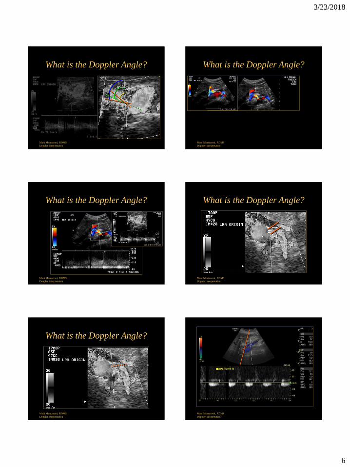
What is the Doppler Angle?
• Angle is the result of
– Doppler line direction
– Cursor correction
• Angle affects velocity accuracy
2 . Ft . cos Θ . Fd
CV =
“ 1 ”
Mani Montazemi, RDMS
Doppler Interpretation
What is the Doppler Angle?
A
3/23/2018
6
Mani Montazemi, RDMS
Doppler Interpretation
What is the Doppler Angle?
Mani Montazemi, RDMS
Doppler Interpretation
What is the Doppler Angle?
Mani Montazemi, RDMS
Doppler Interpretation
What is the Doppler Angle?
Mani Montazemi, RDMS
Doppler Interpretation
What is the Doppler Angle?
Mani Montazemi, RDMS
Doppler Interpretation
What is the Doppler Angle?
Mani Montazemi, RDMS
Doppler Interpretation

3/23/2018
7
Mani Montazemi, RDMS
Doppler Interpretation
Mani Montazemi, RDMS
Doppler Interpretation
Mani Montazemi, RDMS
Doppler Interpretation
Mani Montazemi, RDMS
Doppler Interpretation
In Straight Unbranched Vessels
Blood Flows in Layers (or Laminar)
Where is the Sample Volume?
Mani Montazemi, RDMS
Doppler Interpretation
“ 2 ”
Mani Montazemi, RDMS
Doppler Interpretation

3/23/2018
8
• Size ranges from
0.7 to 15mm
• Larger gate to
search for flow
• Smaller gate for
precise information
What is the Sample Volume Size?
Mani Montazemi, RDMS
Doppler Interpretation
“ 3 ”
Mani Montazemi, RDMS
Doppler Interpretation
What is the Sample Volume Size?
Mani Montazemi, RDMS
Doppler Interpretation
What is the Sample Volume Size? Spectral Doppler - Sample Volume
• What is the recommended size of the
sample volume and why?
The sample volume size should be
no larger than 1/3 of the size of the
vessel. If larger, the sample
volume is capturing slower flow
that occurs near the vessel walls.
Mani Montazemi, RDMS
Doppler Interpretation
Mani Montazemi, RDMS
Doppler Interpretation
What is the Sample Volume Size?
Mani Montazemi, RDMS
Doppler Interpretation
What is the Sample Volume Size?
Too small a gate may
give the false impression
of reduced or even absent
flow

3/23/2018
9
• Where & how was signal obtained?
– What is the angle of insonation
– Where is the sample volume
– What is the sample volume size
• Technical considerations
– Doppler Gain
– Velocity Scale
– Wall Filter
– Sweep Speed
How to Look at a Waveform?
Mani Montazemi, RDMS
Doppler Interpretation
Spectral Doppler – Gain
• Controls the amplification of the returning
Doppler signals
• The Doppler gain should be adjusted to a
level that fills in the gray scale of the
spectral analysis waveform without creating
noise
Mani Montazemi, RDMS
Doppler Interpretation
Mani Montazemi, RDMS
Doppler Interpretation
Spectral Displayeffect of “Doppler Gain”
Mani Montazemi, RDMS
Doppler Interpretation
Angle adjustments are not necessary since the
shape of the waveform, rather than velocity, is
used for interpretation
Mani Montazemi, RDMS
Doppler Interpretation
Θ = 89O
Spectral Doppler - Velocity Scale
• Increasing the scale
– smaller waveform size
• Decreasing the scale
– bigger waveform size
Controls “ PRF ”
(the rate at which the transducer is pulsed per second)
Mani Montazemi, RDMS
Doppler Interpretation

3/23/2018
10
Mani Montazemi, RDMS
Doppler Interpretation
Spectral Doppler - Velocity Scale Spectral Doppler - Velocity Scale
Mani Montazemi, RDMS
Doppler Interpretation
Mani Montazemi, RDMS
Doppler Interpretation
Spectral Doppler - Velocity Scale
Decreased PRF Increased PRF
Mani Montazemi, RDMS
Doppler Interpretation
Spectral Doppler - Velocity Scale
Spectral Doppler - Wall Filter
• Suppress velocities associated with tissue or
wall motion
• Higher setting
– Reduce artifacts
– Can eliminate diagnostic information
Mani Montazemi, RDMS
Doppler Interpretation
Spectral Doppler - Sweep Speed
• Controls how quickly the spectral
information is updated
• Three sweep speeds
– Slow
– Moderate
– Fast
Mani Montazemi, RDMS
OB Doppler

3/23/2018
11
Spectral Doppler - Sweep Speed
Mani Montazemi, RDMS
OB Doppler
Mani Montazemi, RDMS
Doppler Interpretation
How to Look at a Waveform?
• Where & how was signal obtained?
• Flow direction
Mani Montazemi, RDMS
Doppler Interpretation
Diagnostic Challenge
Mani Montazemi, RDMS
Doppler Interpretation
• Where & how was signal obtained?
• Flow direction
• Characterization of signal
How to Look at a Waveform?
Mani Montazemi, RDMS
Doppler Interpretation
Characterization of Signal “Spectral Analysis”
• Site of signal
– What is normal & abnormal
• Shape (edge) of spectrum
– Velocity of blood flow
– Pulsatility
• Structure of spectrum
– Distribution of blood velocities
– Spectral broadening
Mani Montazemi, RDMS
Doppler Interpretation
Characterization of Signal “Spectral Analysis”
• Site of signal
– What is normal & abnormal
• Shape (edge) of spectrum
– Velocity of blood flow
– Pulsatility
• Structure of spectrum
– Distribution of blood velocities
– Spectral broadening
• Diagnosis

3/23/2018
12
Mani Montazemi, RDMS
Doppler Interpretation
Mani Montazemi, RDMS
Doppler Interpretation
What does increased pulsatility in the
hepatic veins suggest?
Mani Montazemi, RDMS
Doppler Interpretation
What does loss of pulsatility in the
hepatic veins suggest?
1. Cirrhosis
2. Compression from mass
3. Partial thrombosis
Mani Montazemi, RDMS
Doppler Interpretation
What does loss of pulsatility in the
portal veins suggest?
Mani Montazemi, RDMS
Doppler Interpretation
What does pulsatile
portal vein suggest?
• Any communication between the systemic and portal veins, (portosystemic shunts, fistulae) may lead to a pulsatile portal vein
• Increased pulsatility of portal venous flow may also be seen with congestion of the liver, especially the passive congestion associated with right-sided cardiac failure and/or tricuspid regurgitation
Mani Montazemi, RDMS
Doppler Interpretation
Portal Vein Gas

3/23/2018
13
Mani Montazemi, RDMS
Doppler Interpretation
Portal vein gas
Ischemic, inflammatory, or infectious bowel diseases
Pediatric age group – Necrotizing enterocolitis
Mani Montazemi, RDMS
Doppler Interpretation
Characterization of Signal
Edge of spectral envelope
• Waveform shape & pulsatility
• Peak velocities
Mani Montazemi, RDMS
Doppler Interpretation
Characterization of Signal
Distribution of blood velocities
• Gray scale distribution of all RBC
Mani Montazemi, RDMS
Doppler Interpretation
Celiac Artery
V189 cm/s
Mani Montazemi, RDMS
Doppler Interpretation
Signs of Stenosis
• Proximal to stenosis
– Change in pulsitility
Important
Mani Montazemi, RDMS
Doppler Interpretation
Signs of Stenosis
• At the stenosis
– Elevated velocities compared
to pre-stenotic segment
– Laminar flow
Important

3/23/2018
14
Mani Montazemi, RDMS
Doppler Interpretation
Signs of Stenosis
• Beyond the stenosis
– Post stenotic turbulence or
disturb flow
– Spectral broadening
– Loss of well defined spectral
edge
Important
Mani Montazemi, RDMS
Doppler Interpretation
Signs of Stenosis
• Distal to stenosis
– Down stream Tardus-Parvus
– Velocity should drop off distal
to stenosis
• Exceptions: long stenosis,
near occlusive lesions
Important
Mani Montazemi, RDMS
Doppler Interpretation
Distribution of
Doppler frequencies
seen in spectrum
“filling of envelope”
Mani Montazemi, RDMS
Doppler Interpretation
Celiac Artery
Mani Montazemi, RDMS
Doppler Interpretation
Celiac Artery
Mani Montazemi, RDMS
Doppler Interpretation

3/23/2018
15
Mani Montazemi, RDMS
Doppler Interpretation
Characterization of Signal
Aorta
Lt Renal Artery
Mani Montazemi, RDMS
Doppler Interpretation
Characterization of Signal
Mani Montazemi, RDMS
Doppler Interpretation
Characterization of Signal
Mani Montazemi, RDMS
Doppler Interpretation
Diastolic Flow
Physiological and Pathological conditions:
– Cardiac and aortic factors
– Vessel compliance
– Downstream resistance
– Venous and arteriovenous connections
– Stenosis at, above or beyond vessel
Mani Montazemi, RDMS
Doppler Interpretation
Increased Diastolic Flow
• Eating affects SMA
• Exercise affects muscles
• Neovascularity
• Inflammatory conditions
• Corpus luteum development
• Menstrual cycle on uterus
• Arteriovenous Shunting
Mani Montazemi, RDMS
Doppler Interpretation
Effect of Eating on Diastole
SMA
Before Meal
SMA
After Meal

3/23/2018
16
Mani Montazemi, RDMS
Doppler Interpretation
Nonspecificity of Neovascularity
Ovarian Cancer Benign Hemorrhagic Cyst
Mani Montazemi, RDMS
Doppler Interpretation
Inflammatory Conditions
“Orchitis”
Mani Montazemi, RDMS
Doppler Interpretation
Uterine Artery Flow
Ovulatory cycles
• There is an increase in
end diastolic flow
velocities between
proliferative & secretory
phases
Mani Montazemi, RDMS
Doppler Interpretation
Uterine Artery
Persistent Notching
• Notch at 25 weeks
implies incomplete
trophoblastic invasion
and is predictive of
preeclampsia and/or
delivering a growth
restricted fetus
Mani Montazemi, RDMS
Doppler Interpretation
Arteriovenous Shunting
• Small connections• tumor vessels, arterioportal shunting in cirrhosis
• Large vessels
– AV Malformations• vein of Galen aneurysm
• uterine AVM
– AV Fistulas• traumatic AVF
Mani Montazemi, RDMS
Doppler Interpretation
• Change of resistance from lower to higher
decreases diastolic flow
– Frequently seen in distal stenosis or occlusive
disease
– Venous outflow obstruction
Decreased Diastolic Flow

3/23/2018
17
Mani Montazemi, RDMS
Doppler Interpretation
Mani Montazemi, RDMS
Doppler Interpretation
Distal Stenosis
Occlusive Disease
Thrombosed Distal Aorta & Common Iliac Arteries
Sagittal Aorta
“Staccato Flow”
Mani Montazemi, RDMS
Doppler Interpretation
Occlusive Disease
Mani Montazemi, RDMS
Doppler Interpretation
Vascular Destruction
• Capillary and vascular destruction obstructs
flow decreasing diastole
– Common sites
• Renal disease
• Placental diseases
Mani Montazemi, RDMS
Doppler Interpretation
• Change of resistance from lower to higher
decreases diastolic flow
– Frequently seen in distal stenosis or occlusive
disease
– Venous outflow obstruction
Decreased Diastolic Flow
Mani Montazemi, RDMS
Doppler Interpretation
Venous Obstruction
Venous outflow affects diastole
• Physiologic
– Erection
• Pathologic
– Renal vein thrombosis

3/23/2018
18
Mani Montazemi, RDMS
Doppler Interpretation
Conclusion
• What effects will proximal or distal disease have on an waveform?
• How to look at a waveform?
• Doppler analysis
• Stenosis profiles
• Diastolic flow
Mani Montazemi, RDMS
Doppler Interpretation
Spectral Doppler
Interpretation
Thank You