why study gene function - cellbiology · (negative selection) recombinants with random insertion...
TRANSCRIPT

1
The Use of Genetically-Modified Mouse Models to Study the
Cytoskeleton
Anthony Kee (PhD)
Neuromuscular and Regenerative Medicine Unit School of Medical Sciences
2012
Why Study Gene Function
• How does development happen?
• Understand the function of gene products in adults
• Genetic disease - when something goes wrong, where, when and how?
Approaches to manipulate the mouse genome
1) Transgenic Ø Random chromosomal integration of foreign DNA
2) Homologous Recombination Ø Site directed disruption (knockout) or
replacement with a modified variant of a gene allele (knock-in).
Ø Conditional KO/KI – tissue specific and inducible
Creation of transgenic mice
Taken from: Strachen & Read Human Molecular Genetics
Fertilised oocyte

2
Transgenic mice – random integration
1) Target protein may be overexpressed: excessive amounts of normal protein expressed in tissues which normally express it, eg GH transgenic.
2) Target protein may be ectopically expressed: protein expressed in tissues which do not normally express it.
3) Mutated protein is expressed to produce: constitutively active (“gain-of-function”) or dominant negative (“loss-of-function”) form of a protein or to mimic a mutated protein observed in a human genetic disease.
Types of transgenics
Context-specific expression of the transgene
Design transgenic construct that contain transcriptional regulatory elements that direct expression to a specific cell type or developmental stage.
Example: Transgenic mouse model of human muscle disease Human skeletal actin promotor Human α-tropomyosinslow(Met9Arg)
Skeletal muscle specific
Early stages of embryonic muscle
Mutant protein causes
Nemaline Myopathy
Issues with random insertion of transgenes
Ø Because of the random nature of the transgene insertion, each resultant founder contains the transgene in a different site in the genome.
Ø The position effect can profoundly affect the expression of both the transgene and the endogenous genes whose regulatory elements may be disrupted.
Ø Transgene may disrupt endogenous genes (insertional mutagenesis) confounding the phenotype.
Variable expression with random insertion of transgenes
4 9 14 20 33
α-Tmslow (Met9Arg)
α-Tmslow
Transgenic lines wt
Nemaline Transgenic Mice
Ø The foreign DNA usually integrates as linear arrays, results in variable levels of gene dosage.

3
Transgenic lines Ø Therefore, it is essential that lines from several
different founder lines be examined before a conclusion relating a specific phenotype to transgene expression is made.
4 9 14 20 33
α-Tmslow (Met9Arg)
α-Tmslow
Transgenic lines
wt
Nemaline Transgenic Mice
Ø To assess dose-response relationships between transgene expression and phenotype, it is also important to assess lines of mice which express the transgene at different levels.
What is a KO/KI mouse
Ø A mouse in which a specific mouse gene has been genetically modified and the modification is transmitted through the germ-line.
Ø KO (knockout) is a modification in which the activity of the gene is eliminated (eg. delete the gene or a key region)
Ø KI (knockin) is a modification in which a specific mutation(s) or rearrangement is introduced and the gene remains functional.
Why make a KO/KI mouse Ø Create mouse models to study pathophysiology of
disease and test therapeutic approaches to disease.
Ø Most useful to mimic recessive disorders (loss of function mutations).
Ø [Traditional transgenics can be used for dominant disorders].

4
How to make a KO mouse • Principle is homologous recombination
Ø A fragment of genomic DNA is introduced into a mammalian cell and it can locate and recombine with the endogenous homologous sequences. This type of homologous recombination is also commonly refer to as gene targeting.
Ø It occurs in yeast, bacteria and certain viruses however it is a rare event in mammalian cells except germ cells.
Ø Transfected DNA most commonly integrates into a random chromosomal site.
Ø The relative frequency of targeted to random integration events will determine the success of generating a KO mouse.
Homologous recombination is normal when germ cells are formed
Homologous recombination increases the genetic variability in germ cells
chromosome from father
chromosome from mother
Making a knockout mouse requires
1. Pluripotent embryonic stem (ES) cells
ES cells can differentiate into all the different types of cells in the body
Ø ES cells are isolated from the Inner Cell Mass of a 3.5 day old mouse embryo.
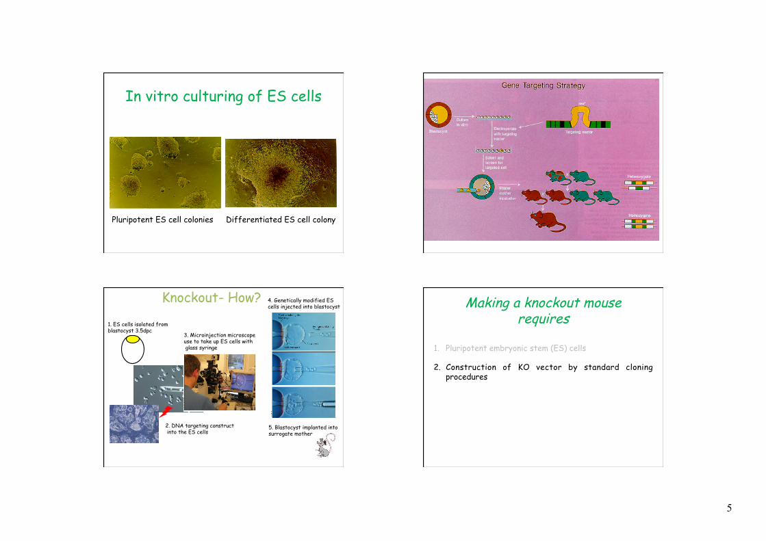
5
In vitro culturing of ES cells
Pluripotent ES cell colonies Differentiated ES cell colony
Knockout- How? 4. Genetically modified ES cells injected into blastocyst
1. ES cells isolated from blastocyst 3.5dpc
2. DNA targeting construct into the ES cells
5. Blastocyst implanted into surrogate mother
3. Microinjection microscope use to take up ES cells with glass syringe
Making a knockout mouse requires
1. Pluripotent embryonic stem (ES) cells
2. Construction of KO vector by standard cloning procedures

6
Mouse genome
DNA vector carrying a mutation
Construction of KO/KI DNA vector
*
Standard molecular biology techniques are used to design and make the targeting DNA vector
Making a knockout mouse requires
1) Pluripotent embryonic stem (ES) cells
2) Construction of KO vector by standard cloning procedures
3) Introducing the KO vector into the ES cells by electroporation
4) Selecting for gene targeting events
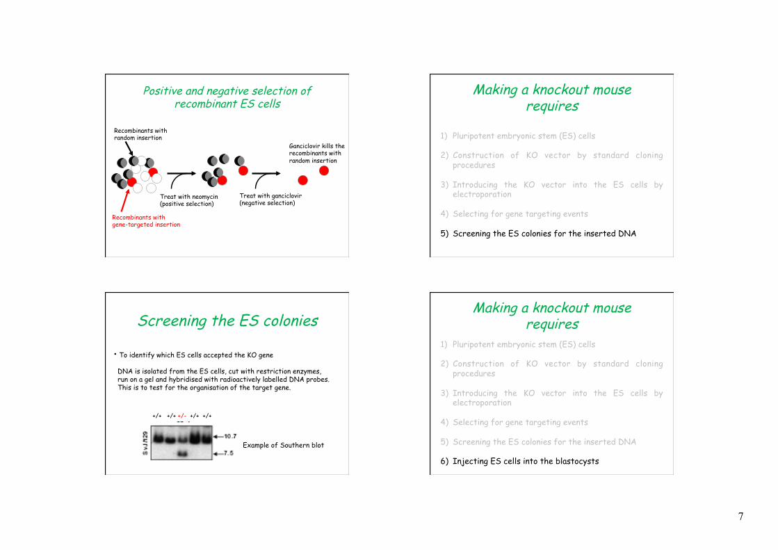
Selecting for gene replacement events
Ø Targeting vector contains marker genes
Ø Positive selectable marker, neomycin phospho-transferase is resistant to the antibiotic neomycin.
Ø Negative selectable marker, thymidine kinase from Herpes Simplex virus. The TK gene confers sensitivity to the chemical gancyclovir.
ES cells with this construct will grow in culture medium containing neomycin but will not survive in the presence of ganciclovir. Resistant to neomycin and sensitive to ganciclovir
ES cells with this construct will grow in culture medium containing neomycin and ganciclovir. Resistant to neomycin and ganciclovir
7
Positive and negative selection of recombinant ES cells
Treat with neomycin (positive selection)
Treat with ganciclovir (negative selection)
Recombinants with random insertion
Recombinants with gene-targeted insertion
Non recombinants Ganciclovir kills the recombinants with random insertion
Making a knockout mouse requires
1) Pluripotent embryonic stem (ES) cells
2) Construction of KO vector by standard cloning procedures
3) Introducing the KO vector into the ES cells by electroporation
4) Selecting for gene targeting events
5) Screening the ES colonies for the inserted DNA
Screening the ES colonies
• To identify which ES cells accepted the KO gene
DNA is isolated from the ES cells, cut with restriction enzymes, run on a gel and hybridised with radioactively labelled DNA probes. This is to test for the organisation of the target gene.
Example of Southern blot
+/+ +/+ +/- +/+ +/+
Making a knockout mouse requires
1) Pluripotent embryonic stem (ES) cells
2) Construction of KO vector by standard cloning procedures
3) Introducing the KO vector into the ES cells by electroporation
4) Selecting for gene targeting events
5) Screening the ES colonies for the inserted DNA
6) Injecting ES cells into the blastocysts

8
ES cells from a brown mouse
Blastocyst from a white mouse Making a knockout mouse
requires 6) Injecting ES cells into the blastocysts
Ø The progeny will be a chimera consisting of both KO and wild type cells
Ø Hopefully some KO cells will contribute to the germ line.
Ø Heterozygous and homozygous progeny for the KO construct can be generated and analysed for phenotypic alterations
Adapted from http://nobelprize.org
Screening of Mice
+/+ -/-
+/-
Time-line for making a knockout mouse

9
KO/KI Versus Transgenic mice
Advantage: Ø Specific insertion of gene at specific location or removal of specific gene
(knockout).
Ø Mimic recessive disorders (loss of function mutations).
Disadvantage: Ø Low level of ES cells with wanted gene inserted
Ø Further breeding necessary to obtain non-chimeric homozygotic animal
Gene targeting
Transgenic mice Advantage: Ø Relative high rate of insertion of the injected gene into the genome. Ø Use for dominant disorders.
Disadvantage: Ø Random insertion- can lead to position effects
Use of genetically modified mice
• KO/KI mice – Cystic fibrosis – Muscular dystrophies
• Transgenic mice – To study dominantly acting alleles of
mutated genes (nemaline myopathy)
Summary • Gene targeting is use to delete or mutate an
existing gene: KO and KI. Mice are generated by the injection into a blastocyst of genetically modified ES cells. Chimeric mice are made.
• Transgenic mice are use to study overexpression of a gene product. Mice are generated by DNA microinjection of fertilized oocytes. Results in random integration of the DNA.
• Both offer a valuable tool for the study of human disorders. ie. Dystrophies.
Transgenic
KO/KI Summary
10
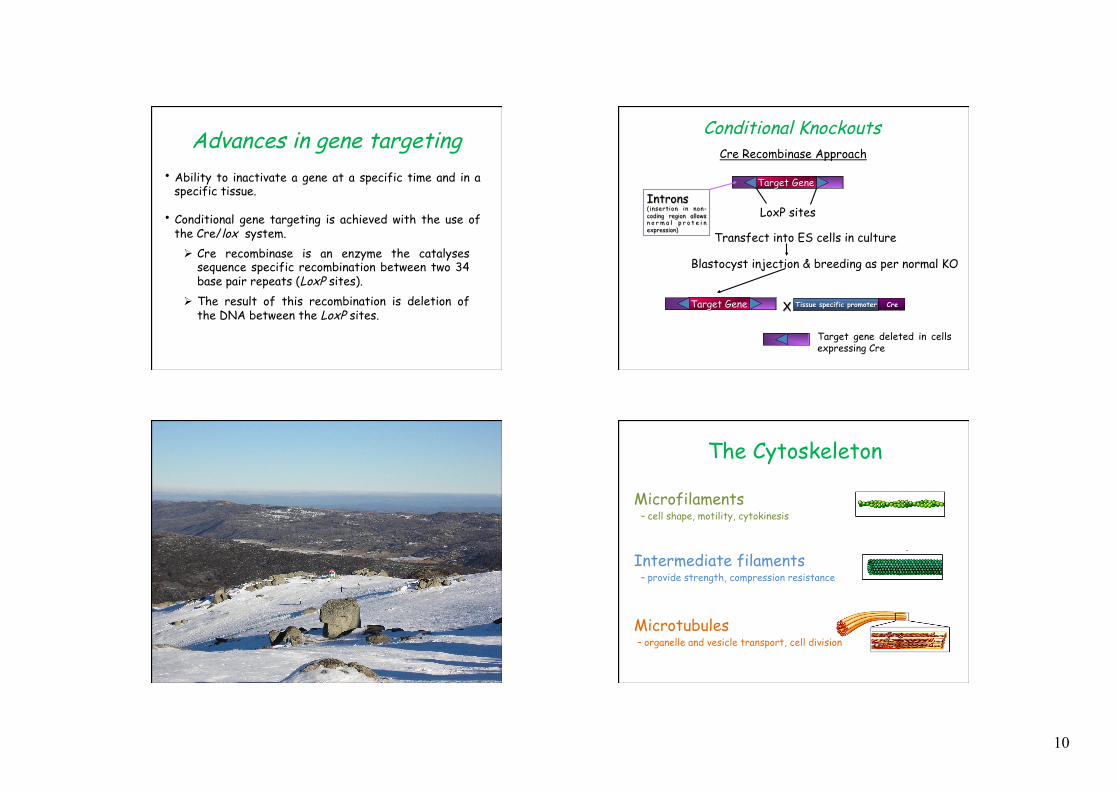
Advances in gene targeting • Ability to inactivate a gene at a specific time and in a
specific tissue. • Conditional gene targeting is achieved with the use of
the Cre/lox system. Ø Cre recombinase is an enzyme the catalyses
sequence specific recombination between two 34 base pair repeats (LoxP sites).
Ø The result of this recombination is deletion of the DNA between the LoxP sites.
Conditional Knockouts Cre Recombinase Approach
Target Gene
LoxP sites
Transfect into ES cells in culture
Blastocyst injection & breeding as per normal KO
Cre Target Gene Tissue specific promoter X
Target gene deleted in cells expressing Cre
Introns (insertion in non-coding region allows n o r m a l p r o t e i n expression)
The Cytoskeleton
Microtubules – organelle and vesicle transport, cell division
Microfilaments – cell shape, motility, cytokinesis
Intermediate filaments – provide strength, compression resistance

11
Microfilaments Intermediate Filaments
Microtubules
Actin monomers (G actin) with the aid of actin binding proteins can polymerize to form actin filaments (F actin)
Within cells an equilibrium exists between monomeric and filamentous actin. This equilibrium is influence by actin binding proteins.
cell shape cell adhesion cell motility vesicle transport endocytosis exocytosis golgi function cytokinesis membrane function
Organization of actin filaments within cells
Organisation Participation
Classes of actin binding proteins
side binding proteins
cross-linking protein (in cell cortex)
capping (end-blocking) protein
severing protein
motor proteins
nucleation protein
monomer- sequestering protein
actin monomers
actin filaments
Tropomyosin

12
Classes of actin binding proteins
side binding proteins
cross-linking protein (in cell cortex)
capping (end-blocking) protein
severing protein
motor proteins
nucleation protein
monomer- sequestering protein
actin monomers
actin filaments
Tropomyosin Tropomyosin
Tropomyosin (Tm)
Ø filamentous protein
Ø Forms dimers
Ø Dimers interact head-to-tail to form a polymer
Ø Tm polymer interacts along the length of the actin filament
Ø Provides stability and regulates the binding of other actin binding protein to the actin filament
Tropomyosins Binds Along the Sides of ActinMicrofilaments
Actin
Tropomyosin (>40 isoforms)
Different Tm isoforms regulate actin filaments by recruiting different actin binding proteins
Stable microfilaments Higher actin filament turnover Shorter filaments
Gunning, Schevzov, Kee, Hardeman. Trend Cell
Biol 2005
Ø Tm5NM1/2 knockout
Using knockout and transgenic mice to understand the function of Tm5NM1 in vivo
– Tm5NM1/2 eliminated in all tissues
Ø hTm5NM1 transgenic (Tg) – Tm5NM1 overexpressed in most tissues
X γTm 1a 1b 4 3 2b 9d
6 b 9c 5
6 a 9a 9b 8 7
Tm5NM1 Tm5NM2
Beta-actin promotor Human Tm5NM1
13
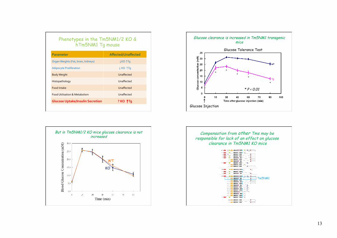
Phenotypes in the Tm5NM1/2 KO & hTm5NM1 Tg mouse
Parameter Affected/Unaffected
Organ Weights (Fat, brain, kidneys) ↓ KO ↑Tg
Adipocyte Proliferation ↓ KO ↑Tg
Body Weight Unaffected
Histopathology Unaffected
Food Intake Unaffected
Food Utilisation & Metabolism Unaffected
Glucose Uptake/Insulin Secretion ? KO ↑Tg
tg
wt
* *
* *
*
* P < 0.01
Glucose Tolerance Test
Glucose Injection
KO
WT
Compensation from other Tms may be responsible for lack of an effect on glucose
clearance in Tm5NM1 KO mice
Tm5NM1
14
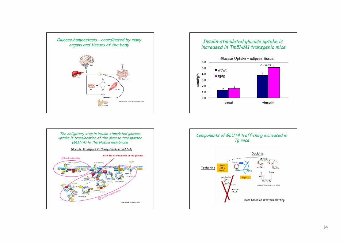
Glucose homeostasis - coordinated by many organs and tissues of the body
Adapted from: Rosen & Spiegelman, 2006
0.0
1.0
2.0
3.0
4.0
5.0
6.0
basal +insulin
umol/g/h
wt/wttg/tg
Insulin-stimulated glucose uptake is increased in Tm5NM1 transgenic mice
Glucose Uptake – adipose tissue P < 0.05
The obligatory step in insulin-stimulated glucose uptake is translocation of the glucose transporter
(GLUT4) to the plasma membrane
Glucose Transport Pathway (muscle and fat)
From: Ramm & James, 2005
GLUT4
GLUT4
1 Insulin signalling
2 GLUT4 translocation
Actin has a critical role in this process
Sec8 Sec 5 Myrip
Wave(1.6)
Tethering
Myo1c
Docking
Tethering
Components of GLUT4 trafficking increased in Tg mice
Actin
Adapted from: Zaid et al., 2008
Data based on Western blotting

15
wt tg
Myo1c
Sec 8
Western blots showing components of GLUT4 trafficking increased in Tg mice
From adipose tissue
Tm5NM1 can produce stable actin filaments
Stable microfilaments Higher actin filament turnover Shorter filaments
Gunning, Schevzov, Kee, Hardeman. Trend Cell
Biol 2005
Can Tm5NM1 increase in actin filaments in the mouse tissues?
Tm5NM1
Tm5NM1 increases filamentous actin in adipose tissue
F-actin staining
16
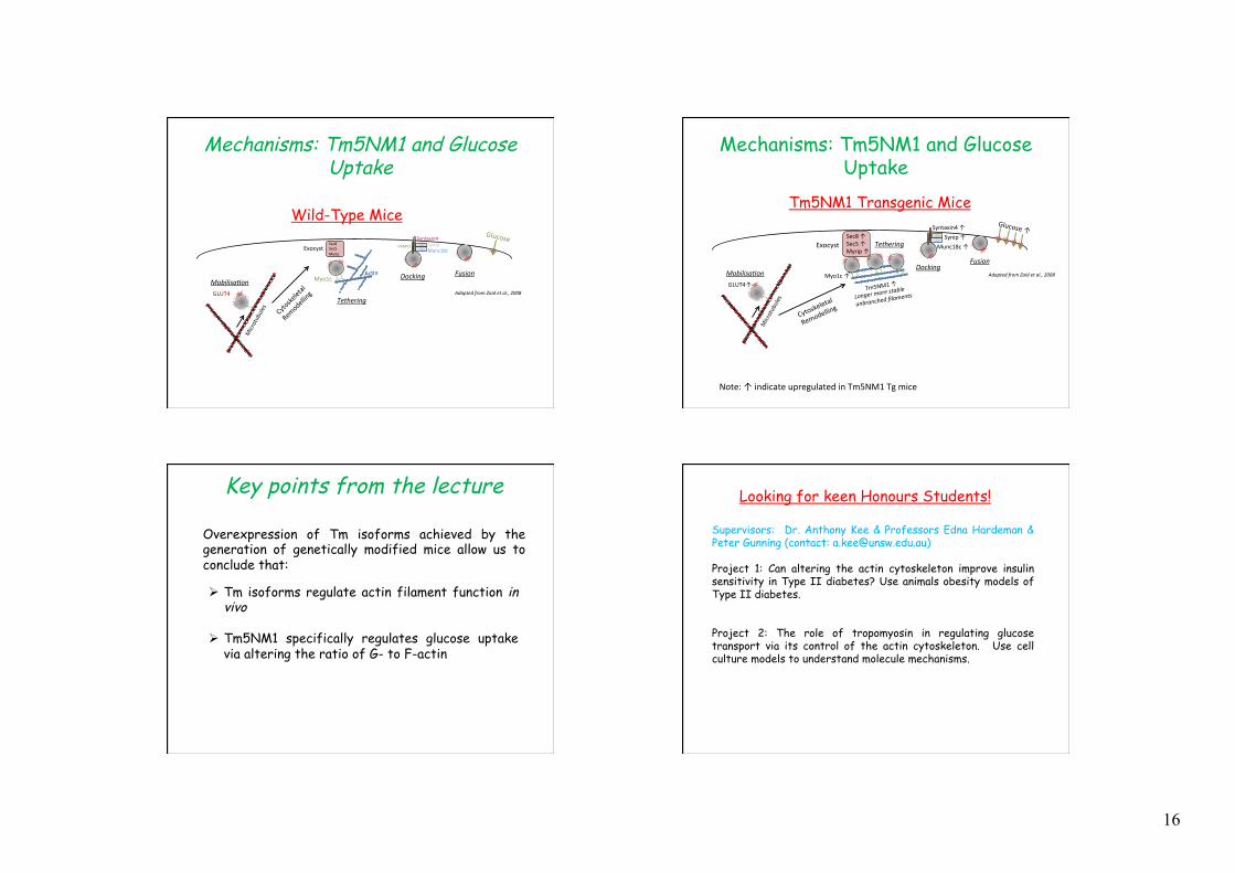
GLUT4
Microtubules
Mobilisa(on
Tethering
Docking Fusion
Syntaxin4 VAMP2 Synip
Munc18c
AcBn
Sec8 Sec5 Myrip
Exocyst
Myo1c
Glucose
Adapted from Zaid et al., 2008
Cytoskeletal
Remodelling
Mechanisms: Tm5NM1 and Glucose Uptake
Wild-Type Mice
Adapted from Zaid et al., 2008
Note: ↑ indicate upregulated in Tm5NM1 Tg mice
GLUT4↑
Microtubules
Mobilisa(on
Tethering
Docking Fusion
Syntaxin4 ↑ Synip ↑
Munc18c ↑
Sec8 ↑ Sec5 ↑ Myrip ↑
Exocyst
Tm5NM1 ↑ Myo1c ↑
Glucose ↑
Cytoskel
etal
Remode
lling
Longer more stable
unbranched filaments
Mechanisms: Tm5NM1 and Glucose Uptake
Tm5NM1 Transgenic Mice
Key points from the lecture
Overexpression of Tm isoforms achieved by the generation of genetically modified mice allow us to conclude that:
Ø Tm isoforms regulate actin filament function in vivo
Ø Tm5NM1 specifically regulates glucose uptake via altering the ratio of G- to F-actin
Looking for keen Honours Students!
Supervisors: Dr. Anthony Kee & Professors Edna Hardeman & Peter Gunning (contact: [email protected]) Project 1: Can altering the actin cytoskeleton improve insulin sensitivity in Type II diabetes? Use animals obesity models of Type II diabetes. Project 2: The role of tropomyosin in regulating glucose transport via its control of the actin cytoskeleton. Use cell culture models to understand molecule mechanisms.