workshop on ion beam therapy - office of science/media/hep/pdf/accelerator-rd... · the workshop on...
TRANSCRIPT

Workshop on Ion Beam Therapy
Summary Report
January 9–11, 2013

2

3
Table of Contents Executive Summary ........................................................................................................................ 5
1. Introduction ............................................................................................................................. 7
2. Identification of Clinical and Radiobiological Applications ................................................... 9
2.1. Clinical Considerations .................................................................................................... 9
2.1.1. Treatment Issues ................................................................................................... 10
2.2. Clinical Trials ................................................................................................................. 11
2.2.1. Desirable Future Facility Capabilities .................................................................. 12
2.3. Radiobiology Considerations ......................................................................................... 13
2.3.1. R&D Issues ........................................................................................................... 15
3. Beam Requirements ............................................................................................................... 16
3.1. Beam Production and Delivery ...................................................................................... 16
3.1.1. Ion Species ............................................................................................................ 16
3.1.2. Beam Energy ......................................................................................................... 16
3.1.3. Intensity................................................................................................................. 17
3.1.4. Scanning and Scattering ........................................................................................ 17
3.1.5. Field Size .............................................................................................................. 17
3.1.6. Beam Geometry .................................................................................................... 17
3.1.7. Spot Size and Position Accuracy .......................................................................... 17
3.1.8. Treatment Time for Various Fractionation Regimens .......................................... 18
3.1.9. Range .................................................................................................................... 18
3.1.10. Dose Accuracy ...................................................................................................... 18
3.2. Treatment Planning ........................................................................................................ 18
4. Beam Delivery System Requirements ................................................................................... 20
4.1. Sources and Accelerators ............................................................................................... 20
4.1.1. Accelerator Types ................................................................................................. 21
4.2. Delivery Systems............................................................................................................ 22
4.2.1. Beam Lines ........................................................................................................... 22
4.2.2. Gantry ................................................................................................................... 22
4.2.3. Imaging and In-vivo Dosimetry ............................................................................ 23
4.2.4. Motion Control...................................................................................................... 24

4
5. R&D Activities ...................................................................................................................... 25
5.1. Specific R&D Needs ...................................................................................................... 25
6. Summary ................................................................................................................................ 27
References ..................................................................................................................................... 29
Appendices .................................................................................................................................... 30
A.1 List of Participants ......................................................................................................... 30
A.2 Workshop Charge ........................................................................................................... 32
A.3 Glossary of Abbreviations .............................................................................................. 34

5
Executive Summary More than 60 participants from the U.S., Sweden, The Netherlands, Japan, Germany, and Italy were charged with addressing four topics that consecutively built a basis for identifying R&D gaps between current accelerator capabilities and future requirements. A pre-workshop survey of ideas was collated to stimulate the participants to consider the range of required parameters. After three opening lectures on clinical, radiobiological and physics aspects covered the current background of the multidisciplinary fields involved in this science, the participants were parceled into subject panels to debate the issues and priorities over two intense days. The conclusions of each break-out panel were presented to all of the participants for further discussion and debate.
Charge 1: Identify pertinent clinical applications and radiobiological requirements for future use of ions in cancer therapy. Charge 2: Assess corresponding beam requirements for future treatment facilities and compare these with the current state-of-the-art. Charge 3: Assess the corresponding beam delivery system requirements for future treatment facilities and compare these with the current state-of-the-art. Charge 4: Identify R&D activities needed to bridge the gap between current capabilities and future requirements and identify which will have the highest near-term performance gain.
What follows are concise statements resulting from the workshop discussions of the four charges:
• A substantial amount of wide-ranging R&D will be required over several years for the U.S. to contribute in a meaningful way to efforts already under way in other countries.
• Fundamental radiobiological research must be conducted in parallel with the development of imaging and accelerator technologies, and should be conducted at all depths of the Bragg curve for tumors and normal tissues to ensure effectiveness and safety.
• Future accelerator and ion beam therapy systems must be reduced in size to the scale of current radiotherapy facilities.
• Components for beam monitoring, treatment planning, quality assurance, and treatment delivery need further developments to improve their speed, accuracy, and capabilities while still ensuring safety and efficacy.
• Future facilities will need multiple ions, at least protons and carbon and one or two others, to permit inter-facility comparisons, to enable needed research with ions other than protons and carbon, and to permit combining ions for optimal patient treatment.
• Short dose deposition times will require rapid scanning both transversely and longitudinally (thus rapid energy modulation will also be needed).

6
• Cost-effective improvements required in beam spill structure, dose rate, multi-energy, and multi-ion extraction will put significant demands on accelerator performance.
• Developments in gantries and beam lines are considered essential to the full realization of ion beam therapy, and will likely require superconducting devices to achieve the desired compactness.
• Real-time measurements of patient parameters will be important to ensure proper beam range and to verify dose deposition; measurement devices must be interfaced with imaging technologies.
• Cooperative research among and between U.S. and international ion beam centers will expedite progress.
7
1. Introduction The cyclotron was invented in America by Ernest Lawrence in 1930 to study the nuclear structure of the atom, for which he received the Nobel Prize in Physics in 1939. Cyclotrons produce high-energy particles that are accelerated outwards in a magnet in a spiral fashion rather than through an extremely long, linear accelerator. Particle beams are unique in that they deposit dose primarily within a “Bragg peak” at the end of their range, unlike conventional radiations whose dose depositions decrease nearly exponentially from the beam entry position. Exploitation of the Bragg peak for cancer therapy was first suggested by Robert Wilson in 1946. In 1952, Cornelius Tobias at the University of California Berkeley performed initial proton and helium ion radiobiology experiments, followed almost immediately by a clinical Phase I/II trial involving 30 patients led by John Lawrence. A subsequent clinical Phase I/II program was conducted at the Lawrence Berkeley National Laboratory in the mid-1970s to 1990s to investigate the clinical efficacy of various light ion beams, with some modest success in treating advanced cancers. Unfortunately, that program was closed for financial reasons in 1992.
Accelerators and related technology are critical to many areas beyond their traditional role in discovery science, and they influence our everyday lives in many ways, including several medical applications. In particular, today’s modern accelerators have become essential for state-of-the-art radiotherapy for cancer, since they can deliver high doses to a deep-seated tumor while sparing surrounding normal tissue. Ten proton accelerators are currently operating in the U.S. for cancer radiotherapy, and more are under construction or planned. However, these and the equivalent accelerators for ion beams have significant limitations in terms of the cost, size, energy, beam delivery, and other design requirements of the machines. Beam scanning methods to “paint” radiation doses within a three-dimensional tumor volume require stringent beam control technologies integrated with high-resolution anatomical imaging. These control technologies need further improvements and confirmation of clinical safety.
In October 2009, the Department of Energy’s Office of High Energy Physics sponsored a symposium and workshop “Accelerators for America’s Future” chaired by Walter Henning and Charles Shank. Its purpose was to elicit the views and opinions of a wide range of accelerator users on the challenges and opportunities for developing and deploying accelerators to meet national needs. The report of this workshop, published in June 2010, has drawn congressional interest in enhancing U.S. stewardship of accelerator science R&D. Currently, accelerator expertise is growing in Asia and Europe, due to national policies in place in some countries that are promoting use of accelerators for medical purposes. Accelerators using ion beams heavier than protons, specifically carbon ion beams1, for cancer radiotherapy in Japan, Germany, and Italy are providing evidence of intriguing clinical responses for radiation-resistant tumors. The U.S. currently lags behind the larger international community in research and development for accelerator applications for medicine.
1 Typically referred to as “light” ions.
8
To assist in preparations for creating an accelerator R&D stewardship strategic plan, Dr. Jim Siegrist, Associate Director of Science for the Department of Energy’s High Energy Physics (HEP) Program within the Office of Science (SC), in consultation with other SC Associate Directors, asked the SLAC National Accelerator Laboratory to convene a community task force, chaired by Dr. Norbert Holtkamp, to provide updated information from a team of experts. This ultimately led to the development and submission to Congress of the U.S. long-term accelerator R&D stewardship strategic plan. One focus of this plan was to develop improved particle therapy beam delivery and control systems for the medical community. Reducing the cost and size of treatment centers is critical to making particle therapy more widely available.
As one of the 27 Institutes and Centers within the National Institutes of Health (NIH), the National Cancer Institute (NCI) has a mandate to fund research related to the prevention, diagnosis and treatment of cancer. Therefore, NCI has played a significant role since the inception of particle therapy in funding research in radiobiology and physics for the conduct of clinical trials.
Although significant progress has been made in the use of particle beams for cancer treatment, an extensive research and development program is needed to maximize the healthcare benefits from these therapies. While it is true that there are no specific NIH Funding Opportunity Announcements directed to particle therapy research, it is also true that the NCI remains open to reviewing any research that is compatible with its established funding mechanisms. However, given the very substantial resources that these facilities currently require, along with the highly competitive economic environment that now exists, it is clear that any research applications need to leverage scientific pursuits, which are the NCI mandate, with the realities of clinical practice, as is also the case for photon radiation research. Such leveraging could be enhanced by the growing opportunities and need for domestic and international collaborations. These collaborations are complicated by the difficulties of separating research (the NCI mission) from clinical practice development and by the fluid nature of the technologies that are being developed and implemented in particle therapy.
The Workshop on Ion Beam Therapy was planned (see Appendices A.1 and A.2) to understand what clinical and research capabilities are currently available, what capabilities are desired for the future, and what R&D activities are needed to bridge the gap. Specific accelerator-related R&D topics were discussed that might benefit future beam therapy programs by enabling therapy with ions heavier than protons and increasing the flexibility and capability of beam delivery systems for this purpose. U.S. and international experts (Appendix A.1) participated in lively discussions with a goal of obtaining ideas for new approaches to deliver particle beams with “gantry” systems that provide maximum flexibility to irradiate the tumor while avoiding the need to move the patient during the treatment. The report that follows summarizes the discussions of the four charges by the workshop participants. For the reader’s convenience, a brief list of abbreviations used can be found in Appendix A.3.
9
2. Identification of Clinical and Radiobiological Applications Charge 1 Identify a set of representative clinical applications that span the range of expected future therapy requirements. These need to include capabilities for performing radiobiological experiments as well as human treatment protocols in order to explore the scientific principles underlying observed clinical results and point the way to promising protocol designs.
2.1. Clinical Considerations Over 10,000 patients have been treated for cancer with ions heavier than protons, mostly in Japan and Germany. At this time, additional light ion beam therapy facilities are in operation or are under construction in Europe and in Asia. We believe therapy with ions heavier than protons should be reintroduced into the United States to evaluate its safety and efficacy in the treatment of cancer patients. Based on clinical experience and radiobiology, the characteristics of tumors most likely to benefit significantly from therapy with ion beams are as follows:
• High risk of local failure after conventional photon or proton radiation therapy
• Radio-unresponsiveness due to histology, hypoxia and/or other factors
• Low risk of systemic failure with the most effective systemic therapies available
• Involvement of critical normal structures that would prevent adequate resection or would require a significant loss of critical organ function (i.e., blindness, paralysis, loss of ambulatory ability, etc.) with a complete surgical resection
• Tumors abutting or in very close proximity to sensitive (critical) normal structures
• Efficient repair of cellular damage
• Recurrent tumors
• Clear imaging delineation between tumor and normal tissues
At the workshop, there was a general view that the analysis performed by ENLIGHT [1] included to a large extent the tumors most likely to benefit from light ion beam therapy. In addition, it was concluded, based on the experience in Japan [2], that osteosarcoma, uveal melanoma and advanced cervical cancer are tumor sites that might benefit from light ion beam therapy. Finally, it was expected that locally advanced non-small-cell lung cancers, and locally unresectable tumors (locally advanced breast cancer, unresectable sarcoma, etc.) may also benefit from ion beam therapy.
10
It is expected that by the time an ion beam therapy facility becomes operational in the United States, physicians will be using yet-to-be-developed specific tumor biomarkers that will help select patients with tumors that respond poorly to low-LET radiation. Such patients would be ideal candidates for treatment with ions heavier than protons. The following is a list of representative tumors that are among those in patients treated to date with ions heavier than protons, along with tumors considered at this time to potentially benefit from ion beam therapy:
• Gliomas
• Non-squamous head and neck malignancies
• Mucosal melanomas
• Chordomas and chondrosarcomas of the skull base and spine/sacrum
• Sarcomas
• Pancreatic and hepatic carcinomas
• Lung cancers
• Renal cell carcinoma
• Locally unresectable tumors (locally advanced breast cancer, and locally advanced or recurrent pelvic malignancies, etc.)
At this time it appears better to treat malignancies in pediatric patients with protons or helium ions rather than heavier charged particles because of concerns about the dose distribution characteristics (e.g., fragmentation tail and large secondary particle halo), and the uncertain late effects of treatment with heavier ions.
2.1.1. Treatment Issues There are a number of special considerations in the treatment of patients with protons and heavier ions. These include density inhomogeneity of tissues in the beam path (resulting in uncertainties in the range of particles in patients), motion of the tumor and normal organs, proximity of tumors to critical normal structures, and the biological characteristics of tumors.
Tumor motion is currently being managed by suppressing respiratory motion, using gated techniques and online monitoring during radiation delivery. Further development of real-time tumor tracking methods currently in clinical use will be even more essential for ion beam therapy than it is for photon therapy. Anesthesia may be the preferred way of controlling respiratory motion, especially for hypofractionated ion beam therapy.
Continued R&D on the range uncertainty of ion beams in patients is essential in the treatment of tumors in close proximity to critical normal tissues. Dual energy CT, megavoltage CT, as well as proton and ion radiography and CT are under investigation and will be very important for ion beam therapy.

11
Planning for the treatment of tumors in close proximity to critical normal structures with ion beams might benefit at this time from the strategy of taking advantage of the extremely sharp lateral penumbra of ion beams. This ion beam property is well defined, whereas knowledge of the range and fragmentation tail of ion beams heavier than protons is less precise. Improved imaging to accurately differentiate between the tumor and normal tissues is essential to precisely delivering the prescribed ion beam dose to the tumor volume (while simultaneously minimizing the radiation dose to adjacent normal organs). Ensuring accurate beam delivery represents an R&D challenge for beam line instrumentation.
Continued R&D on noninvasive methods of imaging hypoxia, adaptive ion therapy, and inversely planned dose painting is essential for achieving future treatment goals. Biologically effective dose intensification, currently only hypothetical, would present the opportunity to use advances in medical imaging to address the inhomogeneity of hypoxia or other causes of relative unresponsiveness of some types of human tumors to radiation. There also exists an urgent need for R&D to identify biomarkers in tumors that are predictive of a greater likelihood of benefit from high-LET ion beam therapy.
2.2. Clinical Trials It is considered paramount that clinical studies be performed to demonstrate the potential advantages of therapy with light ions over proton therapy. It is very important that these clinical trials be well planned and designed so as to be ready for implementation when an ion beam therapy facility becomes operational. Facility throughput sufficient to generate meaningful statistical power in any clinical trial or study is also essential. In the absence of full radiobiological data, protocol development would benefit greatly from early computer simulations, treatment modeling, and comparative biological and physical dose modeling studies based on data that already exist or might be generated by collaboration with existing ion beam therapy facilities.
The expected advantages of ion beam therapy should be explored in Phase I or I/II clinical trials2 incorporating advances in real-time imaging. Determination of ideal dose fractionation schemes and their safety and efficacy should be validated for the various ion species prior to incorporation, as appropriate, into large Phase III clinical trials.
In the eventuality of having one or more ion beam therapy facilities in the U.S., it is anticipated that large scale randomized Phase III trials would be undertaken nationally, and likely internationally, in collaboration with centers throughout the U.S. and with ion beam programs in other countries. In the design of these trials, it will be important to strictly define patient entry criteria based on biomarkers and molecular imaging characteristics. These criteria should be demonstrated in future trials to identify tumors that would most benefit from treatment with ion
2 It may be that the Phase I/II trials could be shortened or eliminated by taking advantage of data already available from our foreign colleagues and earlier U.S. trials, permitting an earlier launch of Phase III trials.

12
beam therapy. In comparing different radiation modalities, it is important that only one variable (e.g., LET) differs between treatment groups. This caveat is magnified by the diversity and rapidity of technology development in particle beam therapy.
In anticipation of having an ion beam therapy clinical program in the United States, significant consideration should be given to establishing an international particle effectiveness repository to include selected treatment planning data, clinical and demographic information, and treatment outcome data (e.g., acute and late toxicities, patterns of failure, and overall survival).
2.2.1. Desirable Future Facility Capabilities Based on discussions at the workshop, it was concluded that accelerator and beam delivery system R&D should lead to ion beam therapy facilities in the United States that are individually capable of delivering treatments with multiple ion species. The ion species should ideally span from protons through neon. However, it was recognized that, in the next 10 years, R&D in accelerator and beam delivery technology may provide facilities in which the upper end of particle species for treatment would be limited to carbon or oxygen. The range of ion beams should extend at least to 30 cm in water, irrespective of the ion species. In addition, a range of 60 cm for ion radiography was considered desirable.
In selected clinical situations it may be advantageous to deliver treatments with multiple ion species within the same treatment course, and ideally within an individual treatment session. This was considered to be particularly advantageous if imaging R&D leads to the ability to define areas in tumors having heterogeneous biological regions or properties, e.g., hypoxia, related to a lack of responsiveness to low-LET radiation therapy. Ion beam therapy system R&D should have as a goal to provide, at a minimum, technical capabilities corresponding to the same dose delivery parameters that will be achievable with the highest technology photon units. This consideration suggests that, to realize the full potential of ion beam therapy, a rotational gantry to deliver that therapy (or other equivalent technology to deliver beams from all directions) would be necessary in at least one room in each ion beam therapy center. However, it is envisioned that, with further R&D as well as further experience, simpler, more compact, and more cost-effective beam delivery solutions should be available for the treatment of patients. Even today, it was concluded that fixed ion beams are certainly acceptable for selected clinical situations.
Among accelerator R&D goals it was considered desirable to provide facilities in which the patient treatment times will be a few minutes or less, and perhaps as short as a few seconds for tumors or normal structures with clinically significant motion. All necessary patient-related activities, including real-time target position and beam delivery verification, target gating or tracking, and dose repainting, should be included within this time interval.
In order to realize the full potential of ion beam therapy when available for patient treatment, the full scope of imaging technologies and motion management capabilities existing in photon therapy facilities should be available on ion beam therapy systems. Additionally, R&D should

13
be performed to develop imaging systems to exploit the properties unique to ions that could be used to verify beam delivery in vivo, to compensate for tumor and normal tissue motion, and to correct for tumor or normal tissue deformation. Ideally, such imaging devices would be available in all treatment rooms at the facility.
2.3. Radiobiology Considerations Although clinical studies with charged particles have been conducted successfully, the limited radiobiology information is a significant impediment to exploiting the full potential of particle therapy. Additional radiation biology knowledge is essential to guide accelerator and beam delivery system R&D. The specific areas of highest importance for ion-related biology research relevant to accelerator development are:
• additional data on biologically-effective doses3 of ions in tumors and normal tissues;
• dose fractionation patterns;
• the role of hypoxia along with other bio-molecular processes; and
• effects of altered dose rate.
Of highest importance is R&D to increase our understanding of the biological response of cells and tissues (in both tumors and normal organs) to irradiation with various ions. It is difficult to design a clinical trial based on the assumption that the same biologically effective dose is being administered in the photon, proton and other light ion arms of the trial without additional knowledge of how to equate those doses. Traditionally, biological and clinical responses to charged particles have been referenced to photon doses. However, there is inadequate knowledge about the responses of various tumor types, as well as early and late responding normal tissues4, to different ion species over a range of radiation doses.
The physical characteristics of particle species, and the energy deposition by the ion beams, change as the particles traverse and finally stop in the patient. Radiation safety and clinical effectiveness require experimental confirmation that the complex interactions of different ionization density along the beam path are inactivating target tumor cells while minimizing damage to normal tissues. To guide the specification of ion species, energies, fluences, etc., that an accelerator should be capable of producing, additional radiation biology studies are needed to quantify:
• the biological response (e.g., relative biological effectiveness or RBE) as a function of ion species (protons through neon);
3 These depend on ion species and on the dose per fraction or dose rate, etc. 4 Early responding normal tissues are rapidly growing tissues, such as skin, that show damage within days to a few weeks after radiation exposure; late responding normal tissues, such as brain and liver, are slower growing and damage in them is not seen until many weeks or months after irradiation.

14
• the importance of dose per fraction, ion energy and depth in tissue for response of both appropriate tumor types and dose-limiting normal tissues.
These studies should include an assessment of the potential importance of high-LET dose effects in normal tissues due to ion beam fragmentation in the distal tail of the Bragg peak (see Fig.1). The most relevant endpoint to be studied is cell inactivation, but other useful information includes DNA damage response, gene expression, molecular and biochemical changes, mode of cell death, intra- and inter-cellular signaling, and neoplastic transformation. Both in vitro and in vivo studies will be necessary to obtain the requisite information. Clinical studies with both photons and charged particles are exploring the safety and efficacy of reducing the number of dose fractions, even down to a single high dose treatment for selected tumor types. Modern accelerators can produce high intensity monoenergetic particle beams that can be used to administer a radiation dose rapidly to the entire volume of a human tumor. The radiobiological effects of single high dose fractions, e.g., 25 Gy, may be substantially different from those of the 1.8–2.0 Gy dose fractions traditionally used in conventional photon radiotherapy. With single high radiation doses, one may see greater damage to vascular structures and tumor stroma. The radiobiological effects of high- and low-LET radiations on tumors and normal tissues over a wide range of dose fraction intensities should be carefully studied. Clinical experience in early neutron studies, and subsequent radiobiology investigations, demonstrated the differential responses of late-responding normal tissues from early-responding tissues and tumors. These differences, and their dependence on ion species as well as radiation dose and fractionation pattern, are in need of further experimentation.
Fig. 1. Calculated and experimental Bragg peaks for 400 MeV/u carbon ions. The “extra” dose due to the fragmentation tail is indicated. [Adapted from E. A. Blakely et al., Radiation Research 80, 122–160 (1979), with permission from Academic Press.]
15
It is well documented in the laboratory that tumors outgrow their oxygen supply and can develop regions of hypoxia that are relatively radio-resistant. Radiobiological studies have shown that the dependence of cell damage on oxygen decreases as the mass of the irradiating ion species increases. However, the dependence of this effect on ion species, energy, dose, and dose rate are in need of further research. Tumor reoxygenation during the prolonged course of a typical photon treatment regimen may, in some tumor types, increase photon treatment efficacy. The roles of hypoxia and reoxygenation in ion beam therapy remain topics where there is need for radiobiology research.
Functional imaging capabilities in humans are improving rapidly. Soon it may be possible to “map” concentrations of oxygen and other metabolites in tumors. R&D in accelerator and beam delivery system designs is needed to incorporate both functional imaging and anatomical imaging to identify beam range in the patient.
The delivery of ion beam therapy via passively scattered beams is being replaced by active beam scanning. Active scanning produces beams with significantly higher instantaneous dose rates (hundreds of Gy/min within a pulse of a few milliseconds) than experienced with traditional dose delivery. This raises the possibility of altered biological responses because of changes in the distribution of free radical types and the potential for cellular oxygen depletion. Therefore, research is needed to define the potential impact of beam spill structure and repetition rate on biological and clinical outcomes, as these features must be considered in the accelerator design.
Most cancer patients now are treated with combined modality regimens that currently include chemotherapy, molecularly-targeted agents, immunotherapy, and/or nanoparticles. It is anticipated that combined modality therapy with ion beams will be important in the future. Although there are pre-clinical and clinical data on interactions of many of these agents with photons, studies of their interactions with charged particles are necessary. Such studies are important, as different biological outcomes may result from high- versus low-LET radiation.
2.3.1. R&D Issues As part of the overall ion therapy accelerator and beam delivery system R&D program, an important issue will be the need to incorporate ancillary instrumentation for patient care, including imaging capabilities immediately prior to treatment, during treatment (to facilitate adaptive radiation treatment), and post-treatment (to confirm dose delivery). Biological research in parallel with instrumentation R&D is needed to develop and validate the use of biomarkers to confirm dose localization as well as tumor and normal tissue responses. This research could include the development of markers for hypoxia, DNA damage and response, cell signaling or cell metabolism. The markers will be detected via PET, MRI, SPECT, or other instrumentation. The design of the accelerator hardware needs to accommodate, in a modular fashion and near the patient, the imaging systems needed to detect biological markers (discussed in more detail in Section 4.2.3).

16
It is worth noting that the radiobiology knowledge gained through these studies will also enhance the use of photons in radiation therapy of cancer patients.
3. Beam Requirements Charge 2 Assess the corresponding beam requirements (e.g., energy range and energy spread, intensity range and pulse-to-pulse intensity jitter, spot size and pulse-to-pulse position jitter, repetition rate, ion species) for future treatment facilities and compare these with today’s state-of-the-art.
3.1. Beam Production and Delivery The clinical requirements from Section 2 translate into the beam production and delivery requirements discussed in the subsections below.
3.1.1. Ion Species Identification of the ion species to be included was discussed in Section 2. Protons and carbon ions should be included in future facilities to provide reference beams for inter-facility comparisons and because of their proven clinical efficacy in selected tumor types. Protons and carbon ions have some significant differences in the biological responses they induce, but may not be the optimal ions, based on biological responses and dose distribution patterns, for all clinical situations. Other ion species, e.g., lithium, may provide clinical advantages in selected situations. This leads to the concept of treating individual patients with multiple types of ions to maximize the clinical benefit. Due to the large number of potential ions, there should be further radiobiological research to guide the choice of ions for patient treatment. The availability of multiple species in a single facility needs to be considered in the accelerator design. In order to be optimally useful, the ability for rapid ion species switching (i.e., within seconds) is needed.
3.1.2. Beam Energy The energy goal is to design for the highest energy needed to penetrate the girth of the largest expected patient. Based on the experiences in Japan, Germany, and Italy, 430 MeV/u, i.e., a range in water of 30 cm, is the required maximum energy for carbon ions. This specification will allow treatment of close to 100% of prospective patients. For other ions, the energy should be scaled accordingly to reach 30 cm depth in water. Higher energies might be required to penetrate through the patient from limited, or all, directions for transmission imaging (radiography or even CT). However, 430 MeV/u may still be sufficient for imaging scans with carbon ions in the head and chest regions. For abdominal or pelvic treatments, it may not be necessary to scan from the direction with the largest diameter. With ions lighter than carbon, of course, the range will not be an issue even for radiography applications.

17
Another goal of accelerator R&D should be to deliver ions with energies low enough that a range of 1 cm in tissue can be achieved.
3.1.3. Intensity A wide intensity range is desirable (105–1010 particles/s) to allow both ion imaging and therapy applications as well as acceptably brief treatment and imaging times across the range of tumor applications. Accelerator and beam line instrumentation must be designed to accommodate this entire range of intensities—a challenging requirement.
3.1.4. Scanning and Scattering The expectation of the workshop participants was that future facilities will provide only for beam scanning. Opportunities exist to optimize scanning technologies for delivery time, precision, and robustness with respect to patient motion. Longitudinal scanning (along the depth direction) must also be considered.
3.1.5. Field Size A field size of 40×40 cm2 is considered to be optimal, with a minimum requirement of 20×20 cm2. This field size must be met by the beam delivery system design.
3.1.6. Beam Geometry As noted in Section 2, at least one patient treatment room in a treatment facility should have a gantry that allows treatments with all ion species. This is not only for flexibility in treatment delivery, but also to allow a proper comparison in clinical trials with current capabilities of proton and photon treatment facilities.
3.1.7. Spot Size and Position Accuracy A spot size less than 5 mm FWHM at isocenter should be achievable for all ion species at all energies. A range of spot sizes would be desirable for flexibility, especially for IMPT (intensity modulated particle therapy). The total number of discrete spot sizes should be kept small to facilitate commissioning of the system. Reasonable values of spot size are 3 mm FWHM, and 10 mm FWHM. For research with small animals (e.g., mice), the spot size should be as small as possible (perhaps <1 mm FWHM). This can be achieved with collimators if need be.
In addition to the different spot sizes, characterization of the shape of the beam spot is important. This provides the ability for the treatment planning system (TPS) to correctly calculate and plan dose deposition based on the exact shape. There are certain circumstances when modifying the shape of the beam spot (e.g., creating a flat edge) is advantageous; a TPS must be able to handle this as well.
Position accuracy pulse-to-pulse should be within ±0.2 mm and should be monitored during beam delivery.

18
3.1.8. Treatment Time for Various Fractionation Regimens For most treatments, a dose rate of 2 Gy/min/liter should be the goal for accelerator design. For delivering high dose in a hypofractionated regime5 and for rapid-scanning applications (especially in cases with organ motion issues), an order of magnitude shorter delivery time, and hence an order of magnitude higher dose rate, is desirable.
3.1.9. Range In addition to the maximum and minimum ranges of the ion beam, which depend on the beam energy, the particular matter through which the beam traverses, and the ion species (as discussed above), the rate at which the range of the beam can be changed is also an important factor in design. Ideally, it would be desirable to change the range of the beam longitudinally at a rate comparable to that achievable laterally.
3.1.10. Dose Accuracy This is the primary consideration for treatment planning. It is very important that the dose be monitored and recorded pulse-by-pulse during each treatment. The critical consideration is to deliver the dose to the target volume with an accuracy within 2.5% of the prescribed dose.
3.2. Treatment Planning Treatment of a patient with ion beam scanning is uniquely tailored for that patient and is quite complex. There are many different parameters involving various systems. Each of these parameters has uncertainties, with the largest uncertainties being patient specific. Treating a patient properly with ion beam therapy requires a TPS that can optimize the radiation delivery by varying these parameters in light of their uncertainties.
A key functionality that the TPS must include is the ability to handle a radiobiological model of the physical interaction of the ions with human tissue, be it normal organs or tumor. Also important is the physiological state of the volume being irradiated (e.g., hypoxia). Each ion species can potentially produce different levels of RBE compared with traditional photon therapy for various normal tissues and different tumor types. In addition, the manner in which the radiation is delivered, e.g., the dose and fractionation schedule, will also impact RBE.
Currently, three approaches are in use for calculating the RBE of carbon (the most widely used light ion) and incorporating it into the TPS:
• the “pseudoconstant clinical RBE,” used in Japan,
• the Local Effect Model (LEM) used in Germany and Italy, and
• the Microdosimetric Kinetic Model (MKM) being developed in Japan.
All of these models have considerable uncertainties due to limited experimental data, but have been used cautiously in the treatment of over 10,000 cancer patients to date. However, the 5 Where, for example, 1–3 fractions with doses of up to 20 Gy each are utilized.
19
uncertainties and limitations of the models have compromised the usable treatment field geometry. In most treatments, sub-optimal parallel-opposed beam arrangements have been used such that some of the biological uncertainties cancel out. Knowing the biology better would open new dimensions in beam delivery.
To take full advantage of such a complex radiobiological model, the TPS must have the capability of accepting and interpreting input from different anatomical, physiological, metabolic, and functional imaging systems. Given that these physical and physiological aspects of a patient and his/her tumor can change on a daily basis, particularly when being treated, the TPS should be able to handle these daily perturbations and re-optimize the radiation delivery schema to best treat the patient. In order to handle the daily perturbations, the TPS should have automatic static and deformable image registration (fusion) and auto-contouring capabilities. Auto-contouring capability would also be helpful in ensuring relative consistency from patient to patient and from physician to physician.
In addition to the biological aspects of a complex radiobiological model, characterization of the physical interactions between the ion and tissue at the near-cellular level is needed. These physical interactions give rise to the linear energy transfer (LET) and to the particle track structure. In order to properly evaluate these phenomena, a Monte Carlo simulation is needed. Simulations of these physical phenomena should be very fast to allow for nearly instantaneous results.
A major uncertainty in the development of a treatment plan is the ability to determine the stopping power of various tissues for a given patient. This can mean a range uncertainty for ion beams of approximately 3–5%6. A better method of determining the stopping power, be it through proton CT or other means, would greatly increase the accuracy of the treatment, particularly when 1 mm range precision is desired. This is an important area for further R&D.
The current state-of-the-art TPS can handle basic radiobiological models. However, current systems lack the ability to fully account for uncertainties, to optimize for different ions, to efficiently adapt treatment plans due to changes in the patient, or to handle complex radiobiological models. These are significant limitations, because it is the TPS that links the physician’s intent with the actual delivery of the radiation treatment. In this area, opportunities exist for R&D work that can greatly improve the current state-of-the-art of ion beam treatment planning. These areas of opportunity include work on:
• reducing stopping power uncertainties,
• decreasing Monte Carlo computation times,
• increasing the capability of optimization engines,
• developing robust radiobiological models, and 6 See IAEA TRS398/2000.

20
• increasing the automation of anatomical segmentation based on various imaging modalities.
Additional R&D in these areas would have a beneficial impact on the efficacy of ion therapy.
4. Beam Delivery System Requirements Charge 3 Assess the corresponding beam delivery system requirements (e.g., energy and position adjustability, time scale for adjustments, footprint size, component mass, transverse and longitudinal acceptance) for future treatment facilities and compare with today’s state-of- the-art.
4.1. Sources and Accelerators The present state-of-the-art for light ion beam therapy is quite advanced. However, further development is required for it to reach its full potential. A number of areas were identified with potential for performance improvements and cost reduction. Some of the important parameters discussed included treating with ions with Z ≤ 8 (maybe 10). The ion species in routine clinical use are protons and carbon ions, but intermediate ions (e.g., helium, lithium and boron) and slightly heavier ones (e.g., oxygen and neon) should be available for research and eventually for future clinical applications.
The beam energies presently used in patient treatment correspond to ranges from approximately 3 to 30 cm for carbon ions, i.e., from 120 MeV/u to 430 MeV/u. A future facility should have the possibility to lower the minimum energy of carbon to about 60 MeV/u, corresponding to a range of 1 cm. If the maximum value (430 MeV/u) is applied to ions lighter than carbon, it corresponds to increased range, which is of interest for performing beam radiography on the patient7. Of course, the available range at 430 MeV/u is somewhat reduced for ions heavier than carbon.
The design dose rate should be at least 2 Gy/liter/minute to keep the treatment time within reasonable limits, but possibly should be increased by an order of magnitude for treatments in a hypofractionation regime, where a high dose is delivered in a single fraction.
The system (accelerator and beam lines for dose delivery) should be capable of fast 3D scanning8 of moving targets, so it needs to include the ability to vary the beam energy rapidly.
Besides these basic considerations, in what follows we describe the current state-of-the-art and where there is room for improvement. 7 In such a case the issue of radioprotection should be adequately addressed. 8 The two directions transverse to the beam axis are handled by fast scanning magnets.

21
.
4.1.1. Accelerator Types
Synchrotron At present the only accelerator type used for ion beam therapy for ions heavier than protons is a slow-extracting synchrotron. Synchrotrons are able to provide the full range of ion beam energies required for patient treatment. They can flexibly accelerate any of the desired ion species. Slow-extracting synchrotrons are cycling machines, with a typical repetition rate of ~0.4 Hz and a spill time duration of ~1 second (during this time the beam is continuous). The charge per dose delivered to each voxel can be changed by a factor of ~1000.
Beam scanning is performed by setting the extraction energy of the synchrotron to a defined value that corresponds to a fixed penetration range at each spill. The fixed range of the beam allows the beam to reach all the points (voxels) of the tumor at the same depth. Taken together, the voxels located at the same depth are referred to as a “layer.” During the spill, scanning magnets rapidly scan the particles on the layer surface. The extracted beam energy is changed spill-by-spill in the synchrotron in order to paint all the tumor layers from the distal to the proximal layer. This means that changing between layers requires about 2 seconds. Here, there is opportunity for accelerator R&D to develop a system that complies (also in terms of longitudinal scanning) with the need for real-time imaging of the tumor position. Such R&D would involve improving the components of the synchrotron, the extraction line, the dose delivery system, and the dose monitoring system.
The synchrotron typically uses an injector linac to pre-accelerate the beam produced by the ion source. There is an opportunity for R&D to improve the design of new, multiple-species ion sources and dedicated pre-injection components in order to simplify the overall injection process for clinical applications.
The commissioning of accelerators and beam lines usually requires significant effort, especially if several beam settings have to be produced to comply with the beam scanning requirements. Developing automatic monitoring and steering systems that enable the transfer lines to perform quality checks to periodically verify the beam settings would represent an opportunity to achieve performance gains compared with today’s facilities.
Present synchrotrons capable of accelerating carbon ions up to 430 MeV/u are ~25 meters in diameter and this size would increase with increasing maximum beam momentum. The average power to run a synchrotron is on the order of 1 MW, with peak power requirements up to 4 MVA due to the ramping of the ring. An energy storage system may be required for transient compensation, especially for repetition rates faster than a few Hz, in order to reduce the impact on the power grid.

22
Other Accelerators There are a number of other types of accelerators that have potential for use in an ion beam therapy center for ions heavier than protons. These include cyclotrons (both isochronous and synchrocyclotrons), rapid cycling synchrotrons, fixed-field alternating gradient(FFAG) rings, cyclotron-linac combinations, dielectric wall accelerators, and laser plasma accelerators. These options are at very different stages of design maturity, but all offer promising design features to offset the shortcomings of current synchrotrons for ion beam therapy. Some of these promising features include:
• fast 3D scanning capabilities; • reduced dimensions and power consumption;
• reduced complexity; and • increased dose rate capability.
4.2. Delivery Systems
4.2.1. Beam Lines When planning the design of a beam line for clinical use there are several parameters that need to be considered: • Virtual-source-to-patient distance
• treatment field size • energy range • scanning modality
• spatial treatment setup9
4.2.2. Gantry As noted in Section 2, the ability to treat from all angles is considered essential, so at least one treatment room in a future state-of-the-art facility should have a gantry. However gantries remain one of the largest and most costly components of a facility, especially for treatment of patients with carbon ions.
The Heidelberg Ion Therapy (HIT) gantry represents the present state-of-the-art for carbon gantries. Its size is 22 m (length) by 13 m (diameter) and its weight is 600 tons. The HIT gantry contains large magnets whose power consumption is ~0.5 MW. The maximum allowable deformation at any rotation angle is specified to be ~0.5 mm. Opportunities exist for improving these parameters. This is a goal that is of interest even for upgrading existing ion therapy centers worldwide.
9 Adequate space around the patient and treatment couch is needed to accommodate the nozzle along with ancillary beam line and patient monitoring instrumentation.
23
A proper gantry design should be adaptable to a variety of accelerator system types and site constraints. The gantry design should include the design of the beam scanning system, which requires careful consideration of the tradeoffs between parameters described above. In particular, a gantry considerably smaller and less massive than the HIT version would be desirable.
Several possible concepts for gantries exist. Typical design features include:
• mobile and fixed isocenters,
• large momentum acceptance (fixed-focusing alternating gradient magnets and achromatic designs),
• combined-function magnets (such as canted cosine theta magnets).
These concepts are not mutually exclusive. Superconducting magnet technology could greatly reduce the size of gantries, and is expected to play a role in the next generation of gantry designs. There is an opportunity for R&D, including prototyping, to explore the possibility that one or more of these gantry concepts could lead to a more functional and less expensive ion beam therapy facility.
4.2.3. Imaging and In-vivo Dosimetry Ion beam therapy comes with unique challenges, requirements and opportunities for imaging. There is a big gap between imaging capabilities of today and imaging needs for future ion beam therapy facilities. This gap needs to be bridged in order to fully exploit the potential of ion beam therapy. For conventional photon therapy, it suffices (to first order) to know the tumor position at every moment in time, whereas ion beam therapy additionally requires dynamic knowledge of the surrounding anatomy, because those tissues affect the range of the ion beam. Ideally, the range of the beam will be directly observed with “range imaging” techniques. Ultimately, in order to capture the temporal-spatial (4D) dose distribution, it would be desirable to know the 4D distribution of stopping power of the tumor and normal tissues with great precision.
It is envisioned that imaging for ion beam therapy in the future will be performed in 3 stages:
Imaging before treatment. Imaging for treatment planning is needed to determine the location of the tumor, the surrounding anatomy, and the distribution of stopping powers of the tissues in the patient. R&D is needed to develop the means to determine the stopping power distribution—a problem unique to ion beam therapy. Potential methods of doing so are dual-energy CT imaging, proton/ion radiography, or proton/ion CT. Range determination by delivering a test low dose ion beam into the target volume and measuring the range with PET or prompt gamma techniques could be used to calibrate the former methods. It is anticipated that imaging at this stage will be supplemented by functional biomarker imaging such as PET/CT imaging.
Imaging during treatment. Currently the only modality that promises true 4D real-time imaging of anatomical information is MRI. For this reason it is worth investigating the practicality of integration of an MRI scanner into an ion beam therapy system. However, MRI does not provide
24
data on stopping power. It is envisioned that several other surrogate imaging techniques will be available for use in combination with a model that deforms the reference image from the pre-treatment stage according to the real-time surrogate measurements. Candidates for surrogate imaging include optical imaging, fluoroscopy, proton/ion radiography perpendicular to the beam direction, and possibly ultrasound. Depending on the duration of the daily treatment, several imaging sessions to acquire range data might be performed during the treatment.
Dose imaging for treatment verification. This is important for confirming the correct deposition of the radiation dose. A candidate is PET/CT imaging based on isotopes produced by the particle beam.
Since imaging technologies evolve rapidly, one important aspect is upgradability of the imaging systems for ion beam therapy. At present, it is felt that imaging for proton therapy is lagging behind the image-guided techniques available for photon radiation therapy, even though proton therapy imaging initially led the way. This is partially due to the constrained implementation of imaging techniques in proton therapy centers, which makes it difficult to upgrade the systems. A more modular approach that is not tightly coupled to the beam delivery system (gantry) is an area of R&D for future ion beam therapy system designs. Possible options include mounting the imaging system to the treatment couch and/or positioning it near the patient by means of a robotic arm in the treatment room.
Another important consideration is the space needed for the various imaging systems that are likely to be used in an ion beam therapy facility. A requirement for new detector R&D could be that such designs can integrate multiple imaging modalities, for example PET, SPECT, and/or prompt gamma imaging.
4.2.4. Motion Control As discussed above, a prerequisite for effective motion control and correction is to have accurate imaging information. Once reliable real-time imaging data are available, motion control becomes relatively easy. Known motion control/correction methods include breath holding, gating, and tracking. All these options should be kept open for use in a future ion beam therapy system. The specific choice will depend on the disease site and characteristics of the patient.
It is clear that with shorter treatment times, motion will become less of an issue. However, the delivery of bursts of radiation of such high intensity in a short time is a challenge for accelerator design, and cannot be taken as a given at this point in time.
There is a need to perform a modeling study that investigates the benefits of very short treatment times (less than a second), or multiple short dose bursts over more standard delivery times on the order of a minute. The modeling study should be based on a bio-mechanical patient model to assess the potential benefits and drawbacks of different delivery time options, for example in terms of margin reductions over tumor tracking methods.
25
A challenge will be the integration of the instrumentation for imaging, motion control, and treatment planning with the instrumentation related to beam delivery and accelerator technologies. To that end, systems engineering R&D should be undertaken to incorporate the design and development of the various technologies essential to successful ion beam therapy.
The ultimate outcome of the developments in the areas of imaging and motion correction should be to enable dose delivery with 1 mm spatial precision for both moving and non-moving targets. This, in turn, will make it possible to fully use the steep distal dose gradient to stop the beam in the tumor and in front of critical normal structures.
5. R&D Activities Charge 4 Identify R&D activities needed to bridge the gap between current capabilities and future requirements; include an assessment of which R&D investments are likely to have the highest near-term performance gains
Successful realization of ion beam therapy will require a substantial amount of R&D over a number of years in order for the U.S. to contribute in a meaningful way to efforts already under way in other countries. These necessary R&D activities are wide ranging.
5.1. Specific R&D Needs Further radiobiology research is needed to understand the biological effectiveness of particle doses in tumors and normal tissues. This information will be used to refine calculations of ion beam dose delivery and to complement real-time imaging to verify that the radiation dose is delivered as specified in the treatment plan. These developments will give physicians confidence in prescribing ion beam therapy. Accelerator designs, old and new, need further development to deliver the ion beam in a manner that is most advantageous for the patient. The physical dimensions of the complete ion beam therapy system need to be reduced to enable siting, construction, and installation in a cost-effective manner. Peripheral components, from treatment planning to beam monitoring, need development to ensure effective and safe delivery of ion beam therapy to the patient.
There are many opportunities for R&D activities to ensure that the promise of ion beam therapy is fully realized. The final sessions of the workshop identified R&D activities necessary to enable the design of a facility capable of delivering the parameters presented above in Sections 2–4.
• Accelerator designs need to be improved to meet the future performance demands of ion beam therapy. Short spills, intense dose rates, and new concepts such as multi-energy

26
extraction place new demands on accelerator performance. New designs need to pay particular attention to issues of safety, efficiency, reliability, maintainability, and fast detection of abnormal events. New approaches in component construction, performance verification, and installation are needed to reduce construction and commissioning times. Ion source characteristics need to match the facility output requirements, and injection lines need to be simpler or possibly eliminated altogether.
• Gantries and efficient beam lines will be essential to realize the complete promise of ion beam therapy. Reduction in their size is necessary to make fabrication cost-effective. Superconductivity appears to be the primary technology that can achieve significant component size reduction. Gantry and beam line components will require extensive R&D, as will their ancillary components, to increase their performance and make them more compact.
• Future facilities will need at least protons and carbon ions to provide reference beams for inter-facility comparison. One or two additional ions, with the potential for combining ion species for an individual patient’s treatment, will be necessary to exploit the full radiobiological potential of ion beam therapy. The existence of multiple ion species in a facility must be accommodated by fast species switching along with associated fast accelerator and beam line adjustments.
• Short dose deposition times will require fast and efficient scanning in all three spatial dimensions. This will place new demands on the accelerator, beam line and detector systems (noted below) to guide and verify dose placement.
• Improvements in beam line instrumentation and analysis software will be necessary to accurately and safely track the beam, to direct it to the correct point in the patient, and to monitor and verify that the dose is deposited where prescribed. The clinical requirements of ion beam therapy drive shorter treatment times, which in turn place severe response time demands on the detectors. Beam line detectors must track the beam through the accelerator and delivery system to the patient with high spatial precision and time resolution, and with negligible beam perturbation. Monitoring the charge over a large dynamic range with appropriate resolution is necessary to deliver the dose accurately and to terminate beam delivery when the desired dose has been achieved (or when beam transport abnormalities are detected).
• Real-time or near real-time measurements of the patient’s anatomical parameters are necessary to ensure correct beam range and to verify the precision of dose deposition. Proton or ion CT could provide direct information that is crucial for dose range calculations. Multi-function imaging systems may be necessary to mitigate congestion in the vicinity of the patient in the treatment room.
27
• Despite the long history of ion beam therapy, there remains fundamental radiobiological information needed for its optimal implementation. The Bragg peak represents the major physical advantage of ion beam therapy. However, the radiobiological response at all points of the Bragg curve in appropriate tumor types and normal tissues for a range of ion species, and as functions of dose and dose rate, must be thoroughly understood to safely and effectively deliver treatment with ion beams.
• Similarly, there are fundamental scientific values whose understanding needs to be completed. These include mean ionization potentials (I) and energy per ion pair (W) for each ion being considered for ion beam therapy. These values are critical both for calculating the range of the ions and for absolute dosimetry.
• A state-of-the-art ion beam therapy system must be a tightly integrated system. The previously mentioned imaging requirements must feed into real-time or nearly real-time treatment planning systems to determine the proper radiobiological dose deposition based on the physical characteristics of the patient during each treatment session.
• The integration of all these components must be carefully done to ensure they function together properly. Information transfer among the components must be timely and reliable. Physical parameters must allow them to coexist without interfering in a limited space near the patient in the treatment room. Standards and protocols, including the possibility of open-source frameworks are necessary to this integration effort and require further R&D. It must also be possible to replace or upgrade components to permit capabilities to evolve over time.
• European centers, as well as those in Japan, have undertaken cooperative efforts to make great strides in the development of ion beam therapy. Participants in the U.S. efforts must embrace a similar sense of cooperation both within the U.S. and with existing international ion beam therapy programs.
6. Summary The U.S. Department of Energy’s Office of High Energy Physics and the National Institutes of Health’s National Cancer Institute sponsored the Workshop on Ion Beam Therapy to identify R&D that can bridge the gaps between current accelerator and delivery system capabilities and future requirements for improving cancer treatment. The goal was to develop an understanding of future accelerator and instrumentation developments that could be carried out under DOE’s accelerator R&D stewardship effort and would contribute to the advancement of ion beam therapy. No ion beam therapy facility for cancer therapy currently exists in the U.S. that uses ions heavier than protons, despite the fact that the original technology was invented in the U.S. At present, Japan, Germany, and Italy all have ion beam therapy medical facilities, and several other countries have facilities under construction or planned. Experts from these overseas

28
facilities participated in the workshop, providing valuable experience and perspective on the current limitations and future directions of ion beam therapy.
A substantial amount of R&D on a number of fronts will be required over several years for the U.S. to contribute in a meaningful way to efforts already under way in other countries. Fundamental radiobiological research must be conducted in parallel with the development of imaging, treatment planning, and accelerator technologies to ensure effectiveness and safety. The physical size of the accelerator and the therapy systems must be reduced to be compatible with footprints that could be accommodated by conventional radiotherapy clinics. Components for beam monitoring, treatment planning, quality assurance, and treatment delivery need further development to ensure effectiveness and safety. In addition, ion species between protons and carbon (or beyond) may be needed to exploit the full radiobiological potential of ion beam therapy. Cost-effective improvements required in spill structure, dose rate, multi-species, and multi-energy extraction will make significant demands on accelerator performance. These stringent demands are also placed on the beam line and gantry. Gantries are essential to the full realization of ion beam therapy and will likely require superconducting magnets to reduce their size. Improvements in detectors and other instrumentation will be needed to ensure compatibility with short treatment times. Real-time measurements of patient parameters will be important to ensure proper beam range and to verify dose deposition. Such measurements need to be interfaced with imaging technologies. Cooperative research among and between U.S. and international ion beam therapy centers will expedite progress.
With the proper focus and support, the U.S. can once again become a leader in this very important and unique area where physics, radiobiology, and cancer therapy converge.

29
References 1. Pommier P, Lievens Y, Feschet F, Borras JM, Baron MH, Shtiliyanova A, Pijls-
Johannesma M. “Simulating demand for innovative radiotherapies: an illustrative model based on carbon ion and proton radiotherapy.” Radiother. Oncol. 2010 Aug;96(2):243-9.
2. Matsunobu A, Imai R, Kamada T, Imaizumi T, Tsuji H, Tsujii H, Shioyama Y, Honda H, Tatezaki S; Working Group for Bone and Soft Tissue Sarcomas. “Impact of carbon ion radiotherapy for unresectable osteosarcoma of the trunk.” Cancer. 2012 Sep 15;118(18):4555-63.

30
Appendices A.1 List of Participants
Table A.1-1. Invited Participants. Name Affiliation Dr. Chris J. Beltran† Mayo Clinic Dr. Eleanor Blakely Lawrence Berkeley National Laboratory Prof. Thomas Bortfeld† Massachusetts General Hospital Prof. Stefan Both University of Pennsylvania Dr. Anders Brahme Karolinska Institutet Dr. Kevin Camphausen National Cancer Institute Dr. Hak Choy University of Texas Southwestern Medical Center Dr. Eric Colby SLAC National Accelerator Laboratory Prof. Theodore DeWeese Johns Hopkins Cancer Center Dr. Lei Dong Scripps Proton Therapy Center Prof. Marco Durante GSI Helmholtzentrum für Schwerionenforschung Dr. Delnora Erickson†† Walter Reed National Military Medical Center Mr. Dennis Falkenstein iOnTrends, LLC Prof. Jay Flanz Massachusetts General Hospital Dr. Lester Greer†† Walter Reed National Military Medical Center Prof. Thomas Haberer Heidelberg Ion Beam Therapy Center Dr. Stephen Hahn University of Pennsylvania Perelman School of Medicine Dr. Kathryn Held† Massachusetts General Hospital Prof. Richard Hoppe Stanford School of Medicine Prof. Oliver Jäkel German Cancer Research Center Dr. Carol Johnstone Fermi National Accelerator Laboratory Dr. Nathan Jones†† Walter Reed National Military Medical Center Dr. Tadashi Kamada National Institute of Radiological Sciences Dr. Aparna Kesarwala†† National Cancer Institute Prof. Ritsuko Komaki MD Anderson Cancer Center Dr. Thomas Kroc† Fermi National Accelerator Laboratory Prof. Zuofeng Li University of Florida Proton Therapy Institute Prof. T. Rockwell Mackie University of Wisconsin Dr. Felix Marti Michigan State University Dr. Jim McDonough University of Pennsylvania Dr. Nancy Mendenhall University of Florida Prof. Robert Miller Mayo Clinic Dr. Joe Minervini Massachusetts Institute of Technology Dr. James Mitchell National Cancer Institute Prof. Radhe Mohan University of Texas MD Anderson Cancer Center Dr. Koji Noda National Institute of Radiological Sciences Dr. John O'Connell† Walter Reed National Military Medical Center Dr. Peter Ostroumov Argonne National Laboratory Prof. Harald Paganetti Massachusetts General Hospital Prof. Katia Parodi Ludwig Maximilians University Munich †Organizing committee member ††Parallel session scribe
31
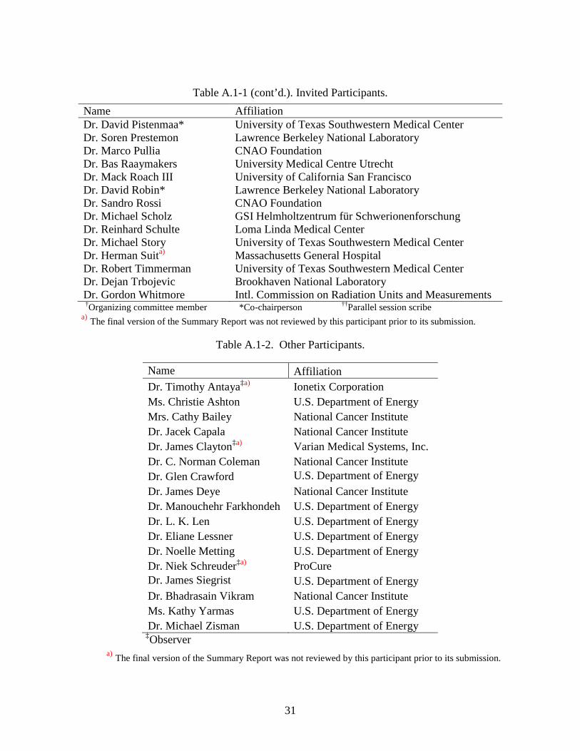
Table A.1-1 (cont’d.). Invited Participants.
Name Affiliation Dr. David Pistenmaa* University of Texas Southwestern Medical Center Dr. Soren Prestemon Lawrence Berkeley National Laboratory Dr. Marco Pullia CNAO Foundation Dr. Bas Raaymakers University Medical Centre Utrecht Dr. Mack Roach III University of California San Francisco Dr. David Robin* Lawrence Berkeley National Laboratory Dr. Sandro Rossi CNAO Foundation Dr. Michael Scholz GSI Helmholtzentrum für Schwerionenforschung Dr. Reinhard Schulte Loma Linda Medical Center Dr. Michael Story University of Texas Southwestern Medical Center Dr. Herman Suita) Massachusetts General Hospital Dr. Robert Timmerman University of Texas Southwestern Medical Center Dr. Dejan Trbojevic Brookhaven National Laboratory Dr. Gordon Whitmore Intl. Commission on Radiation Units and Measurements †Organizing committee member *Co-chairperson ††Parallel session scribe
a) The final version of the Summary Report was not reviewed by this participant prior to its submission.
Table A.1-2. Other Participants.
Name Affiliation Dr. Timothy Antaya‡a) Ionetix Corporation Ms. Christie Ashton U.S. Department of Energy Mrs. Cathy Bailey National Cancer Institute Dr. Jacek Capala National Cancer Institute Dr. James Clayton‡a) Varian Medical Systems, Inc. Dr. C. Norman Coleman National Cancer Institute Dr. Glen Crawford U.S. Department of Energy Dr. James Deye National Cancer Institute Dr. Manouchehr Farkhondeh U.S. Department of Energy Dr. L. K. Len U.S. Department of Energy Dr. Eliane Lessner U.S. Department of Energy Dr. Noelle Metting U.S. Department of Energy Dr. Niek Schreuder‡a) ProCure Dr. James Siegrist U.S. Department of Energy Dr. Bhadrasain Vikram National Cancer Institute Ms. Kathy Yarmas U.S. Department of Energy Dr. Michael Zisman U.S. Department of Energy
‡Observer a) The final version of the Summary Report was not reviewed by this participant prior to its submission.

32
A.2 Workshop Charge August 24, 2012
Joint DOE-NCI Workshop on Ion Beam Therapy
January 2013 Bethesda, MD
Background Even with less than fully optimized treatment techniques, there have been reports of impressive local control rates using particle beam therapy for difficult-to-treat cancers such as liver, pancreas, lung and head and neck, and pediatric neoplasms. Although lower-cost proton beam options are starting to appear, today’s proton beam facilities are costly to build and thus are not widely available. Based on a number of case reports and on their potential biological advantage, there is now increasing medical interest in exploring the use of other light ions for therapy; typically beams up to carbon are considered. While beam delivery (“gantry”) systems for proton beams have been designed and constructed previously, they are typically large, massive, and costly. Accommodating heavier beams, up to 12C, with similar technology requires even more massive delivery systems. To develop requirements for next-generation ion beam therapy production and delivery systems, the Department of Energy Office of Science’s High-Energy Physics (HEP) program, along with the National Cancer Institute are hosting a Community Input Workshop on Ion Beam Therapy. This workshop will identify the required features of next-generation beam production and delivery systems, such as spot size, pulse intensity, positional control and stability, energy control and stability, and beam halo criteria. Technological challenges of providing the required features will be discussed, with the aim of applying modern accelerator design approaches to their solution, especially to improve gantry system design. Attendance at the workshop is limited and will be by invitation. Interested parties can contact the Workshop co-chairs requesting an invitation. Workshop Charge This workshop is asked to:
• Identify a set of representative clinical applications that span the range of expected future therapy requirements. These need to include capabilities for performing radiobiological experiments as well as human treatment protocols in order to explore the scientific principles underlying observed clinical results and point the way to promising protocol designs.
• Assess the corresponding beam requirements (e.g., energy range and energy spread, intensity range and pulse-to-pulse intensity jitter, spot size and pulse-to-pulse position jitter, repetition rate, ion species) for future treatment facilities and compare these with today’s state-of-the-art

33
• Assess the corresponding beam delivery system requirements (e.g., energy and position adjustability, time scale for adjustments, size of footprint, component mass, transverse and longitudinal acceptance) for future treatment facilities and compare these with today’s state-of-the-art
• Identify R&D activities needed to bridge the gap between current capabilities and future requirements; include an assessment of which R&D investments are likely to have the highest near-term performance gains
While the importance of radiation biology is recognized, it is not a primary focus of this workshop. However, this workshop will emphasize the importance of the physical ability to study the biology of the light ions. The workshop outcome will consist of a concise report describing what accelerator and beam delivery system developments are needed to support the full range of radiotherapy research requirements, the R&D activities needed to carry out such developments, and a rough timeline indicating when such developments are needed to support a world-class treatment program.
34
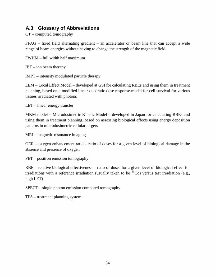
A.3 Glossary of Abbreviations CT – computed tomography
FFAG – fixed field alternating gradient – an accelerator or beam line that can accept a wide range of beam energies without having to change the strength of the magnetic field.
FWHM – full width half maximum
IBT – ion beam therapy
IMPT – intensity modulated particle therapy
LEM – Local Effect Model – developed at GSI for calculating RBEs and using them in treatment planning, based on a modified linear-quadratic dose response model for cell survival for various tissues irradiated with photons
LET – linear energy transfer
MKM model – Microdosimetric Kinetic Model – developed in Japan for calculating RBEs and using them in treatment planning, based on assessing biological effects using energy deposition patterns in microdosimetric cellular targets
MRI – magnetic resonance imaging
OER – oxygen enhancement ratio – ratio of doses for a given level of biological damage in the absence and presence of oxygen
PET – positron emission tomography
RBE – relative biological effectiveness – ratio of doses for a given level of biological effect for irradiations with a reference irradiation (usually taken to be 60Co) versus test irradiation (e.g., high LET)
SPECT – single photon emission computed tomography
TPS – treatment planning system