world journal of - bsdwebstorage.blob.core.windows.net · badri m shrestha, sheffield nestor...
TRANSCRIPT

World Journal of NephrologyWorld J Nephrol 2016 January 6; 5(1): 1-124
ISSN 2220-6124 (online)
Published by Baishideng Publishing Group Inc

World Journal of NephrologyW J N
EDITOR-IN-CHIEFAnil K Mandal, St. Augustine
ASSOCIATE EDITORSLi Zuo, BeijingHiroyuki Kobori, KagawaHiromichi Suzuki, SaitamaJuan F Navarro-Gonzalez, Santa Cruz de Tenerife
GUEST EDITORIAL BOARD MEMBERS Wen-Chi Chen, TaipeiYi-Wen Chiu, KaohsiungRu-Lan Hsieh, TaipeiYH Hsu, TaipeiPo-Chang Lee, TainanChing-Yuang Lin, TaichungChi-Feng Liu, TaipeiKuo-Cheng Lu, New TaipeiPei-Jung Lu, TainanYee-Yung Ng, TaipeiJunne-Ming Sung, TainanJiunn-Jong Wu, TainanTzung-Hai Yen, Taipei
MEMBERS OF THE EDITORIAL BOARD
AlgeriaKhedidja Mekki, Oran
ArgentinaCarlos Guido Musso, TemperleyHernan Trimarchi, Buenos AiresL Trumper, Rosario
Patricia G Valles, Mendoza
AustraliaNeil C Boudville, WaRobert Gordon Fassett, QueenslandHelen Grania Healy, BrisbaneMohamed Saleem, AdelaideIbrahim M Salman, New South WalesDavid Alan Vesey, BrisbaneHuiling Wu, SydneyGuoping Zheng, Sydney
Belgium
Maarten Naesens, LeuvenBenjamin A Vervaet, Antwerp
Bosnia and Herzegovina
Halima Resic, Sarajevo
Brazil
Liborio Braga Alexandre, FortalezaNiels OS Camara, Cidade UniversitáriaJozélio Freire de Carvalho, Salvador-BahiaSilvia Maria De Oliveira Titan, Sao PauloJose Mario F de Oliveira, Rio de JaneiroMaria Franco, Sao PauloJosé AR Gontijo, CampinasSonia M Oliani, CEP Leonardo O Reis, UnicampNestor Schor, Sao Paulo
Canada
Shao-Bin Duan, ChangshaPaul A Keown, VancouverMarcel Lebel, QuebecOzgur Mete, Ontario
Chile
Guillermo E Lema, Santiago
China
Chia-Chu Chang, ChanghuaFeng Ding, ShanghaiHua-Feng Liu, ZhanjiangFei-Zhang Shou, HangzhouYan-Qing Tong, ChangchunAngela Yee-Moon Wang, Hong KongDan-Xia Zheng, Beijing
Croatia
Dean Markic, RijekaDrasko Pavlovic, ZagrebVladimir Trkulja, Zagreb
Czech Republic
Sylvie Opatrná, PilsenVladimír Tesar, Prague
I
Editorial Board2016-2019
The World Journal of Nephrology Editorial Board consists of 282 members representing a team of worldwide experts in nephrology. They are from 49 countries, Afghanistan (1), Algeria (1), Argentina (4), Australia (1), Belgium (1), Bosnia and Herzegovina (1), Brazil (10), Canada (4), Chile (1), China (21), Croatia (3), Czech Repoublic (2), Denmark (2), Egypt (6), Finland (1), France (1), Germany (4), Greece (14), Hungary (4), India (12), Iran (9), Ireland (1), Israel (2), Italy (21), Japan (15), Jordan (1), Malaysia (1), Mexico (2), Netherlands (4), Morocco (1), Nigeria (2), Oman (1), Pakistan (2), Palestine (1), Poland (6), Portugal (4), Qatar (1), Romania (1), Serbia (2), Singapore (3), South Africa (1), South Korea (3), Spain (10), Sweden (3), Thailand (5), Turkey (9), United Arab Emirates (1), United Kingdom (11), and United States (59).
March 2, 2016WJN|www.wjgnet.com

Denmark
Robert A Fenton, AarhusErling B Pedersen, Holstebro
Egypt
Ahmed I Akl, MansouraMohammad Al-Haggar, MansouraAmgad El Agroudy, BahrainOsama Gheith, MansouraHussein A Sheashaa, MansouraNeveen A Soliman, Cairo
Finland
Sanna Helena Lehtonen, Helsinki
France
Dominique Guerrot, Rouen
Germany
Wolfgang E Jelkmann, LuebeckNadezda Koleganova, HeidelbergJan Menne, HannoverPeter Schemmer, Heidelberg
Greece
Dimitra Bacharaki, RiminiGrapsa Eirini, AthensTheodoros Eleftheriadis, LarissaMoses S Elisaf, IoanninaDimitrios Karakitsos, AthensDimitrios A Kirmizis, ThessalonikiAikaterini Papagianni, ThessalonikiKosmas I Paraskevas, AthensPloumis Stavros Passadakis, AlexandroupolisGiorgos K Sakkas, ThessalyPantelis A Sarafidis, ThessalonikiAristeidis Stavroulopoulos, KallitheaGeorgios Tsaνgalis, maroussi-attikisParaskevi Tseke, Athens
Hungary
Miklos Zsolt Molnar, BudapestJános Nemcsik, BudapestTaha EL Hadj Othmane, BudapestLaszlo Rosivall, Budapest
India
Sanjay K Agarwal, New DelhiAnish Bhattacharya, ChandigarhSanjay D’Cruz, ChandigarhAmit K Dinda, New DelhiVivekanand Jha, Chandigarh
Madhu Khullar, Chandigarh Chitra Madiwale, MumbaiShivanand Karopadi Nayak, HyderabadMayoor V Prabhu, MangaloreSidharth Kumar Sethi, HaryanaRajiv Sinha, KolkataKushaljit S Sodhi, Chandigarh
Iran
Mohammadreza Ardalan, TabrizBehzad Einollahi, TehranSeyed-Mohammad Fereshtehnejad, TehranPatricia Khashayar, TehranHamid Tayebi Khosroshahi, TabrizFarzaneh Montazerifar, ZahedanHasan Otukesh, TehranAmir Keshvari Persian, TehranSaeed Taheri, Tehran
Ireland
Harry Holthofer, Dublin
Israel
Farid M Nakhoul, Lower GalileeOded Olsha, Jer United Stateslem
Italy
Gianni Bellomo, FolignoCristina Costa, TorinoPaolo Cravedi, Ranica BergamoBiagio Raffaele Di Iorio, SolofraLuciana Ghio, MilanoAndrea Giusti, GenovaAntonio Granata, AgrigentoFrancesco Locatelli, LeccoLorenzo S Malatino, CataniaGiancarlo Marenzi, MilanPiergiorgio Messa, MilanNicola Perrotti, CatanzaroFrancesco Perticone, CatanzaroGiorgina B Piccoli, TorinoPierangela Presta, CatanzaroClaudio Ronco, VicenzaDomenico Santoro, MessinaRoberto Scarpioni, PiacenzaVincenzo Sepe, PaviaGiovanni Luigi Tripepi, Reggio CalabriaLuca V Valenti, V Valenti, Milano
Japan
Yoshihide Fujigaki, HamamatsuKeiju Hiromura, GunmaKazunari Kaneko, OsakaSatoshi Morimoto, HirakataToshio Nishikimi, KyotoNaro Ohashi, HamamatsuTakashi Oite, NiigataGeorge Seki, Shizuoka prefectureAkira Shimizu, Tokyo
Kouichi Tamura, YokohamaH Tanaka, HirosakiToru Watanabe, NiigataNoriaki Yorioka, Hiroshima
Jordan
Mohammad Khassawneh, Irbid
Malaysia
Bak-Leong Goh, Kajang
Mexico
Gustavo Martínez-Mier, MéxicoAlejandro Trevino-Becerra, Mexico
Morocco
Faissal Tarrass, Casablanca
Netherlands
Sebastian Dolff, EssenPeter JH Smak Gregoor, DordrechtJoris Hubertus Robben, NijmegenJoris JTH Roelofs, Amsterdam
Nigeria
Martin A Nzegwu, EnuguWasiu Adekunle Olowu, Ile-Ife
Pakistan
Ali AA Lanewala, KarachiMuhammed Mubarak, Karachi
Palestine
Mahmoud M Othman, Nablus
Poland
Alicja E Grzegorzewska, PoznańAndrzej Jaroszynski, KielceJerzy Konstantynowicz, BialystokMariusz Kusztal, WroclawJacek Wiktor Manitiu, BydgoszczMarcin Tkaczyk, Lodz
Portugal
Márcia Carvalho, PortoElísio Costa, PortoLa Salete S Martins, Oporto
II March 2, 2016WJN|www.wjgnet.com

III March 2, 2016WJN|www.wjgnet.com
Manuel Pestana Vasconcelos, Porto
Qatar
Khaled Mohamed Mahmoud, Doha
Romania
Gheorghe Nechifor, Bucharest
Serbia
Amira Peco-Antic, BelgradeRadojica V Stolic, Kragujevac
Singapore
Tan Ban Hock, SingaporeAnselm Mak, SingaporeWoo Keng Thye, Singapore
South Africa
Rajendra Bhimma, Congella
South Korea
Byoung Soo Cho, SeoulChan Kyo Kim, Gangnam-gu SeoulJae IL Shin, Seoul
Spain
Miguel A Arrabal-Polo, GranadaRicardo J Bosch, Alcala de HenaresJosep M Campistol, BarcelonaJavier Fernandez de Canete, MalagaVictor M Garcia-Nieto, Santa Cruz de TenerifeFrancisco J Lopez-Hernandez, SalamancaAlberto Ortiz, MadridKatia Lopez Revuelta, MadridFernando Santos, Oviedo
Sweden
Peter Bárány, Stockholm
Per Magnusson, Linkoping
Thailand
Pornanong Aramwit, BangkokSinee Disthabanchong, BangkokSomchai Eiam-Ong, BangkokPrasit Futrakul, BangkokViroj Wiwanitkit, Bangkok
Turkey
Turgay Akgul, OsmaniyeFiliz Akyuz, IstanbulMustafa Arici, AnkaraOzgu Aydogdu, NigdeEsra Güzeldemir-Akcakanat, YuvacikMehmet Kanbay, IstanbulSalih Kavukcu, Balcova IzmirAhmet Kiykim, MersinHabibe Sahin Sahin, Kayseri
United Arab Emirates
Bassam Bernieh, Arabe Emirates
United Kingdom
Jacob A Akoh, PlymouthRodney D Gilbert, SouthamptonColin Andrew Hutchison, BirminghamJonathon Olsburgh, LondonDipen S Parikh, DurhamAdrian W Philbey, GlasgowBhusana Premande, LondonBadri M Shrestha, SheffieldNestor Velasco, KilmarnockAlexander Woywodt, PrestonQihe Xu, London
United States
Horacio J Adrogué, HoustonAnil K Agarwal, ColumbusPatrick D Brophy, Hawkins Drive IowaYiqiang Cai, New HavenDaniel J Canter, Atlanta
Oscar A Carretero, DetroitJames CM Chan, PortlandBrian S Decker, IndianapolisJames V Donadio, RochesterAmy C Dwyer, LouisvilleEwa Elenberg, HoustonKevin Finkel, HoustonEA Friedman, BrooklynCA Gadegbeku, Ann ArborClaudia Gragnoli, HersheyParta Hatamizadeh, Ann ArborAdriana M Hung, NashvillePedro A Jose, WashingtonTheodoros Kelesidis, Los AngelesBruce C Kone, HoustonRajesh Kumar, TempleDaniel L Landry, SpringfieldKrista Lentine, St LouisYan Chun Li, ChicagoYouhua Liu, PittsburghMaria-Luisa SS Lopez, Charlottesville John K Maesaka, Mineola Joseph Keith Melancon, WashingtonTibor Nadasdy, ColumbusMacaulay Onuigbo, Eau ClaireIsak Prohovnik, New YorkAmanda C Raff, New YorkArmin Rashidi, NorfolkAnjay Rastogi, Los AngelesMohammed S Razzaque, BostonJeff M Sands, AtlantaMartin J Schreiber, ClevelandGerald Schulman, TennesseeJames Alan Shayman, MichiganAndrey Staruschenko, MilwaukeeAlison Leah Steiber, ClevelandTheodore I Steinman, BostonJames D Stockand, San AntonioMingming Su, KannapolisYunxia Tao, AmarilloGeorge Christos Tsokos, BostonJaime Uribarri, New YorkUlka Vaishampayan, DetroitVolker Vallon, San DiegoPaul A Voziyan, NashvilleBradford Lee West, IllinoisMark E Williams, BostonAnna Woodbury, AtlantaRobert Peter Woroniecki, BronxJ Ruth Wu-Wong, ChicagoDu Yong, HoustonRubin Zhang, LouisianaXin-Jin Zhou, Dallas

W JContents
IWJN|www.wjgnet.com January 6, 2016|Volume 5|Issue 1|
World Journal of NephrologyN
Bimonthly Volume 5 Number 1 January 6, 2016
EDITORIAL
1 ReceptoractivatorofnuclearfactorκB ligand/osteoprotegerin axis and vascular calcifications in patients
withchronickidneydisease
Spartalis M, Papagianni A
FRONTIER
6 Updateonimmunoglobulinanephropathy.PartⅡ:Clinical,diagnosticandtherapeuticalaspects
Salvadori M, Rosso G
REVIEW
20 Howdokinasescontributetotonicity-dependentregulationofthetranscriptionfactorNFAT5?
Zhou X
33 Water, electrolytes, and acid-base alterations in human immunodeficiency virus infected patients
Musso CG, Belloso WH, Glassock RJ
43 Adultstemcellsasatoolforkidneyregeneration
Suzuki E, Fujita D, Takahashi M, Oba S, Nishimatsu H
53 Baroreflex dysfunction in chronic kidney disease
Kaur M, Chandran DS, Jaryal AK, Bhowmik D, Agarwal SK, Deepak KK
MINIREVIEWS
66 Secondary amyloidosis in autoinflammatory diseases and the role of inflammation in renal damage
Scarpioni R, Ricardi M, Albertazzi V
76 Roleofcalciuminpolycystickidneydisease:Fromsignalingtopathology
Mangolini A, de Stephanis L, Aguiari G
84 Percutaneousnephrolithotomyinpediatricagegroup:Assessmentofeffectivenessandcomplications
Ozden E, Mercimek MN
90 Centralbloodpressureandchronickidneydisease
Ohno Y, Kanno Y, Takenaka T

IIWJN|www.wjgnet.com January 6, 2016|Volume 5|Issue 1|
Contents
ORIGINAL ARTICLE
Retrospective Study
101 IncidenceandprevalenceofhepatitisBandhepatitisCvirusesinhemodialysispatientsinLebanon
Abou Rached A, El Khoury L, El Imad T, Geara AS, Jreijiry J, Ammar W
108 RenalandperinephricabscessesinWestChinaHospital:10-yearretrospective-descriptivestudy
Liu XQ, Wang CC, Liu YB, Liu K
META-ANALYSIS
115 SexbiasinresponsetohepatitisBvaccinationinend-stagerenaldiseasepatients:Meta-analysis
Khedmat H, Aghaei A, Ghamar-Chehreh ME, Agah S
World Journal of NephrologyVolume 5 Number 1 January 6, 2016

ContentsWorld Journal of Nephrology
Volume 5 Number 1 January 6, 2016
EDITORS FOR THIS ISSUE
Responsible Assistant Editor: Xiang Li Responsible Science Editor: Shui QiuResponsible Electronic Editor: Xiao-Kang Jiao Proofing Editorial Office Director: Xiu-Xia SongProofing Editor-in-Chief: Lian-Sheng Ma
EDITORIALOFFICEJin-Lei Wang, DirectorXiu-Xia Song, Vice DirectorWorld Journal of NephrologyRoom 903, Building D, Ocean International Center, No. 62 Dongsihuan Zhonglu, Chaoyang District, Beijing 100025, ChinaTelephone: +86-10-85381891Fax: +86-10-85381893E-mail: [email protected] Desk: http://www.wjgnet.com/esps/helpdesk.aspxhttp://www.wjgnet.com
PUBLISHERBaishideng Publishing Group Inc8226 Regency Drive, Pleasanton, CA 94588, USATelephone: +1-925-223-8242Fax: +1-925-223-8243E-mail: [email protected] Desk: http://www.wjgnet.com/esps/helpdesk.aspxhttp://www.wjgnet.com
PUBLICATIONDATEJanuary 6, 2016
COPYRIGHT© 2016 Baishideng Publishing Group Inc. Articles pub-lished by this Open-Access journal are distributed under the terms of the Creative Commons Attribution Non-commercial License, which permits use, distribution, and reproduction in any medium, provided the original work is properly cited, the use is non commercial and is otherwise in compliance with the license.
SPECIALSTATEMENTAll articles published in journals owned by the Baishideng Publishing Group (BPG) represent the views and opin-ions of their authors, and not the views, opinions or policies of the BPG, except where otherwise explicitly indicated.
INSTRUCTIONSTOAUTHORSFull instructions are available online at http://www.wjgnet.com/2218-6220/g_info_20100722175812.htm.
ONLINESUBMISSIONhttp://www.wjgnet.com/esps/
IIIWJN|www.wjgnet.com
ABOUT COVER
AIM AND SCOPE
FLYLEAF
January 6, 2016|Volume 5|Issue 1|
NAMEOFJOURNALWorld Journal of Nephrology
ISSNISSN 2220-6124 (online)
LAUNCHDATEFebruary 6, 2012
FREQUENCYBimonthly
EDITORS-IN-CHIEFJosep M Campistol, Professor, ICNU Director, Hos-pital Clínic, Universitat de Barcelona, c/Villarroel, 170 ESC 12-5, 08036 Barcelona, Spain
Anil K Mandal, MB, BS, Professor, Department of Medicine, University of Florida, Gainesville, Florida; Mandal Diabetes Research Foundation, 105 Southpark Blvd., Suite B-202, Saint Augustine, FL 32086, United States
EditorialBoardMemberofWorldJournalofNephrology ,EliAFriedman,MD,Professor,DepartmentofMedicine,DownstateMedicalCenter,Brooklyn,NY11203,UnitedStates
World Journal of Nephrology (World J Nephrol, WJN, online ISSN 2220-6124, DOI: 10.5527) is a peer-reviewed open access academic journal that aims to guide clinical practice and improve diagnostic and therapeutic skills of clinicians.
WJN covers topics concerning kidney development, renal regeneration, kidney tumors, therapy of renal disease, hemodialysis, peritoneal dialysis, kidney transplanta-tion, diagnostic imaging, evidence-based medicine, epidemiology and nursing. Priority publication will be given to articles concerning diagnosis and treatment of nephrology diseases. The following aspects are covered: Clinical diagnosis, laboratory diagnosis, dif-ferential diagnosis, imaging tests, pathological diagnosis, molecular biological diagnosis, immunological diagnosis, genetic diagnosis, functional diagnostics, and physical diagno-sis; and comprehensive therapy, drug therapy, surgical therapy, interventional treatment, minimally invasive therapy, and robot-assisted therapy.
We encourage authors to submit their manuscripts to WJN. We will give priority to manuscripts that are supported by major national and international foundations and those that are of great basic and clinical significance.
World Journal of Nephrology is now indexed in PubMed Central, PubMed, and Digital
Object Identifier.
I-III EditorialBoard
INDExING/ABSTRACTING

Receptor activator of nuclear factor κB ligand/osteoprotegerin axis and vascular calcifications in patients with chronic kidney disease
Michalis Spartalis, Aikaterini Papagianni
Michalis Spartalis, Aikaterini Papagianni, Department of Nephrology, Aristotle University of Thessaloniki, “Hippokration” General Hospital, 54642 Thessaloniki, Greece
Author contributions: Sartalis M made the literature search and wrote the first draft of the manuscript; Papagianni A critically reviewed the literature and wrote the final form of the manuscript.
Conflict-of-interest statement: The authors declare no conflict of interest.
Open-Access: This article is an open-access article which was selected by an in-house editor and fully peer-reviewed by external reviewers. It is distributed in accordance with the Creative Commons Attribution Non Commercial (CC BY-NC 4.0) license, which permits others to distribute, remix, adapt, build upon this work non-commercially, and license their derivative works on different terms, provided the original work is properly cited and the use is non-commercial. See: http://creativecommons.org/licenses/by-nc/4.0/
Correspondence to: Aikaterini Papagianni, MD, PhD, Associate Professor, Department of Nephrology, Aristotle University of Thessaloniki, “Hippokration” General Hospital, Papanastasiou 50 Str, 54642 Thessaloniki, Greece. [email protected] Telephone: +30-2310-992856Fax: +30-2310-892382
Received: May 31, 2015Peer-review started: June 4, 2015First decision: August 14, 2015Revised: September 23, 2015Accepted: October 20, 2015 Article in press: October 27, 2015Published online: January 6, 2016
AbstractVascular calcifications are commonly observed in patients with chronic kidney disease (CKD) and contri-
bute to the excessive cardiovascular morbidity and mortality rates observed in these patients populations. Although the pathogenetic mechanisms are not yet fully elucidated, recent evidence suggests a link between bone metabolism and the development and progression of vascular calcifications. Moreover, accumulating data indicate that receptor activator of nuclear factor κB ligand/osteoprotegerin axis which plays essential roles in the regulation of bone metabolism is also involved in extra-osseous bone formation. Further studies are required to establish the prognostic significance of the above biomarkers as predictors of the presence and severity of vascular calcifications in CKD patients and of cardiovascular morbidity and mortality. Moreover, randomized clinical trials are needed to clarify whether inhibition of osteoclast activity will protect from vascular calcifications.
Key words: Arterial stiffness; Bone turnover; Chronic kidney disease; Osteoprotegerin; RANK ligand; Receptor activator nuclear factor κB; Vascular calcifications
© The Author(s) 2016. Published by Baishideng Publishing Group Inc. All rights reserved.
Core tip: Vascular calcifications are commonly observed in chronic kidney disease patients and recently mounting evidence suggest that Receptor activator of nuclear factor κB ligand/osteoprotegerin axis controls both bone metabolism and extra-osseous bone formation. Further studies are required to establish the role of these biomarkers as predictors of the presence and severity of vascular calcifications and of cardiovascular morbidity and mortality. Moreover, randomized clinical trials are needed to clarify whether inhibition of osteoclast activity will protect from vascular calcifications.
Spartalis M, Papagianni A. Receptor activator of nuclear factor κB ligand/osteoprotegerin axis and vascular calcifications in patients
EDITORIAL
1 January 6, 2016|Volume 5|Issue 1|WJN|www.wjgnet.com
World Journal of NephrologyW J N
Submit a Manuscript: http://www.wjgnet.com/esps/Help Desk: http://www.wjgnet.com/esps/helpdesk.aspxDOI: 10.5527/wjn.v5.i1.1
World J Nephrol 2016 January 6; 5(1): 1-5ISSN 2220-6124 (online)
© 2016 Baishideng Publishing Group Inc. All rights reserved.

with chronic kidney disease. World J Nephrol 2016; 5(1): 1-5 Available from: URL: http://www.wjgnet.com/2220-6124/full/v5/i1/1.htm DOI: http://dx.doi.org/10.5527/wjn.v5.i1.1
INTRODUCTIONCardiovascular disease (CVD) remains the leading cause of excessive morbidity and mortality for patients with chronic kidney disease (CKD) and particularly those with end-stage renal disease (ESRD) on renal replacement therapy with either hemodialysis or peritoneal dialysis[1]. Vascular calcifications are also commonly observed in CKD and are now considered part of the syndrome chronic kidney disease-mineral and bone disorder (CKD-MBD), the pathogenesis of which has accumulated great research interest the last years. In CKD patients calcifications in both intimal (atherosclerotic) and medial lamina (arteriosclerotic) often coexist, appear early and follow an accelerated course. Particularly the latter is an almost ubiquitous feature of arterial tree in chronic uremia and a major contributor to the accelerated ar-teriosclerosis and to the increased all-cause and CVD mortality in these patients populations[2].
The presence of vascular calcifications in CKD has been associated with a number of traditional risk factors including older age, hypertension, dyslipidemia and diabetes mellitus which are highly prevalent in this population, as well as uremia-related risk factors including chronic inflammation, oxidative stress and mineral and bone disorders which are currently under investigation. Of note, mineral alterations (hyperca-lcemia, hyperphosphatemia) and disorders of bone metabolism (both secondary hyperparathyroidism and adynamic bone disease) are mainly associated with the development and progression of medial but not intimal calcifications[3].
It is now well recognized that vascular calcification is not simply a passive physicochemical process of calcium phosphate deposition but a highly regulated active process similar to normal bone modeling. Moreover, recent evidence suggests that the phenotypic trans-differentiation of vascular smooth muscle cells (VSMCs) into osteoblast-like cells is a key pathogenetic event in the osteogenesis of the vascular wall. A variety of regulatory factors and molecular pathways of this process which regulate bone turnover and/or mineralization have been identified[3]. Although their relative importance in different disease states appear still incompletely understood emerging evidence suggest that the receptor activator of nuclear factor κB (RANK)/RANK Ligand (RANKL)/osteoprotegerin (OPG) system that plays essential roles in the regulation of bone metabolism is also involved in extra-osseous bone formation[4].
RANK/RANKL/OPG PATHWAYRANK, RANKL and OPG are members of the tumor
necrosis factor (TNF) superfamily which were originally studied as factors involved in bone tissue and immune system physiology. However, recent studies revealed that they also constitute a link between bone metabolism and vascular pathophysiology by controlling simult-aneously bone remodeling and vascular calcification mechanisms[5].
RANK/RANKL/OPG signaling pathway regulates osteoclast differentiation and activation. RANKL is a transmembrane protein consisted of 316 aminoacids, which is expressed on osteoblasts, stromal and T cells in areas of bone remodeling. RANKL binds to its receptor RANK, a 616 amino acid type Ⅰ transmembrane protein which is expressed on the surface of myeloid cell lineages like osteoclasts, monocytic and dendritic cells. The above activates multiple intracellular signals, including activation of the c-jun N-terminal kinase and nuclear factor κB pathways that regulate the differen-tiation, function and survival of these cells. A variety of factors including hormones, cytokines and growth factors regulate its expression. Thus, parathormone (PTH), TNF-α, calcium, corticosteroids and several interleukins (IL-6,-11,-17) increase RANKL expression on osteoblasts, whereas transforming growth factor-β (TGF-β) decreases it[3,5]. OPG is a soluble glycoprotein widely expressed in most human tissues including bone (osteoblests, mesenchymal stem cells), immune cells (T and B cells) and vessels (endothelial and VSMCs). It acts as a decoy receptor and binds to RANKL, thereby not allowing the activation of RANK and inhibits its regulatory effects on inflammation, skeletal and vascular systems[5]. OPG also has anti-apoptotic actions, as it binds and deactivates the TNF related apoptosis-inducing ligand (TRAIL), which is expressed by many cell types, including VSMCs, and can also lead to ectopic mineralization[5]. OPG’s expression is increased by vitamin D, TNF-α, IL-1α, IL-6, IL-11 and IL-17, bone morphogenic protein-2, TGF-β and estrogens, whereas it is reduced by PTH, corticosteroids and prostaglandin E2[5].
Mounting evidence suggests that the RANK/RANKL/OPG axis exerts actions simultaneously on endothelial cells and VSMCs and participate in multiple processes that regulate vascular calcification. The implication of this system in vascular pathophysiology is supported by the expression of these molecules in the normal cardiovascular system (heart, arteries and veins). Both endothelial cells and VSMCs constitutively express OPG, and their levels are particularly high in aortic and renal arteries. Furthermore, OPG is physically associated with factor Ⅷ-von Willebrand factor complex localized in the Weibel–Palade bodies of endothelial cells, it is rapidly secreted in response to inflammatory stimuli and inhibits osteoclastogenesis and promotes endothelial cell survival[6] through neutralization of pro-apoptotic TRAIL[5]. In contrast, RANKL and RANK are frequently undetectable in normal vessels and non-calcified arteries or valves[5]. However, osteoclast-like RANK(+) cells were found close to VSMCs on calcified vascular walls and
Spartalis M et al . RANKL/OPG and vascular calcifications in CKD
2 January 6, 2016|Volume 5|Issue 1|WJN|www.wjgnet.com

expression of both RANK and RANKL was reported on the vascular wall, in calcified areas[7].
Several studies suggested that OPG’s expression might reflect a protective mechanism against the vascular calcifications. Thus, over-expression of OPG leads to osteopetrosis, while its gene deletion, increases bone metabolism and leads to osteoporosis and medial calcifications in the aorta and the renal arteries[8,9]. Moreover, OPG administration in laboratory animals, was reported to potently reduce both bone resorption activity and medial arterial calcifications induced by administration of toxic doses of vitamin D or warfarin[10]. These findings, together with the fact that OPG is expressed in the vascular wall under normal circumstances, indicate that the endogenous production of OPG prevents the ossification of the vascular wall and favors bone mineralization. In accordance with the above, OPG was detected in matrix vesicles, nanopa-rticles that are released from VSMCs with the capacity to nucleate mineral, which directly inhibited deposition of hydroxyapatite in the vascular wall[11].
OPG AND VASCULAR CALCIFICATIONS IN CKD PATIENTSIn the general population, studies have demonstrated that high levels of OPG are correlated with cardiovascular risk[12]. In CKD patients, it has been shown that OPG levels significantly increase along with the decline in GFR and are reduced after a successful renal transplan-tation[13]. However, studies examining the association of OPG levels with the presence and extent of cardiovascular calcifications are relatively limited and their results were sometimes inconsistent. Thus, in non-dialyzed CKD patients a cut-off value of OPG level was found to predict the presence of coronary artery calcifications (CAC) assessed by chest multidetector computed tomography[14]. In addition, a very recent study in CKD patients reported a significant association between high OPG levels and CAC independently of other risk factors including age, gender, diabetes, body mass index and smoking habits[15]. In transplanted patients CAC at baseline, but not 1 year after renal transplantation, was found to be independently associated with base-line OPG whereas post-transplant CAC progression was predicted by baseline CAC score[16]. In contrast, another study in transplanted patients reported that OPG levels were significantly and independently associated with the progression of aortic calcification index (ACI) assessed by lateral lumbar x-ray during a two-year follow-up period[17]. In adults and children with ESRD on hemodialysis, studies demonstrated a significant independent correlation between OPG levels and CAC[18,19]. Similarly, another study showed an association between OPG and ACI assessed by computed tomography scans independently of traditional and uremia-related risk factors[20]. In addition, high OPG levels have been found to correlate with faster progression of
aortic calcifications during a 5-year follow-up[21]. Finally, several studies in patients with various CKD stages as well as in renal transplant recipients demonstrated that OPG levels were a significant independent predictor of all-cause and cardiovascular mortality during the follow-up period[22-26]. However, one study in ESRD and pre-dialysis CKD patients showed that renal function rather than OPG levels were mostly associated with the progression of aortic and coronary calcifications[27], whereas another one correlated elevated OPG levels only with moderate CAC[28].
Regarding the association of OPG with markers of medial calcifications such as arterial stiffness and pulse wave velocity (PWV), the results are also sometimes controversial. A study in non-dialyzed CKD patients demonstrated a strong relationship between serum OPG and arterial stiffness independent of many potential confounders including traditional cardiovascular risk factors, abnormal bone and mineral metabolism, and inflammation[29]. Similarly, in hemodialysis patients OPG levels were found to be strongly associated with aortic or carotid-to-femoral PWV independently of traditional and uremia-related risk factors including markers of inflammation[25,26,30]. However, other studies in adults and children on hemodialysis treatment were unable to confirm the above findings[19,31].
As it was previously noted, the exact role of OPG in the VC process remains unclear. Since OPG inhibits osteoclast activity and OPG knockout mice develop arterial calcifications, it appears reasonable to assume that it plays some regulatory and/or inhibitory role against ectopic calcifications and thus its increased levels could be interpreted as an attempt to compensate for the ongoing calcification process[9-11]. The above speculation contradicts the reported association of high OPG levels with cardiovascular mortality[22,23,25,26] and moreover, increase of OPG levels could be due to its production by calcified vascular cells in conditions of diffuse calcification. Thus, it remains to be clarified whether the elevated OPG levels induce arterial wall sclerosis or represent a compensatory mechanism to prevent further arterial damage or are just a marker of initiation of vascular calcification process[4].
RANKL AND VASCULAR CALCIFICATIONS IN CKD PATIENTSThe exact role of RANKL in the development of cardio-vascular calcifications and CVD remains to be identified. Some studies showed a correlation of RANKL levels with future cardiovascular events[32], but the probable association between vascular calcifications and RANKL levels has been scarcely investigated so far. A prospective study in 3250 Framingham Study participants reported that RANKL concentrations were inversely associated with multiple cardiovascular risk factors, including smoking, diabetes, and antihypertensive treatment, but that were not related with CAC or incident CVD or mortality during
3 January 6, 2016|Volume 5|Issue 1|WJN|www.wjgnet.com
Spartalis M et al . RANKL/OPG and vascular calcifications in CKD
necrosis factor-related apoptosis-inducing ligand, and receptor activator of nuclear factor-kappaB ligand in Mönckeberg’s sclerosis and atherosclerosis. J Clin Endocrinol Metab 2004; 89: 4104-4112 [PMID: 15292354 DOI: 10.1210/jc.2003-031432]
8 Simonet WS, Lacey DL, Dunstan CR, Kelley M, Chang MS, Lüthy R, Nguyen HQ, Wooden S, Bennett L, Boone T, Shimamoto G, DeRose M, Elliott R, Colombero A, Tan HL, Trail G, Sullivan J, Davy E, Bucay N, Renshaw-Gegg L, Hughes TM, Hill D, Pattison W, Campbell P, Sander S, Van G, Tarpley J, Derby P, Lee R, Boyle WJ. Osteoprotegerin: a novel secreted protein involved in the regulation of bone density. Cell 1997; 89: 309-319 [PMID: 9108485 DOI: 10.1016/S0092-8674(00)80209-3]
9 Bucay N, Sarosi I, Dunstan CR, Morony S, Tarpley J, Capparelli C, Scully S, Tan HL, Xu W, Lacey DL, Boyle WJ, Simonet WS. Osteoprotegerin-deficient mice develop early onset osteoporosis and arterial calcification. Genes Dev 1998; 12: 1260-1268 [PMID: 9573043 DOI: 10.1101/gad.12.9.1260]
10 Price PA, June HH, Buckley JR, Williamson MK. Osteoprotegerin inhibits artery calcification induced by warfarin and by vitamin D. Arterioscler Thromb Vasc Biol 2001; 21: 1610-1616 [PMID: 11597934 DOI: 10.1161/hq1001.097102]
11 Schoppet M, Kavurma MM, Hofbauer LC, Shanahan CM. Crystallizing nanoparticles derived from vascular smooth muscle cells contain the calcification inhibitor osteoprotegerin. Biochem Biophys Res Commun 2011; 407: 103-107 [PMID: 21371424 DOI: 10.1016/j.bbrc.2011.02.117]
12 Kiechl S, Schett G, Wenning G, Redlich K, Oberhollenzer M, Mayr A, Santer P, Smolen J, Poewe W, Willeit J. Osteoprotegerin is a risk factor for progressive atherosclerosis and cardiovascular disease. Circulation 2004; 109: 2175-2180 [PMID: 15117849 DOI: 10.1161/01.CIR.0000127957.43874.BB]
13 Bargnoux AS, Dupuy AM, Garrigue V, Deleuze S, Cristol JP, Mourad G. Renal transplantation decreases osteoprotegerin levels. Transplant Proc 2006; 38: 2317-2318 [PMID: 16980077 DOI: 10.1016/j.transproceed.2006.06.126]
14 Morena M, Dupuy AM, Jaussent I, Vernhet H, Gahide G, Klouche K, Bargnoux AS, Delcourt C, Canaud B, Cristol JP. A cut-off value of plasma osteoprotegerin level may predict the presence of coronary artery calcifications in chronic kidney disease patients. Nephrol Dial Transplant 2009; 24: 3389-3397 [PMID: 19574342 DOI: 10.1093/ndt/gfp301]
15 Morena M, Jaussent I, Dupuy AM, Bargnoux AS, Kuster N, Chenine L, Leray-Moragues H, Klouche K, Vernhet H, Canaud B, Cristol JP. Osteoprotegerin and sclerostin in chronic kidney disease prior to dialysis: potential partners in vascular calcifications. Nephrol Dial Transplant 2015; 30: 1345-1356 [PMID: 25854266 DOI: 10.1093/ndt/gfv081]
16 Bargnoux AS, Dupuy AM, Garrigue V, Jaussent I, Gahide G, Badiou S, Szwarc I, Deleuze S, Vernhet H, Cristol JP, Mourad G. Evolution of coronary artery calcifications following kidney transplantation: relationship with osteoprotegerin levels. Am J Transplant 2009; 9: 2571-2579 [PMID: 19775319 DOI: 10.1111/j.1600-6143.2009.02814.x]
17 Meneghini M, Regalia A, Alfieri C, Barretta F, Croci D, Gandolfo MT, Vettoretti S, Rastaldi MP, Messa P. Calcium and osteoprotegerin levels predict the progression of the abdominal aortic calcifications after kidney transplantation. Transplantation 2013; 96: 42-48 [PMID: 23812001 DOI: 10.1097/TP.0b013e3182934cee]
18 Cianciolo G, La Manna G, Donati G, Persici E, Dormi A, Cappuccilli ML, Corsini S, Fattori R, Russo V, Nastasi V, Colì L, Wratten M, Stefoni S. Coronary calcifications in end-stage renal disease patients: a new link between osteoprotegerin, diabetes and body mass index? Blood Purif 2010; 29: 13-22 [PMID: 19816015 DOI: 10.1159/000245042]
19 Shroff RC, Shah V, Hiorns MP, Schoppet M, Hofbauer LC, Hawa G, Schurgers LJ, Singhal A, Merryweather I, Brogan P, Shanahan C, Deanfield J, Rees L. The circulating calcification inhibitors, fetuin-A and osteoprotegerin, but not matrix Gla protein, are associated with vascular stiffness and calcification in children on dialysis. Nephrol Dial Transplant 2008; 23: 3263-3271 [PMID: 18463323 DOI:
a mean follow-up of 4.6 years[33]. However, a study in hemodialysis patients found that change in OPG levels after 1-year were an independent predictor of CAC score progression during the same period[34]. Of note, RANKL levels, in contrast with OPG, are low in CKD patients[35]. Considering the fact that OPG and RANKL have opposite functions in bone resorption, OPG/RANKL ratio could be considered a better biomarker of bone metabolism and consequently a better predictor of the presence and severity of vascular calcifications[35]. In agreement with the above hypothesis, the aforementioned study in hemodialysis patients, showed that baseline OPG/RANKL ratio was significantly higher in patients whose coronary calcifications progressed during the one year follow up period[34].
CONCLUSIONDespite the progress and the knowledge acquired within the previous years, the pathogenesis of vascular calcifications remains to be fully elucidated. Recently, mounting evidence suggest that RANKL/RANK/OPG system which controls bone metabolism plays a signi-ficant role in this process. Alterations of RANKL/OPG axis appear a promising prognostic biomarker of the initiation and progression of vascular calcifications in CKD patients and of cardiovascular morbidity and mortality. Further studies are required to establish this theory and to identify the exact role of these two biomarkers in CKD patients. Moreover, randomized clinical trials are needed to clarify whether inhibition of osteoclast activity will protect from vascular calcifications.
REFERENCES1 Go AS, Chertow GM, Fan D, McCulloch CE, Hsu CY. Chronic
kidney disease and the risks of death, cardiovascular events, and hospitalization. N Engl J Med 2004; 351: 1296-1305 [PMID: 15385656 DOI: 10.1056/NEJMoa041031]
2 Mizobuchi M, Towler D, Slatopolsky E. Vascular calcification: the killer of patients with chronic kidney disease. J Am Soc Nephrol 2009; 20: 1453-1464 [PMID: 19478096 DOI: 10.1681/ASN.2008 070692]
3 Román-García P, Rodríguez-García M, Cabezas-Rodríguez I, López-Ongil S, Díaz-López B, Cannata-Andía JB. Vascular calcification in patients with chronic kidney disease: types, clinical impact and pathogenesis. Med Princ Pract 2011; 20: 203-212 [PMID: 21454988 DOI: 10.1159/000323434]
4 Evrard S, Delanaye P, Kamel S, Cristol JP, Cavalier E. Vascular calcification: from pathophysiology to biomarkers. Clin Chim Acta 2015; 438: 401-414 [PMID: 25236333 DOI: 10.1016/j.cca.2014.0 8.034]
5 Baud’huin M, Lamoureux F, Duplomb L, Rédini F, Heymann D. RANKL, RANK, osteoprotegerin: key partners of osteoimmunology and vascular diseases. Cell Mol Life Sci 2007; 64: 2334-2350 [PMID: 17530461 DOI: 10.1007/s00018-007-7104-0]
6 Baud’huin M, Duplomb L, Téletchéa S, Charrier C, Maillasson M, Fouassier M, Heymann D. Factor VIII-von Willebrand factor complex inhibits osteoclastogenesis and controls cell survival. J Biol Chem 2009; 284: 31704-31713 [PMID: 19758994 DOI: 10.1074/jbc.M109.030312]
7 Schoppet M, Al-Fakhri N, Franke FE, Katz N, Barth PJ, Maisch B, Preissner KT, Hofbauer LC. Localization of osteoprotegerin, tumor
4 January 6, 2016|Volume 5|Issue 1|WJN|www.wjgnet.com
Spartalis M et al . RANKL/OPG and vascular calcifications in CKD

10.1093/ndt/gfn226]20 Nitta K, Akiba T, Uchida K, Otsubo S, Takei T, Yumura W, Kabaya
T, Nihei H. Serum osteoprotegerin levels and the extent of vascular calcification in haemodialysis patients. Nephrol Dial Transplant 2004; 19: 1886-1889 [PMID: 15128884 DOI: 10.1093/ndt/gfh263]
21 Nitta K, Akiba T, Uchida K, Kawashima A, Yumura W, Kabaya T, Nihei H. The progression of vascular calcification and serum osteoprotegerin levels in patients on long-term hemodialysis. Am J Kidney Dis 2003; 42: 303-309 [PMID: 12900812 DOI: 10.1016/S0272-6386(03)00655-3]
22 Morena M, Terrier N, Jaussent I, Leray-Moragues H, Chalabi L, Rivory JP, Maurice F, Delcourt C, Cristol JP, Canaud B, Dupuy AM. Plasma osteoprotegerin is associated with mortality in hemodialysis patients. J Am Soc Nephrol 2006; 17: 262-270 [PMID: 16280472 DOI: 10.1681/ASN.2005030260]
23 Hjelmesaeth J, Ueland T, Flyvbjerg A, Bollerslev J, Leivestad T, Jenssen T, Hansen TK, Thiel S, Sagedal S, Røislien J, Hartmann A. Early posttransplant serum osteoprotegerin levels predict long-term (8-year) patient survival and cardiovascular death in renal transplant patients. J Am Soc Nephrol 2006; 17: 1746-1754 [PMID: 16687626 DOI: 10.1681/ASN.2005121368]
24 Nascimento MM, Hayashi SY, Riella MC, Lindholm B. Elevated levels of plasma osteoprotegerin are associated with all-cause mortality risk and atherosclerosis in patients with stages 3 to 5 chronic kidney disease. Braz J Med Biol Res 2014; 47: 995-1002 [PMID: 25296363 DOI: 10.1590/1414-431X20144007]
25 Speer G, Fekete BC, El Hadj Othmane T, Szabó T, Egresits J, Fodor E, Kiss I, Logan AG, Nemcsik J, Szabó A, Németh ZK, Szathmári M, Tislér A. Serum osteoprotegerin level, carotid-femoral pulse wave velocity and cardiovascular survival in haemodialysis patients. Nephrol Dial Transplant 2008; 23: 3256-3262 [PMID: 18450827 DOI: 10.1093/ndt/gfn242]
26 Nakashima A, Carrero JJ, Qureshi AR, Hirai T, Takasugi N, Ueno T, Taniguchi Y, Lindholm B, Yorioka N. Plasma osteoprotegerin, arterial stiffness, and mortality in normoalbuminemic Japanese hemodialysis patients. Osteoporos Int 2011; 22: 1695-1701 [PMID: 20812007 DOI: 10.1007/s00198-010-1377-0]
27 Mesquita M, Demulder A, Wolff F, Mélot C, Damry N, Dratwa M, Bergmann P. Osteoprotegerin and progression of coronary and aortic calcifications in chronic kidney disease. Transplant Proc 2010; 42: 3444-3449 [PMID: 21094794 DOI: 10.1016/j.transproceed.2010.09.129]
28 Morena M, Jaussent I, Halkovich A, Dupuy AM, Bargnoux AS,
Chenine L, Leray-Moragues H, Klouche K, Vernhet H, Canaud B, Cristol JP. Bone biomarkers help grading severity of coronary calcifications in non dialysis chronic kidney disease patients. PLoS One 2012; 7: e36175 [PMID: 22567137 DOI: 10.1371/journal.pone.0036175]
29 Scialla JJ, Leonard MB, Townsend RR, Appel L, Wolf M, Budoff MJ, Chen J, Lustigova E, Gadegbeku CA, Glenn M, Hanish A, Raj D, Rosas SE, Seliger SL, Weir MR, Parekh RS. Correlates of osteoprotegerin and association with aortic pulse wave velocity in patients with chronic kidney disease. Clin J Am Soc Nephrol 2011; 6: 2612-2619 [PMID: 21940840 DOI: 10.2215/CJN.03910411]
30 Pateinakis P, Papagianni A, Douma S, Efstratiadis G, Memmos D. Associations of fetuin-A and osteoprotegerin with arterial stiffness and early atherosclerosis in chronic hemodialysis patients. BMC Nephrol 2013; 14: 122 [PMID: 23758931 DOI: 10.1186/1471-2369-14-122]
31 Schlieper G, Brandenburg V, Djuric Z, Damjanovic T, Markovic N, Westenfeld R, Dimkovic N, Ketteler M, Floege J. [Is there a correlation between C-reactive protein and calcification inhibitors with cardiovascular parameters and risk factors in hemodialysis patients?]. Dtsch Med Wochenschr 2007; 132: 1820-1824 [PMID: 17726653 DOI: 10.1055/s-2007-984969]
32 Kiechl S, Schett G, Schwaiger J, Seppi K, Eder P, Egger G, Santer P, Mayr A, Xu Q, Willeit J. Soluble receptor activator of nuclear factor-kappa B ligand and risk for cardiovascular disease. Circulation 2007; 116: 385-391 [PMID: 17620507 DOI: 10.1161/CIRCULATIONAHA.106.686774]
33 Lieb W, Gona P, Larson MG, Massaro JM, Lipinska I, Keaney JF, Rong J, Corey D, Hoffmann U, Fox CS, Vasan RS, Benjamin EJ, O’Donnell CJ, Kathiresan S. Biomarkers of the osteoprotegerin pathway: clinical correlates, subclinical disease, incident cardiovascular disease, and mortality. Arterioscler Thromb Vasc Biol 2010; 30: 1849-1854 [PMID: 20448212 DOI: 10.1161/ATVBAHA.109.199661]
34 Ozkok A, Caliskan Y, Sakaci T, Erten G, Karahan G, Ozel A, Unsal A, Yildiz A. Osteoprotegerin/RANKL axis and progression of coronary artery calcification in hemodialysis patients. Clin J Am Soc Nephrol 2012; 7: 965-973 [PMID: 22490874 DOI: 10.2215/CJN.11191111]
35 Persy V, D’Haese P. Vascular calcification and bone disease: the calcification paradox. Trends Mol Med 2009; 15: 405-416 [PMID: 19733120 DOI: 10.1016/j.molmed.2009.07.001]
P- Reviewer: Kusztal M S- Editor: Tian YL L- Editor: A E- Editor: Jiao XK
5 January 6, 2016|Volume 5|Issue 1|WJN|www.wjgnet.com
Spartalis M et al . RANKL/OPG and vascular calcifications in CKD

by different clinical manifestations and by long-term different outcomes. Major problem for the physicians is to understanding which patients are at risk of a disease evolution and to prescribe the right therapy to the right patients. Indeed, in addition to patients with a stable disease with no trend to evolution or even with a spontaneous recovery, patients with an active disease and patients with a rapidly evolving glomeru-lonephritis are described. Several histopathological, biological and clinical markers have been described and are currently used to a better understanding of patients at risk, to suggest the right therapy and to monitor the therapy effect and the IgAN evolution over time. The clinical markers are the most reliable and allow to divide the IgAN patients into three categories: The low risk patients, the intermediate risk patients and the high risk patients. Accordingly, the therapeutic measures range from no therapy with the only need of repeated controls, to supportive therapy eventually associated with low dose immunosuppression, to immunosuppressive treat-ment in the attempt to avoid the evolution to end stage renal disease. However the current evidence about the different therapies is still matter of discussion. New drugs are in the pipeline and are described. They are object of randomized controlled trials, but studies with a number of patients adequately powered and with a long follow up are needed to evaluate efficacy and safety of these new drugs.
Key words: IgA nephropathy prevention and control; IgA nephropathy; IgA nephropathy diagnosis; IgA nephropathy prognosis; IgA nephropathy classification; IgA nephropathy therapy
© The Author(s) 2016. Published by Baishideng Publishing Group Inc. All rights reserved.
Core tip: Primary immunoglobulin A nephropathy (IgAN) is the most frequent glomerulonephritis. The IgAN is a relatively benign disease however, the long term prognosis should not be considered mild, because, after
Update on immunoglobulin a nephropathy. Part Ⅱ: Clinical, diagnostic and therapeutical aspects
Maurizio Salvadori, Giuseppina Rosso
Maurizio Salvadori, Department of Renal Transplantation and Renal Diseases, Careggi University Hospital, 50139 Florence, Italy
Giuseppina Rosso, Division of Nephrology, San Luca Hospital, 55100 Lucca, Italy
Author contributions: Salvadori M designed the research and wrote the manuscript; Rosso G collected the literature data, helped in writing and revised the manuscript.
Conflict-of-interest statement: The authors do not have any conflict of interest.
Open-Access: This article is an open-access article which was selected by an in-house editor and fully peer-reviewed by external reviewers. It is distributed in accordance with the Creative Commons Attribution Non Commercial (CC BY-NC 4.0) license, which permits others to distribute, remix, adapt, build upon this work non-commercially, and license their derivative works on different terms, provided the original work is properly cited and the use is non-commercial. See: http://creativecommons.org/licenses/by-nc/4.0/
Correspondence to: Maurizio Salvadori, MD, Department of Renal Transplantation and Renal Diseases, Careggi University Hospital, viale Pieraccini 18, 50139 Florence, Italy. [email protected]: +39-55-597151Fax: +39-55-597151
Received: October 20, 2015Peer-review started: October 21, 2015First decision: November 6, 2015Revised: November 13, 2015Accepted: December 3, 2015Article in press: December 4, 2015Published online: January 6, 2016
AbstractImmunoglobulin A nephropathy (IgAN) is characterized
FRONTIER
6 January 6, 2016|Volume 5|Issue 1|WJN|www.wjgnet.com
World Journal of NephrologyW J N
Submit a Manuscript: http://www.wjgnet.com/esps/Help Desk: http://www.wjgnet.com/esps/helpdesk.aspxDOI: 10.5527/wjn.v5.i1.6
World J Nephrol 2016 January 6; 5(1): 6-19ISSN 2220-6124 (online)
© 2016 Baishideng Publishing Group Inc. All rights reserved.

20 years of disease evolution, 25% of the patients are going into chronic renal failure. It is essential to find out the risk factors predicting the evolution to end-stage renal disease (ESRD) and to select those patients who may benefit from immunosuppressive treatment. For all patients, it is essential to have a regular clinical control to check any disease evolution, in order to avoid or delay the disease progression to ESRD.
Salvadori M, Rosso G. Update on immunoglobulin a nephropathy. Part II: Clinical, diagnostic and therapeutical aspects. World J Nephrol 2016; 5(1): 6-19 Available from: URL: http://www.wjgnet.com/2220-6124/full/v5/i1/6.htm DOI: http://dx.doi.org/10.5527/wjn.v5.i1.6
INTRODUCTIONPreviously[1] we highlighted that, the diagnosis of immunoglobulin A nephropathy (IgAN) is principally based on a renal biopsy because there are different histological image results and different clinical presentation.
As the clinical presentations may be extremely different a tentative clinical classification aimed for the best therapy may be particularly useful.
Floege et al[2] categorized the clinical scenarios of IgAN into four classes: (1) patients diagnosed accidentally while looking for clinical manifestations such as reduced glomerular filtration rate (GFR), hypertension and urinary abnormalities. Such patients may be affected by IgAN and are called the silent majority; (2) patients affected by recurrent macroscopic hematuria strictly connected with acute infective diseases occurring in the upper respiratory tract. Such patients are also called typical IgAN patients and, in addition to hematuria, may be affected by proteinuria, hypertension and reduced GFR, which represent signs of disease evolution[3]; (3) patients presenting atypical signs such as nephritic syndrome and acute or rapidly progressive renal disease; and (4) patients with IgAN recurrence after kidney transplantation.
In addition to these categories, there are patients presenting acute kidney injury (AKI) accompanying macroscopic hematuria, due to acute tubular necrosis and intratubular erythrocyte casts. The relevance of this IgAN presentation is represented by the good prognosis not characterized by a disease progression.
Finally, IgAN may affect subjects who are otherwise healthy as documented by biopsies of kidneys suitable for transplantation or by autopsies of subjects not affected by renal diseases[4,5].
A further difficulty in decision making on therapeutic approaches is related to the fact that most subjects with IgAN may have a benign course or even disease resolution as documented by cohorts of patients followed for 10 years after diagnosis in China and Spain[6,7].
Such extremely variable clinical presentations and disease evolution have two principal consequences: (1)
most guidelines concerning IgAN, such as the Kidney Disease Improving Global Outcomes (KDIGO)[8], are based on a low level of evidence and are often based on opinions. As a consequence, there are few guidelines based on recommendations and the majority are only suggestions. Indeed, whether to treat and the beneficial effects of many treatments remain to be better validated[9]; and (2) There is a need for research on histological, biological and clinical markers that are able to predict the risk of IgAN progression and to guide therapeutic decisions and monitor therapeutic results.
Indeed, only a fraction of IgAN patients require treatment to prevent disease progression, and predicting which patients are at risk of progression is of overwhelming importance. Different histological, biological and clinical markers of prognosis have been identified, and other markers will likely be validated[10].
RESEARCH METHODOLOGYWe have analyzed the available papers on IgAN diagnosis, IgAN prognosis and IgAN therapy by a review of the currently available papers. A literature search was performed using PubMed (NCBI/NIH) with the search words “IgAN diagnosis”, “IgAN prognosis”, “IgAN biomarkers”, “IgAN classification”, and IgAN therapy”. As first line research the papers published in the last three years were examined. Paper selection has been mede according the relevance of the journal, the authors, the dimension of the study and the novelty of the findings. So doing 40 papers recently published have been selected, then we proceeded in a backward way and studies previously published have also been included. Studies currently under way were searched for in “clinical trial.gov” and the European EUDRACT register. As clinical trial.gov also includes studies that to date are either closed or have not started, we selected only randomized controlled trials (RCTs) that are active and enrolling patients. So doing we report 15 RCTs out of the 68 that may be found on clinical.trial.gov. The RCTs exluded are either terminated or closed or not enrolling patients.
DIAGNOSIS AND PROGNOSIS Histological markersThe glomerular histopathology in the IgAN is extremely variable, and its identification and reproducibility among different observers is essential to establish any relationship between renal pathology and disease evolution[11].
The glomerular abnormalities range from minimal abnormalities to mesangial hypercellularity, endocapillary hypercellularity, extra capillary hypercellularity, and segmental glomerulosclerosis.
The tubulointerstitial lesions may be near normal, but in some patients a tubular injury resulting in a fibro proliferative peritubular response is observed. In addition, several clinicopathological correlations have reported that the tubular atrophy is the most reliable
Salvadori M et al . Update on therapy of IgA nephropathy
7 January 6, 2016|Volume 5|Issue 1|WJN|www.wjgnet.com

marker of an adverse outcome[11].Several histological classifications have been
proposed in an attempt to provide a valuable grading of histological damage and a clinicopathological relationship. Until recently, the classifications from Lee et al[12], Haas et al[13] and Wakai et al[14] has been used the most. All of these classifications have the weakness of not distinguishing between the histological markers of acute activity and chronic activity of the disease. As a consequence, they fail to provide useful information concerning therapy for the acute and evolving phase of the disease.
Later on, an international working group of over 40 pathologists and nephrologists developed an evidencebased and reproducible classification for IgAN[15]. Data were obtained from 265 patients affected by IgAN who were followed for 5 years. Four histological variables had an independent value in predicting renal outcomes: Mesangial hypercellularity scores (M), segmental glomerulosclerosis (S), endocapillary hypercellularity (E) and tubular atrophy/interstitial fibrosis (T). This study led to the formulation of the Oxford classification (Table 1).
The Oxford classification has some limitations that should be remarked as the authors themselves recognize. The study is retrospective and the material
comes from different countries and different centers, each with a specific and different method of evaluating renal function. In addition, the median number of glomeruli with crescents was only 9% and no patient had more than 55% of glomeruli with crescents. As a consequence, as remarked by other studies[16] in this cohort the prognostic significance of crescents is poor. Other limitations of the Oxford classification is the lack of immunohistochemical findings as the authors recognize in a further study[17]. This lack in addition to other points claims for the need of more validation studies[18].
Indeed, the prognostic value of the Oxford classification required validation and, since the Oxford classification was published, at least 17 validation studies have been reported. Eight of these studies were able to validate the classification (Table 2), principally highlighting the relevance of T, S and M scores[1926]. Five more studies apparently did not validate completely the Oxford classification[2731].
Validation of the Oxford classification of the IgA (VALIGA) is one of the more recent validation studies[26]. This study involved 1147 patients from 13 European countries. The principal conclusions of the study were that M, S and T lesions independently predicted eGFR loss and lower survival rates, but the addition of M, S
� January 6, 2016|Volume 5|Issue 1|WJN|www.wjgnet.com
Variable Definition Score
Mesangial hypercellularity
< 4 Mesangial cells/mesangial area = 0 M0 < 0.54-5 Mesangial cells/mesangial area = 1 M1 > 0.56-7 Mesangial cells/mesangial area = 2> � Mesangial cells/mesangial area = 3
Segmental glomerulosclerosis
Any amount of the tuft involved in sclerosis, but not involving the whole tuft or the presence of an adhesion S0 = absentS1 = present
Endocapillary hypercellularity
Hypercellularity due to increased number of cells within glomerular capillary lumina causing narrowing of the lumina
E0 = absentE1 = present
Tubular atrophy/interstitial fibrosis
Percentage of cortical area involved by the tubular atrophy or interstitial fibrosis, whichever is greater 0%-25% - T026%-50% - T1
> 50% - T2
Table 1 Definitions of pathological variables used in the Oxford classification of immunoglobulin a nephropathy
Study Patients (n ) End point Univariate analysis Multivariate analysis
Coppo et al[19] 206 A, 59 C Rate of eGFR decline M, E, S T M, E, S, THerzenberg et al[20] 143 A, 44 C Rate of eGFR decline Not done E, S, TKatafuchi et al[21] 702 A, C ESRD Not done S, TZeng et al[22] 1026 A Rate of eGFR decline M, S, T M, TShi et al[23] 410 A ESRD M, S, T S, TEdström Halling et al[24] 99 C GFR reduction > 50%, ESRD M, E, T EShima et al[25] 161 C eGFR < 60% mL/min per 1.73m2 M, T M, TCoppo et al[26] 973 A, 174 C Rate of eGFR decline M, E, S, T S, TAlamartine et al[27] 1�3 A Doubling of SCr or ESRD E, S, T NoneEl Karoui et al[2�] 12� A Rate of eGFR decline Not done TLee et al[29] 69 A GFR reduction > 50%, ESRD E, T EKang et al[30] 197 A GFR reduction > 50%, ESRD T TLe et al[31] 21� C eGFR reduction > 50%, ESRD T, S T
Table 2 Summary of studies correlating the Oxford classification for immunoglobulin a nephropathy with clinical outcomes
A: Adults; C: Children; eGFR: Estimated glomerular filtration rate; E: Endothelial hypercellularity; ESRD: End-stage renal disease; GFR: Glomerular filtration rate; M: Mesangial hypercellularity; S: Segmental sclerosis; Scr: Serum creatinine; T: Tubular atrophy/interstitial fibrosis.
Salvadori M et al . Update on therapy of IgA nephropathy

Serum galactose deficient immunoglobulin A1 Galactose deficient immunoglobulin A1 (GdIgA1) represents a core antigen of the pathogenic IgA1 immunocomplexes and leads to activation of mesangial cells. Principally, GdIgA1 represents a diagnostic marker. Data from studies considering GdIgA1 a prognostic marker are discordant. In one study, the serum levels of GdIgA1 were associated with disease progression[40]. In another study, the serum levels of GdIgA1 did not correlate with proteinuria and eGFR decline[41].
Serum anti-glycan antibodiesThis biomarker correlates with the urine protein/creatinine ratio[42] and with disease progression towards ESRD[43].
Serum breakdown of complement C3 productsComplement activation is upregulated in 50% of patients and correlates with a decrease in renal function[4446]. Additionally preliminary studies have documented in IgAN the association of glomerular C4d deposition with serum creatinine, proteinuria and histological damage[47]
Fibroblast growth factor 23Fibroblast growth factor 23 (FGF23) is a circulating hormone involved in phosphate homeostasis. In a recent study, FGF23 levels were significantly associated with IgAN progression[48].
CD89-IgA complexesThe deposition of CD89IgA complexes may facilitate mesangial cell activation. A study reported that IgAN patients without disease progression had high levels of soluble CD89, whereas patients with disease progression had low levels of soluble CD89[49].
In addition to the serum biomarkers, urinary biomarkers may also be useful both in the diagnosis of IgAN and the prognosis .
and T lesions to clinical variables predicted progression only in patients not receiving immunosuppressive treatment.
Overall, although the studies to validate the Oxford classification system led to divergent findings, this classification offers physicians a simple tool to distinguish between active and chronic lesions[32] and is the only classification system created in a truly evidencebased manner[33]. In addition, the Oxford classification system should be considered a working classification, and meetings are been held to clarify the discrepancies among the different validation studies. Waiting for further results and clarifications, to date, the KDIGO guidelines[34] do not recommend the use of pathological findings to guide therapy and predict prognosis.
The addition of clinical data to the histological findings improved the ability to predict outcomes. Indeed, in a recent study[35], a new rule to predict the risk of developing ESRD in IgAN patients was developed and validated using clinical measures together with the Oxford classification.
Biological markersSerum and urine biomarkers may be useful both for diagnostic and prognostic purposes.
Several authors[36,37] have formulated the “four hits” theory to explain the IgAN pathogenesis. Accordingly, in a four steps fashion, after an increase of galactose deficient circulating IgA1 (GdIgA1), there is an antibody production against these GdIgA1. Later on immunocomplexes are formed and may deposit in the kidney. Finally an inflammatory response is activated.
According to the four hits theory of IgAN pathogenesis, the diagnostic biomarker’s usefulness decreases from hit 1 to hit 4, while, on the contrary, the prognostic value increases[38].
Table 3 summarizes the different biomarkers and their rationale in the diagnosis[39].
9 January 6, 2016|Volume 5|Issue 1|WJN|www.wjgnet.com
Biologics Source Rationale
Galactose deficient IgA1 Serum Core antigen of the pathogenic IgA1 immune complex; leads to activation of mesangial cells and glomerulonephritis
Glycan-specific IgG Serum Form glycan-dependent complex with galactose-deficient IgA1; alanine to serine substitution in complementary-determining region 3 of IgG heavy chain; able to differentiate IgA nephropathy patients from controls with 88% specificity and 95% sensitivity
Activated complement C3 Serum Up-regulated level in 30% of patients; correlated with deteriorating renal functionFGF 23 Serum FGF23 serum levels are significantly associated with IgAN progressionSoluble CD�9 Serum Low levels in patients with disease progression compared with those without disease progressionMannose-binding lectin Urine Significantly higher in patients than healthy controls; associated with histopathologic aggravations such as
mesangial hypercellularity, tubular atrophy, interstitial fibrosisEGF and MCP-1 Urine An EGF/MCP-1 ratio greater than 366.66 extends renal survival to at least �4 mo in a cohort of 44 patientsProteomic pattern Urine High throughput characterization of 2000 polypeptide using capillary electrophoresis on-line coupled to a
mass spectrometermicroRNA profile Urine Sequencing identified microRNA profiling that is specific to IgA nephropathy
Table 3 Potential biomarkers for immunoglobulin a nephropathy
IgA1: Immunoglobulin A1; IgG: Immunoglobulin G; FGF23: Fibroblast growth factor 23; IgAN: Immunoglobulin a nephritis; RNA: Ribonucleic acid; EGF: Epidermal growth factor; MCP-1: Monocyte chemotactic peptide-1.
Salvadori M et al . Update on therapy of IgA nephropathy

The urinary mannosebinding lectin[50] is a biomarker for predicting IgAN progression. Indeed, it is associated with the worsening of histopathologic lesions such as mesangial hypercellularity, tubular atrophy and interstitial fibrosis.
In a small cohort[51], a urinary epidermal growth factor/monocyte chemotactic peptide ratio greater than 366.66 was related to an improvement in the renal survival rate over the long term.
The relevance of urine proteomics as an alternative to single biomarkers has been evaluated[52,53]. The usefulness of proteomics as a diagnostic tool has been documented, but its value as a prognostic factor remains to be evaluated.
Several studies have evaluated the role of small microRNAs in the diagnosis and the prognosis of IgAN[54].
MicroRNAs are short, noncoding RNA molecules that regulate gene expression. Micro RNAs such as 185 p, 29 c, 133 a, 133 b, 148 b, 185, 192 and 200 c have been documented to exert a role in the pathogenesis of IgAN. Their level in urinary excretion may be elevated in the course of the disease and may represent a useful diagnostic tool. The prognostic value remains to be evaluated, even though the relationship between the urinary levels of miRNA 146 and miRNA 155 and proteinuria and lower GFR have recently been documented[55].
Many biological markers have been described principally as a possible diagnostic tool. Some papers have also reported their usefulness in prognosis and have described their correlation with disease evolution. However, none of these approaches has been properly confirmed as a valuable predictor of clinical outcomes, and their superiority with respect to the clinical markers is still to be proven.
Clinical markersTo date, clinical prognostic markers remain as the most reliable predictors of IgAN evolution.
Principally, they include an impaired GFR, sustained hypertension and proteinuria[56,57]. Longitudinal trends in blood pressure (BP) and proteinuria are both associated with disease progression[58,59]. In a prospective study on 332 IgAN patients[59], proteinuria > 1 g/d, and hypertension > 140/90 mmHg, when associated with severe histological lesions, allowed the calculation of a risk score predicting death or ESRD 10 years to 20 years after disease onset. Another study, based on retrospective data from 600 IgAN Chinese patients[60], identified four baseline variables with an independent risk of ESRD evolution; i.e., GFR, serum albumin, hemoglobin and systolic BP. Recently, looking for the IgAN outcome predictors, a study on a multiethnic United States cohort documented that the baseline eGFR was the strongest predictor of ESRD[61]. High body mass index and smoking have also been identified as predictors of poorer outcomes in IgAN[62,63]. These factors, however, are not specific for IgAN, but are common to any glomerulonephritis.
By contrast, the degree of hematuria, which is a typical manifestation of IgAN, does not have a predictive value. As already mentioned the clinical presentation with AKI accompanying macroscopic hematuria doesn’t necessarily mean crescentic IgAN but may be the expression of acute tubular necrosis spontaneously resolving.
In summary, several histological, biological and clinical markers have been proposed as predictors of IgAN outcomes and, as a consequence, are useful for suggesting therapeutic measures and monitoring their effects. However, to date, neither histological nor biological markers have documented a clear superiority over the more simple clinical markers[10].
THERAPYFrom a therapeutic point of view, IgAN patients at diagnosis should be divided into three groups[10] and the therapeutic approaches differ according IgAN groups. (1) low risk patients: These are subjects with normal GFR, no hypertension and minor urinary abnormalities (proteinuria < 0.5 g/d +/ isolated microhematuria). These patients do not require treatment but should be checked annually or biannually for at least 10 years. Monitoring is recommended to check any disease evolution. In the case of disease evolution, therapeutic measures should be adopted as described below; (2) intermediate risk patients have a proteinuria > 0.51 g/d that may be associated with hypertension and a reduced GFR. These patients should receive optimized supportive therapy and should be strictly monitored. A corticosteroids course and/or immunosuppressive treatment might be added if proteinuria increases or GFR declines; and (3) high risk patients show a rapid decrease in the GFR that may be associated with nephritic syndrome or crescentic glomerulonephritis. These findings may be already present at IgAN diagnosis or may develop during the disease evolution. In addition to supportive treatment, corticosteroids and immunosuppression should be considered for these high risk patients.
Supportive careSupportive care is recommended by KDIGO guidelines[34] for any IgAN patient at risk of disease evolution.
The supportive care includes several measures aimed to control the progression of any glomerulonephritis, among which is IgAN (Table 4)[64].
The mainstay of supportive treatment in IgAN is the control of BP and control of the reninangiotensin system (RAS)[65].
A review of 11 RCTs, documented that treatment with angiotensin converting enzyme inhibitors (ACEI) or with angiotensin receptor blockers (ARB) significantly reduced proteinuria and had a renoprotective effect with respect to the controls[66]. These data were confirmed by a metaanalysis that reviewed 6 RCTs[67].
10 January 6, 2016|Volume 5|Issue 1|WJN|www.wjgnet.com
Salvadori M et al . Update on therapy of IgA nephropathy

More recently, the beneficial effect of Aliskiren, a direct renin inhibitor, has been documented by two studies[68,69]. Its protective effect principally is a consequence of proteinuria reduction.
In addition, a wide Cochrane review of 56 RCTs including 2838 IgAN patients[70] documented that antihypertensive agents, in particular the RAS inhibitors were more powerful renoprotective agents among the nonimmunosuppressive therapies. Indeed, the effect of antihypertensive agents was compared with treatments such as fish oil supplementation, antiplatelets/anticoagulants agents and other treatments such as statins, phenytoin, herbal medicine, vitamin E and sodium cromoglycate.
Other controversial non-immunosuppressive treatmentsFish oil supplementation is an old therapy with varied results.
In a metaanalysis of fish oil therapies, no significant beneficial result was observed[71]. In the largest RCT with fish oil, an improvement in disease evolution was observed in treated patients[72], but these results were not confirmed in a more recent RCT[73].
Antiplatelet and anticoagulant based therapy is widely used in Asia. A small study documented some efficacy with dipiridamole and warfarin, but the study did not have a control group[74].
In a recent study[75] a beneficial effect was observed using statins. The study was small, not controlled, and the effect of statins on IgAN remained unclear.
In summary, as documented by the above mentioned Cochrane review[70] and after comparing the different nonimmunosuppressive treatments, the only documented beneficial effect is exerted by the antihypertensive drugs, and this effect seems to be mediated by proteinuria reduction. In a recent metaanalysis[76], combination therapy with ACEI and ARB seems to achieve more benefits, even if the longterm effects still need to be documented.
TonsillectomyThe efficacy of tonsillectomy alone or associated with immunosuppression has been a matter of discussion, and discordant results have been reported for a long time. The rationale of tonsillectomy in IgAN prevention and/or treatment is the elimination of an important source of pathogens by removing tonsil crypts. Indeed, a recent study[77] has indicated that palatine tonsils are probably a major site of GdIgA1 producing cells. In some patients these cells may be largely present in other lymphoid organs, and this fact might explain the diverging results of tonsillectomy.
Tonsillectomy associated with pulse steroids or other immunosuppressants is largely used in Japan, as documented by several retrospective studies[78,79]. In addition, a recent metaanalysis of seven nonrandomized studies (6 in Japan and 1 in China) documented an overall beneficial effect of tonsillectomy plus corticosteroids[80]. In another metaanalysis from China, of 14 studies including 1794 patients[81], the authors concluded that tonsillectomy may induce clinical remission, but the adjustment for confounding variables could not be performed because the majority of the studies included retrospective cohorts of patients.
Recently, the first national multicenter RCT from Japan failed to demonstrate any superior effect of tonsillectomy associated with pulse steroids over pulse steroids alone[82].
Because other studies on Chinese[83] and Caucasian patients[84,85] did not confirm the tonsillectomy beneficial effect, waiting for an adequately powered RCT tonsillectomy should not be recommended. The KDIGO suggested that tonsillectomy should not be performed to treat IgAN[8]. A retrospective study on 1147 European patients with IgAN failed to demonstrate a significant correlation between tonsillectomy and renal function decline[86].
CorticosteroidsTo date, the KDIGO guidelines[34] suggest a 6 mo course
11 January 6, 2016|Volume 5|Issue 1|WJN|www.wjgnet.com
Level 1 Control blood pressure (sitting systolic BP in the 120 s)ACE inhibitor or ARB therapy with up-titration of dosage or combination ACE inhibitor and ARB therapy
Level 2 Control protein intakeRestrict NaCl intake/institute diuretic therapyControl each component of the metabolic syndromeAldosterone antagonist therapyBeta-blocker therapySmoking cessation
Other measures Allopurinol therapyEmpiric NaHCO3 therapy, independent of whether metabolic acidosis is present or notAvoid NSAIDs altogether, or no more than once or twice weekly at mostAvoid prolonged severe hypokalemiaAvoid phosphate catharticsErgocalciferol therapy to correct vitamin D deficiencyControl hyperphosphatemia and hyperparathyroidism
Table 4 Supportive therapy of immunoglobulin a nephropathy
ACE: Angiotensin-converting enzyme; ARB: Angiotensin receptor blocker; NaCl: Sodium chloride; NaHCO3: Sodium bicarbonate; NSAID: Non-steroidal anti-inflammatory drug.
Salvadori M et al . Update on therapy of IgA nephropathy

of corticosteroids only for those patients at intermediate risk of having persisting proteinuria > 1 g/d and with a GFR between 30 mL/min per 1.73 m2 and 50 mL/min per 1.73 m2, after optimization of supportive therapy. Several studies have been performed to evaluate the usefulness of corticosteroid therapy in IgAN. According to several studies[8790], steroids have a renoprotective effect. In some of these studies, the beneficial effect seems to be related to a long course therapy or to a higher dose. Other studies did not confirm a steroid related beneficial effect[91] or highlight the problem of corticosteroid side effects[92].
A Cochrane review on immunosuppressive therapy in IgAN[93] analyzed 32 studies comprising 1781 patients. Six of these studies analyzed the effects of steroids. A renoprotective effect was observed comparing steroids vs placebo or no treatment. Unfortunately, all the aforementioned studies did not answer a number of questions such as the following: Were steroids also effective for patients with a GFR < 30 mL/min per 1.73 m2? What is the best steroid dosage and regimen to avoid side effects? RCTs that are ongoing such as the Supportive Versus Immunosuppressive Therapy of Progressive IgA Nephropathy (STOP IgAN)[94] and the Therapeutic Evaluation of Steroids in IgA Nephropathy (TESTING) study[95] might provide definitive evidence for a role of corticosteroids in the treatment of IgAN.
Recently, the VALIGA study retrospectively evaluated the role of corticosteroids in IgAN[96]. The authors observed that corticosteroids reduced proteinuria and the rate of renal function decline. In addition, these benefits also involved patients with an eGFR < 50 mL/min. The results of this study should encourage nephrologists to further investigate corticosteroids efficacy in patients with low baseline GFR[97].
Corticosteroids in association with other therapiesThe already cited Cochrane review[93] highlighted the higher efficacy of corticosteroids given in association with ARB with respect to corticosteroids alone or ARB alone.
Other studies[80] documented the higher efficacy of tonsillectomy plus steroids with respect to tonsillectomy alone or steroid therapies alone.
The association of steroids with other immunosuppressants has been principally used for high risk patients.
Association of cyclophosphamide and corticosteroids offered different resultsThe association of cyclophosphamide and corticosteroids has been principally examined in studies concerning patients with progressive renal deterioration or with crescentic IgAN[98100]. The combined cyclophosphamide/steroid therapy may benefit patients at a high risk of renal failure. The limitation of these studies is that they are small, often retrospective, and side effects represent a serious concern. The KDIGO guidelines[34] do not recommend such treatment for the vast majority
of IgAN patients. A possible role is suggested by the guidelines only for patients with crescentic IgAN and rapidly decreasing renal function.
Similarly, the use of azathioprine (AZA) in addition to corticosteroids is not recommended. Indeed, in two studies from Pozzi et al[101,102] the addition of AZA to corticosteroids did not provide any beneficial result in patients with ongoing severe chronic renal failure.
The aforementioned Cochrane review on immunosuppressants in the treatment of IgAN highlighted that the use of such treatments had low evidence and was not powerful to guide clinical practice. In addition, evidence on mortality, infections and cancers is sparse or of low quality.
The use of calcineurine inhibitors in addition to corticosteroids has been tested in some recent small RCTs[103,104]. Some benefit has been reported for the reduction of proteinuria, but the addition of cyclosporine in some patients caused a serum creatinine increase and a higher infection incidence.
Other immunosuppressantsIn a recent study, Kim et al[105] compared tacrolimus (TAC) with ACEI/ARB therapy. In this small study, TAC reduced proteinuria in IgAN patients, but the followup was too short to draw any conclusion.
Mycophenolic mofetil (MMF), in addition to its immunosuppressive action on lymphocytes, has been documented to reverse IgA1 aberrant glycosylation, upregulating the core 1 beta 3 GalTspecific molecular chaperone that is impaired in IgAN[106].
The first RCT of MMF was conducted on Chinese patients with severe IgAN[107]. The effects on proteinuria were significant at 18 mo. At the same time, two other European studies failed to document a beneficial effect of MMF[108,109]. These data raised the possibility of a different response to MMF in different ancestral cohorts.
Later on, three other Chinese studies reported an improved outcome in IgAN patients treated by MMF[110112]. In addition to the improved outcomes of patients treated with MMF, the study by Tang et al[111] documented that MMF inhibited IgA binding to mesangial cells. Diverging results have also been reported in more recent studies. In an Italian study, MMF and steroids reduced proteinuria and improved outcomes in IgAN patients at risk for progression[113]. In another study, MMF therapy was effective for IgAN children with nephritic syndrome and resistant to steroid treatment[114].
A recent study from the United States was not able to document any MMF related beneficial effect[115], but the study had the limitation of enrolling few patients and had a short followup.
A Chinese review on the efficacy and safety of MMF treatment in IgAN recognized that high quality RCTs with large sample sizes and a long followup are needed to evaluate the MMF efficacy in IgAN[116]. To date, the KDIGO guidelines do not recommend the use of MMF in IgAN patients.
12 January 6, 2016|Volume 5|Issue 1|WJN|www.wjgnet.com
Salvadori M et al . Update on therapy of IgA nephropathy

Therapy for recurrence of IgAN after kidney transplantationPosttransplant recurrence of IgAN is common. As prevention and treatment of acute and chronic rejection is continuously improving, renal disease recurrence on the graft may become a relevant cause of graft loss over the long term[117]. However, none of the current available immunosuppressive drugs are able to prevent the histological recurrence of IgAN[118,119]. Patients with recurrent IgAN after transplantation should be given optimized supportive care. A Japanese study suggested that a preoperative tonsillectomy might not affect the recurrence of IgAN[120].
A study from Berthoux et al[121] suggested that an induction therapy with ATG might have a protective role against IgAN recurrence, but these results have not been confirmed. Registry data from the Australia and New Zealand Dialysis and Transplant Registry documented that the corticosteroids given continuously after transplantation significantly reduced the risk of IgAN recurrence[122]. An analysis of the United States Renal Data System similarly documented a protective effect of corticosteroids after IgAN recurrence in renal transplant patients[123]. A retrospective study documented no benefit using MMF instead of AZA as an antimetabolite drug after transplantation[124].
New therapies and ongoing clinical trials New therapies: In vitro studies documented that peroxisome proliferatoractivated receptorgamma (PPARgamma) agonist attenuates inflammation in tubular epithelial cells in IgAN[125]. The additive effect of PPARgamma agonist and ARB has been confirmed in an animal model of IgAN[126].
A new enteric formulation of the locally acting glucocorticoid budesonide, designed to release the active compound in the ileocecal region, has been used to treat IgAN and was effective in reducing proteinuria and slightly increasing eGFR[127] . The aim of a locally releasing compound is to limit the corticosteroid side effects. Based on these data, a multicenter phase Ⅱb trial (NEFIGAN) is currently ongoing in Europe.
Complement activation is involved in IgAN tissue injury. Rituximab has been successfully used as rescue therapy in IgAN with rapid progression[128] . However, in another study, rituximab, given as a single dose at the beginning of the therapy, failed to reduce proteinuria and to inhibit GFR decline[129].
Bortezomib is a proteasome inhibitor approved for the treatment of multiple myeloma and tested to decrease antibody levels in hyperimmune patients in renal transplantation. The rationale for using Bortezomib in the treatment of IgAN relies on the fact that, in IgAN, a switch from proteasome (PS) to immune PS has been observed, suggesting a hyperactivation of the PS system. In addition, an increased nuclear translocation of the p50 active subunit of NFκB has been observed in these patients[130,131]. A phase Ⅲ clinical trial is to date ongoing.
Spleen tyrosine kinase (SYK) is an intracellular protein tyrosine kinase involved in cell signaling downstream of the immunoreceptors. Recently, the involvement of the SYK in the inhibition of IgA1 stimulation of human mesangial cells and in the pathogenesis of IgAN has been documented[132]. A RCT with a selective oral SYK inhibitor in patients with IgAN is currently ongoing.
Recently in China the efficacy and safety of Leflunomide given in association with steroids has been evaluated. In a first RCT the efficacy of Leflunomide was evaluated in IgAN patients affected by nephritic syndrome[133]. In this context leflunomide resulted a safe and effective drug for the treatment of IgAN. More recently a larger number of IgAN patients were enrolled in a RCT to receive Valsartan combined with clopidogrel and/or leflunomide for the treatment of progressive IgAN[134]. The treatment with Valsartan combined with clopidogrel and leflunomide resulted in a reduction of proteinuria and of renal function deterioration.
Ongoing clinical trials for IgAN treatment: Several clinical trials for IgAN are ongoing. As mentioned previously, only the active ongoing clinical trials that are recruiting patients will be discussed.
Clinical trials may involve old drugs given with new strategies or new drugs not yet on the market.
Two RCTs are testing the efficacy of MMF in patients with IgAN. One RCT[135] includes patients with proteinuria > 1 g/d already in treatment with ARB. The purpose of the RCT is to evaluate the efficacy of MMF in reducing proteinuria and preserving renal function compared to corticosteroids. The other trial (MAIN)[136] is enrolling patients with advanced IgAN. The purpose of the study is to evaluate MMF compared to losartan alone in patients treated with the maximum tolerated daily dose of losartan.
Four RCTs are evaluating the effects of corticosteroids on IgAN.
Apart from the already cited TESTING study[95], a Chinese RCT[137] is evaluating the efficacy and safety of steroids in IgAN patients with active pathological lesions. The TOPplusIgAN RCT[138] is evaluating the effects of prednisone plus cyclophosphamide in patients with advanced stage IgAN and is evaluating combination therapy with respect to corticosteroids alone. The first available data from the STOPIgA[94] reported that appropriate supportive care blunted the effect of immunosuppression in proteinuric IgAN patients.
The adrenocorticotropic hormone (ACTH) has been used in RCTs for the treatment of several diseases, among which is glomerulonephritis. Indeed, ACTH seems to exert a nonspecific antiproteinuric effect rather than a specific effect. Bomback et al[139] treated several proteinuric patients, among which 5 patients were affected by IgAN with proteinuria resistant to other therapies.
To date, two studies are testing the gel formulation of ACTH in the treatment of IgAN at a high risk of progression[140].
13 January 6, 2016|Volume 5|Issue 1|WJN|www.wjgnet.com
Salvadori M et al . Update on therapy of IgA nephropathy

As mentioned previously, rituximab has been used in the treatment of IgAN. To our knowledge, the only RCT on rituximab[141] is not enrolling patients.
CCX168 is an orally administered, specific small molecule inhibitor of the C5a receptor. Trials with CCX168 are ongoing in the treatment of the atypical hemolytic uremic syndrome and antineutrophils cytoplasmic antibodies vasculitis. A phase Ⅱ study is enrolling patients to evaluate CCX168 efficacy in reducing proteinuria in IgAN with persistent proteinuria despite supportive therapy with a maximally tolerated RAS blocker[142].
Blisibimod is a selective antagonist of the Bcell activating factor and is being tested in lupus nephritis. A RCT (BRIGHTSC) is evaluating blisibimod in a phase Ⅱ/Ⅲ trial in proteinuric patients affected by IgAN[143].
The aforementioned enteric budesonide is being evaluated for the treatment of IgAN in a European multicenter RCT[144].
A pilot study on Velcade (bortezomib)[145] in IgAN has the purpose of investigating the ability of bortezomib to induce complete or partial remission in patients with severe IgAN.
Fostanatimib is a selective inhibitor of SYK that is involved in the pathogenesis of IgAN. A phase Ⅱ RCT is, to date, ongoing with the purpose of determining whether fostanatimib is safe and effective in the treatment of IgA nephropathy[146].
Finally, two Chinese RCTs are evaluating the efficacy of two traditional Chinese medicines; i.e., Abelmoschus Manihot[147] and Tripterygium Wilfordii HOOK[148], in the treatment of IgAN. The former RCT is comparing the study drug with losartan, and the latter with MMF.
CONCLUSIONPatients affected by IgAN may present extremely different clinical aspects at diagnosis. The disease evolution also may differ ranging from a stable course of disease with no evolution to a disease rapidly evolving to ESRD. Accordingly, the therapeutic approaches may vary from only the need for frequent controls to check for disease evolution to careful supportive care for patients with clinical signs, from urinary abnormalities, hypertension and reduced GFR to intensive treatment in patients with rapid evolution.
Because the so called “silent majority” does not have any disease evolution, the major problem is to identify those patients who will have disease evolution in the future. Histological and biological markers have been proposed in an attempt to identify such patients, but, to date, the clinical markers represent the optimal tool for monitoring IgAN patients.
Patients with stable disease with no sign of disease evolution only need to be monitored. Patients with slow evolving disease and low level proteinuria, in addition to being monitored, need optimal supportive care. In recent years, treatment with corticosteroids may be useful for such patients and is recommended by the guidelines.
Intensive treatment with corticosteroids and other immunosuppressants should only be reserved for patients with rapidly progressive disease or with a histological picture of extracapillary glomerulonephritis or with nephrotic proteinuria.
Several RCTs concerning new drugs are included in the international registries, but only some trials are enrolling patients.
In any case, these new drugs should be reserved for high risk patients and should not be used until validated in large studies for a long period of time.
REFERENCES1 Salvadori M, Rosso G. Update on immunoglobulin A nephropathy,
Part I: Pathophysiology. World J Nephrol 2015; 4: 455-467 [PMID: 26380197 DOI: 10.5527/wjn.v4.i4.455]
2 Floege J, Eitner F. Current therapy for IgA nephropathy. J Am Soc Nephrol 2011; 22: 1785-1794 [PMID: 21903997 DOI: 10.1681/ASN.2011030221]
3 D’Amico G. Natural history of idiopathic IgA nephropathy and factors predictive of disease outcome. Semin Nephrol 2004; 24: 179-196 [PMID: 15156525 DOI: 10.1016/j.semnephrol.2004.01.001]
4 Waldherr R, Rambausek M, Duncker WD, Ritz E. Frequency of mesangial IgA deposits in a non-selected autopsy series. Nephrol Dial Transplant 1989; 4: 943-946 [PMID: 2516884]
5 Suzuki K, Honda K, Tanabe K, Toma H, Nihei H, Yamaguchi Y. Incidence of latent mesangial IgA deposition in renal allograft donors in Japan. Kidney Int 2003; 63: 2286-2294 [PMID: 12753320 DOI: 10.1046/j.1523]
6 Szeto CC, Lai FM, To KF, Wong TY, Chow KM, Choi PC, Lui SF, Li PK. The natural history of immunoglobulin a nephropathy among patients with hematuria and minimal proteinuria. Am J Med 2001; 110: 434-437 [PMID: 11331053 DOI: 10.1016/S0002-9343(01) 00659-3]
7 Gutiérrez E, Zamora I, Ballarín JA, Arce Y, Jiménez S, Quereda C, Olea T, Martínez-Ara J, Segarra A, Bernis C, García A, Goicoechea M, García de Vinuesa S, Rojas-Rivera J, Praga M. Long-term outcomes of IgA nephropathy presenting with minimal or no proteinuria. J Am Soc Nephrol 2012; 23: 1753-1760 [PMID: 22956820 DOI: 10.1681/ASN.2012010063]
8 Radhakrishnan J, Cattran DC. The KDIGO practice guideline on glomerulonephritis: reading between the (guide)lines--application to the individual patient. Kidney Int 2012; 82: 840-856 [PMID: 22895519 DOI: 10.1038/ki.2012.280]
9 Wyatt RJ, Julian BA. IgA nephropathy. N Engl J Med 2013; 368: 2402-2414 [PMID: 23782179 DOI: 10.56/NEJMra1206793]
10 Floege J, Feehally J. Treatment of IgA nephropathy and Henoch-Schönlein nephritis. Nat Rev Nephrol 2013; 9: 320-327 [PMID: 23545591 DOI: 10.1038/nrneph.2013.59]
11 Roberts IS. Pathology of IgA nephropathy. Nat Rev Nephrol 2014; 10: 445-454 [PMID: 24861083 DOI: 10.1038/nrneph.2014.92]
12 Lee SM, Rao VM, Franklin WA, Schiffer MS, Aronson AJ, Spargo BH, Katz AI. IgA nephropathy: morphologic predictors of progressive renal disease. Hum Pathol 1982; 13: 314-322 [PMID: 7076216]
13 Haas M. Histologic subclassification of IgA nephropathy: a clinicopathologic study of 244 cases. Am J Kidney Dis 1997; 29: 829-842 [PMID: 9186068]
14 Wakai K, Kawamura T, Endoh M, Kojima M, Tomino Y, Tamakoshi A, Ohno Y, Inaba Y, Sakai H. A scoring system to predict renal outcome in IgA nephropathy: from a nationwide prospective study. Nephrol Dial Transplant 2006; 21: 2800-2808 [PMID: 16822793 DOI: 10.1093/ndt/gfl342]
15 Cattran DC, Coppo R, Cook HT, Feehally J, Roberts IS, Troyanov S, Alpers CE, Amore A, Barratt J, Berthoux F, Bonsib S, Bruijn JA, D’Agati V, D’Amico G, Emancipator S, Emma F, Ferrario F, Fervenza FC, Florquin S, Fogo A, Geddes CC, Groene HJ, Haas
14 January 6, 2016|Volume 5|Issue 1|WJN|www.wjgnet.com
Salvadori M et al . Update on therapy of IgA nephropathy

M, Herzenberg AM, Hill PA, Hogg RJ, Hsu SI, Jennette JC, Joh K, Julian BA, Kawamura T, Lai FM, Leung CB, Li LS, Li PK, Liu ZH, Mackinnon B, Mezzano S, Schena FP, Tomino Y, Walker PD, Wang H, Weening JJ, Yoshikawa N, Zhang H. The Oxford classification of IgA nephropathy: rationale, clinicopathological correlations, and classification. Kidney Int 2009; 76: 534-545 [PMID: 19571791 DOI: 10.1038/ki.2009.243]
16 Nasri H, Mubarak M. Extracapillary proliferation in IgA nephropathy; recent findings and new ideas. J Nephropathol 2015; 4: 1-5 [PMID: 25657978 DOI: 10.12860/jnp.2015.01]
17 Bellur SS, Troyanov S, Cook HT, Roberts IS. Immunostaining findings in IgA nephropathy: correlation with histology and clinical outcome in the Oxford classification patient cohort. Nephrol Dial Transplant 2011; 26: 2533-2536 [PMID: 21273233 DOI: 10.1093/ndt/gfq812]
18 Mubarak M. Significance of immunohistochemical findings in Oxford classification of IgA nephropathy: The need for more validation studies. J Nephropathol 2013; 2: 210-213 [PMID: 24475452 DOI: 10.12860/JNP.2013.34]
19 Coppo R, Troyanov S, Camilla R, Hogg RJ, Cattran DC, Cook HT, Feehally J, Roberts IS, Amore A, Alpers CE, Barratt J, Berthoux F, Bonsib S, Bruijn JA, D’Agati V, D’Amico G, Emancipator SN, Emma F, Ferrario F, Fervenza FC, Florquin S, Fogo AB, Geddes CC, Groene HJ, Haas M, Herzenberg AM, Hill PA, Hsu SI, Jennette JC, Joh K, Julian BA, Kawamura T, Lai FM, Li LS, Li PK, Liu ZH, Mezzano S, Schena FP, Tomino Y, Walker PD, Wang H, Weening JJ, Yoshikawa N, Zhang H. The Oxford IgA nephropathy clinicopathological classification is valid for children as well as adults. Kidney Int 2010; 77: 921-927 [PMID: 20200498 DOI: 10.1038/ki.2010.43]
20 Herzenberg AM, Fogo AB, Reich HN, Troyanov S, Bavbek N, Massat AE, Hunley TE, Hladunewich MA, Julian BA, Fervenza FC, Cattran DC. Validation of the Oxford classification of IgA nephropathy. Kidney Int 2011; 80: 310-317 [PMID: 21544062 DOI: 10.1038/ki.2011.126]
21 Katafuchi R, Ninomiya T, Nagata M, Mitsuiki K, Hirakata H. Validation study of oxford classification of IgA nephropathy: the significance of extracapillary proliferation. Clin J Am Soc Nephrol 2011; 6: 2806-2813 [PMID: 22157710]
22 Zeng CH, Le W, Ni Z, Zhang M, Miao L, Luo P, Wang R, Lv Z, Chen J, Tian J, Chen N, Pan X, Fu P, Hu Z, Wang L, Fan Q, Zheng H, Zhang D, Wang Y, Huo Y, Lin H, Chen S, Sun S, Wang Y, Liu Z, Liu D, Ma L, Pan T, Zhang A, Jiang X, Xing C, Sun B, Zhou Q, Tang W, Liu F, Liu Y, Liang S, Xu F, Huang Q, Shen H, Wang J, Shyr Y, Phillips S, Troyanov S, Fogo A, Liu ZH. A multicenter application and evaluation of the oxford classification of IgA nephropathy in adult chinese patients. Am J Kidney Dis 2012; 60: 812-820 [PMID: 22819700 DOI: 10.1053/j.ajkd.2012.06.011]
23 Shi SF, Wang SX, Jiang L, Lv JC, Liu LJ, Chen YQ, Zhu SN, Liu G, Zou WZ, Zhang H, Wang HY. Pathologic predictors of renal outcome and therapeutic efficacy in IgA nephropathy: validation of the oxford classification. Clin J Am Soc Nephrol 2011; 6: 2175-2184 [PMID: 21852672 DOI: 10.2215/CJN.11521210]
24 Edström Halling S, Söderberg MP, Berg UB. Predictors of outcome in paediatric IgA nephropathy with regard to clinical and histopathological variables (Oxford classification). Nephrol Dial Transplant 2012; 27: 715-722 [PMID: 21750154 DOI: 10.1093/ndt/gfr339]
25 Shima Y, Nakanishi K, Hama T, Mukaiyama H, Togawa H, Hashimura Y, Kaito H, Sako M, Iijima K, Yoshikawa N. Validity of the Oxford classification of IgA nephropathy in children. Pediatr Nephrol 2012; 27: 783-792 [PMID: 22134880 DOI: 10.1007/s00467-011-2061-0]
26 Coppo R, Troyanov S, Bellur S, Cattran D, Cook HT, Feehally J, Roberts IS, Morando L, Camilla R, Tesar V, Lunberg S, Gesualdo L, Emma F, Rollino C, Amore A, Praga M, Feriozzi S, Segoloni G, Pani A, Cancarini G, Durlik M, Moggia E, Mazzucco G, Giannakakis C, Honsova E, Sundelin BB, Di Palma AM, Ferrario F, Gutierrez E, Asunis AM, Barratt J, Tardanico R, Perkowska-Ptasinska A. Validation of the Oxford classification of IgA
nephropathy in cohorts with different presentations and treatments. Kidney Int 2014; 86: 828-836 [PMID: 24694989 DOI: 10.1038/ki.2014.63]
27 Alamartine E, Sauron C, Laurent B, Sury A, Seffert A, Mariat C. The use of the Oxford classification of IgA nephropathy to predict renal survival. Clin J Am Soc Nephrol 2011; 6: 2384-2388 [PMID: 21885791 DOI: 10.2215/CJN.01170211]
28 El Karoui K, Hill GS, Karras A, Moulonguet L, Caudwell V, Loupy A, Bruneval P, Jacquot C, Nochy D. Focal segmental glomerulosclerosis plays a major role in the progression of IgA nephropathy. II. Light microscopic and clinical studies. Kidney Int 2011; 79: 643-654 [PMID: 21178978 DOI: 10.1038/ki.2010.460]
29 Lee H, Yi SH, Seo MS, Hyun JN, Jeon JS, Noh H, Han DC, Hwang SD, Jin SY, Kwon SH. Validation of the Oxford classification of IgA nephropathy: a single-center study in Korean adults. Korean J Intern Med 2012; 27: 293-300 [PMID: 23019394 DOI: 10.3904/kjim.2012.27.3.293]
30 Kang SH, Choi SR, Park HS, Lee JY, Sun IO, Hwang HS, Chung BH, Park CW, Yang CW, Kim YS, Choi YJ, Choi BS. The Oxford classification as a predictor of prognosis in patients with IgA nephropathy. Nephrol Dial Transplant 2012; 27: 252-258 [PMID: 21606384 DOI: 10.1093/ndt/gfr295]
31 Le W, Zeng CH, Liu Z, Liu D, Yang Q, Lin RX, Xia ZK, Fan ZM, Zhu G, Wu Y, Xu H, Zhai Y, Ding Y, Yang X, Liang S, Chen H, Xu F, Huang Q, Shen H, Wang J, Fogo AB, Liu ZH. Validation of the Oxford classification of IgA nephropathy for pediatric patients from China. BMC Nephrol 2012; 13: 158 [PMID: 23181565 DOI: 10.1186/1471-2369-13-158]
32 Troyanov S, Fervenza FC. Validating the oxford classification of IgA nephropathy. Clin J Am Soc Nephrol 2011; 6: 2335-2336 [PMID: 21979911 DOI: 10.2215/CJN.08440811]
33 Haas M, Rastaldi MP, Fervenza FC. Histologic classification of glomerular diseases: clinicopathologic correlations, limitations exposed by validation studies, and suggestions for modification. Kidney Int 2014; 85: 779-793 [PMID: 24088958 DOI: 10.1038/ki.2013.375]
34 Chapter 10: Immunoglobulin A nephropathy. Kidney Int Suppl (2011) 2012; 2: 209-217 [PMID: 25018935 DOI: 10.1038/kisup.20 12.23]
35 Tanaka S, Ninomiya T, Katafuchi R, Masutani K, Tsuchimoto A, Noguchi H, Hirakata H, Tsuruya K, Kitazono T. Development and validation of a prediction rule using the Oxford classification in IgA nephropathy. Clin J Am Soc Nephrol 2013; 8: 2082-2090 [PMID: 24178970 DOI: 10.2215/CJN.03480413]
36 Kiryluk K, Novak J. The genetics and immunobiology of IgA nephropathy. J Clin Invest 2014; 124: 2325-2332 [PMID: 24892706 DOI: 10.1172/JCI74475]
37 Suzuki H, Kiryluk K, Novak J, Moldoveanu Z, Herr AB, Renfrow MB, Wyatt RJ, Scolari F, Mestecky J, Gharavi AG, Julian BA. The pathophysiology of IgA nephropathy. J Am Soc Nephrol 2011; 22: 1795-1803 [PMID: 21949093 DOI: 10.1681/ASN.2011050464]
38 Hastings MC, Moldoveanu Z, Suzuki H, Berthoux F, Julian BA, Sanders JT, Renfrow MB, Novak J, Wyatt RJ. Biomarkers in IgA nephropathy: relationship to pathogenetic hits. Expert Opin Med Diagn 2013; 7: 615-627 [PMID: 24175678 DOI: 10.1517/17530059.2013.856878]
39 Zhang C, Zeng X, Li Z, Wang Z, Li S. Immunoglobulin A nephr-opathy: current progress and future directions. Transl Res 2015; 166: 134-144 [PMID: 25797891 DOI: 10.1016/j.trsl.2015.02.007]
40 Zhao N, Hou P, Lv J, Moldoveanu Z, Li Y, Kiryluk K, Gharavi AG, Novak J, Zhang H. The level of galactose-deficient IgA1 in the sera of patients with IgA nephropathy is associated with disease progression. Kidney Int 2012; 82: 790-796 [PMID: 22673888 DOI: 10.1038/ki.2012.197]
41 Camilla R, Suzuki H, Daprà V, Loiacono E, Peruzzi L, Amore A, Ghiggeri GM, Mazzucco G, Scolari F, Gharavi AG, Appel GB, Troyanov S, Novak J, Julian BA, Coppo R. Oxidative stress and galactose-deficient IgA1 as markers of progression in IgA nephropathy. Clin J Am Soc Nephrol 2011; 6: 1903-1911 [PMID: 21784819 DOI: 10.2215/CJN.11571210]
15 January 6, 2016|Volume 5|Issue 1|WJN|www.wjgnet.com
Salvadori M et al . Update on therapy of IgA nephropathy

42 Suzuki H, Fan R, Zhang Z, Brown R, Hall S, Julian BA, Chatham WW, Suzuki Y, Wyatt RJ, Moldoveanu Z, Lee JY, Robinson J, Tomana M, Tomino Y, Mestecky J, Novak J. Aberrantly glycosylated IgA1 in IgA nephropathy patients is recognized by IgG antibodies with restricted heterogeneity. J Clin Invest 2009; 119: 1668-1677 [PMID: 19478457 DOI: 10.1172/JCI38468]
43 Berthoux F, Suzuki H, Thibaudin L, Yanagawa H, Maillard N, Mariat C, Tomino Y, Julian BA, Novak J. Autoantibodies targeting galactose-deficient IgA1 associate with progression of IgA nephropathy. J Am Soc Nephrol 2012; 23: 1579-1587 [PMID: 22904352 DOI: 10.1681/ASN.2012010053]
44 Zwirner J, Burg M, Schulze M, Brunkhorst R, Götze O, Koch KM, Floege J. Activated complement C3: a potentially novel predictor of progressive IgA nephropathy. Kidney Int 1997; 51: 1257-1264 [PMID: 9083294]
45 Zhang J, Wang C, Tang Y, Peng H, Ye ZC, Li CC, Lou TQ. Serum immunoglobulin A/C3 ratio predicts progression of immunoglobulin A nephropathy. Nephrology (Carlton) 2013; 18: 125-131 [PMID: 23134230 DOI: 10.1111/nep.12010]
46 Kim SJ, Koo HM, Lim BJ, Oh HJ, Yoo DE, Shin DH, Lee MJ, Doh FM, Park JT, Yoo TH, Kang SW, Choi KH, Jeong HJ, Han SH. Decreased circulating C3 levels and mesangial C3 deposition predict renal outcome in patients with IgA nephropathy. PLoS One 2012; 7: e40495 [PMID: 22792353 DOI: 10.1371/journal.pone.0040495]
47 Nasri H, Ahmadi A, Rafieian-Kopaei M, Bashardoust B, Nasri P, Mubarak M. Association of glomerular C4d deposition with various demographic data in IgA nephropathy patients; a preliminary study. J Nephropathol 2015; 4: 19-23 [PMID: 25657981 DOI: 10.12860/jnp.2015.04]
48 Lundberg S, Qureshi AR, Olivecrona S, Gunnarsson I, Jacobson SH, Larsson TE. FGF23, albuminuria, and disease progression in patients with chronic IgA nephropathy. Clin J Am Soc Nephrol 2012; 7: 727-734 [PMID: 22383747 DOI: 10.2215/CJN.10331011]
49 Vuong MT, Hahn-Zoric M, Lundberg S, Gunnarsson I, van Kooten C, Wramner L, Seddighzadeh M, Fernström A, Hanson LÅ, Do LT, Jacobson SH, Padyukov L. Association of soluble CD89 levels with disease progression but not susceptibility in IgA nephropathy. Kidney Int 2010; 78: 1281-1287 [PMID: 20811333 DOI: 10.1038/ki.2010.314]
50 Liu LL, Jiang Y, Wang LN, Liu N. Urinary mannose-binding lectin is a biomarker for predicting the progression of immunoglobulin (Ig)A nephropathy. Clin Exp Immunol 2012; 169: 148-155 [PMID: 22774989 DOI: 10.1111/j.1365-2249.2012.04604.x]
51 Torres DD, Rossini M, Manno C, Mattace-Raso F, D’Altri C, Ranieri E, Pontrelli P, Grandaliano G, Gesualdo L, Schena FP. The ratio of epidermal growth factor to monocyte chemotactic peptide-1 in the urine predicts renal prognosis in IgA nephropathy. Kidney Int 2008; 73: 327-333 [PMID: 17943082 DOI: 10.1038/sj.ki.5002621]
52 Rocchetti MT, Centra M, Papale M, Bortone G, Palermo C, Centonze D, Ranieri E, Di Paolo S, Gesualdo L. Urine protein profile of IgA nephropathy patients may predict the response to ACE-inhibitor therapy. Proteomics 2008; 8: 206-216 [PMID: 18095357 DOI: 10.1002/pmic.200700492]
53 Haubitz M, Wittke S, Weissinger EM, Walden M, Rupprecht HD, Floege J, Haller H, Mischak H. Urine protein patterns can serve as diagnostic tools in patients with IgA nephropathy. Kidney Int 2005; 67: 2313-2320 [PMID: 15882273 DOI: 10.1111/j.1523-1755.2005.00335.x]
54 Szeto CC, Li PK. MicroRNAs in IgA nephropathy. Nat Rev Nephrol 2014; 10: 249-256 [PMID: 24709842 DOI: 10.1038/nrneph.2014.50]
55 Wang G, Kwan BC, Lai FM, Chow KM, Li PK, Szeto CC. Elevated levels of miR-146a and miR-155 in kidney biopsy and urine from patients with IgA nephropathy. Dis Markers 2011; 30: 171-179 [PMID: 21694443]
56 Radford MG, Donadio JV, Bergstralh EJ, Grande JP. Predicting renal outcome in IgA nephropathy. J Am Soc Nephrol 1997; 8: 199-207 [PMID: 9048338]
57 D’Amico G, Minetti L, Ponticelli C, Fellin G, Ferrario F, Barbiano
di Belgioioso G, Imbasciati E, Ragni A, Bertoli S, Fogazzi G. Prognostic indicators in idiopathic IgA mesangial nephropathy. Q J Med 1986; 59: 363-378 [PMID: 3749442]
58 Reich HN, Troyanov S, Scholey JW, Cattran DC. Remission of proteinuria improves prognosis in IgA nephropathy. J Am Soc Nephrol 2007; 18: 3177-3183 [PMID: 17978307 DOI: 10.1681/ASN.2007050526]
59 Berthoux F, Mohey H, Laurent B, Mariat C, Afiani A, Thibaudin L. Predicting the risk for dialysis or death in IgA nephropathy. J Am Soc Nephrol 2011; 22: 752-761 [PMID: 21258035 DOI: 10.1681/ASN.2010040355]
60 Xie J, Kiryluk K, Wang W, Wang Z, Guo S, Shen P, Ren H, Pan X, Chen X, Zhang W, Li X, Shi H, Li Y, Gharavi AG, Chen N. Predicting progression of IgA nephropathy: new clinical progression risk score. PLoS One 2012; 7: e38904 [PMID: 22719981 DOI: 10.1371/journal.pone.0038904]
61 Arroyo AH, Bomback AS, Butler B, Radhakrishnan J, Herlitz L, Stokes MB, D’Agati V, Markowitz GS, Appel GB, Canetta PA. Predictors of outcome for severe IgA Nephropathy in a multi-ethnic U.S. cohort. Clin Nephrol 2015; 84: 145-155 [PMID: 26226949 DOI: 10.5414/CN108556]
62 Kataoka H, Ohara M, Shibui K, Sato M, Suzuki T, Amemiya N, Watanabe Y, Honda K, Mochizuki T, Nitta K. Overweight and obesity accelerate the progression of IgA nephropathy: prognostic utility of a combination of BMI and histopathological parameters. Clin Exp Nephrol 2012; 16: 706-712[PMID: 22350469 DOI: 10.1007/s10157-012-0613-7]
63 Yamamoto R, Nagasawa Y, Shoji T, Iwatani H, Hamano T, Kawada N, Inoue K, Uehata T, Kaneko T, Okada N, Moriyama T, Horio M, Yamauchi A, Tsubakihara Y, Imai E, Rakugi H, Isaka Y. Cigarette smoking and progression of IgA nephropathy. Am J Kidney Dis 2010; 56: 313-324 [PMID: 20471735 DOI: 10.1053/j.ajkd.2010.02.351]
64 Wilmer WA, Rovin BH, Hebert CJ, Rao SV, Kumor K, Hebert LA. Management of glomerular proteinuria: a commentary. J Am Soc Nephrol 2003; 14: 3217-3232 [PMID: 14638920]
65 Lai KN, Leung JCK, Tang SCW The treatment of IgA nephropathy. Kidney Dis 2015; 1: 19-26 [DOI: 10.1159/000381508]
66 Cheng J, Zhang W, Zhang XH, He Q, Tao XJ, Chen JH. ACEI/ARB therapy for IgA nephropathy: a meta analysis of randomised controlled trials. Int J Clin Pract 2009; 63: 880-888 [PMID: 19490198 DOI: 10.1111/j.1742-1241.2009.02038.x]
67 Bahado-Singh RO, Akolekar R, Mandal R, Dong E, Xia J, Kruger M, Wishart DS, Nicolaides K. Metabolomics and first-trimester prediction of early-onset preeclampsia. J Matern Fetal Neonatal Med 2012; 25: 1840-1847 [PMID: 22494326 DOI: 10.1111/j.1742-1241.2012.02970.x]
68 Tang SC, Lin M, Tam S, Au WS, Ma MK, Yap DY, Ho YW, Lai KN. Aliskiren combined with losartan in immunoglobulin A nephropathy: an open-labeled pilot study. Nephrol Dial Transplant 2012; 27: 613-618 [PMID: 21680850 DOI: 10.1093/ndt/gfr349]
69 Szeto CC, Kwan BC, Chow KM, Leung CB, Li PK. The safety and short-term efficacy of aliskiren in the treatment of immunoglobulin a nephropathy--a randomized cross-over study. PLoS One 2013; 8: e62736 [PMID: 23675422 DOI: 10.1371/journal.pone.0062736]
70 Reid S, Cawthon PM, Craig JC, Samuels JA, Molony DA, Strippoli GF. Non-immunosuppressive treatment for IgA nephropathy. Cochrane Database Syst Rev 2011; (3): CD003962 [PMID: 21412884 DOI: 10.1002/14651858.CD003962.pub2]
71 Dillon JJ. Fish oil therapy for IgA nephropathy: efficacy and interstudy variability. J Am Soc Nephrol 1997; 8: 1739-1744 [PMID: 9355077]
72 Donadio JV, Grande JP, Bergstralh EJ, Dart RA, Larson TS, Spencer DC. The long-term outcome of patients with IgA nephro-pathy treated with fish oil in a controlled trial. Mayo Nephrology Collaborative Group. J Am Soc Nephrol 1999; 10: 1772-1777 [PMID: 10446945]
73 Hogg RJ, Lee J, Nardelli N, Julian BA, Cattran D, Waldo B, Wyatt R, Jennette JC, Sibley R, Hyland K, Fitzgibbons L, Hirschman G, Donadio JV, Holub BJ. Clinical trial to evaluate omega-3 fatty acids
16 January 6, 2016|Volume 5|Issue 1|WJN|www.wjgnet.com
Salvadori M et al . Update on therapy of IgA nephropathy

and alternate day prednisone in patients with IgA nephropathy: report from the Southwest Pediatric Nephrology Study Group. Clin J Am Soc Nephrol 2006; 1: 467-474 [PMID: 17699247 DOI: 10.2215/CJN.01020905]
74 Lee GSL, Choong HL, Chiang GSC, Woo KT. Three-year randomized controlled trial of dipyridamole and low-dose warfarin in patients with IgA nephropathy and renal impairment. Nephrology (Carlton) 1997; 3: 117-121[DOI: 10.1111/j.1440-1797.1997.tb00201.x]
75 Moriyama T, Oshima Y, Tanaka K, Iwasaki C, Ochi A, Itabashi M, Takei T, Uchida K, Nitta K. Statins stabilize the renal function of IgA nephropathy. Ren Fail 2014; 36: 356-360 [PMID: 24341619 DOI: 10.3109/0886022X.2013.866512]
76 Cheng J, Zhang X, Tian J, Li Q, Chen J. Combination therapy an ACE inhibitor and an angiotensin receptor blocker for IgA nephropathy: a meta-analysis. Int J Clin Pract 2012; 66: 917-923 [PMID: 22994326]
77 Nakata J, Suzuki Y, Suzuki H, Sato D, Kano T, Yanagawa H, Matsuzaki K, Horikoshi S, Novak J, Tomino Y. Changes in nephritogenic serum galactose-deficient IgA1 in IgA nephropathy following tonsillectomy and steroid therapy. PLoS One 2014; 9: e89707 [PMID: 24586974 DOI: 10.1371/journal.pone.0089707]
78 Xie Y, Nishi S, Ueno M, Imai N, Sakatsume M, Narita I, Suzuki Y, Akazawa K, Shimada H, Arakawa M, Gejyo F. The efficacy of tonsillectomy on long-term renal survival in patients with IgA nephropathy. Kidney Int 2003; 63: 1861-1867 [PMID: 12675864 DOI: 10.1046/j.1523-1755.2003.00935.x]
79 Hotta O, Miyazaki M, Furuta T, Tomioka S, Chiba S, Horigome I, Abe K, Taguma Y. Tonsillectomy and steroid pulse therapy significantly impact on clinical remission in patients with IgA nephropathy. Am J Kidney Dis 2001; 38: 736-743 [PMID: 11576876 DOI: 10.1053/ajkd.2001.27690]
80 Wang Y, Chen J, Wang Y, Chen Y, Wang L, Lv Y. A meta-analysis of the clinical remission rate and long-term efficacy of tonsillectomy in patients with IgA nephropathy. Nephrol Dial Transplant 2011; 26: 1923-1931 [PMID: 21081691 DOI: 10.1093/ndt/gfq674]
81 Liu LL, Wang LN, Jiang Y, Yao L, Dong LP, Li ZL, Li XL. Tonsille-ctomy for IgA nephropathy: a meta-analysis. Am J Kidney Dis 2015; 65: 80-87 [PMID: 25446024 DOI: 10.1053/j.ajkd.2014.06.036]
82 Kawamura T, Yoshimura M, Miyazaki Y, Okamoto H, Kimura K, Hirano K, Matsushima M, Utsunomiya Y, Ogura M, Yokoo T, Okonogi H, Ishii T, Hamaguchi A, Ueda H, Furusu A, Horikoshi S, Suzuki Y, Shibata T, Yasuda T, Shirai S, Imasawa T, Kanozawa K, Wada A, Yamaji I, Miura N, Imai H, Kasai K, Soma J, Fujimoto S, Matsuo S, Tomino Y. A multicenter randomized controlled trial of tonsillectomy combined with steroid pulse therapy in patients with immunoglobulin A nephropathy. Nephrol Dial Transplant 2014; 29: 1546-1553 [PMID: 24596084 DOI: 10.1093/ndt/gfu020]
83 Chen Y, Tang Z, Wang Q, Yu Y, Zeng C, Chen H, Liu ZH, Li LS. Long-term efficacy of tonsillectomy in Chinese patients with IgA nephropathy. Am J Nephrol 2007; 27: 170-175 [PMID: 17337885 DOI: 10.1159/000100431]
84 Rasche FM, Schwarz A, Keller F. Tonsillectomy does not prevent a progressive course in IgA nephropathy. Clin Nephrol 1999; 51: 147-152 [PMID: 10099887]
85 Piccoli A, Codognotto M, Tabbi MG, Favaro E, Rossi B. Influence of tonsillectomy on the progression of mesangioproliferative glomerulonephritis. Nephrol Dial Transplant 2010; 25: 2583-2589 [PMID: 20200005 DOI: 10.1093/ndt/gfq107]
86 Vergano L, Loiacono E, Albera R, Coppo R, Camilla R, Peruzzi L, Amore A, Donadio ME, Chiale F, Boido A, Mariano F, Mazzucco G, Ravera S, Cancarini G, Magistroni R, Beltrame G, Rollino C, Stratta P, Quaglia M, Bergia R, Cravero R, Cusinato S, Benozzi L, Savoldi S, Licata C. Can tonsillectomy modify the innate and adaptive immunity pathways involved in IgA nephropathy? J Nephrol 2015; 28: 51-58 [PMID: 24756968 DOI: 10.1007/s40620-014-0086-8]
87 Kobayashi Y, Fujii K, Hiki Y, Tateno S. Steroid therapy in IgA nephropathy: a prospective pilot study in moderate proteinuric cases. Q J Med 1986; 61: 935-943 [PMID: 3628707]
88 Pozzi C, Bolasco PG, Fogazzi GB, Andrulli S, Altieri P, Ponticelli
C, Locatelli F. Corticosteroids in IgA nephropathy: a randomised controlled trial. Lancet 1999; 353: 883-887 [PMID: 10093981 DOI: 10.1016/S0140-6736(98)03563-6]
89 Zhou YH, Tang LG, Guo SL, Jin ZC, Wu MJ, Zang JJ, Xu JF, Wu CF, Qin YY, Cai Q, Gao QB, Zhang SS, Yu DH, He J. Steroids in the treatment of IgA nephropathy to the improvement of renal survival: a systematic review and meta-analysis. PLoS One 2011; 6: e18788 [PMID: 21533280 DOI: 10.1371/journal.pone.0018788]
90 Lv J, Xu D, Perkovic V, Ma X, Johnson DW, Woodward M, Levin A, Zhang H, Wang H. Corticosteroid therapy in IgA nephropathy. J Am Soc Nephrol 2012; 23: 1108-1116 [PMID: 22539830 DOI: 10.1681/ASN.2011111112]
91 Lai KN, Lai FM, Ho CP, Chan KW. Corticosteroid therapy in IgA nephropathy with nephrotic syndrome: a long-term controlled trial. Clin Nephrol 1986; 26: 174-180 [PMID: 3536231]
92 Cheng J, Zhang X, Zhang W, He Q, Tao X, Chen J. Efficacy and safety of glucocorticoids therapy for IgA nephropathy: a meta-analysis of randomized controlled trials. Am J Nephrol 2009; 30: 315-322 [PMID: 19546530 DOI: 10.1159/000226129]
93 Vecchio M, Bonerba B, Palmer SC, Craig JC, Ruospo M, Samuels JA, Molony DA, Schena FP, Strippoli GF. Immunosuppressive agents for treating IgA nephropathy. Cochrane Database Syst Rev 2015; 8: CD003965 [PMID: 26235292 DOI: 10.1002/14651858]
94 Eitner F, Ackermann D, Hilgers RD, Floege J. Supportive Versus Immunosuppressive Therapy of Progressive IgA nephropathy (STOP) IgAN trial: rationale and study protocol. J Nephrol 2008; 21: 284-289 [PMID: 18587715]
95 The George Institute. Therapeutic Evaluation of Steroids in IgA Nephropathy Global Study (TESTING Study). In: ClinicalTrials.gov [Internet]. Bethesda (MD): National Library of Medicine (US). Available from: URL: https://www.clinicaltrials.gov/ct2/show/NCT01560052 NLM Identifier: NCT01560052
96 Tesar V, Troyanov S, Bellur S, Verhave JC, Cook HT, Feehally J, Roberts IS, Cattran D, Coppo R. Corticosteroids in IgA Nephropathy: A Retrospective Analysis from the VALIGA Study. J Am Soc Nephrol 2015; 26: 2248-2258 [PMID: 25677392 DOI: 10.1681/ASN.201407069]
97 Ponticelli C, Glassock RJ. IgA Nephritis with Declining Renal Function: Treatment with Corticosteroids May Be Worthwhile. J Am Soc Nephrol 2015; 26: 2071-2073 [PMID: 25677390 DOI: 10.1681/ASN.2015010030]
98 Ballardie FW, Roberts IS. Controlled prospective trial of prednisolone and cytotoxics in progressive IgA nephropathy. J Am Soc Nephrol 2002; 13: 142-148 [PMID: 11752031]
99 Tumlin JA, Lohavichan V, Hennigar R. Crescentic, proliferative IgA nephropathy: clinical and histological response to methylpre-dnisolone and intravenous cyclophosphamide. Nephrol Dial Transplant 2003; 18: 1321-1329 [PMID: 12808169 DOI: 10.1093/ndt/gfg081]
100 Mitsuiki K, Harada A, Okura T, Higaki J. Histologically advanced IgA nephropathy treated successfully with prednisolone and cyclophosphamide. Clin Exp Nephrol 2007; 11: 297-303 [PMID: 18085391 DOI: 10.1007/s.10157-007-0497-0]
101 Pozzi C, Andrulli S, Pani A, Scaini P, Roccatello D, Fogazzi G, Pecchini P, Rustichelli R, Finocchiaro P, Del Vecchio L, Locatelli F. IgA nephropathy with severe chronic renal failure: a randomized controlled trial of corticosteroids and azathioprine. J Nephrol 2013; 26: 86-93 [PMID: 22460183 DOI: 10.5301/jn.5000110]
102 Pozzi C, Andrulli S, Pani A, Scaini P, Del Vecchio L, Fogazzi G, Vogt B, De Cristofaro V, Allegri L, Cirami L, Procaccini AD, Locatelli F. Addition of azathioprine to corticosteroids does not benefit patients with IgA nephropathy. J Am Soc Nephrol 2010; 21: 1783-1790 [PMID: 20634300 DOI: 10.1681/ASN.2010010117]
103 Liu H, Xu X, Fang Y, Ji J, Zhang X, Yuan M, Liu C, Ding X. Comparison of glucocorticoids alone and combined with cyclosporine a in patients with IgA nephropathy: a prospective randomized controlled trial. Intern Med 2014; 53: 675-681 [PMID: 24694475 DOI: 10.2169/internalmedicine.53.1136]
104 Xu L, Liu ZC, Guan GJ, Lv XA, Luo Q. Cyclosporine A combined with medium/low dose prednisone in progressive IgA nephropathy.
17 January 6, 2016|Volume 5|Issue 1|WJN|www.wjgnet.com
Salvadori M et al . Update on therapy of IgA nephropathy

Kaohsiung J Med Sci 2014; 30: 390-395 [PMID: 25002376 DOI: 10.1016/j.kjms.2014.04.002]
105 Kim YC, Chin HJ, Koo HS, Kim S. Tacrolimus decreases albuminuria in patients with IgA nephropathy and normal blood pressure: a double-blind randomized controlled trial of efficacy of tacrolimus on IgA nephropathy. PLoS One 2013; 8: e71545 [PMID: 23977072 DOI: 10.1371/journal.pone.0071545]
106 Xie L, Tan C, Fan J, Fu P, Tang Y, Tao Y, Qin W. Mycophenolic acid reverses IgA1 aberrant glycosylation through up-regulating Cosmc expression in IgA nephropathy. Int Urol Nephrol 2013; 45: 571-579 [PMID: 23136028 DOI: 10.1007/s11255-012-0313-y]
107 Chen X, Chen P, Cai G, Wu J, Cui Y, Zhang Y, Liu S, Tang L. [A randomized control trial of mycophenolate mofeil treatment in severe IgA nephropathy]. Zhonghua Yi Xue Za Zhi 2002; 82: 796-801 [PMID: 12126522]
108 Maes BD , Oyen R, Claes K, Evenepoel P, Kuypers D, Vanwalleghem J, Van Damme B, Vanrenterghem YF. Myco-phenolate mofetil in IgA nephropathy: results of a 3-year prospective placebo-controlled randomized study. Kidney Int 2004; 65: 1842-1849 [PMID: 15086925 DOI: 10.1111/j.1523-1755.2004.00588.x]
109 Frisch G, Lin J, Rosenstock J, Markowitz G, D’Agati V, Radha-krishnan J, Preddie D, Crew J, Valeri A, Appel G. Mycophenolate mofetil (MMF) vs placebo in patients with moderately advanced IgA nephropathy: a double-blind randomized controlled trial. Nephrol Dial Transplant 2005; 20: 2139-2145 [PMID: 16030050 DOI: 10.1093/ndt/gfh974]
110 Tang S, Leung JC, Chan LY, Lui YH, Tang CS, Kan CH, Ho YW, Lai KN. Mycophenolate mofetil alleviates persistent proteinuria in IgA nephropathy. Kidney Int 2005; 68: 802-812 [PMID: 16014059 DOI: 10.1111/j.1523-1755.2005.00460.x]
111 Tang SC, Tang AW, Wong SS, Leung JC, Ho YW, Lai KN. Long-term study of mycophenolate mofetil treatment in IgA nephropathy. Kidney Int 2010; 77: 543-549 [PMID: 20032964 DOI: 10.1038/ki.2009.499]
112 Liu X, Dewei D, Sun S, Xu G, Liu H, He L, Zhang P. Treatment of severe IgA nephropathy: mycophenolate mofetil/prednisone compared to cyclophosphamide/prednisone. Int J Clin Pharmacol Ther 2014; 52: 95-102 [PMID: 24161158 DOI: 10.5414/CP201887]
113 Roccatello D, Rossi D, Marletto F, Naretto C, Sciascia S, Baldovino S, Piras D, Giachino O. Long-term effects of methylprednisolone pulses and mycophenolate mofetil in IgA nephropathy patients at risk of progression. J Nephrol 2012; 25: 198-203 [PMID: 21725921 DOI: 10.5301/JN.2011.8452]
114 Kang Z, Li Z, Duan C, Wu T, Xun M, Ding Y, Zhang Y, Zhang L, Yin Y. Mycophenolate mofetil therapy for steroid-resistant IgA nephropathy with the nephrotic syndrome in children. Pediatr Nephrol 2015; 30: 1121-1129 [PMID: 25773534 DOI: 10.1007/s00467-014-3041-y]
115 Hogg RJ, Bay RC, Jennette JC, Sibley R, Kumar S, Fervenza FC, Appel G, Cattran D, Fischer D, Hurley RM, Cerda J, Carter B, Jung B, Hernandez G, Gipson D, Wyatt RJ. Randomized Controlled Trial of Mycophenolate Mofetil in Children, Adolescents, and Adults With IgA Nephropathy. Am J Kidney Dis 2015; 66: 783-791 [PMID: 26209543 DOI: 10.1053/j.ajkd.2015.06.013]
116 Chen Y, Li Y, Yang S, Li Y, Liang M. Efficacy and safety of mycophenolate mofetil treatment in IgA nephropathy: a systematic review. BMC Nephrol 2014; 15: 193 [PMID: 25475967 DOI: 10.1186/1471-2369-15-193]
117 Moroni G, Longhi S, Quaglini S, Gallelli B, Banfi G, Montagnino G, Messa P. The long-term outcome of renal transplantation of IgA nephropathy and the impact of recurrence on graft survival. Nephrol Dial Transplant 2013; 28: 1305-1314 [PMID: 23229925 DOI: 10.1093/ndt/gfs472]
118 Floege J. Recurrent IgA nephropathy after renal transplantation. Semin Nephrol 2004; 24: 287-291 [PMID: 15156532 DOI: 10.1016/j.semnephrol.2004.01.008]
119 Chandrakantan A, Ratanapanichkich P, Said M, Barker CV, Julian BA. Recurrent IgA nephropathy after renal transplantation despite immunosuppressive regimens with mycophenolate mofetil. Nephrol
Dial Transplant 2005; 20: 1214-1221 [PMID: 15797890 DOI: 10.1093/ndt/gfh773]
120 Sato Y, Ishida H, Shimizu T, Tanabe K. Evaluation of tonsillectomy before kidney transplantation in patients with IgA nephropathy. Transpl Immunol 2014; 30: 12-17 [PMID: 24246415 DOI: 10.1016/j.trim.2013.11.001]
121 Berthoux F, El Deeb S, Mariat C, Diconne E, Laurent B, Thibaudin L. Antithymocyte globulin (ATG) induction therapy and disease recurrence in renal transplant recipients with primary IgA nephropathy. Transplantation 2008; 85: 1505-1507 [PMID: 18497694 DOI: 10.1097/TP.0b013e3181705ad4]
122 Clayton P, McDonald S, Chadban S. Steroids and recurrent IgA nephropathy after kidney transplantation. Am J Transplant 2011; 11: 1645-1649 [PMID: 21797974 DOI: 10.1111/j.1600-6143.2011.03667.x]
123 Mulay AV, van Walraven C, Knoll GA. Impact of immuno-suppressive medication on the risk of renal allograft failure due to recurrent glomerulonephritis. Am J Transplant 2009; 9: 804-811 [PMID: 19353768 DOI: 10.1111/j.1600-6143]
124 Pham PT, Pham PC. The impact of mycophenolate mofetil versus azathioprine as adjunctive therapy to cyclosporine on the rates of renal allograft loss due to glomerular disease recurrence. Nephrol Dial Transplant 2012; 27: 2965-2971 [PMID: 22207327 DOI: 10.1093/ndt/gfr731]
125 Xiao J, Leung JC, Chan LY, Tang SC, Lai KN. Crosstalk between peroxisome proliferator-activated receptor-gamma and angiotensin II in renal tubular epithelial cells in IgA nephropathy. Clin Immunol 2009; 132: 266-276 [PMID: 19443277 DOI: 10.1016/j.clim.2009.04.004]
126 Lai KN, Chan LY, Guo H, Tang SC, Leung JC. Additive effect of PPAR-γ agonist and ARB in treatment of experimental IgA nephropathy. Pediatr Nephrol 2011; 26: 257-266 [PMID: 21125406 DOI: 10.1007/s00467-010-1703-y]
127 Smerud HK, Bárány P, Lindström K, Fernström A, Sandell A, Påhlsson P, Fellström B. New treatment for IgA nephropathy: enteric budesonide targeted to the ileocecal region ameliorates proteinuria. Nephrol Dial Transplant 2011; 26: 3237-3242 [PMID: 21378156 DOI: 10.1093/ndt/gfr052]
128 Rosenblad T, Rebetz J, Johansson M, Békássy Z, Sartz L, Karpman D. Eculizumab treatment for rescue of renal function in IgA nephropathy. Pediatr Nephrol 2014; 29: 2225-2228 [PMID: 24924752 DOI: 10.1007/s00467-014-2863-y]
129 Sugiura H, Takei T, Itabashi M, Tsukada M, Moriyama T, Kojima C, Shiohira T, Shimizu A, Tsuruta Y, Amemiya N, Ogawa T, Uchida K, Tsuchiya K, Nitta K. Effect of single-dose rituximab on primary glomerular diseases. Nephron Clin Pract 2011; 117: c98-105 [PMID: 20693810 DOI: 10.1159/000319656]
130 Coppo R. Proteasome inhibitors in progressive renal diseases. Nephrol Dial Transplant 2014; 29 Suppl 1: i25-i30 [PMID: 24493867 DOI: 10.1093/ndt/gft271]
131 Coppo R, Camilla R, Alfarano A, Balegno S, Mancuso D, Peruzzi L, Amore A, Dal Canton A, Sepe V, Tovo P. Upregulation of the immunoproteasome in peripheral blood mononuclear cells of patients with IgA nephropathy. Kidney Int 2009; 75: 536-541 [PMID: 19037255 DOI: 10.1038/ki.2008.579]
132 Kim MJ, McDaid JP, McAdoo SP, Barratt J, Molyneux K, Masuda ES, Pusey CD, Tam FW. Spleen tyrosine kinase is important in the production of proinflammatory cytokines and cell proliferation in human mesangial cells following stimulation with IgA1 isolated from IgA nephropathy patients. J Immunol 2012; 189: 3751-3758 [PMID: 22956578 DOI: 10.4049/jimmunol.1102603]
133 Liu XW, Li DM, Xu GS, Sun SR. Comparison of the therapeutic effects of leflunomide and mycophenolate mofetil in the treatment of immunoglobulin A nephropathy manifesting with nephrotic syndrome. Int J Clin Pharmacol Ther 2010; 48: 509-513 [PMID: 20650041]
134 Cheng G, Liu D, Margetts P, Liu L, Zhao Z, Liu Z, Tang L, Fang Y, Li H, Guo Y, Chen F, Liu F. Valsartan combined with clopidogrel and/or leflunomide for the treatment of progressive immunoglobulin A nephropathy. Nephrology (Carlton) 2015; 20: 77-84 [PMID:
1� January 6, 2016|Volume 5|Issue 1|WJN|www.wjgnet.com
Salvadori M et al . Update on therapy of IgA nephropathy

25358874 DOI: 10.1111/nep.12359]135 Sun Yat-sen University. Mycophenolate Mofetil (MMF) in Patients
With IgA Nephropathy (IgAN). In: ClinicalTrials.gov [Internet]. Bethesda (MD): National Library of Medicine (US). Available from: URL: https://www.clinicaltrials.gov/ct2/show/NCT00657059 NLM Identifier: NCT00657059
136 Fan Fan Hou. The Effects of Mycophenolate Mofetil (MMF) on Renal Outcomes in Advanced Immunoglobulin A (IgA) Nephropathy Patients (MAIN). In: ClinicalTrials.gov [Internet]. Bethesda (MD): National Library of Medicine (US). Available from: URL: https://www.clinicaltrials.gov/ct2/show/NCT01854814 NLM Identifier: NCT01854814
137 Sun Yat-sen University. A Controlled Study of Steroids Therapy for Patients of IgA Nephropathy With Active Pathological Changes. In: ClinicalTrials.gov [Internet]. Bethesda (MD): National Library of Medicine (US). Available from: URL: https://www.clinicaltrials.gov/ct2/show/NCT02160132 NLM Identifier: NCT02160132
138 Guangdong General Hospital. Treatment of Prednisone Plus Cyclophosphamide in Patients With Advanced-stage IgA Nephropathy (TOPplus-IgAN). In: ClinicalTrials.gov [Internet]. Bethesda (MD): National Library of Medicine (US). Available from: URL: https://www.clinicaltrials.gov/ct2/show/NCT01758120 NLM Identifier: NCT01758120
139 Bomback AS, Canetta PA, Beck LH, Ayalon R, Radhakrishnan J, Appel GB. Treatment of resistant glomerular diseases with adrenocorticotropic hormone gel: a prospective trial. Am J Nephrol 2012; 36: 58-67 [PMID: 22722778 DOI: 10.1159/000339287]
140 Mayo Clinic. Pilot Study of ACTH in the Treatment of IgA Nephropathy at High Risk of Progression. In: ClinicalTrials.gov [Internet]. Bethesda (MD): National Library of Medicine (US). Available from: URL: https://www.clinicaltrials.gov/ct2/show/NCT02282930 NLM Identifier: NCT02282930
141 Mayo Clinic. Rituximab in Progressive IgA Nephropathy. In: ClinicalTrials.gov [Internet]. Bethesda (MD): National Library of Medicine (US). Available from: URL: https://www.clinicaltrials.gov/ct2/show/NCT00498368 NLM Identifier: NCT00498368
142 ChemoCentryx. Open-Label Study to Evaluate Safety and Efficacy of CCX168 in Subjects With Immunoglobulin A Nephropathy on Stable RAAS Blockade. In: ClinicalTrials.gov [Internet]. Bethesda (MD): National Library of Medicine (US). Available from: URL: https://www.clinicaltrials.gov/ct2/show/NCT02384317 NLM Identifier: NCT02384317
143 Anthera Pharmaceuticals. BRIGHT-SC: Blisibimod Response in IgA Nephropathy Following At-Home Treatment by Subcutaneous Administration. In: ClinicalTrials.gov [Internet]. Bethesda (MD): National Library of Medicine (US). Available from: URL: https://www.clinicaltrials.gov/ct2/show/NCT02062684 NLM Identifier: NCT02062684
144 Pharmalink AB. Primary IgA nephropathy patients at risk of developing end stage renal disease. In: European Union Clinical Trials Register [Internet]. Available from: URL: https://www.clinicaltrialsregister.eu/ctr-search/search?query=2012-001923-11 EudraCT Number: 2012-001923-11
145 The Rogosin Institute. Pilot Study of Velcade® in IgA Nephropathy. In: ClinicalTrials.gov [Internet]. Bethesda (MD): National Library of Medicine (US). Available from: URL: https://www.clinicaltrials.gov/ct2/show/NCT01103778 NLM Identifier: NCT01103778
146 Rigel Pharmaceuticals. Safety and Efficacy Study of Fostamatinib to Treat Immunoglobin A (IgA) Nephropathy. In: ClinicalTrials.gov [Internet]. Bethesda (MD): National Library of Medicine (US). Available from: URL: https://www.clinicaltrials.gov/ct2/show/NCT02112838 NLM Identifier: NCT02112838
147 Chen Xiangmei. Efficacy and Safety of Abelmoschus Manihot for IgA Nephropathy. In: ClinicalTrials.gov [Internet]. Bethesda (MD): National Library of Medicine (US). Available from: URL: https://www.clinicaltrials.gov/ct2/show/NCT02231125 NLM Identifier: NCT02231125
148 Second Xiangya Hospital of Central South University. Treatment of IgAN With Multi-glycoside of Tripterygium Wilfordii HOOK. In: ClinicalTrials.gov [Internet]. Bethesda (MD): National Library of Medicine (US). Available from: URL: https://www.clinicaltrials.gov/ct2/show/NCT02187900 NLM Identifier: NCT02187900
P- Reviewer: Mubarak M, Revuelta KL, Yong D S- Editor: Qi Y L- Editor: A E- Editor: Jiao XK
19 January 6, 2016|Volume 5|Issue 1|WJN|www.wjgnet.com
Salvadori M et al . Update on therapy of IgA nephropathy

How do kinases contribute to tonicity-dependent regulation of the transcription factor NFAT5?
Xiaoming Zhou
Xiaoming Zhou, Division of Nephrology, Department of Medicine, Uniformed Services University, Bethesda, MD 20814, United States
Author contributions: Zhou X drafted, edited and approved the manuscript.
Conflict-of-interest statement: The author declares no conflict-of-interest for this manuscript.
Open-Access: This article is an open-access article which was selected by an in-house editor and fully peer-reviewed by external reviewers. It is distributed in accordance with the Creative Commons Attribution Non Commercial (CC BY-NC 4.0) license, which permits others to distribute, remix, adapt, build upon this work non-commercially, and license their derivative works on different terms, provided the original work is properly cited and the use is non-commercial. See: http://creativecommons.org/licenses/by-nc/4.0/
Correspondence to: Xiaoming Zhou, PhD, Division of Nephrology, Department of Medicine, Uniformed Services University, 4301 Jones Bridge Road, Bethesda, MD 20814, United States. [email protected]: +1-301-2959604Fax: +1-301-2953557
Received: September 5, 2015Peer-review started: September 8, 2015First decision: September 29, 2015Revised: October 19, 2015Accepted: December 9, 2015Article in press: December 11, 2015Published online: January 6, 2016
AbstractNFAT5 plays a critical role in maintaining the renal functions. Its dis-regulation in the kidney leads to or is associated with certain renal diseases or disorders, most notably the urinary concentration defect. Hypertonicity, which the kidney medulla is normally exposed to,
activates NFAT5 through phosphorylation of a signaling molecule or NFAT5 itself. Hypotonicity inhibits NFAT5 through a similar mechanism. More than a dozen of protein and lipid kinases have been identified to contribute to tonicity-dependent regulation of NFAT5. Hypertonicity activates NFAT5 by increasing its nuclear localization and transactivating activity in the early phase and protein abundance in the late phase. The known mechanism for inhibition of NFAT5 by hypotonicity is a decrease of nuclear NFAT5. The present article reviews the effect of each kinase on NFAT5 nuclear localization, transactivation and protein abundance, and the relationship among these kinases, if known. Cyclosporine A and tacrolimus suppress immune reactions by inhibiting the phosphatase calcineurin-dependent activation of NFAT1. It is hoped that this review would stimulate the interest to seek explanations from the NFAT5 regulatory pathways for certain clinical presentations and to explore novel therapeutic approaches based on the pathways. On the basic science front, this review raises two interesting questions. The first one is how these kinases can specifically signal to NFAT5 in the context of hypertonicity or hypotonicity, because they also regulate other cellular activities and even opposite activities in some cases. The second one is why these many kinases, some of which might have redundant functions, are needed to regulate NFAT5 activity. This review reiterates the concept of signaling through cooperation. Cells need these kinases working in a coordinated way to provide the signaling specificity that is lacking in the individual one. Redundancy in regulation of NFAT5 is a critical strategy for cells to maintain robustness against hypertonic or hypotonic stress.
Key words: Tonicity enhancer binding protein; Osmotic response element binding protein; Phosphorylation; Kidney; Urinary concentration; Signal transduction; Nephropathy; Hypertonicity; Hypotonicity
© The Author(s) 2016. Published by Baishideng Publishing
REVIEW
20 January 6, 2016|Volume 5|Issue 1|WJN|www.wjgnet.com
World Journal of NephrologyW J N
Submit a Manuscript: http://www.wjgnet.com/esps/Help Desk: http://www.wjgnet.com/esps/helpdesk.aspxDOI: 10.5527/wjn.v5.i1.20
World J Nephrol 2016 January 6; 5(1): 20-32ISSN 2220-6124 (online)
© 2016 Baishideng Publishing Group Inc. All rights reserved.

Group Inc. All rights reserved.
Core tip: NFAT5 is critical for kidney functions. Its dis-regulation results in or is associated with the renal diseases and disorders. More than a dozen of kinases have been identified to contribute to tonicity-dependent regulation of NFAT5. The present review is focused on how these kinases regulate NFAT5 activity under the context of hypertonicity or hypotonicity. Understanding these regulatory mechanisms will have therapeutic implications. A precedent example is that recognition of the cyclosporine immunosuppressive effect resulted from inhibition of the phosphatase calcineurin-dependent activation of NFAT1 allows combination use of cyclo-sporine with other mechanistically different immuno-suppressants to improve their therapeutic efficacy and reduce their side effects.
Zhou X. How do kinases contribute to tonicity-dependent regulation of the transcription factor NFAT5? World J Nephrol 2016; 5(1): 20-32 Available from: URL: http://www.wjgnet.com/2220-6124/full/v5/i1/20.htm DOI: http://dx.doi.org/10.5527/wjn.v5.i1.20
INTRODUCTIONFunctions of NFAT5 in the kidneyThe kidney medulla contributes to maintaining body fluid and electrolyte balance through concentration of urine. In order to achieve this goal, the medulla must establish two pre-requisites: Adequate water permeability alone the renal tubules and hypertonicity and hyperosmolality in the renal medullary interstitial fluid, which provide an osmolar gradient driving water absorption. NFAT5, nuclear factor of activated T cells 5[1] also named as TonEBP[2] and OREBP[3], is the primary transcription factor that is activated by hypertonicity in the mammalian system and plays a pivotal role in establishing these two conditions. NFAT5 activates expression of water channels aquaporin-2 (AQP-2), which dictates the apical water permeability of the collecting ducts[4-6] and aquaporin-1 (AQP-1), an important gene for water trafficking across the proximal tubules and descending limb of the loop of Henle[7], and urea transporter 1 (UTA1), a critical contributor for hyperosmolality in the renal medullary interstitium[5,6,8], and osmoprotective genes like betaine/glycine transporter 1 (BGT1), sodium-dependent myo-inositol transporter (SMIT) and aldose reductase (AR)[1-3,9], which are essential for the kidney medulla to survive in the hypertonic environment. Expression of a dominant negative mutant of NFAT5 in the kidney epithelial cells reduces expression of AQP-2 and UTA1 and impairs urinary concentration[5]. A majority of homozygous NFAT5 knockouts die embryonically[5], probably due to impaired development and function of cardiomycytes[10]. The survived knockouts have profound renal medullary hypotrophy with reduced expression
of the osmoprotective gene[9]. Thus, NFAT5 is tightly regulated in the kidney medulla to ensure normal process of urinary concentration. Hypokalemia, cyclosporine A and lipopolysaccharides-induced urinary concentration defect is associated with reduced NFAT5 activity in the region[6,11,12]. Water restriction induces an increase of urinary excretion of sodium to prevent hypernatremia and rise in extracellular tonicity. In the primary rat renal medullary cells, NFAT5 is necessary for hypertonicity-induced increase of serum- and glucocorticoid-inducible kinase-dependent expression of the type A natriuretic peptide receptor[13]. This cascade might be a mechanism for dehydration-induced natriuresis[13].
Besides activation by hypertonicity, NFAT5 is also activated by hypoxia[14,15]. Renal ischemia for 30 min increases the mouse medullary mRNA abundance of NFAT5, which is protective against ischemia/reperfusion-induced acute kidney injury[14]. However, ischemia for 45 min in the rat kidney decreases NFAT5 mRNA and protein abundance in the medulla[16], but the functional consequence of the effect remains unknown[16]. NFAT5 mRNA is up-regulated in the kidney by unilateral ureteral obstruction[17]. NFAT5 involves in diabetic nephropathy. Haplotype association analysis of 718 type 1 diabetic patients reveals a significant association of NFAT5 with nephropathy[18]. High glucose increases NFAT5 transcriptional activity more in the peripheral blood mononuclear cells isolated from type 1 diabetes patients with nephropathy than in the cells isolated from the patients without nephropathy[19].
Phosphorylation of NFAT5NFAT5 belongs to the family of the Rel transcription factors, including NFAT1-4 and NF-κB[1-3]. It is best known for its essential role in protecting cells from hypertonic stress. However, it has become clear that NFAT5 also has important functions outside hyperto-nicity[20]. Therefore, it is not surprising that NFAT5 is also expressed in the tissues that are not normally exposed to hypertonicity[21]. Hypertonicity activates NFAT5 by increasing its transactivation, nuclear localization and DNA binding and protein abundance[22]. Like many other biological processes, phosphorylation of NFAT5 regulates NFAT5 activation. High NaCl rapidly increases phosphorylation of NFAT5. NFAT5 has 216 serines, 15 tyrosines, and 111 threonines, all of which could be phosphorylated[22]. Through mass spectrometry, DNA mutation, immunocytochemistry and Western analyses, NFAT5 tyrosine 143 (Y143), threonine 135 (T135), serine 155 and 158 (S155 and S158) have been identified so far as the phosphorylation sites and play a critical role in regulation of NFAT5 activity. High NaCl increases phosphorylation of NFAT5-Y143, leading to increase of NFAT5 nuclear localization in cell culture[23-25], and phosphorylation of NFAT5-Y143 is increased in the normal rat renal inner medulla and the Brattleboro rat inner medulla treated with vasopressin, known to increase the renal medullary tonicity[25]. The similar
Zhou X. Kinases regulate NFAT5 activity
21 January 6, 2016|Volume 5|Issue 1|WJN|www.wjgnet.com

phenomena are also observed with NFAT5-T135[26]. On the other hand, low NaCl increases phosphorylation of NFAT5-S155 and then S158, leading to reduced NFAT5 nuclear accumulation[26,27]. In contrast to the demon-stration of regulation of NFAT5 nuclear distribution by direct phosphorylation, how phosphorylation regulates NFAT5 transactivating activity is elusive. Although a majority of kinases contributes to tonicity-dependent increase of NFAT5 transactivation (Figure 1), none of phosphorylation sites in the transactivation domain has been definitively identified. NFAT5-S1197, S1247 and S1367 lie in the NFAT5 transactivation domain. Over expression of the alanine mutants of these serine residues in HEK293 cells or AT cells, which have inactive ATM kinase, reduces NFAT5 transcriptional activity under isotonicity and/or hypertonicity[28]. However, whether high NaCl increases phosphorylation of these serine residues remains unknown.
Signaling regulation through coordinationConsistent with the observations that phosphorylation regulates NFAT5 activation are that more than a dozen of kinases (Figure 1) and a few phosphatases have been identified to regulate NFAT5 transcriptional activity[22,23,29-32]. However, all of these kinases and phosphatases also regulate other cellular activities and even opposite activities. For example, p38 and ERK1/2 contribute to hypertonicity-induced activation of NFAT5, but hypotonicity, which is known to inhibit NFAT5 activity, also increases phosphorylation (activation) of these two kinases[30]. The pleiotropic effects of these kinases and phosphatases raise a question concerning how they can selectively signal to NFAT5 in the context of hyperto-nicity. Another question is why many signaling molecules are needed to regulate NFAT5 activity. Signaling through
cooperation/committee might be a plausible explanation. This concept was originally put forward to describe how protein kinases and phosphatases in budding yeast capture and relay information in a coordinated way responding to a signal[33]. This concept can be viewed as that cells have a specific committee tasked for a specific perturbation. Each member in the committee is pre-decided when, where and how to act, so that cells can respond to the perturbation in a coordinated way[34]. The committee members are like different and redundant instrument players in an orchestra in which each one plays his/her instrument, maybe viewed as activation of a signaling molecule, in a coordinated way with other players for a specific music piece signaled by a conductor. This theory explains why each of the identified kinases is necessary for full activation of NFAT5, but none alone, is sufficient[22], why CDK5 is only required in the early phase of NFAT5 activation[26], and why over expression of catalytically active PKA only increases NFAT5 activity under isotonicity but not under hypertonicity[35], because neither a single player nor an over active player can play an orchestra piece. Numerous signaling molecules are required in order to form redundancy in signaling hypertonic stress. Redundancy is a critical strategy for cells to maintain robustness against internal and external perturbations[36], as multiple players are needed to produce desired volume from a particular instrument in orchestrating a music piece.
Potential clinical significance of this reviewThe present review is focused on how kinases contribute to tonicity-dependent activation/inactivation of NFAT5, since this area is the most studied one. Understanding these regulatory pathways will have therapeutic im-plications. A good example is the mechanism by which cyclosporine A and tacrolimus suppress immune reactions. These two medications inhibit the phospha-tase calcineurin, which leads to inhibition of nuclear translocation of NFAT1, NFAT2 and NFAT4, resulting in suppression of expression of the proinflamatory cytokines[37]. This mechanism has helped understanding both the therapeutic and side effects including renal toxic effects of the medications[37] and is critical for the combined use of cyclosporine A and tacrolimus with other mechanistically different immunosuppressants to improve their therapeutic efficacy and reduce their side effects[38]. Interestingly, cyclosporine A induces urinary concentration defect, which is ascribed to the decrease in NFAT5 activity in the rat kidney[12,39]. The effect is mediated through inhibition of calcineurin remains unknown. Another example is that the anti-diabetes medication metformin was shown to induce apoptosis in the kidney medulla of both normally hydrated and dehydrated type 2 diabetic mice, probably by inhibition of NFAT5 through activation of 5’-AMP-activated protein kinase[40]. This observation raises safety concern for metformin in the dehydrated diabetic patients[41].
Diabetic nephropathy is one of the most severe
22 January 6, 2016|Volume 5|Issue 1|WJN|www.wjgnet.com
P13K
PKCα PKA AKT1
mTORERK1/2 GSK-3β
Fyn
ATM
NFAT5
TA c-Abl
PAFAK
PKCμ
CDK5NL
NL, T
A
CK1p38δp38αMKK6MEKK3
MKK3
NL, TATATA
TA TA NL
Figure 1 Summary of kinases known to regulate tonicity-dependent activation/inactivation of NFAT5 through an increase/decrease of its transactivating activity, nuclear localization and/or protein abundance. If none of these three steps appears with an arrow, this means that the mechanism is unknown. mTOR: Mammalian target of rapamycin; TA: Transactivating activity; NL: Nuclear localization; PA: Protein abundance; PKA: Protein kinase A; PKC: Protein kinase C; ERK: Extracellular signal-regulated kinase; CDK: Cyclin dependent kinase.
Zhou X. Kinases regulate NFAT5 activity

context of tonicity-dependent regulation of NFAT5, the exact mechanism underlying this effect is far from clear. Whether p38 is critical for tonicity-dependent activa-tion of the transcription factor is even questionable. Knockdown of Rac1 or OSM by its siRNAs reduces high NaCl-induced NFAT5 transcriptional activity, but increases phosphorylation of p38 at both basal and hypertonic levels in HEK293 cells[66]. It should be noted that an opposite effect of knockdown of Rac1 or OSM on phosphorylation of p38 in the same type of cells was reported[68]. Although whether activation of p38 is regulated by cell volume or intracellular ionic strength remains unclear, hypotonicity, which reduces nuclear NFAT5, presumably NFAT5 activity[27,71], also activates p38 in various types of cells[72-75]. These observations call for more attention to which isoform of p38 when the effect of the kinase on NFAT5 is examined.
The chemical inhibitors of MEK-ERK1/2 PD98059 and U-0126 inhibit high NaCl-induced activation of NFAT5 in nucleus pulposus cells[62], renal carcinoma cells[55] and possibly in mIMCD3 cells[76]. ERK2 siRNA reduces high NaCl-dependent NFAT5 transcriptional activity in nucleus pulposus[62] and in HEK293 cells[29]. It is reasonably concluded that ERK1/2, or at least ERK2, contributes to tonicity-dependent activation of NFAT5[30], although it is not clear why PD98059 fails to inhibit NFAT5 transcri-ptional activity in the primary splenocytes[61]. The effect of JNK on tonicity-dependent activation of NFAT5 is elusive and also least studied. Both lack of effect[64] and a positive effect of JNK1/2[55] on NFAT5 have been reported.
Like the effect on p38, hypotonicity also increases phosphorylation of ERK1/2 in human keratinocytes[73], mIMCD3 cells[77], renal epithelial A6 cells[72], although inhibition of ERK by hypotonic stress in A6 cells was also reported[78]. Therefore, the mechanism for how ERK1/2 contributes to tonicity-dependent activation of NFAT5 remains to be elucidated. In an overly simplified term, p38 and ERK1/2 can signal both hypertonic and hypotonic responses, depending on which committee they are in.
AGC protein kinases Based on sequence alignments of the catalytic domains, the term AGC kinase was first used in 1995 to define a subgroup of serine/threonine protein kinases that were most related to cAMP-dependent protein kinase 1 (PKA; also known as PKAC), cGMP-dependent protein kinase (PKG; also known as CGK1α) and protein kinase C (PKC)[79]. It was later realized that the group of AGC protein kinases includes more than 60 protein kinases in the human genome, classified into 14 families: PDK1, AKT/PKB, SGK, PKA, PKG, PKC, PKN/PRK, RSK, NDR, MAST, YANK, DMPK, GRK and SGK494[80]. AGC kinases regulate a wide array of important cellular functions. Therefore, their mutation and dysregulation contribute to the pathogenesis of various human diseases, including kidney diseases[80].
complications of diabetes with resultant increases of morbidity and mortality. Its treatment has posed a formidable challenge to the medical and scientific communities. Numerous novel therapeutic approaches promisingly found in animal studies have not been successfully translated into clinical practices[42]. For example, pyridoxamine, which showed a potent effect in blocking formation of advanced glycosylated end product in animal models has failed in clinical trials[42,43]. AR, a transcriptional target of NFAT5, is a rate-limiting enzyme of the polyol pathway, which plays a crucial role in the pathogenesis of diabetic complications including diabetic nephropathy[44,45]. Thus, targeting the regulatory network of NFAT5 may be an alternative approach to treat diabetic nephropathy. In this regard, the extract from plant Aralia elata has been recently shown to prevent neuronal death by downregulating NFAT5 and AR in mice with diabetic retinopathy, although which regulatory pathway it affects remains unknown[46].
Mitogen-activated protein kinases Mitogen-activated protein kinases (MAPKs) have three major families: p38, extracellular signal-regulated kinases (ERKs) and c-Jun NH2-terminal protein kinases (JNKs). Each family has multiple isoforms. They are the most studied kinases in tonicity-dependent activation of NFAT5, which was recently reviewed[30]. The present review only summarizes the salient points of that review. p38 has four isoforms: p38α[47], p38β[48], p38γ[49] and p38δ[50]. p38α contributes to tonicity-dependent activation of NFAT5, whereas p38δ does the opposite[51]. The imidazole derivatives such as SB203580 inhibit p38α and p38β, but not p38δ[50,52]. SB203580, its analogs, p38α dominant negative mutant or siRNAs uniformly inhibits hypertonicity-induced NFAT5 transcriptional activity[53-63]. Because p38α is also called p38, it has been often concluded that p38 signals hypertonicity-induced activation of NFAT5. However, this conclusion causes confusion for interpretation of the effect of the p38 upstream kinase MKK3 and phosphatase MKP-1 on NFAT5 activity. Over expression of MKK3 dominant negative mutant[64] or MKP-1[51] inhibits p38 without significantly affecting NFAT5 transcriptional activity. This paradox can be explained by the interpretation that inhibition of the positive effect of p38α by SB203580, a dominant negative mutant[54] or its siRNAs[51] unmasks an inhibitory effect of p38δ, whereas the dominant negative mutant of MKK3[50,65] and MKP-1[51] reduce both p38α and p38δ activities, therefore, causing no significant change in NFAT5 activity[51,64]. Based on this theory, it is not surprising that another p38 upstream kinase MKK6[13,66] and the MKK3 and MKK6 upstream kinase MEKK3[67] have been demonstrated to contribute to tonicity-dependent activation of NFAT5, because although both MKK3 and MKK6 activate p38α under hypertonicity[68,69], MKK3 strongly activates p38δ, whereas MKK6 does not[70].
Although p38 is the most studied MAPKs in the
23 January 6, 2016|Volume 5|Issue 1|WJN|www.wjgnet.com
Zhou X. Kinases regulate NFAT5 activity

(1) PKA exists as a heterotetramer composed of two regulatory subunits and two catalytic subunits. A pseudosubstrate motif in the regular subunits binds to the substrate-binding site of the catalytic domain. Upon activation, two molecules of cAMP bind to each regulatory subunit, allowing the release of active catalytic subunits[80]. PKA is the first AGC kinase demonstrated contributing to tonicity-dependent activation of NFAT5[35]. Hypertonicity induced by high NaCl increases PKA activity. An inhibitor of PKA (H89, 10 μmol/L) and dominant-negative PKA catalytic subunit reduce NFAT5 transcriptional activity associated with a decrease of NFAT5 transactivating activity in HepG2 cells[35]. Further, overexpression of the catalytic subunit of PKA (PKAc) alone increases NFAT5 transactivating and transcriptional activities under the isotonic condition[35]. Subsequent studies indicate that PKA contributes to tonicity-dependent activation of NFAT5 by suppressing the negative effect of GSK-3β on the transcription factor through increasing the inhibitory phosphorylation of GSK-3β at serine 9[81]. PKA has also been suggested to contribute to tonicity-dependent activation of NFAT5 in the primary splenocytes, based on the inhibitory effect of H89[61]. H89 at 2 μmol/L failed to inhibit high NaCl-induced increase of protein abundance of HSP70, a transcriptional target of NFAT5[82,83], in NIH3T3 cells[84]. This is probably due to that the concentration of H89 is too low. Whether hypertonicity-induced activation of PKA requires cAMP is not clear. High NaCl does not significantly alter cAMP level in HepG2 cells where the effect of PKA on NFAT5 is observed[35] or in LLC-PK1 cells[85], but increases cAMP level in mIMCD3 cells[86] and neutrophils[87]. Yet, the lack of the effect of forskolin (increasing intracellular cAMP) or dibutyryl-cAMP (a mimic of cAMP) on NFAT5 transcriptional activity in HepG2 cells let investigators conclude that the effect of PKA on NFAT5 is cAMP-in-dependent[35]. A precedent example is that activation of NF-κB by PKA is independent of cAMP[35]. It is not clear why hypertonicity does not increase PKA activity in mpkCCD14 cells[88].
(2) The PKC family has 10 isoforms and can be divided into three categories based on their structure and biochemical properties: classical or conventional PKC (cPKC), including PKCα, PKCβⅠ, PKCβⅡ, and PKCγ; novel PKC (nPKC), including PKCδ, PKCε, PKCη, and PKCθ; and atypical PKC (aPKC), including PKCζ and PKCλ. PKC is a primary target of diacylglycerol, which is produced by phospholipase C (PLC)-catalyzed hydrolysis of lipid phosphatidylinositol-(4,5)-bisphosphate [Ptd-Ins(4,5)P2]. Diacylglycerol binds a conserved C1 domain in PKC, resulting in the plasma membrane translocation and activation of the kinase[89]. High NaCl increases PLCγ1 activity[24], diacylglycerol and total PKC activity[90]. PKC inhibitors reduce NFAT5 transcriptional activity in mIMCD3[91] and NIH3T3 cells[84]. We recently identified PKCα involved in regulation of NFAT5 activity[29]. Acute hypertonic stress with high NaCl increases PKCα activity in HEK293 cells. Knockdown of PKCα by its siRNAs decreases NFAT5 transcriptional activity mediated by
reduction of NFAT5 transactivating activity, but not by NFAT5 nuclear localization or protein abundance[29]. More interestingly, PKCα activity is elevated in the kidney inner medulla due to increase of its protein abundance. Knockout of PKCα reduces expression of NFAT5-targeted genes AR and betain/glycine transporter 1, associated with reduced expression of NFAT5 protein abundance[29]. This is the first demonstration showing that a signaling molecule regulates NFAT5 in the kidney inner medulla. The effect of PKCα on NFAT5 is relayed by ERK1/2 in HEK293 cells and possible in the kidney inner medulla, since knockdown of PKCα attenuates high NaCl-induced phosphorylation of ERK1/2 and has no additional inhibi-tion on NFAT5 in the presence of ERK2 siRNAs in HEK293 cells, and knockout of the kinase reduces phosphorylation of ERK1/2 in the kidney inner medulla[29]. PKCα was previously demonstrated to contribute to regulation of urinary concentration[92,93], possibly by increasing high NaCl-dependent phosphorylation of urea transporters[93] and urea permeability[94] in the inner medullary collecting ducts. Our recent observations provide a possible additional mechanism for the effect of PKC on urinary concentration[29].
PKD1, also called PKCmu, is one of three members of PKD kinase family that is closely related to PKC. PKC activates PKD through direct phosphorylation of S744 and S748 in the activation loop of PKD[95]. PKD is highly mobile and functions as a “communicator”between different subcellular compartments[95]. General PKC inhibitors, Go6976 and GF109203X, and siRNA-mediated knockdown of PKD1 reduce high NaCl-induced increase of protein abundance of HSP70[84]. The general inhibitors reduce high NaCl-induced NFAT5 mobility shift and have no significant effect on NFAT5 nuclear localization[84]. The latter effect is consistent with the lack of effect of PKCα and ERK1/2 on NFAT5 nuclear accumulation. PKD acts upstream of ERK1/2 under certain contexts[95]. However, it remains unclear whether PKD1 involves in PKCα-ERK1/2 signaling activation of NFAT5, since elimination of high NaCl-induced phosphorylation of ERK1/2 by PD98059 (20 micromol/L) does not reduce tonicity-dependent increase of HSP70 protein abundance[84].
(3) The AKT protein kinase family comprises three highly related isoforms encoded by different genes. Despite the shared common, multi-step mechanism of activation downstream of class IA PI3 kinases, these isoforms play different roles in signaling, as revealed by distinct phenotypes displayed by genetically modified animals, identification of isoform-specific substrates and association with discrete subcellular locations[96]. Inhibition of phosphorylation of AKT1-S473 by a general AKT inhibitor, triciribine, or by a PI3K inhibitor wortmannin reduces high NaCl-induced expression of AR, BGT1 and SMIT[97]. Co-expression of the catalytically active AKT1 with GSK3 in the GSK3-/- mouse embryonic fibroblasts reverses the inhibitory effect of GSK3β on NFAT5. These data indicate that AKT1 contributes to tonicity-dependent activation of NFAT5 by attenuating the inhibitory effect of GSK3[81]. Whether
24 January 6, 2016|Volume 5|Issue 1|WJN|www.wjgnet.com
Zhou X. Kinases regulate NFAT5 activity

hypertonicity/hyperosmolality activates AKT remains controversial. Hypertonicity activates AKT, including direct measurements of increased AKT activity in NIH 3T3 and CHO cells[98]. High NaCl increases phosphorylation (activation) of AKT1-S473 in mCCDcl1 and HepG2 cells[97]. Also, high NaCl increases phosphorylation of AKT-S174 in Madin-Darby canine kidney cells, so does dehydration in the rat inner medulla[99]. In contrast, high sorbitol decreases the kinase activity in HEK293 and COS cells[100], and high sucrose decreases the kinase activity in Swiss 3T3 cells, despite increases of phosphatidylinositol (3,4,5)-trisphosphate (PIP3) abundance and PI3K activity[101]. In the latter study, failure of the increased PIP3 to activate AKT was ascribed to concomitant activation of an inhibitory pathway. It is worthwhile to note that these studies were done with different hyperto-nicity/hyperosmolality-inducers in different types of cells.
Ataxia telangiectasia-mutated, c-Abl and phosphatidylinositide 3-kinase-IAAtaxia telangiectasia-mutated (ATM) is a DNA damage-inducible serine/threonine kinase belonging to the PI3K-like kinase family[102]. PI3K is a family of lipid kinases that phosphorylate the 3’-position hydroxyl of the D-myo-inositol head group to generate specific phosphoinositide forms[103]. Based on their in vitro lipid substrate specificity, structure, and mode of regu-lation, PI3Ks can be divided into three main classes. Class Ⅰ, which has class ⅠA and B, synthesizes pho-sphatidylinositol (3,4)-bisphosphate [PtdIns(3,4)P2] and phosphatidylinositol (3,4,5)-trisphosphate [PtdIns (3,4,5) P3][103]. It is a heterodimer composed of a p110 catalytic subunit and a p85 regulatory subunit[103]. c-Abl belongs to a family of non-receptor tyrosine kinases, which has two members, c-Abl and Arg (Abl-related gene)[104]. These three different types of kinases are reviewed together, because evidence already exists that they act in coordination to regulate high NaCl-induced activation of NFAT5. It has been proposed for a while that hypertonicity/hyperosmolality-induced damages interplay with hypertonicity/hyperosmolality-induced responses[22,105,106]. The role of ATM in regulation of NFAT5 activity is an example of this theory. High NaCl damages DNA[107]. High NaCl activates ATM, most likely through high NaCl-induced DNA damage, although it is difficult to directly approve it[28]. ATM contributes to high NaCl-induced activation of NFAT5 through increasing NFAT5 transactivating activity[28] and nuclear localization[108]. Phosphatidylinositide 3-kinase-IA (PI3K-IA) contributes to tonicity-dependent activation of NFAT5 by increasing its transactivation, since over expression of a dominant negative mutant of p85 or by siRNA-mediated knockdown of p110α reduces NFAT5 transcriptional and transactivating activities[109]. PI3K-IA acts as an upstream kinase to mediate high NaCl- and ionizing radiation-induced activation of ATM as measured by the stimulatory phosphorylation of ATM[109]. Since NaCl-induced increase of NFAT5 activity is reduced equally by inhibition of ATM and PI3K-IA, and the effects
are not additive, it is concluded that the effect of PI3K-IA on tonicity-dependent activation of NFAT5 is mediated by ATM[109]. However, it is not clear why PI3K-IA is not involved in high NaCl-induced increase of nuclear NFAT5[109]. High NaCl increases c-Abl kinase activity. Like ATM, c-Abl regulates tonicity-dependent activation of NFAT5 through increasing NFAT5 transactivating activity and nuclear localization[25]. The effect of c-Abl on NFAT5 nuclear distribution is also mediated by direct phosphorylation of NFAT5-Y143[25]. Over expression of a c-Abl kinase dead mutant abolishes high NaCl-induced phosphorylation (activation) of S1981 of ATM, and high NaCl-induced NFAT5 nuclear accumulation is greatly enhanced in AT cells, which lack active ATM, when wild-type ATM is transfected[25]. These data indicate that c-Abl regulates NFAT5 activity through ATM. However, it is unlikely that the protein tyrosine kinase c-Abl directly phosphorylates ATM-S1981. Further, the relationship between PI3K-IA and c-Abl in signaling activation of NFAT5 remains unknown.
Mammalian target of rapamycinMammalian target of rapamycin (mTOR) is a serine-threonine kinase belonging to the phosphatidylinositol kinase-related kinase family[110]. It has two multi-protein complex isoforms, mTORC1 and mTORC2. The mTORC1 is composed of regulatory-associated protein of mTOR (Raptor), PRAS40 (also known as Akt substrate 1) and mLST8. mTORC1 is rapamycin-sensitive. mTORC2 combines rapamycin-insensitive companion of mTOR, mSIN1, Protor and mLST8[110]. mTOR controls cell gro-wth and division in part through regulating ribosomal p70 S6 kinase and the eukaryotic translation initiation factor 4E binding proteins[110]. It is well-known that mTORC1 is activated by PI3K-AKT axis[110]. Whether this mechanism is also present in the hypertonic setting is not clear. High NaCl increases PI3K-IA kinase activity in HEK293 cells[109] and phosphorylation (activation) of AKT1-S473 in mCCDcl1 or HepG2 cells[97], but analyses of diagnostic substrates downstream mTORC1 by phosphorylated-S235/236 in the ribosomal subunit S6, and phosphorylation-dependent electrophoretic mobility shift of 4E-BP1 and mTORC2 by phosphorylation of S473 of AKT show that hypertonicity partially inhibits both complexes in the immortalized wild-type adenosine monophosphate-activated protein kinase (AMPK) mouse embryonic fibroblasts[111]. The discrepancy could be due to different types of cells used. Nevertheless, based on the inhibitory effects of the mTOR inhibitors, torin1 and rapamycin, on high NaCl-induced expression of NFAT5-targeted genes and NFAT5 transcriptional reporter activity, it is concluded that mTOR contributes to tonicity-dependent activation of NFAT5. The effect of mTOR is probably due to facilitating a transcription-permissive condition for NFAT5 by enhancing histone H4 acetylation and the recruitment of RNA polymerase Ⅱ[111]. It should be pointed out that in human colon cancer cell lines under an isotonic condition, NFAT5 activates expression of a DNA damage-response kinase, REDD1, which in
25 January 6, 2016|Volume 5|Issue 1|WJN|www.wjgnet.com
Zhou X. Kinases regulate NFAT5 activity
turn inhibits mTOR signaling[112].
Src family kinasesSrc kinase is a family of non-receptor tyrosine kinases that regulate a wide variety of cellular activities such as cell adhesion and motility, carcinogenesis, immune cell function, and even learning and memory. This family has 12 members: c-Src, Fyn, Yes, Yrk, Lyn, Hck, Fgr, Blk, Lck, Brk, Srm, and Frk (with Frk/Rak and Iyk/Bsk subfamilies), 11 of which are found in humans[113]. Src family kinases exhibit a common modular architecture dominated by so-called “SRC homology,” or SH domain. SH1 is the catalytic domain. In the inactive state, a key tyrosine in this domain (Y416) blocks the substrate binding site. When autophosphorylated, this residue is displaced and substrate access is unimpeded. SH2 and SH3 are protein-protein interaction domains shared not only among the members but also with many other signaling proteins[114]. The effects of hypertonicity on activities of Src kinases are heterogeneous. Hypertoni-city increases Fyn activity and phosphorylation of its targets[115,116], whereas it inhibits c-Src activity[116]. The involvement of Src family kinases in regulation of NFAT5 was suggested by the observation that a Src family kinase inhibitor PP2 reduces NFAT5 transactivating activity and protein abundance in the colon cancer cells[117]. More convincing evidence comes from studies of Fyn. Using PP2, Fyn dominant negative mutant and Fyn null cells, Ko et al[54] demonstrated that Fyn contributes to hypertonicity-induced activation of NFAT5 by increasing its transactivating activity.
Focal adhesion kinaseFocal adhesion kinase (FAK) is a mechanosensitive non-receptor protein tyrosine kinase that is widely expressed. In response to integrin engagement as occurs in hypertonicity-induced cell shrinkage, FAK is autophosphorylated and activated at Y397, which entails diverse intracellular events. This function makes FAK a central signaling component downstream of integrin[118]. FAK is abundant in the renal papilla, and furosemide, known to reduce the renal medullary interstitial tonicity, decreases phosphorylation of FAK-Y397 in the region[32]. Hypertonicity increases time-dependent phosphorylation of FAK-Y397 in HEK293 cells[32]. FAK contributes to hypertonicity-induced increase of NFAT5 transcriptional activity[32]. The mechanism underlying this effect is unique, because FAK affects neither hypertonicity-induced increase of nuclear NFAT5 nor NFAT5 transa-ctivating activity. Instead, the effect is mediated by contribution of FAK to hypertonicity-induced increase of NFAT5 protein abundance through stabilizing its mRNA, which depends on NFAT5 3′-UTR[32]. Integrin alpha1beta1 is necessary for hypertonicity-induced full activation of NFAT5 in the inner medullary collecting duct cells[119]. Integrin alpha1-null mice have impaired ability to accumulate organic osmolytes in the inner medulla due to decreased expression of NFAT5-targeted
osmoprotective genes and develop early tubular necro-sis and increased apoptosis of renal medullary cells following dehydration[119]. Although integrin regulates NFAT5 activity in renal cells and possible in the renal medulla[119] and carcinoma cells[32,117,120,121], whether the effect is through FAK remains to be determined. Besides autophosphorylation at Y397, FAK can be also phos-phorylated at multiple tyrosine residues by Src family kinases[118]. FAK is constitutively active in a renal cell carcinoma cell line Caki-1 under an isotonic condition. This is probably due to a high Src kinase activity in the cells[55]. The high activities of Src and FAK in Caki-1 cells are in part responsible for the high basal activity of NFAT5 in the cells as compared with that in the non-cancerous proximal tubule cell line HK-2[55].
Cyclin dependent kinasesThe human kinome reveals that the serine/threonine kinase Cyclin dependent kinase (CDK) family has 26 members, of which 21 are classified as CDKs and five form a more distant group of CDK-like kinases[122,123]. CDKs regulate the cell division cycle, apoptosis, transcription and differentiation. Each CDK serves its function by recognizing its specific substrate or other protein effector through the divergent spots located in an overall conserved architecture[122,123]. In HEK293 cells, high NaCl activates CDK5, which directly phosphorylates NFAT5-T135. Phosphorylation of NFAT5-T135 is also increased in the rat renal inner medulla[26]. Inhibition of CDK5 by its siRNA or an inhibitor reduces the increase in NFAT5 transcriptional activity that has occurred by 4 h after NaCl is raised, associated with inhibition of NFAT5 nuclear accumulation at that time, but does not reduce either NFAT5 activity or nuclear NFAT5 after 16 h. This is because high NaCl increases the overall abundance of NFAT5 protein at the later time, which eventually raises its effective level in the nucleus, but the early effect of high NaCl on NFAT5 nuclear localization requires CDK5[26]. CDK5 has no significant effect on NFAT5 transactivating activity. This is special, because a majority of signaling molecules identified so far affects NFAT5 transactivation activity without altering NFAT5 nuclear localization (reviewed above). Besides CDK5, CDK9 also regulates NFAT5. The targeted proteomics shows that CDK9 is physically associated with DDX5/17, a RNA helicase important in alternative RNA splicing of NFAT5. CDK9 is necessary for DDX5 recruitment to NFAT5 as measured by chromatin immunoprecipit-ation[124].
Glycogen synthase kinase 3β , Casein kinase 1 and 5’-AMPK In contrast to kinases reviewed above that contribute to high NaCl-induced activation of NFAT5, Glycogen synthase kinase 3β (GSK3β), Casein kinase 1 (CK1) and AMPK actually inhibit tonicity-dependent activation of NFAT5. Therefore, these three kinases are reviewed together. GSK3β is a ubiquitously expressed serine/thre-
26 January 6, 2016|Volume 5|Issue 1|WJN|www.wjgnet.com
Zhou X. Kinases regulate NFAT5 activity

onine kinase originally characterized as phosphorylating and inactivating glycogen synthase, the rate-limiting enzyme of glycogen synthesis[125]. Since then, GSK3β has been found to regulate a wide variety of biological processes such as function of neurons[126], immunological responses[127], cardiac hypertrophy[128] and cancer[129]. The pleiotropic effects of GSK3β involve regulation of many transcription factors, such as cAMP response element-binding protein, neurogenin 2, SMAD1, c-Jun, β-catenin[126] and NFAT1-4[127]. GSK3β is unique be-cause unlike most other protein kinases it is most active in cells’ resting state, contributing to inhibition of its target transcription factors. When the cells are stimulated, GSK3β is inhibited, resulting in activation of its substrates. The activity of GSK3β is inhibited by phosphorylation of serine residues, of which, serine 9 is most studied[126]. This mechanism is not exceptional in tonicity-dependent activation of NFAT5. GSK3β inhibits NFAT5 transcriptional activity by reducing NFAT5 transactivating activity and protein abundance under the normal tonicity. High NaCl increases phosphorylation of GSK3β-S9 and decreases GSK3β activity, which results in an increase of NFAT5 transcriptional activity mediated by the increment of NFAT5 transactivating activity, but not by NFAT5 nuclear localization or pro-tein abundance[81]. The lack of the effect of GSK3β on NFAT5 nucleo-cytoplasmic trafficking is in contrast to its effect on NFAT1-4. GSK3β phosphorylates the serines in serine-proline repeats, conserved in the amino terminus of NFAT1-4, resulting in promotion of nuclear exit of NFAT1-4 and inhibition of NFAT1-4 transcriptional activity[130]. Unlike NFAT1-4, NFAT5 does not contain serine-proline repeats in its amino terminus[2,131]. Instead, its nucleo-cytoplasmic distribution is regulated by phosphorylation of other amino acids in the terminus such as tyrosine 143[23-25], threonine 135[26] and serines 155 and 158[27]. The difference in amino acid composition explains why GSK-3β affects nuclear localization of NFAT5 differently from that of NFAT1-4.
The stimulatory effect of PKA, PI3K and AKT1 on NFAT5 is dependent on their attenuation of the GSK3β inhibitory effect on the transcription factor[81]. Therefore, GSK3β integrates, at least in part, the effects of PKA, PI3K and AKT1 on NFAT5. However, GSK3β is not involved in the effect of p38α on NFAT5, because co-expression of p38α and its constitutively active upstream kinase MKK6 does not increase phosphorylation of GSK3β-S9 or reverse the inhibitory effect of GSK3β-S9 on NFAT5[81], despite the observations in other settings that p38α inhibits GSK3β activity[132]. On the other hand, low NaCl reduces the inhibitory phosphorylation of GSK3β-S9, which leads to reduction of NFAT5 mRNA and protein abundance in the mouse inner medullary colle-cting duct cells[133]. It is worth noting that the inhibitory effect of high NaCl on GSK3β may be cell-dependent, because high NaCl reduces the phosphorylation of GSK3β-S9 and increases the kinase activity in several tumor cell lines[134] and decreases the phosphorylation of GSK3β-S9 in the renal medullary interstitial cells[135]. It
would be interesting to know the effect of high NaCl on NFAT5 activity in these cells.
CK is a group of serine/threonine kinases that can be divided into CK1 and CK2 families based on their high homology in their catalytic domains[136]. In verte-brates, seven CK1 isoforms (α, β, γ1, γ2, γ3, δ and ε) and several splice variants for CK1α, δ, ε and γ3 have been identified[136]. This family of kinases has been shown to phosphorylate key regulatory molecules involved in a wide array of cellular activities such as cell cycle, cytokinesis, chromosome and microtubule dynamics and transcription and translation[136]. NFAT5 nucleocytoplasmic trafficking is regulated by the dual phosphorylation of serine 155 and 158[27]. Hypotonicity increases phosphorylation of NFAT5-S155, which primes the phosphorylation of serine 158, leading to reduction of nuclear NFAT5[27]. Unlike GSK3β, which has no significant effect on NFAT5 cellular trafficking[81], CK1α1L increases phosphorylation of NFAT5-S158, contributing to hypotonicity-induced decrease of nuclear NFAT5[27].
The serine/threonine kinase AMPK is a major cellular energy sensor that exists as a heterotrimer composed of a catalytic α subunit and each of regulatory β and γ subunits[137]. A high level of AMP or a low level of ATP activates AMPK through phosphorylation of the kinase, resulting in inhibition of energy consumption and stimulation of energy production, which leads to restoration of energy homeostasis[137]. Hypertonicity inhibits the kinase as measured by phosphorylation of the enzyme in the renal medullary interstitial cells (RMIC)[138]. Pharmacological activators of AMPK reduce high NaCl-induced NFAT5 nuclear localization and expression of NFAT5-targeted genes in the cultured RMIC and increases dehydration-induced apoptosis in the mice medulla, suggesting that AMPK inhibits tonicity-dependent activation of NFAT5[138]. Further, the anti-diabetes medication metformin activates AMPK and inhibits NFAT5 transcriptional activity in RMIC and increases RMIC apoptosis in both normally hydrated and dehydrated type 2 diabetes mice[40]. However, since metformin and the pharmacological activators have other effects besides activation of AMPK, whether AMPK inhibits tonicity-dependent activation of NFAT5 needs to be confirmed with a specific way of manipulating the kinase.
Summary and perspectiveNFAT5 is clearly critical for kidney functions. Emerging evidence has shown that its dis-regulation results in or is associated with the renal diseases and disorders. Figure 1 summarizes currently known protein and lipid kinases that involve in regulation of tonicity-dependent activation of NFAT5. More are expected to come. Cells need these kinases working together to orchestrate a specific signal to NFAT5 in response to hypertonic or hypotonic perturbation. These kinases could fulfill their assignments by their different activation duration and strength, and their network with each other as well as with other signaling molecules and scaffolds in a
27 January 6, 2016|Volume 5|Issue 1|WJN|www.wjgnet.com
Zhou X. Kinases regulate NFAT5 activity

specific subcellular location and time. Further work is needed to provide direct pieces of evidence to support this hypothesis. A vast majority of these kinases were identified in cultured cells. They need to be tested directly in the kidney to determine whether they have the same functions in vivo. Inhibition of NFAT5 results in the urinary concentration defect, indicative of a decrease in the renal medullary interstitial tonicity[5]. The decrease of the renal medullary tonicity inhibits NFAT5 activity[139]. It is difficult to dissect whether the effect of knockout of a kinase, even when it is done in the kidney epithelium-specific manner, on NFAT5 is from the direct effect on the transcription factor or from an indirect effect secondary to alteration of tonicity in the renal medullary interstitium. This challenge calls for a new technology to address how NFAT5 is regulated in the kidney medulla.
ACKNOWLEDGMENTSThe author thanks Ms. Yao Xiao for her secretarial assistance in preparation of this review.
REFERENCES1 Lopez-Rodríguez C, Aramburu J, Rakeman AS, Rao A. NFAT5,
a constitutively nuclear NFAT protein that does not cooperate with Fos and Jun. Proc Natl Acad Sci USA 1999; 96: 7214-7219 [PMID: 10377394 DOI: 10.1073/pnas.96.13.7214]
2 Miyakawa H, Woo SK, Dahl SC, Handler JS, Kwon HM. Tonicity-responsive enhancer binding protein, a rel-like protein that stimulates transcription in response to hypertonicity. Proc Natl Acad Sci USA 1999; 96: 2538-2542 [PMID: 10051678 DOI: 10.1073/pnas.96.5.2538]
3 Ko BC, Turck CW, Lee KW, Yang Y, Chung SS. Purification, identification, and characterization of an osmotic response element binding protein. Biochem Biophys Res Commun 2000; 270: 52-61 [PMID: 10733904 DOI: 10.1006/bbrc.2000.2376]
4 Li SZ, McDill BW, Kovach PA, Ding L, Go WY, Ho SN, Chen F. Calcineurin-NFATc signaling pathway regulates AQP2 expression in response to calcium signals and osmotic stress. Am J Physiol Cell Physiol 2007; 292: C1606-C1616 [PMID: 17166937 DOI: 10.1152/ajpcell.00588.2005]
5 Lam AK, Ko BC, Tam S, Morris R, Yang JY, Chung SK, Chung SS. Osmotic response element-binding protein (OREBP) is an essential regulator of the urine concentrating mechanism. J Biol Chem 2004; 279: 48048-48054 [PMID: 15347663 DOI: 10.1074/jbc.M407224200]
6 Küper C, Fraek ML, Müller HH, Beck FX, Neuhofer W. Sepsis-induced urinary concentration defect is related to nitric oxide-dependent inactivation of TonEBP/NFAT5, which downregulates renal medullary solute transport proteins and aquaporin-2. Crit Care Med 2012; 40: 1887-1895 [PMID: 22610191 DOI: 10.1097/CCM.0b013e31824e1186]
7 Lanaspa MA, Andres-Hernando A, Li N, Rivard CJ, Cicerchi C, Roncal-Jimenez C, Schrier RW, Berl T. The expression of aquaporin-1 in the medulla of the kidney is dependent on the transcription factor associated with hypertonicity, TonEBP. J Biol Chem 2010; 285: 31694-31703 [PMID: 20639513 DOI: 10.1074/jbc.M109.093690]
8 Nakayama Y, Peng T, Sands JM, Bagnasco SM. The TonE/TonEBP pathway mediates tonicity-responsive regulation of UT-A urea transporter expression. J Biol Chem 2000; 275: 38275-38280 [PMID: 10995747 DOI: 10.1074/jbc.M004678200]
9 López-Rodríguez C, Antos CL, Shelton JM, Richardson JA, Lin F, Novobrantseva TI, Bronson RT, Igarashi P, Rao A, Olson EN. Loss
of NFAT5 results in renal atrophy and lack of tonicity-responsive gene expression. Proc Natl Acad Sci USA 2004; 101: 2392-2397 [PMID: 14983020 DOI: 10.1073/pnas.0308703100]
10 Mak MC, Lam KM, Chan PK, Lau YB, Tang WH, Yeung PK, Ko BC, Chung SM, Chung SK. Embryonic lethality in mice lacking the nuclear factor of activated T cells 5 protein due to impaired cardiac development and function. PLoS One 2011; 6: e19186 [PMID: 21765887 DOI: 10.1371/journal.pone.0019186]
11 Jeon US, Han KH, Park SH, Lee SD, Sheen MR, Jung JY, Kim WY, Sands JM, Kim J, Kwon HM. Downregulation of renal TonEBP in hypokalemic rats. Am J Physiol Renal Physiol 2007; 293: F408-F415 [PMID: 17409277 DOI: 10.1152/ajprenal.00502.2006]
12 Lim SW, Ahn KO, Sheen MR, Jeon US, Kim J, Yang CW, Kwon HM. Downregulation of renal sodium transporters and tonicity-responsive enhancer binding protein by long-term treatment with cyclosporin A. J Am Soc Nephrol 2007; 18: 421-429 [PMID: 17202415 DOI: 10.1681/ASN.2006060664]
13 Chen S, Grigsby CL, Law CS, Ni X, Nekrep N, Olsen K, Humphreys MH, Gardner DG. Tonicity-dependent induction of Sgk1 expression has a potential role in dehydration-induced natriuresis in rodents. J Clin Invest 2009; 119: 1647-1658 [PMID: 19436108 DOI: 10.1172/JCI35314]
14 Hao S, Bellner L, Zhao H, Ratliff BB, Darzynkiewicz Z, Vio CP, Ferreri NR. NFAT5 is protective against ischemic acute kidney injury. Hypertension 2014; 63: e46-e52 [PMID: 24379188 DOI: 10.1161/HYPERTENSIONAHA.113.02476]
15 Villanueva S, Suazo C, Santapau D, Pérez F, Quiroz M, Carreño JE, Illanes S, Lavandero S, Michea L, Irarrazabal CE. NFAT5 is activated by hypoxia: role in ischemia and reperfusion in the rat kidney. PLoS One 2012; 7: e39665 [PMID: 22768306 DOI: 10.1371/journal.pone.0039665]
16 Cha SA, Park BM, Jung YJ, Kim SM, Kang KP, Kim W, Kim SH. Regional heterogeneity of expression of renal NPRs, TonEBP, and AQP-2 mRNAs in rats with acute kidney injury. Peptides 2015; 69: 33-39 [PMID: 25858778 DOI: 10.1016/j.peptides.2015.03.026]
17 Feger M, Alesutan I, Castor T, Mia S, Musculus K, Voelkl J, Lang F. Inhibitory effect of nh4cl treatment on renal Tgfβ1 signaling following unilateral ureteral obstruction. Cell Physiol Biochem 2015; 37: 955-964 [PMID: 26394244 DOI: 10.1159/000430222]
18 Kavanagh DH, Savage DA, Patterson CC, McKnight AJ, Crean JK, Maxwell AP, McKay GJ. Haplotype association analysis of genes within the WNT signalling pathways in diabetic nephropathy. BMC Nephrol 2013; 14: 126 [PMID: 23777469 DOI: 10.1186/1471-2369-14-126]
19 Yang B, Hodgkinson AD, Oates PJ, Kwon HM, Millward BA, Demaine AG. Elevated activity of transcription factor nuclear factor of activated T-cells 5 (NFAT5) and diabetic nephropathy. Diabetes 2006; 55: 1450-1455 [PMID: 16644704 DOI: 10.2337/db05-1260]
20 Halterman JA, Kwon HM, Wamhoff BR. Tonicity-independent regulation of the osmosensitive transcription factor TonEBP (NFAT5). Am J Physiol Cell Physiol 2012; 302: C1-C8 [PMID: 21998140 DOI: 10.1152/ajpcell.00327.2011]
21 Zhang Z, Ferraris JD, Brooks HL, Brisc I, Burg MB. Expression of osmotic stress-related genes in tissues of normal and hyposmotic rats. Am J Physiol Renal Physiol 2003; 285: F688-F693 [PMID: 12824075 DOI: 10.1152/ajprenal.00028.2003]
22 Burg MB, Ferraris JD, Dmitrieva NI. Cellular response to hyperosmotic stresses. Physiol Rev 2007; 87: 1441-1474 [PMID: 17928589 DOI: 10.1152/physrev.00056.2006]
23 Zhou X, Gallazzini M, Burg MB, Ferraris JD. Contribution of SHP-1 protein tyrosine phosphatase to osmotic regulation of the transcription factor TonEBP/OREBP. Proc Natl Acad Sci USA 2010; 107: 7072-7077 [PMID: 20351292 DOI: 10.1073/pnas.1002795107]
24 Irarrazabal CE, Gallazzini M, Schnetz MP, Kunin M, Simons BL, Williams CK, Burg MB, Ferraris JD. Phospholipase C-gamma1 is involved in signaling the activation by high NaCl of the osmoprotective transcription factor TonEBP/OREBP. Proc Natl Acad Sci USA 2010; 107: 906-911 [PMID: 20080774 DOI: 10.1073/pnas.0913415107]
25 Gallazzini M, Yu MJ, Gunaratne R, Burg MB, Ferraris JD. c-Abl
28 January 6, 2016|Volume 5|Issue 1|WJN|www.wjgnet.com
Zhou X. Kinases regulate NFAT5 activity

mediates high NaCl-induced phosphorylation and activation of the transcription factor TonEBP/OREBP. FASEB J 2010; 24: 4325-4335 [PMID: 20585028 DOI: 10.1096/fj.10-157362]
26 Gallazzini M, Heussler GE, Kunin M, Izumi Y, Burg MB, Ferraris JD. High NaCl-induced activation of CDK5 increases phosphorylation of the osmoprotective transcription factor TonEBP/OREBP at threonine 135, which contributes to its rapid nuclear localization. Mol Biol Cell 2011; 22: 703-714 [PMID: 21209322 DOI: 10.1091/mbc.E10-08-0681]
27 Xu S, Wong CC, Tong EH, Chung SS, Yates JR, Yin Y, Ko BC. Phosphorylation by casein kinase 1 regulates tonicity-induced osmotic response element-binding protein/tonicity enhancer-binding protein nucleocytoplasmic trafficking. J Biol Chem 2008; 283: 17624-17634 [PMID: 18411282 DOI: 10.1074/jbc.M800281200]
28 Irarrazabal CE, Liu JC, Burg MB, Ferraris JD. ATM, a DNA damage-inducible kinase, contributes to activation by high NaCl of the transcription factor TonEBP/OREBP. Proc Natl Acad Sci USA 2004; 101: 8809-8814 [PMID: 15173573 DOI: 10.1073/pnas.0403062101]
29 Wang H, Ferraris JD, Klein JD, Sands JM, Burg MB, Zhou X. PKC-α contributes to high NaCl-induced activation of NFAT5 (TonEBP/OREBP) through MAPK ERK1/2. Am J Physiol Renal Physiol 2015; 308: F140-F148 [PMID: 25391900 DOI: 10.1152/ajprenal.00471]
30 Zhou X. Regulation of tonicity-dependent activation of NFAT5 by mitogen-activated protein kinases. Abdomen 2015; 2: e767 [DOI: 10.14800/Abdomen]
31 Zhou X, Wang H, Burg MB, Ferraris JD. High NaCl-induced inhibition of PTG contributes to activation of NFAT5 through attenuation of the negative effect of SHP-1. Am J Physiol Renal Physiol 2013; 305: F362-F369 [PMID: 23720348 DOI: 10.1152/ajprenal.00218.2013]
32 Neuhofer W, Küper C, Lichtnekert J, Holzapfel K, Rupanagudi KV, Fraek ML, Bartels H, Beck FX. Focal adhesion kinase regulates the activity of the osmosensitive transcription factor TonEBP/NFAT5 under hypertonic conditions. Front Physiol 2014; 5: 123 [PMID: 24772088 DOI: 10.3389/fphys.2014.00123]
33 Breitkreutz A, Choi H, Sharom JR, Boucher L, Neduva V, Larsen B, Lin ZY, Breitkreutz BJ, Stark C, Liu G, Ahn J, Dewar-Darch D, Reguly T, Tang X, Almeida R, Qin ZS, Pawson T, Gingras AC, Nesvizhskii AI, Tyers M. A global protein kinase and phosphatase interaction network in yeast. Science 2010; 328: 1043-1046 [PMID: 20489023]
34 Levy ED, Landry CR, Michnick SW. Cell signaling. Signaling through cooperation. Science 2010; 328: 983-984 [PMID: 20489011 DOI: 10.1126/science.1190993]
35 Ferraris JD, Persaud P, Williams CK, Chen Y, Burg MB. cAMP-independent role of PKA in tonicity-induced transactivation of tonicity-responsive enhancer/ osmotic response element-binding protein. Proc Natl Acad Sci USA 2002; 99: 16800-16805 [PMID: 12482947 DOI: 10.1073/pnas.222659799]
36 Kitano H. Biological robustness. Nat Rev Genet 2004; 5: 826-837 [PMID: 15520792 DOI: 10.1038/nrg1471]
37 Azzi JR, Sayegh MH, Mallat SG. Calcineurin inhibitors: 40 years later, can’t live without. J Immunol 2013; 191: 5785-5791 [PMID: 24319282 DOI: 10.4049/jimmunol.1390055]
38 Aliabadi A, Cochrane AB, Zuckermann AO. Current strategies and future trends in immunosuppression after heart transplantation. Curr Opin Organ Transplant 2012; 17: 540-545 [PMID: 22941325 DOI: 10.1097/MOT.0b013e328358000c]
39 Lim SW, Li C, Sun BK, Han KH, Kim WY, Oh YW, Lee JU, Kador PF, Knepper MA, Sands JM, Kim J, Yang CW. Long-term treatment with cyclosporine decreases aquaporins and urea transporters in the rat kidney. Am J Physiol Renal Physiol 2004; 287: F139-F151 [PMID: 14871880 DOI: 10.1152/ajprenal.00240.2003]
40 Zheng S, Liu J, Han Q, Huang S, Su W, Fu J, Jia X, Du S, Zhou Y, Zhang X, Guan Y. Metformin induces renal medullary interstitial cell apoptosis in type 2 diabetic mice. J Diabetes 2014; 6: 132-146 [PMID: 24405721 DOI: 10.1111/1753-0407.12105]
41 von Mach MA, Gauer M, Meyer S, Omogbehin B, Schinzel H,
Kann PH, Weilemann LS. Antidiabetic medications in overdose: a comparison of the inquiries made to a regional poisons unit regarding original sulfonylureas, biguanides and insulin. Int J Clin Pharmacol Ther 2006; 44: 51-56 [PMID: 16502763 DOI: 10.5414/CPP44051]
42 Burney BO, Kalaitzidis RG, Bakris GL. Novel therapies of diabetic nephropathy. Curr Opin Nephrol Hypertens 2009; 18: 107-111 [PMID: 19434047 DOI: 10.1097/MNH.0b013e3283249c51]
43 Fernández Fernández B, Elewa U, Sánchez-Niño MD, Rojas-Rivera JE, Martin-Cleary C, Egido J, Ortiz A. 2012 update on diabetic kidney disease: the expanding spectrum, novel pathogenic insights and recent clinical trials. Minerva Med 2012; 103: 219-234 [PMID: 22805616]
44 Brownlee M. The pathobiology of diabetic complications: a unifying mechanism. Diabetes 2005; 54: 1615-1625 [PMID: 15919781 DOI: 10.2337/diabetes.54.6.1615]
45 Iso K, Tada H, Kuboki K, Inokuchi T. Long-term effect of epalrestat, an aldose reductase inhibitor, on the development of incipient diabetic nephropathy in Type 2 diabetic patients. J Diabetes Complications 2001; 15: 241-244 [PMID: 11522497 DOI: 10.1016/S1056-8727(01)00160-X]
46 Kim SJ, Yoo WS, Kim H, Kwon JE, Hong EK, Choi M, Han Y, Chung I, Seo S, Park J, Yoo JM, Choi WS. Aralia elata prevents neuronal death by downregulating tonicity response element binding protein in diabetic retinopathy. Ophthalmic Res 2015; 54: 85-95 [PMID: 26278282 DOI: 10.1159/000437356]
47 Han J, Lee JD, Bibbs L, Ulevitch RJ. A MAP kinase targeted by endotoxin and hyperosmolarity in mammalian cells. Science 1994; 265: 808-811 [PMID: 7914033]
48 Jiang Y, Chen C, Li Z, Guo W, Gegner JA, Lin S, Han J. Characterization of the structure and function of a new mitogen-activated protein kinase (p38beta). J Biol Chem 1996; 271: 17920-17926 [PMID: 8663524 DOI: 10.1074/jbc.271.30.17920]
49 Li Z, Jiang Y, Ulevitch RJ, Han J. The primary structure of p38 gamma: a new member of p38 group of MAP kinases. Biochem Biophys Res Commun 1996; 228: 334-340 [PMID: 8920915 DOI: 10.1006/bbrc.1996.1662]
50 Jiang Y, Gram H, Zhao M, New L, Gu J, Feng L, Di Padova F, Ulevitch RJ, Han J. Characterization of the structure and function of the fourth member of p38 group mitogen-activated protein kinases, p38delta. J Biol Chem 1997; 272: 30122-30128 [PMID: 9374491 DOI: 10.1074/jbc.272.48.30122]
51 Zhou X, Ferraris JD, Dmitrieva NI, Liu Y, Burg MB. MKP-1 inhibits high NaCl-induced activation of p38 but does not inhibit the activation of TonEBP/OREBP: opposite roles of p38alpha and p38delta. Proc Natl Acad Sci USA 2008; 105: 5620-5625 [PMID: 18367666 DOI: 10.1073/pnas.0801453105]
52 Avitzour M, Diskin R, Raboy B, Askari N, Engelberg D, Livnah O. Intrinsically active variants of all human p38 isoforms. FEBS J 2007; 274: 963-975 [PMID: 17241234 DOI: 10.1111/j.1742-4658.2007.05644.x]
53 Nadkarni V, Gabbay KH, Bohren KM, Sheikh-Hamad D. Osmotic response element enhancer activity. Regulation through p38 kinase and mitogen-activated extracellular signal-regulated kinase kinase. J Biol Chem 1999; 274: 20185-20190 [PMID: 10400634 DOI: 10.1074/jbc.274.29.20185]
54 Ko BC, Lam AK, Kapus A, Fan L, Chung SK, Chung SS. Fyn and p38 signaling are both required for maximal hypertonic activation of the osmotic response element-binding protein/tonicity-responsive enhancer-binding protein (OREBP/TonEBP). J Biol Chem 2002; 277: 46085-46092 [PMID: 12359721 DOI: 10.1074/jbc.M208138200]
55 Küper C, Beck FX, Neuhofer W. NFAT5-mediated expression of S100A4 contributes to proliferation and migration of renal carcinoma cells. Front Physiol 2014; 5: 293 [PMID: 25152734 DOI: 10.3389/fphys.2014.00293]
56 Kleinewietfeld M, Manzel A, Titze J, Kvakan H, Yosef N, Linker RA, Muller DN, Hafler DA. Sodium chloride drives autoimmune disease by the induction of pathogenic TH17 cells. Nature 2013; 496: 518-522 [PMID: 23467095 DOI: 10.1038/nature11868]
29 January 6, 2016|Volume 5|Issue 1|WJN|www.wjgnet.com
Zhou X. Kinases regulate NFAT5 activity

57 Hsin YH, Tang CH, Lai HT, Lee TH. The role of TonEBP in regulation of AAD expression and dopamine production in renal proximal tubule cells upon hypertonic challenge. Biochem Biophys Res Commun 2011; 414: 598-603 [PMID: 21982764 DOI: 10.1016/j.bbrc.2011.09.128]
58 Kojima R, Taniguchi H, Tsuzuki A, Nakamura K, Sakakura Y, Ito M. Hypertonicity-induced expression of monocyte chemoattractant protein-1 through a novel cis-acting element and MAPK signaling pathways. J Immunol 2010; 184: 5253-5262 [PMID: 20368270 DOI: 10.4049/jimmunol.0901298]
59 Kino T, Takatori H, Manoli I, Wang Y, Tiulpakov A, Blackman MR, Su YA, Chrousos GP, DeCherney AH, Segars JH. Brx mediates the response of lymphocytes to osmotic stress through the activation of NFAT5. Sci Signal 2009; 2: ra5 [PMID: 19211510 DOI: 10.1126/scisignal.2000081]
60 Lee JH, Kim M, Im YS, Choi W, Byeon SH, Lee HK. NFAT5 induction and its role in hyperosmolar stressed human limbal epithelial cells. Invest Ophthalmol Vis Sci 2008; 49: 1827-1835 [PMID: 18436816 DOI: 10.1167/iovs.07-1142]
61 Morancho B, Minguillón J, Molkentin JD, López-Rodríguez C, Aramburu J. Analysis of the transcriptional activity of endogenous NFAT5 in primary cells using transgenic NFAT-luciferase reporter mice. BMC Mol Biol 2008; 9: 13 [PMID: 18221508 DOI: 10.1186/1471-2199-9-13]
62 Tsai TT, Guttapalli A, Agrawal A, Albert TJ, Shapiro IM, Risbud MV. MEK/ERK signaling controls osmoregulation of nucleus pulposus cells of the intervertebral disc by transactivation of TonEBP/OREBP. J Bone Miner Res 2007; 22: 965-974 [PMID: 17371162 DOI: 10.1359/jbmr.070322]
63 Gallazzini M, Karim Z, Bichara M. Regulation of ROMK (Kir 1.1) channel expression in kidney thick ascending limb by hypertonicity: role of TonEBP and MAPK pathways. Nephron Physiol 2006; 104: 126-135 [PMID: 17003571 DOI: 10.1159/000095855]
64 Kültz D, Garcia-Perez A, Ferraris JD, Burg MB. Distinct regulation of osmoprotective genes in yeast and mammals. Aldose reductase osmotic response element is induced independent of p38 and stress-activated protein kinase/Jun N-terminal kinase in rabbit kidney cells. J Biol Chem 1997; 272: 13165-13170 [PMID: 9148932 DOI: 10.1074/jbc.272.20.13165]
65 Enslen H, Raingeaud J, Davis RJ. Selective activation of p38 mitogen-activated protein (MAP) kinase isoforms by the MAP kinase kinases MKK3 and MKK6. J Biol Chem 1998; 273: 1741-1748 [PMID: 9430721 DOI: 10.1074/jbc.273.3.1741]
66 Zhou X, Izumi Y, Burg MB, Ferraris JD. Rac1/osmosensing scaffold for MEKK3 contributes via phospholipase C-gamma1 to activation of the osmoprotective transcription factor NFAT5. Proc Natl Acad Sci USA 2011; 108: 12155-12160 [PMID: 21712438 DOI: 10.1073/pnas.1108107108]
67 Padda R, Wamsley-Davis A, Gustin MC, Ross R, Yu C, Sheikh-Hamad D. MEKK3-mediated signaling to p38 kinase and TonE in hypertonically stressed kidney cells. Am J Physiol Renal Physiol 2006; 291: F874-F881 [PMID: 16684924 DOI: 10.1152/ajprenal.00377.2005]
68 Uhlik MT, Abell AN, Johnson NL, Sun W, Cuevas BD, Lobel-Rice KE, Horne EA, Dell’Acqua ML, Johnson GL. Rac-MEKK3-MKK3 scaffolding for p38 MAPK activation during hyperosmotic shock. Nat Cell Biol 2003; 5: 1104-1110 [PMID: 14634666 DOI: 10.1038/ncb1071]
69 Cuevas BD, Abell AN, Johnson GL. Role of mitogen-activated protein kinase kinase kinases in signal integration. Oncogene 2007; 26: 3159-3171 [PMID: 17496913 DOI: 10.1038/sj.onc.1210409]
70 Remy G, Risco AM, Iñesta-Vaquera FA, González-Terán B, Sabio G, Davis RJ, Cuenda A. Differential activation of p38MAPK isoforms by MKK6 and MKK3. Cell Signal 2010; 22: 660-667 [PMID: 20004242 DOI: 10.1016/j.cellsig.2009.11.020]
71 Woo SK, Dahl SC, Handler JS, Kwon HM. Bidirectional regulation of tonicity-responsive enhancer binding protein in response to changes in tonicity. Am J Physiol Renal Physiol 2000; 278: F1006-F1012 [PMID: 10836989]
72 Taruno A, Niisato N, Marunaka Y. Hypotonicity stimulates renal
epithelial sodium transport by activating JNK via receptor tyrosine kinases. Am J Physiol Renal Physiol 2007; 293: F128-F138 [PMID: 17344192 DOI: 10.1152/ajprenal.00011.2007]
73 Kippenberger S, Loitsch S, Guschel M, Müller J, Kaufmann R, Bernd A. Hypotonic stress induces E-cadherin expression in cultured human keratinocytes. FEBS Lett 2005; 579: 207-214 [PMID: 15620715 DOI: 10.1016/j.febslet.2004.11.077]
74 Tilly BC, Gaestel M, Engel K, Edixhoven MJ, de Jonge HR. Hypo-osmotic cell swelling activates the p38 MAP kinase signalling cascade. FEBS Lett 1996; 395: 133-136 [PMID: 8898080 DOI: 10.1016/0014-5793(96)01028-9]
75 Niisato N, Taruno A, Marunaka Y. Involvement of p38 MAPK in hypotonic stress-induced stimulation of beta- and gamma-ENaC expression in renal epithelium. Biochem Biophys Res Commun 2007; 358: 819-824 [PMID: 17506993 DOI: 10.1016/j.bbrc.2007.04.192]
76 Umenishi F, Schrier RW. Hypertonicity-induced aquaporin-1 (AQP1) expression is mediated by the activation of MAPK pathways and hypertonicity-responsive element in the AQP1 gene. J Biol Chem 2003; 278: 15765-15770 [PMID: 12600999 DOI: 10.1074/jbc.M209980200]
77 Zhang Z , Yang XY, Cohen DM. Hypotonicity activates transcription through ERK-dependent and -independent pathways in renal cells. Am J Physiol 1998; 275: C1104-C1112 [PMID: 9755064]
78 Niisato N, Ohta M, Eaton DC, Marunaka Y. Hypotonic stress upregulates β- and γ-ENaC expression through suppression of ERK by inducing MKP-1. Am J Physiol Renal Physiol 2012; 303: F240-F252 [PMID: 22573375 DOI: 10.1152/ajprenal.00198.2011]
79 Hanks SK, Hunter T. Protein kinases 6. The eukaryotic protein kinase superfamily: kinase (catalytic) domain structure and classification. FASEB J 1995; 9: 576-596 [PMID: 7768349]
80 Pearce LR, Komander D, Alessi DR. The nuts and bolts of AGC protein kinases. Nat Rev Mol Cell Biol 2010; 11: 9-22 [PMID: 20027184 DOI: 10.1038/nrm2822]
81 Zhou X, Wang H, Burg MB, Ferraris JD. Inhibitory phosphorylation of GSK-3β by AKT, PKA, and PI3K contributes to high NaCl-induced activation of the transcription factor NFAT5 (TonEBP/OREBP). Am J Physiol Renal Physiol 2013; 304: F908-F917 [PMID: 23324178 DOI: 10.1152/ajprenal.00591.2012]
82 Woo SK, Lee SD, Na KY, Park WK, Kwon HM. TonEBP/NFAT5 stimulates transcription of HSP70 in response to hypertonicity. Mol Cell Biol 2002; 22: 5753-5760 [PMID: 12138186 DOI: 10.1128/MCB.22.16.5753-5760.2002]
83 Heo JI, Lee MS, Kim JH, Lee JS, Kim J, Park JB, Lee JY, Han JA, Kim JI. The role of tonicity responsive enhancer sites in the transcriptional regulation of human hsp70-2 in response to hypertonic stress. Exp Mol Med 2006; 38: 295-301 [PMID: 16819288 DOI: 10.1038/emm.2006.35]
84 Lim YS, Lee JS, Huang TQ, Seo JS. Protein kinase Cmu plays an essential role in hypertonicity-induced heat shock protein 70 expression. Exp Mol Med 2008; 40: 596-606 [PMID: 19116445 DOI: 10.3858/emm.2008.40.6.596]
85 Hasler U, Nunes P, Bouley R, Lu HA, Matsuzaki T, Brown D. Acute hypertonicity alters aquaporin-2 trafficking and induces a MAPK-dependent accumulation at the plasma membrane of renal epithelial cells. J Biol Chem 2008; 283: 26643-26661 [PMID: 18664568 DOI: 10.1074/jbc.M801071200]
86 Lee SM, Lee YJ, Yoon JJ, Kang DG, Lee HS. Effect of Poria cocos on hypertonic stress-induced water channel expression and apoptosis in renal collecting duct cells. J Ethnopharmacol 2012; 141: 368-376 [PMID: 22414475 DOI: 10.1016/j.jep.2012.02.048]
87 Orlic T, Loomis WH, Shreve A, Namiki S, Junger WG. Hypertonicity increases cAMP in PMN and blocks oxidative burst by PKA-dependent and -independent mechanisms. Am J Physiol Cell Physiol 2002; 282: C1261-C1269 [PMID: 11997240 DOI: 10.1152/ajpcell.00479.2001]
88 Hasler U, Vinciguerra M, Vandewalle A, Martin PY, Féraille E. Dual effects of hypertonicity on aquaporin-2 expression in cultured renal collecting duct principal cells. J Am Soc Nephrol 2005; 16:
30 January 6, 2016|Volume 5|Issue 1|WJN|www.wjgnet.com
Zhou X. Kinases regulate NFAT5 activity

1571-1582 [PMID: 15843469 DOI: 10.1681/ASN.2004110930]89 Newton AC. Protein kinase C: poised to signal. Am J Physiol
Endocrinol Metab 2010; 298: E395-E402 [PMID: 19934406 DOI: 10.1152/ajpendo.00477.2009]
90 Zhuang S, Hirai SI, Ohno S. Hyperosmolality induces activation of cPKC and nPKC, a requirement for ERK1/2 activation in NIH/3T3 cells. Am J Physiol Cell Physiol 2000; 278: C102-C109 [PMID: 10644517]
91 Zhao H, Tian W, Cohen DM. Rottlerin inhibits tonicity-dependent expression and action of TonEBP in a PKCdelta-independent fashion. Am J Physiol Renal Physiol 2002; 282: F710-F717 [PMID: 11880333 DOI: 10.1152/ajprenal.00303.2001]
92 Yao L, Huang DY, Pfaff IL, Nie X, Leitges M, Vallon V. Evidence for a role of protein kinase C-alpha in urine concentration. Am J Physiol Renal Physiol 2004; 287: F299-F304 [PMID: 15039142 DOI: 10.1152/ajprenal.00274.2003]
93 Klein JD, Martin CF, Kent KJ, Sands JM. Protein kinase C-α mediates hypertonicity-stimulated increase in urea transporter phosphorylation in the inner medullary collecting duct. Am J Physiol Renal Physiol 2012; 302: F1098-F1103 [PMID: 22301620 DOI: 10.1152/ajprenal.00664.2011]
94 Wang Y, Klein JD, Froehlich O, Sands JM. Role of protein kinase C-α in hypertonicity-stimulated urea permeability in mouse inner medullary collecting ducts. Am J Physiol Renal Physiol 2013; 304: F233-F238 [PMID: 23097465 DOI: 10.1152/ajprenal.00484.2012]
95 Wang QJ. PKD at the crossroads of DAG and PKC signaling. Trends Pharmacol Sci 2006; 27: 317-323 [PMID: 16678913 DOI: 10.1016/j.tips.2006.04.003]
96 Toker A, Marmiroli S. Signaling specificity in the Akt pathway in biology and disease. Adv Biol Regul 2014; 55: 28-38 [PMID: 24794538 DOI: 10.1016/j.jbior.2014.04.001]
97 Roth I, Leroy V, Kwon HM, Martin PY, Féraille E, Hasler U. Osmoprotective transcription factor NFAT5/TonEBP modulates nuclear factor-kappaB activity. Mol Biol Cell 2010; 21: 3459-3474 [PMID: 20685965 DOI: 10.1091/mbc.E10-02-0133]
98 Konishi H, Matsuzaki H, Tanaka M, Ono Y, Tokunaga C, Kuroda S, Kikkawa U. Activation of RAC-protein kinase by heat shock and hyperosmolarity stress through a pathway independent of phosphatidylinositol 3-kinase. Proc Natl Acad Sci USA 1996; 93: 7639-7643 [PMID: 8755528]
99 Terada Y, Inoshita S, Hanada S, Shimamura H, Kuwahara M, Ogawa W, Kasuga M, Sasaki S, Marumo F. Hyperosmolality activates Akt and regulates apoptosis in renal tubular cells. Kidney Int 2001; 60: 553-567 [PMID: 11473638 DOI: 10.1046/j.1523-1755.2001.060002553.x]
100 Meier R , Thelen M, Hemmings BA. Inact ivat ion and dephosphorylation of protein kinase Balpha (PKBalpha) promoted by hyperosmotic stress. EMBO J 1998; 17: 7294-7303 [PMID: 9857186]
101 Van der Kaay J, Beck M, Gray A, Downes CP. Distinct phosphatidylinositol 3-kinase lipid products accumulate upon oxidative and osmotic stress and lead to different cellular responses. J Biol Chem 1999; 274: 35963-35968 [PMID: 10585485 DOI: 10.1074/jbc.274.50.35963]
102 Sirbu BM, Cortez D. DNA damage response: three levels of DNA repair regulation. Cold Spring Harb Perspect Biol 2013; 5: a012724 [PMID: 23813586 DOI: 10.1101/cshperspect.a012724]
103 Jean S, Kiger AA. Classes of phosphoinositide 3-kinases at a glance. J Cell Sci 2014; 127: 923-928 [PMID: 24587488 DOI: 10.1242/jcs.093773]
104 Sirvent A, Benistant C, Roche S. Cytoplasmic signalling by the c-Abl tyrosine kinase in normal and cancer cells. Biol Cell 2008; 100: 617-631 [PMID: 18851712 DOI: 10.1042/BC20080020]
105 Lamitina T, Huang CG, Strange K. Genome-wide RNAi screening identifies protein damage as a regulator of osmoprotective gene expression. Proc Natl Acad Sci USA 2006; 103: 12173-12178 [PMID: 16880390 DOI: 10.1073/pnas.0602987103]
106 Choe KP, Strange K. Genome-wide RNAi screen and in vivo protein aggregation reporters identify degradation of damaged proteins as an essential hypertonic stress response. Am J Physiol
Cell Physiol 2008; 295: C1488-C1498 [PMID: 18829898 DOI: 10.1152/ajpcell.00450.2008]
107 Dmitrieva NI, Cai Q, Burg MB. Cells adapted to high NaCl have many DNA breaks and impaired DNA repair both in cell culture and in vivo. Proc Natl Acad Sci USA 2004; 101: 2317-2322 [PMID: 14983007 DOI: 10.1073/pnas.0308463100]
108 Zhang Z, Ferraris JD, Irarrazabal CE, Dmitrieva NI, Park JH, Burg MB. Ataxia telangiectasia-mutated, a DNA damage-inducible kinase, contributes to high NaCl-induced nuclear localization of transcription factor TonEBP/OREBP. Am J Physiol Renal Physiol 2005; 289: F506-F511 [PMID: 15840767 DOI: 10.1152/ajprenal.00417.2004]
109 Irarrazabal CE, Burg MB, Ward SG, Ferraris JD. Phosphatidy-linositol 3-kinase mediates activation of ATM by high NaCl and by ionizing radiation: Role in osmoprotective transcriptional regulation. Proc Natl Acad Sci USA 2006; 103: 8882-8887 [PMID: 16728507 DOI: 10.1073/pnas.0602911103]
110 Yip PY. Phosphatidylinositol 3-kinase-AKT-mammalian target of rapamycin (PI3K-Akt-mTOR) signaling pathway in non-small cell lung cancer. Transl Lung Cancer Res 2015; 4: 165-176 [PMID: 25870799 DOI: 10.3978/j.issn.2218-6751.2015.01.04]
111 Ortells MC, Morancho B, Drews-Elger K, Viollet B, Laderoute KR, López-Rodríguez C, Aramburu J. Transcriptional regulation of gene expression during osmotic stress responses by the mammalian target of rapamycin. Nucleic Acids Res 2012; 40: 4368-4384 [PMID: 22287635 DOI: 10.1093/nar/gks038]
112 Zhou Y, Wang Q, Weiss HL, Evers BM. Nuclear factor of activated T-cells 5 increases intestinal goblet cell differentiation through an mTOR/Notch signaling pathway. Mol Biol Cell 2014; 25: 2882-2890 [PMID: 25057011 DOI: 10.1091/mbc.E14-05-0998]
113 Puls LN, Eadens M, Messersmith W. Current status of SRC inhibitors in solid tumor malignancies. Oncologist 2011; 16: 566-578 [PMID: 21521831 DOI: 10.1634/theoncologist.2010-0408]
114 Cohen DM. SRC family kinases in cell volume regulation. Am J Physiol Cell Physiol 2005; 288: C483-C493 [PMID: 15692147 DOI: 10.1152/ajpcell.00452.2004]
115 Kapus A, Di Ciano C, Sun J, Zhan X, Kim L, Wong TW, Rotstein OD. Cell volume-dependent phosphorylation of proteins of the cortical cytoskeleton and cell-cell contact sites. The role of Fyn and FER kinases. J Biol Chem 2000; 275: 32289-32298 [PMID: 10921917 DOI: 10.1074/jbc.M003172200]
116 Kapus A, Szászi K, Sun J, Rizoli S, Rotstein OD. Cell shrinkage regulates Src kinases and induces tyrosine phosphorylation of cortactin, independent of the osmotic regulation of Na+/H+ exchangers. J Biol Chem 1999; 274: 8093-8102 [PMID: 10075710 DOI: 10.1074/jbc.274.12.8093]
117 Chen M, Sastry SK, O’Connor KL. Src kinase pathway is involved in NFAT5-mediated S100A4 induction by hyperosmotic stress in colon cancer cells. Am J Physiol Cell Physiol 2011; 300: C1155-C1163 [PMID: 21289293 DOI: 10.1152/ajpcell.00407.2010]
118 Mitra SK, Schlaepfer DD. Integrin-regulated FAK-Src signaling in normal and cancer cells. Curr Opin Cell Biol 2006; 18: 516-523 [PMID: 16919435 DOI: 10.1016/j.ceb.2006.08.011]
119 Moeckel GW, Zhang L, Chen X, Rossini M, Zent R, Pozzi A. Role of integrin alpha1beta1 in the regulation of renal medullary osmolyte concentration. Am J Physiol Renal Physiol 2006; 290: F223-F231 [PMID: 16106035 DOI: 10.1152/ajprenal.00371.2004]
120 Jauliac S, López-Rodriguez C, Shaw LM, Brown LF, Rao A, Toker A. The role of NFAT transcription factors in integrin-mediated carcinoma invasion. Nat Cell Biol 2002; 4: 540-544 [PMID: 12080349 DOI: 10.1038/ncb816]
121 Chen M, Sinha M, Luxon BA, Bresnick AR, O’Connor KL. Integrin alpha6beta4 controls the expression of genes associated with cell motility, invasion, and metastasis, including S100A4/metastasin. J Biol Chem 2009; 284: 1484-1494 [PMID: 19011242 DOI: 10.1074/jbc.M803997200]
122 Endicott JA, Noble ME. Structural characterization of the cyclin-dependent protein kinase family. Biochem Soc Trans 2013; 41: 1008-1016 [PMID: 23863171 DOI: 10.1042/BST20130097]
123 Lolli G. Structural dissection of cyclin dependent kinases regulation
31 January 6, 2016|Volume 5|Issue 1|WJN|www.wjgnet.com
Zhou X. Kinases regulate NFAT5 activity

and protein recognition properties. Cell Cycle 2010; 9: 1551-1561 [PMID: 20372077]
124 Yang J, Zhao Y, Kalita M, Li X, Jamaluddin M, Tian B, Edeh CB, Wiktorowicz JE, Kudlicki A, Brasier AR. Systematic Determination of Human Cyclin Dependent Kinase (CDK)-9 Interactome Identifies Novel Functions in RNA Splicing Mediated by the DEAD Box (DDX)-5/17 RNA Helicases. Mol Cell Proteomics 2015; 14: 2701-2721 [PMID: 26209609]
125 Hughes K, Pulverer BJ, Theocharous P, Woodgett JR. Baculovirus-mediated expression and characterisation of rat glycogen synthase kinase-3 beta, the mammalian homologue of the Drosophila melanogaster zeste-white 3sgg homeotic gene product. Eur J Biochem 1992; 203: 305-311 [PMID: 1346104 DOI: 10.1111/j.1432-1033.1992.tb19860.x]
126 Hur EM, Zhou FQ. GSK3 signalling in neural development. Nat Rev Neurosci 2010; 11: 539-551 [PMID: 20648061 DOI: 10.1038/nrn2870]
127 Wang H, Brown J, Martin M. Glycogen synthase kinase 3: a point of convergence for the host inflammatory response. Cytokine 2011; 53: 130-140 [PMID: 21095632 DOI: 10.1016/j.cyto.2010.10.009]
128 Cheng H, Woodgett J, Maamari M, Force T. Targeting GSK-3 family members in the heart: a very sharp double-edged sword. J Mol Cell Cardiol 2011; 51: 607-613 [PMID: 21163265 DOI: 10.1016/j.yjmcc.2010.11.020]
129 Mills CN, Nowsheen S, Bonner JA, Yang ES. Emerging roles of glycogen synthase kinase 3 in the treatment of brain tumors. Front Mol Neurosci 2011; 4: 47 [PMID: 22275880 DOI: 10.3389/fnmol.2011.00047]
130 Beals CR, Sheridan CM, Turck CW, Gardner P, Crabtree GR. Nuclear export of NF-ATc enhanced by glycogen synthase kinase-3. Science 1997; 275: 1930-1934 [PMID: 9072970 DOI: 10.1126/science.275.5308.1930]
131 Graef IA, Gastier JM, Francke U, Crabtree GR. Evolutionary relationships among Rel domains indicate functional diversification
by recombination. Proc Natl Acad Sci USA 2001; 98: 5740-5745 [PMID: 11344309 DOI: 10.1073/pnas.101602398]
132 Kaidanovich-Beilin O, Woodgett JR. GSK-3: Functional Insights from Cell Biology and Animal Models. Front Mol Neurosci 2011; 4: 40 [PMID: 22110425 DOI: 10.3389/fnmol.2011.00040]
133 Quadri S, Siragy HM. Regulation of (pro)renin receptor expression in mIMCD via the GSK-3β-NFAT5-SIRT-1 signaling pathway. Am J Physiol Renal Physiol 2014; 307: F593-F600 [PMID: 24990896 DOI: 10.1152/ajprenal.00245.2014]
134 Perez-Pinera P, Menendez-Gonzalez M, del Valle M, Vega JA. Hypertonicity activates GSK3beta in tumor cells. Mol Cell Biochem 2006; 291: 93-100 [PMID: 16767496 DOI: 10.1007/s11010-006-9201-z]
135 Rao R, Hao CM, Breyer MD. Hypertonic stress activates glycogen synthase kinase 3beta-mediated apoptosis of renal medullary interstitial cells, suppressing an NFkappaB-driven cyclooxygenase-2-dependent survival pathway. J Biol Chem 2004; 279: 3949-3955 [PMID: 14607840 DOI: 10.1074/jbc.M309325200]
136 Schittek B, Sinnberg T. Biological functions of casein kinase 1 isoforms and putative roles in tumorigenesis. Mol Cancer 2014; 13: 231 [PMID: 25306547 DOI: 10.1186/1476-4598-13-231]
137 Hardie DG. AMP-activated protein kinase: a cellular energy sensor with a key role in metabolic disorders and in cancer. Biochem Soc Trans 2011; 39: 1-13 [PMID: 21265739 DOI: 10.1042/BST0390001]
138 Han Q, Zhang X, Xue R, Yang H, Zhou Y, Kong X, Zhao P, Li J, Yang J, Zhu Y, Guan Y. AMPK potentiates hypertonicity-induced apoptosis by suppressing NFκB/COX-2 in medullary interstitial cells. J Am Soc Nephrol 2011; 22: 1897-1911 [PMID: 21903993 DOI: 10.1681/ASN.2010080822]
139 Sheen MR, Kim JA, Lim SW, Jung JY, Han KH, Jeon US, Park SH, Kim J, Kwon HM. Interstitial tonicity controls TonEBP expression in the renal medulla. Kidney Int 2009; 75: 518-525 [PMID: 19052532 DOI: 10.1038/ki.2008.60S1]
P- Reviewer: de Oliveira JMF, Friedman EA, Kelesidis T S- Editor: Qiu S L- Editor: A E- Editor: Jiao XK
32 January 6, 2016|Volume 5|Issue 1|WJN|www.wjgnet.com
Zhou X. Kinases regulate NFAT5 activity

AbstractThe clinical spectrum of human immunodeficiency virus (HIV) infection associated disease has changed significantly over the past decade, mainly due to the wide availability and improvement of combination antiretroviral therapy regiments. Serious complications associated with profound immunodeficiency are nowadays fortunately rare in patients with adequate access to care and treatment. However, HIV infected patients, and particularly those with acquired immune deficiency syndrome, are predisposed to a host of different water, electrolyte, and acidbase disorders (sometimes with opposite characteristics), since they have a modified renal physiology (reduced free water clearance, and relatively increased fractional excretion of calcium and magnesium) and they are also exposed to infectious, inflammatory, endocrinological, oncological variables which promote clinical conditions (such as fever, tachypnea, vomiting, diarrhea, polyuria, and delirium), and may require a variety of medical interventions (antiviral medication, antibiotics, antineoplastic agents), whose combination predispose them to undermine their homeostatic capability. As many of these disturbances may remain clinically silent until reaching an advanced condition, high awareness is advisable, particularly in patients with late diagnosis, concomitant inflammatory conditions and opportunistic diseases. These disorders contribute to both morbidity and mortality in HIV infected patients.
Key words: Human immunodeficiency virus; Acquired immune deficiency syndrome; Salt; water; potassium; Acidbase
© The Author(s) 2016. published by Baishideng publishing Group Inc. All rights reserved.
Core tip: Human immunodeficiency virus infected patients, and particularly those with acquired immune
Water, electrolytes, and acid-base alterations in human immunodeficiency virus infected patients
Carlos G Musso, waldo H Belloso, Richard J Glassock
Carlos G Musso, Division of Nephrology, Department of Medicine, Hospital Italiano de Buenos Aires, Buenos Aires C1199ABB, Argentina
waldo H Belloso, Infectology Section, Division of Internal Medicine, Department of Medine, Hospital Italiano de Buenos Aires, Buenos Aires C1199ABB, Argentina
Richard J Glassock, Department of Medicine, Geffen School of Medicine, UCLA, Los Angeles, CA 90095, United States
Author contributions: Musso CG and Belloso WH collected the data and wrote the paper; Glassock RJ reviewed and edited the paper.
Conflict-of-interest statement: Authors declare no conflict of interests for this article.
OpenAccess: This article is an open-access article which was selected by an in-house editor and fully peer-reviewed by external reviewers. It is distributed in accordance with the Creative Commons Attribution Non Commercial (CC BY-NC 4.0) license, which permits others to distribute, remix, adapt, build upon this work non-commercially, and license their derivative works on different terms, provided the original work is properly cited and the use is non-commercial. See: http://creativecommons.org/licenses/by-nc/4.0/
Correspondence to: Carlos G Musso, MD, phD, Division of Nephrology, Department of Medicine, Hospital Italiano de Buenos Aires, Gascón 450, Buenos Aires C1199ABB, Argentina. [email protected]: +54-9-1149590200-8724Fax: +54-9-1149590200-8664
Received: June 3, 2015peerreview started: June 7, 2015First decision: August 16, 2015Revised: September 5, 2015Accepted: November 13, 2015Article in press: November 17, 2015published online: January 6, 2016
REVIEW
33 January 6, 2016|Volume 5|Issue 1|WJN|www.wjgnet.com
World Journal of NephrologyW J N
Submit a Manuscript: http://www.wjgnet.com/esps/Help Desk: http://www.wjgnet.com/esps/helpdesk.aspxDOI: 10.5527/wjn.v5.i1.33
World J Nephrol 2016 January 6; 5(1): 33-42ISSN 2220-6124 (online)
© 2016 Baishideng Publishing Group Inc. All rights reserved.

deficiency syndrome, are predisposed to different water, electrolyte, and acidbase disorders since they have a modified renal physiology and they also are exposed to infectious, inflammatory, endocrinological, oncological, and pharmacological variables whose combination undermine their homeostatic capability. we herein discuss each of these internal milieu alterations usually observed in this group.
Musso CG, Belloso WH, Glassock RJ. Water, electrolytes, and acid-base alterations in human immunodeficiency virus infected patients. World J Nephrol 2016; 5(1): 33-42 Available from: URL: http://www.wjgnet.com/2220-6124/full/v5/i1/33.htm DOI: http://dx.doi.org/10.5527/wjn.v5.i1.33
INTRODUCTIONThe clinical spectrum of human immunodeficiency virus (HIV) infection associated disease has changed significantly over the past decade, mainly due to the wide availability and improvement of combination antiretroviral therapy regiments. Serious complications associated with profound immunodeficiency are nowadays fortunately rare in patients with adequate access to care and treatment. Currently, most complications observed in patients with HIV infection are derived from serious but nonacquired immune deficiency syndrome (AIDS) defining clinical events, which are more frequent due to the chronic inflammatory status promoted by the virus itself and are further aggravated by the use of some antiretroviral agents[1,2].
Renal disorders have been increasingly reported in the context of human retroviral infection, particularly the decrease over time of estimated glomerular filtration rate (eGFR), nephrotic syndrome, and proximal tubular deficiency associated with the use of tenofovir disoproxilfumarate (TDF) and some protease inhibitors such as lopinavir/ritonavir and atazanavir[3]. Although periodic evaluation of renal function (e.g., serum creatinine, eGFR) and proteinuria are routinely recommended in the care of these patients, much less is known or published about specific renal water handling abnormalities, electrolyte disturbances and alterations of acidbase balance in patients with HIV infection[35].
HIV infected patients, in particular those with advanced disease may be affected by infectious, autoimmune and oncologic diseases that promote clinical conditions (such as fever, tachypnea, vomiting, diarrhea, polyuria, and delirium), and may require a variety of medical interventions (antiviral medication, antibiotics, antineoplastic agents), whose combination predispose them to develop different sort of electrolytes disorders[6,7].
In the present report, renal water, electrolyte, and acidbase disorders in the HIV infected patients are analyzed.
RENAL WATER AND ELECTROLYTES HANDLING IN HIV PATIENTSRenal water and electrolytes handling in HIV patients on different therapeutic regimens including with tenofovir, with nontenofovir, and without antiretroviral drugs (naïve). These exploratory renal physiology studies (urine concentration and dilution tests) found no significant differences in sodium, potassium, chloride, phosphorus, calcium, magnesium, glucose, urea, and uric acid renal handling between healthy volunteers and stable HIV patients with normal renal function, independently whether or not they were receiving antiretroviral therapy. However, a significant reduction in maximal urine concentration dilution capability in stable HIV patients compared to healthy volunteers was consistently documented. In this study maximum free water clearance showed values three times lower in HIV patients than in the healthy volunteers despite normal osmolar clearance[8,9]. This finding may explain the reason why HIV patients were slightly hyponatremic during the dilution test. The urine concentrationdilution defect was attributed to a dysfunction in the thick ascending limb of the loop of Henle (TAHL). Since, HIV has been detected in renal tubular cells, this suggests that either the infection itself or the associated inflammatory process may produce direct tubular damage which appears to be independent of the presence of antiretroviral treatment. This finding also means that there may be an increased risk of developing hyponatremia in stable HIVinfected patients who undergo a water load or receive hyponatremia inducing drugs, as well as dehydration when they are exposed to settings of water loss and impaired thirst or intake of water[810]. A recent study has shown that in a setting of volume expansion, where tubule reabsorption is reduced because of the high urinary flux, there was a significant reduction in serum calcium and magnesium values, as well as a concomitant and significant increase in their urinary fractional excretion in stable HIVpositive patients compared to healthy volunteers[9]. Since calcium and magnesium are importantly reabsorbed in TAHL, and this segment show dysfunction in this population, a basal TAHL reabsorption defect worsened by the increased urinary flux was suggested. This finding means that there is an increased risk for developing hypocalcemia or hypomagnesemia in stable HIVinfected patients who undergo volume expansion or who receive hypocalcemia or hypomagnesemiainducing drugs[911].
SALT AND WATER BALANCE IN HIV INFECTION AND AIDSSalt and water imbalances can induce abnormalities in extracellular volume status and/or serum sodium depending on the nature of this alteration (increase or decrease), its absolute magnitude (mild or severe), and
Musso CG et al . Internal milieu alterations in HIV
34 January 6, 2016|Volume 5|Issue 1|WJN|www.wjgnet.com
its relative magnitude (body sodium content relative to body water content)[12]. A significant salt and water depletion generates real hypovolemia, and if this depletion involves an excess of hypotonic fluid loss, it can generate hypernatremia (serum sodium > 145 mmol/L), while if the loss of salt is in excess of water it may generate hyponatremia (serum sodium < 135 mmol/L)[12].
Salt and water retention induces an increase in ECF that, depending on its pathophysiologic mechanism, it may appear either as hypervolemia and edema (e.g., renal failure) or effective arterial hypovolemia and edema (e.g., cirrhosis, cardiac failure, some of nephrotic syndromes). Another factor that can modify the sodium/water ratio is body potassium content since its intracellular depletion induces hyponatremia by at least two mechanisms: A shift of sodium to the intracellular space, and possibly by aberrant vasopressin release. Edelman summarized these concepts in the following equation[13]: Serum sodium = [body (exchangeable) sodium content + body (exchangeable) potassium content]/total body water content.
Additionally, there are two infrequent causes of hyponatremia: First, a hyponatremia secondary to an overtly excessive water intake which overcomes renal capability of free water excretion and is associated with fully suppressed vasopressin secretion, especially in states of low osmolar excretion. This type of hyponatremia has been documented in AIDS patients who suffered from dementia and primary polydipsia[14]. Second, a reset osmostat hyponatremia, usually found in malnourished chronicallyill AIDS patients[12]. Based on the above mentioned pathophysiological mechanisms, hyponatremia is currently classified depending on patient’s plasma tonicity level into: Hypertonic, normotonic, or hypotonic hyponatremia. In addition, hypotonic hyponatremia is classified depending on patient’s extracellular fluid (ECF) status with low, normal or high ECF[12]. Each type of hyponatremia in AIDS patients was described as follows:
HYPONATREMIANormotonic hyponatremiaNormotonic hyponatremia or pseudohyponatremia (PSH) consist of a low serum sodium value in a context of normal plasma tonicity, since it is a measurement artifact caused by an increase in the solid fraction of plasma, usually due to hyperlipidemia or hyperproteinemia[4]. A direct ionsensitive electrode potentiometrybased estimation can avoid this error[15]. Also, the addition of a nonelectrolyte osmoles (sorbitol, manitol, sucrose) to the extracellular space with redistribution of sodiumdeficient water from the intracellular space can cause this finding[14]. PSH has been described in HIV patients who have important hypergammaglobulinemia which may be related to disease progression or its response to antiretroviral therapy. Besides, polyclonal
hypergammaglobulinemia in this population it may also be secondary to a coinfection with hepatitis C. It is important to identify PSH since treating it as hypotonic hyponatremia can cause severe dehydration and even death[14,16].
Hypertonic hyponatremiaSince in absence of renal failure, plasma osmolality (Posm) is mainly determined by serum sodium and glucose level (Calculated Posm = serum sodium × 2 + glycemia/18 + uremia/6), hypertonic hyponatremia is observed in hypertonic variety of uncontrolled diabetes mellitus with severe hyperglycemia. Hyperglycemia increases extracellular tonicity which extracts sodiumdeficient water out of the intracellular space diluting the serum sodium concentration in the extracellular space, inducing hyponatremia[12]. Other solutes, like sorbitol and manitol can behave similarly.
Hypotonic hyponatremiaPatients with cardiac, hepatic, renal, lung, intracranial, and endocrine diseases can develop hypotonic hyponatremia secondary to an excess of water consumed voluntarily or administered iatrogenically, when urine free water excretion is impaired due to a decreased circulatory delivery of fluid to diluting segments (cardiac failure), altered TALH segment function (tubulopathy), and/or (inappropriate or appropiate) vasopressin release[12].
Since impairment of the function of the afore mentioned organs is frequent in the context of AIDS and associated complications, hyponatremia is not surprisingly the most frequent electrolyte abnormality (23.5%75%) seen both in nonhospitalized and hospitalized patients with HIV infection and AIDS[1720]. Hyponatremic patients with AIDS are more prone to morbidity and mortality and frequently manifest complicating opportunistic infectionrelated illnesses (particularly Pneumocystis jiroveci and cytomegalovirus)[19]. However, this poor prognosis has not been attributed to this electrolyte disorder since most of the patients were normonatremic at death, and their higher mortality has been attributed to the severity of their immunecompromised state: For instance, severe hyponatremia (serum sodium < 125 mmol/L) was associated to a lower CD4 T cell count than in AIDS patients who did not have hyponatremia[8,17,21,22].
Dao et al[23] also reported a higher mortality rate among women who showed hyponatremia and hypochloremia (in that context it means a serum chloride value significantly lower respect to the expected one for hyponatremia) compared with women who only had one electrolyte abnormality. This observation suggests that a combination of both disorders (hyponatremia + hypochloremia) may suggest a more profound clinical disturbance in a HIV patient, such as the one secondary to subclinical tuberculosis or cryptococcal lung or cerebral infection. Each type of hypotonic hyponatremia in AIDS patients has been described as follows:
35 January 6, 2016|Volume 5|Issue 1|WJN|www.wjgnet.com
Musso CG et al . Internal milieu alterations in HIV

Several hyponatremiainducing mechanisms have been described in patients suffering from hypothyroidism, such as reduced function of the nephron diluting segment due to low renal perfusion secondary to decrease cardiac output, inappropriate vasopressin secretion, and increased urinary salt loss[2936].
The most frequent cause of hypothyroidism in AIDS patients is the “low T3 syndrome” which shows a normal thyroid production of T3, but an impaired peripheral conversion of T4 to T3, since 80% of serum T3 usually comes from T4 deodination in peripheral tissues. Another cause of reduction in thyroid function in this population is centrallyinduced hypothyroidism secondary to pituitary infections caused by Pneumocystis, cytomegalovirus, toxoplasmosis, neurosyphilis, and HIV itself; or decrease in hypothalamic thyrotropin releasing hormone due to the wasting syndrome induced by AIDS (nonthyroidal illness syndrome)[25,36]. Finally, hypothyroidism secondary to Hashimoto’s thyroiditis (autoimmunity induced by increased B cell activation), and antifungal agents such as miconazole have been described[36,37].
Hyponatremia secondary to glucocorticoid deficiency: Since cortisol exerts a negative effect on neurophysiological vasopressin secretion, its deficit can promote an inappropriate vasopressin release, and consequently an increased trend to hyponatremia[37]. The isolated cortisol deficit can be generated by any infectious, immunologic, or oncologic damage in the glucocorticoid axis[38,39].
Hyponatremia with low ECFThe most common cause of hyponatremia in the AIDS population is one caused by volume depletion secondary to vomiting, diarrhea, or tubular disorders[8]. Volume depletion can induce hyponatremia by stimulating the nonosmotic vasopressin release, an appropriate response for protecting the intravascular volume, in a setting of an adequate or excessive oral water (hypotonic
Hyponatremia with normal ECF (Table 1) Syndrome of inappropriate antidiuretic hormone release: This is an entity induced by free water retention secondary to an inappropriate (for the level of serum osmolality) vasopressin hormone release or an excessive response of its receptor (V2 receptor) in the collecting tubules, in the context of normal GFR, normal thyroid and adrenal gland function, and in the absence of hyponatremia inducing drugs[12,24].
Syndrome of inappropriate antidiuretic hormone release (SIADH) may be present in up to 36% of patients with advanced HIV infection and it can by induced by infection (neurosyphilis, etc.) neoplasm of the lungs or central nervous system[8,25]. SIADH must be differentiated (not always easy) from cerebral salt wasting syndrome (CSW) since both entities can appear in AIDS patients, and may present as hyponatremia with high urinary sodium, and elevated circulating natriuretic peptide and vasopressin levels. However, CSW patients show clinical signs of hypovolemia, increased serum urea:creatinine ratio, normal or high serum uric acid, lower fractional excretion of uric acid, and very high urinary sodium levels, while SIADH patients show slight hypervolemia, low urea: Creatinine ratio, low serum uric acid, higher fractional excretion of uric acid, and high urinary sodium levels[12,26].
Central pontine myelinolysis, a severe neurological disease that may be observed in hyponatremia and its overly rapid correction, has also been documented in patients with AIDS, particularly in those with advanced HIV infection, prolonged hyponatremia, anorexia, hypoalbuminemia, chronic alcoholism, disseminated malignancy, and in those patients treated with systemic chemotherapy. The clinical presentation varies between rapidly evolving spastic paraparesis with pseudobulbar palsy, and changes in mental state such as confusion or coma[2628].
Hyponatremia secondary to hypothyroidism:
36 January 6, 2016|Volume 5|Issue 1|WJN|www.wjgnet.com
Hyponatremia with normal ECF SIADH: Lungs or central nervous system infection or neoplasm Hypothyroidism: Low T3 syndrome, pituitary infections, thyroiditis and miconazole Glucocorticoid deficiency: Glucocorticoid axis damagedHyponatremia with low ECF (volume depletion) Digestive losses: vomiting, diarrhea Renal losses: CSW, interstitial nephritis, cortisol resistance and adrenal insufficiency Hyponatremia with high ECF (edematous states) Non-renal causes: cirrhosis, heart failure Renal causes: acute tubular necrosis, intra-tubular obstruction, interstitial nephritis, nephrocalcinosis, hemolytic-uremic syndrome, collapsing focal and segmental glomerulosclerosis Hyponatremia secondary to drugs Renal insufficiency Interstitial nephritis Impair maximal urinary dilution capability by direct tubular effect Cortisol deficiency SIADH effect
Table 1 Causes of hypotonic hyponatremia in human immunodeficiency virus infected patients
ECF: Extracellular fluid; SIADH: Syndrome of inappropriate antidiuretic hormone release; CSW: Cerebral salt wasting.
Musso CG et al . Internal milieu alterations in HIV

solution) intake[40]. Sodium losses lead to hypovolemia and consequently induce adequate vasopressin secretion, thus hyponatremia is promoted in this case by a double mechanism: A reduction in body sodium content (sodium loss) and an increase in body water content (water retention). Negative sodium balance is worsened in settings where sodium reabsorption is ineffective, as is the case in CSW, interstitial nephritis, adrenal insufficiency[12] (Table 1).
Gastrointestinal losses: This is the second most common cause of hyponatremia in patients with HIV infection and AIDS, particularly when it is represented by diarrhea (induced by HIV or other organisms) in a setting of low electrolyte content fluid replacement[8,12].
CSW: CSW is an uncommon disorder characterized by hyponatremia, volume depletion and clinical response to water and salt replacement[26]. The etiology of this entity has been attributed to a decrease in the sympathetic nervous system outflow leading to decrease sodium reabsorption in proximal tubules, inhibition of RAAS, and also release of natriuretic peptides (e.g., atrial and brain natriuretic peptides)[26,39]. CSW occurs in patients with a central nervous system insults, and its similarity with SIADH makes crucial its recognition as water restriction, a SIADHoriented treatment, is detrimental to patients with unrecognized CSW[21,22]. Even though, differentiation between CSW and SIADH is not so simple, CSW tends to be characterized by the presence of clinical hypovolemia, normal or increased serum urea and uric acid levels, and polyuria with much more higher sodium excretion compared to SIADH[39,40,41].
Interstitial nephritis: Interstitial nephritis represents another potential cause of urine loss of salt which can induce volume depletion in AIDS patients since they are exposed to polypharmacy and/or autoimmunity disorders[38].
Adrenal insufficiency: The prevalence of adrenal insufficiency is up to 22% in AIDS patients[42,43]. Both HIV itself as well as concomitant disseminated tuberculosis can cause suppression of hypothalamus pituitaryadrenal axis and destruction of adrenal gland; and this may lead to adrenal insufficiency and subsequent hyponatremia. Other opportunistic organisms that can induce hypoadrenalism in HIV patients are Cytomegalovirus, Cryptococcus neoformans, Mycobacterium aviumintracellulare, Pneumocystis jiroveci, Histoplasma capsulatum, and Blastomyces dermatitidis. Moreover, hypoadrenalism in this population can be induced by adrenal gland damage due to Kaposi’s sarcoma, lymphoma, or adrenocortical hemorrhage secondary to a coagulopathy, as well as to pharmacological intervention, as is the case of ketoconazole (inhibition of steroids synthesis), rifampicin, and phenytoin (increased cortisol metabolism)[3944].
Cortisol resistance: In this entity, patients suffering from advanced HIV infection present clinical features suggestive of hypoadrenalism, such as asthenia, mucocutaneous melanosis, hypovolemic hyponatremia, but serum testing reveal high serum cortisol and normal/high adrenocorticotropic hormone levels. This particular clinical setting of cortisol resistance characteristically improves with high doses of glucocorticoids[4547]. In this case the presence of hyperkalemia with low potassium excretion can help to differentiate this entity from CSW[38].
Hyponatremia with high ECFThis sort of hyponatemia is observed in severe edematous states secondary to cardiac, hepatic or renal insufficiency, as well as uncommonly in severe nephrotic syndrome.
In clinical settings of effective hypovolemia such as severe cardiac or hepatic insufficiency, and some nephrotic syndromes, hypotonic hyponatremia appears as a consequence of an impaired circulatory delivery to diluting segments, in combination with adequate vasopressin release (effective hypovolemia)[12]. On the other hand, in clinical settings of hypervolemia such as severe renal insufficiency, hypotonic hyponatremia appears as a consequence of an impair capability of free water excretion due to a significantly decrease in GFR (lower than 5 mL/min per 1.73 m²)[12].
Renal insufficiency is a wellknown complication in HIV positive patients, usually induced by a heterogeneous collection of miscellaneous mechanisms: Acute tubular necrosis (toxic, ischemic), intratubular obstruction from uric acid or phosphate (tumor destruction), different type of glomerular (glomerulonephritis, etc.), tubulointerstitial (interstitial nephritis, nephrocalcinosis, etc.), and vascular diseases (atypical hemolyticuremic syndrome), and also a particular type of focal and segmental glomerulosclerosis only found in this population called HIV associated nephropathy often of a collapsing variant[8,22,26,48] (Table 1).
Hyponatremia secondary to drugsDrug induced hyponatremia is the third most common cause of hyponatremia in AIDS patients[8].
Medication can induce hyponatremia by different mechanisms, and therefore this type of hyponatremia described here separately. AIDS patients may frequently receive medications that can induce hyponatremia by promoting water retention and/or sodium loss[17,49,50]: (1) renal insufficiency (cotrimoxazole); (2) interstitial nephritis (trimethoprim, loop diuretics, thiazides); (3) impair maximal urinary dilution capability by direct tubular effect (thiazides); (4) cortisol deficiency (rifampin, ketoconazol, suramin); (5) SIADH effect (pyrazinamide, ethambutol, narcotics, lopinavir, ritonavir); and (6) undefined mechanism (amphotericin B, pentamidine) (Table 1).
37 January 6, 2016|Volume 5|Issue 1|WJN|www.wjgnet.com
Musso CG et al . Internal milieu alterations in HIV

HYPERNATREMIAThis disorder occurs when a large loss of free water is combined with an inadequate amount of water ingestion or insufficient iatrogenic provision of water in unconscious patients, and it was reported in up to 31% of patients with very advanced disease[4951] Among the main causes of free water loss in AIDS patients are[26,48]: (1) fever with insensible water losses through the lung and skin; (2) digestive water losses: Vomiting, diarrhea; (3) central diabetes insipidus secondary to toxoplasmosis or cytomegalovirus encephalitis; and (4) nephrogenic diabetes insipidus secondary to nephrocalcinosis, tubuleinterstitial diseases caused by infections (cytomegalovirus, Mycobacterium avium intracellulare, systemic mycoses), tumors (lymphoma), or medication, such as rifampin, foscarnet, and amphotericin B.
Regarding hypernatremia secondary to low water intake in AIDS patients, it has been described in unconscious patients affected by a neurological disorder, or in those patients suffering from adipsia. The latter is a rare hypothalamic condition in which a conscious patient develops serum hyperosmolality secondary to reduced water intake because he/she have no thirst. This disorder commonly is associated with lack of vasopressin release, which was attributed to vascular, neoplastic, or granulomatous destruction of the osmoreceptor and thirst center[52] (Table 2).
POTASSIUM IMBALANCE IN AIDSPotassium is the main cation in the intracellular space, its total body content in healthy adults is around 3700 mmol, and muscle tissues represent its main body reserve. Potassium has two significant balances: The external balance between the organism and the environment, and the internal balance between the intracellular compartment and the extracellular compartment within the organism[5355]. The external balance depends on nutrition as well as colonic (20%) and renal (80%) potassium excretion, and this excretion depends both on GFR and potassium distal tubule secretion, which is mainly stimulated by aldosterone hormone. The internal balance depends on the potassium shifts between intracellular and extracellular compartments. Insulin and the adrenergic system are the main stimuli for its intracellular shift along with metabolic alkalosis, plasma hypotonicity and betaadrenergic sympathetic tone, while the main stimuli for its extracellular shift are
glucagon, metabolic acidosis, plasma hypertonicity, and alphaadrenergic sympathetic tone[5356].
Hypokalemia Hypokalemia (serum potassium < 3.5 mmol/L) has been reported in about 19% of patients with AIDS[54]. The main causes of hypokalemia in this population are gastrointestinal potassium losses, usually induced by profuse diarrhea secondary to intestinal infection, intestinal tumor, or AIDSassociated enteropathy[8,57]. Vomiting is another important cause of hypokalemia, not only by direct potassium loss (emesis) but also increasing urinary potassium excretion by inducing hypovolemia, bicarbonaturia and consequently secondary hyperaldosteronism[55]. Urinary potassium wasting can also accompany tubule injury secondary to direct toxic effect of nephrotoxic drugs (e.g., amphotericin B, aminoglycosides) or interstitial nephritis or secondary to some antibiotics (e.g., sulfonamides, cephalosporins) or nonsteroidal antiinflammatory (NSAIDs) drugs[5662]. Anorexia and low potassium intake, sarcopenia and myopathy (low potassium body reserves) observed in HIVassociated wasting syndrome exacerbate the risk of hypokalemia in this population[8,63]. In addition, acquired tubulopathies can also induce urinary electrolytes wasting, and as a consequence hypomagnesemia, hypocalcemia, and hypophosphatemia (Fanconi syndrome) can develop in this population[6470]. Among the main tubulopathy inducing drugs in AIDS patients are: TDF, foscarnet, zidovudine and didanosine[6772] (Table 3).
HyperkalemiaHyperkalemia (serum potassium > 5.5 mmol/L) has been reported in 5%53% of AIDS patients[55,73]. Two main mechanisms of hyperkalemia have been described in these patients. First, reduced urinary potassium excretion (external balance), such as the one observed with severe renal failure (GFR < 5 mL/min per 1.73 m²), hyperkalemia inducing drugs (ACEIs, NSAIDs, trimethoprim), adrenal insufficiency, and hyporeninemic hypoaldosteronism[21,3943,51,7477]. Second, increased shift of potassium from the intracellular compartment to the extracellular compartment (internal balance), such as, rhabdomyolysis, tumor lysis syndrome after chemotherapy in AIDS patients affected by malignancies, and diabetes mellitus[7780]. In this case plasma hypertonicity induced by severe hyperglycemia, develop hyperkalemia through osmotically induced water and potassium shifts from the intracellular compartment to
38 January 6, 2016|Volume 5|Issue 1|WJN|www.wjgnet.com
Hypernatremia
Increased insensible water losses: Fever and tachypneaIncreased digestive water losses: Vomiting, diarrhea Increased urinary water losses: Central diabetes insipidus, nephrogenic diabetes insipidus secondary to nephrocalcinosis or tubule-interstitial damage caused by infection, tumors, drugsReduced water intake: Unconsciousness, adipsia: Thirst´s center destruction by a vascular, neoplastic or infectious cause
Table 2 Causes of hypernatremia in human immunodeficiency virus infected patients
Musso CG et al . Internal milieu alterations in HIV

the extracellular (intravascular) compartment[12] (Table 3).
ACID-BASE DISORDERSAcidbase imbalance generates different sort of internal milieu disorders such as acidosis, alkalosis, or their combination (double or triple acidbase disorders). Acidosis is the pathophysiologic process characterized by either a primary acid gain or a primary alkali loss, while acedemia indicates an increased H+ concentration in the blood (blood pH < 7.36). Conversely, alkalosis is the pathophysiologic process characterized by either a primary acid loss or primary alkali gain, and alkalemia indicates a decreased H+ concentration in the blood (blood pH > 7.44). Additionally, acidosis is usually classified depending on its pathophysiologic mechanism in respiratory acidosis (carbon dioxide retention), normochloremic or high aniongap metabolic acidosis (bicarbonate conversion), and hyperchloremic or normal aniongap metabolic acidosis (bicarbonate loss). On the other hand, alkalosis is usually classified depending on their pathophysiologic mechanism in respiratory alkalosis (carbon dioxide high excretion) and metabolic alkalosis (bicarbonate gain)[81].
In AIDS patients the main cause of hyperchloremic (normal aniongap) metabolic acidosis is bicarbonate loss through profuse diarrhea or renal tubule dysfunction induced by drugs (TDF, pentamidine, amphotericin, B, rifampicin, ethambutol, cidofovir, adefovir, abacavir or nelfinavir), hypergammaglobulinaemia, renal diseases (acute tubular necrosis, atopic or infectious interstitial nephritis), adrenal insufficiency (type Ⅳ distal tubule acidosis), and even HIV direct tubular cytopathic effect[11,51,56,74,75,8291].
On the other hand, normochloremic (high anion
gap) metabolic acidosis has been documented in AIDS during severe renal failure (uremic acidosis), diabetic acidosis (ketoacidosis) secondary to pentamidineinduced pancreatic damage, and in sepsis, systemic inflammatory response syndrome, or nonHodgkin lymphoma (hypoxic lactic acidosis: Type A)[8,22,26,82,83]. It is worth mentioning that lactic acidosis secondary to lymphoma is considered a paraneoplastic syndrome of poor prognosis, since lactate production increases as the aggressive tumor outgrows its blood supply resulting in local hypoxia in the absence of any systemic hypoxia or hypoperfusion. As pathophysiological mechanism an increased glycolytic activity causing an increase in lactic acid generation, overexpression of the glycolytic enzyme hexokinase Ⅱ or increased IGFbinding protein activity, has been proposed[83].
A particular type of nonhypoxic lactic acidosis (type B) has been described with the use of antiretroviral drugs that are no longer recommended, such as zalcitabine, stavudine, didanosine or zidobudine. This entity is explained mainly by mitochondrial toxicity and reveals hyperlactataemia without lactic acidosis to overt lifethreatening lactic acidosis[8697]. These antiretroviral drugs are nucleosidic inhibitors of viral reverse transcriptase which alter mitochondrial function by inhibiting the mitochondrial DNA polymerase gamma (the enzyme responsible for the replication of mitochondrial DNA). The diminution in this DNA content provokes a diminished synthesis of respiratory chain enzymes[94]. Metabolic alkalosis is frequently induced in these patients by volume contraction secondary to gastrointestinal (vomiting, diarrhea) or urinary losses (diuretics, polyuria, etc.)[8,8486,90]. Opportunistic infections (e.g., histoplasmosis, etc.) and malignancies affecting the respiratory tract, the central nervous system, or/and liver function can stimulate hyperventilation and as a
39 January 6, 2016|Volume 5|Issue 1|WJN|www.wjgnet.com
Acidosis Hyperchloremic metabolic acidosis: Diarrhea, tubular damage secondary to drugs, hypergammaglobulinaemia, acute tubular necrosis, interstitial nephritis, HIV High anion gap metabolic acidosis: Uremia, diabetic ketoacidosis, lactic acidosis (type A or B) Alkalosis Metabolic alkalosis (volume contraction): Gastro-intestinal losses, urinary losses Respiratory alkalosis (hyperventilation): Central nervous system alteration, altered liver function, lung opportunistic infections and malignancies
Table 4 Causes of acid-Base disorders in human immunodeficiency virus infected patients
HIV: Human immunodeficiency virus.
Hypokalemia Increased gastrointestinal K+ losses: Diarrhea: Infection, tumor or AIDS-associated enteropathy Increased urinary K+ losses: Vomits, tubule toxicity, interstitial nephritis Low K+ body content: Low potassium intake, sarcopenia and myopathy Hyperkalemia Reduced urinary K+ excretion: Drugs, adrenal insufficiency, hyporeninemic hypoaldosteronism Increased K+ shift to EC: Rhabdomyolysis, tumor lysis syndrome, hyperglucemia
Table 3 Causes of dyskalemia in human immunodeficiency virus infected patients
K+: Potassium; EC: Extracellular compartment; AIDS: Acquired immune deficiency syndrome.
Musso CG et al . Internal milieu alterations in HIV

consequence induce respiratory alkalosis[98,99] (Table 4).
CONCLUSIONHIV infected patients, and particularly those with AIDS, are predisposed to a host of different water, electrolyte, and acidbase disorders (sometimes with opposing effects), since they are exposed to infectious, inflammatory, oncological, and pharmacological variables whose combination undermine their homeostatic capability. As many of these disturbances may remain clinically silent until reaching an advanced condition, high awareness is advisable, particularly in patients with late diagnosis, concomitant inflammatory conditions and opportunistic diseases. These disorders contribute to both morbidity and mortality in HIV infected patients.
REFERENCES1 Hsu DC, Sereti I, Ananworanich J. Serious Non-AIDS events:
Immunopathogenesis and interventional strategies. AIDS Res Ther 2013; 10: 29 [PMID: 24330529 DOI: 10.1186/1742-6405-10-29]
2 Serrano-Villar S, Pérez-Elías MJ, Dronda F, Casado JL, Moreno A, Royuela A, Pérez-Molina JA, Sainz T, Navas E, Hermida JM, Quereda C, Moreno S. Increased risk of serious non-AIDS-related events in HIV-infected subjects on antiretroviral therapy associated with a low CD4/CD8 ratio. PLoS One 2014; 9: e85798 [PMID: 24497929 DOI: 10.1371/journal.pone.0085798]
3 Fux CA, Simcock M, Wolbers M, Bucher HC, Hirschel B, Opravil M, Vernazza P, Cavassini M, Bernasconi E, Elzi L, Furrer H. Tenofovir use is associated with a reduction in calculated glomerular filtration rates in the Swiss HIV Cohort Study. Antivir Ther 2007; 12: 1165-1173 [PMID: 18240857]
4 Cooper RD, Wiebe N, Smith N, Keiser P, Naicker S, Tonelli M. Systematic review and meta-analysis: renal safety of tenofovir disoproxil fumarate in HIV-infected patients. Clin Infect Dis 2010; 51: 496-505 [PMID: 20673002 DOI: 10.1086/655681]
5 Labarga P, Barreiro P, Martin-Carbonero L, Rodriguez-Novoa S, Solera C, Medrano J, Rivas P, Albalater M, Blanco F, Moreno V, Vispo E, Soriano V. Kidney tubular abnormalities in the absence of impaired glomerular function in HIV patients treated with tenofovir. AIDS 2009; 23: 689-696 [PMID: 19262355 DOI: 10.1097/QAD.0b013e3283262a64]
6 Maggi P, Montinaro V, Mussini C, Di Biagio A, Bellagamba R, Bonfanti P, Calza L, Cherubini C, Corsi P, Gargiulo M, Montella F, Rusconi S. Novel antiretroviral drugs and renal function monitoring of HIV patients. AIDS Rev 2014; 16: 144-151 [PMID: 25102336]
7 Estrella MM, Fine DM. Screening for chronic kidney disease in HIV-infected patients. Adv Chronic Kidney Dis 2010; 17: 26-35 [PMID: 20005486 DOI: 10.1053/j.ackd.2009.07.014]
8 Perazella MA, Brown E. Electrolyte and acid-base disorders associated with AIDS: an etiologic review. J Gen Intern Med 1994; 9: 232-236 [PMID: 8014731 DOI: 10.1007/BF02600132]
9 Belloso WH, de Paz Sierra M, Navarro M, Sanchez ML, Perelsztein AG, Musso CG. Impaired Urine Dilution Capability in HIV Stable Patients. Int J Nephrol 2014; 2014: 381985 [PMID: 24800076 DOI: 10.1155/2014/381985]
10 Musso C, Bellizzi V, Belloso W. Altered water renal handling in patients with HIV infection. J Nephrol 2014; 27: 107 [PMID: 24519868 DOI: 10.1007/s40620-013-0004-5]
11 Laing CM, Roberts R, Summers S, Friedland JS, Lightstone L, Unwin RJ. Distal renal tubular acidosis in association with HIV infection and AIDS. Nephrol Dial Transplant 2006; 21: 1420-1422 [PMID: 16449292 DOI: 10.1093/ndt/gfk055]
12 Adrogué H, Wesson D. Salt & water. Houston. Libra & Gemini. 1993
13 Edelman IS, Leibman J, O’Meara MP, Birkenfeld LW. Interrelations between serum sodium concentration, serum osmolarity and total exchangeable sodium, total exchangeable potassium and total body water. J Clin Invest 1958; 37: 1236-1256 [PMID: 13575523 DOI: 10.1172/JCI103712]
14 Bevilacqua M. Hyponatraemia in AIDS. Baillieres Clin Endocrinol Metab 1994; 8: 837-848 [PMID: 7811225 DOI: 10.1016/S0950-351X(05)80304-0]
15 Garibaldi BT, Cameron SJ, Choi M. Pseudohyponatremia in a patient with HIV and hepatitis C coinfection. J Gen Intern Med 2008; 23: 202-205 [PMID: 17994269 DOI: 10.1007/s11606-007-0446-3]
16 Nguyen MK, Ornekian V, Butch AW, Kurtz I. A new method for determining plasma water content: application in pseudohy-ponatremia. Am J Physiol Renal Physiol 2007; 292: F1652-F1656 [PMID: 17299138 DOI: 10.1152/ajprenal.00493.2006]
17 Tolaymat A, al-Mousily F, Sleasman J, Paryani S, Neiberger R. Hyponatremia in pediatric patients with HIV-1 infection. South Med J 1995; 88: 1039-1042 [PMID: 7481960 DOI: 10.1097/00007611-199510000-00008]
18 Vitting KE, Gardenswartz MH, Zabetakis PM, Tapper ML, Gleim GW, Agrawal M, Michelis MF. Frequency of hyponatremia and nonosmolar vasopressin release in the acquired immunodeficiency syndrome. JAMA 1990; 263: 973-978 [PMID: 2299765 DOI: 10.1001/jama.1990.03440070061033]
19 Cusano AJ, Thies HL, Siegal FP, Dreisbach AW, Maesaka JK. Hyponatremia in patients with acquired immune deficiency syndrome. J Acquir Immune Defic Syndr 1990; 3: 949-953 [PMID: 2398458]
20 Glassock RJ, Cohen AH, Danovitch G, Parsa KP. Human immunodeficiency virus (HIV) infection and the kidney. Ann Intern Med 1990; 112: 35-49 [PMID: 2403474 DOI: 10.7326/0003-4819-112-1-35]
21 Wen Y, Zhou Y, Wang W, Wang Y, Lu X, Sun CM, Cui W, Liu J, Geng WQ, Shang H, Liu P. Baseline factors associated with mortality within six months after admission among hospitalized HIV-1 patients in Shenyang, China. Intern Med 2014; 53: 2455-2461 [PMID: 25366003 DOI: 10.2169/internalmedicine.53.2502]
22 Xu L, Ye H, Huang F, Yang Z, Zhu B, Xu Y, Qiu Y, Li L. Moderate/Severe hyponatremia increases the risk of death among hospitalized Chinese human immunodeficiency virus/acquired immunodeficiency syndrome patients. PLoS One 2014; 9: e111077 [PMID: 25360785 DOI: 10.1371/journal.pone.0111077]
23 Dao CN, Peters PJ, Kiarie JN, Zulu I, Muiruri P, Ong’ech J, Mutsotso W, Potter D, Njobvu L, Stringer JS, Borkowf CB, Bolu O, Weidle PJ. Hyponatremia, hypochloremia, and hypoalbuminemia predict an increased risk of mortality during the first year of antiretroviral therapy among HIV-infected Zambian and Kenyan women. AIDS Res Hum Retroviruses 2011; 27: 1149-1155 [PMID: 21417949 DOI: 10.1089/AID.2010.0345]
24 Decaux G, Vandergheynst F, Bouko Y, Parma J, Vassart G, Vilain C. Nephrogenic syndrome of inappropriate antidiuresis in adults: high phenotypic variability in men and women from a large pedigree. J Am Soc Nephrol 2007; 18: 606-612 [PMID: 17229917 DOI: 10.1681/ASN.2006090987]
25 Milger K, Fleig V, Kohlenberg A, Discher T, Lohmeyer J. Neurosy-philis manifesting with unilateral visual loss and hyponatremia: a case report. BMC Infect Dis 2011; 11: 17 [PMID: 21235811 DOI: 10.1186/1471-2334-11-17]
26 Ti LK, Kang SC, Cheong KF. Acute hyponatraemia secondary to cerebral salt wasting syndrome in a patient with tuberculous meningitis. Anaesth Intensive Care 1998; 26: 420-423 [PMID: 9743858]
27 Miller RF, Harrison MJ, Hall-Craggs MA, Scaravilli F. Central pontine myelinolysis in AIDS. Acta Neuropathol 1998; 96: 537-540 [PMID: 9829820 DOI: 10.1007/s004010050931]
28 Apoola A, Ross J, Duddy MJ, Mudaliar V, Jones EL, Huengsberg M, Miller RF. Central pontine myelinolysis complicating treatment of multicentric Castleman’s disease and Kaposi’s sarcoma in a patient with AIDS. Sex Transm Infect 2003; 79: 179-184 [PMID: 12794197
40 January 6, 2016|Volume 5|Issue 1|WJN|www.wjgnet.com
Musso CG et al . Internal milieu alterations in HIV

DOI: 10.1136/sti.79.3.179]29 Makino Y, Fujii T, Kuroda S, Inenaga T, Kawano Y, Takishita S.
Exacerbation of renal failure due to hypothyroidism in a patient with ischemic nephropathy. Nephron 2000; 84: 267-269 [PMID: 10720898 DOI: 10.1159/000045587]
30 Cadnapaphornchai MA, Kim YW, Gurevich AK, Summer SN, Falk S, Thurman JM, Schrier RW. Urinary concentrating defect in hypothyroid rats: role of sodium, potassium, 2-chloride co-transporter, and aquaporins. J Am Soc Nephrol 2003; 14: 566-574 [PMID: 12595491 DOI: 10.1097/01.ASN.0000053417.33945.63]
31 Woodrow G, Brownjohn AM, Turney JH. Acute-on-chronic renal failure and hyponatraemia associated with severe hypothyroidism. Nephrol Dial Transplant 1993; 8: 557-559 [PMID: 8394539]
32 Schmitt R, Klussmann E, Kahl T, Ellison DH, Bachmann S. Renal expression of sodium transporters and aquaporin-2 in hypothyroid rats. Am J Physiol Renal Physiol 2003; 284: F1097-F1104 [PMID: 12569081 DOI: 10.1152/ajprenal.00368.2002]
33 Nakahama H, Sakaguchi K, Horita Y, Sasaki O, Nakamura S, Inenaga T, Takishita S. Treatment of severe hypothyroidism reduced serum creatinine levels in two chronic renal failure patients. Nephron 2001; 88: 264-267 [PMID: 11423759 DOI: 10.1159/000046000]
34 Kreisman SH, Hennessey JV. Consistent reversible elevations of serum creatinine levels in severe hypothyroidism. Arch Intern Med 1999; 159: 79-82 [PMID: 9892334 DOI: 10.1001/archinte.159.1.79]
35 Shah M, Quigley R, Baum M. Maturation of proximal straight tubule NaCl transport: role of thyroid hormone. Am J Physiol Renal Physiol 2000; 278: F596-F602 [PMID: 10751220]
36 Musso CG, Macías Núñez JF, Imperiali N, Algranati L. Low frac-cional excretion of urea in hypothyroidism induced hyponatremia. Electron J Biomed 2005; 1: 64-67
37 Carta S, Olivieri A, Sorcini M, Medda E, Fazzini C, Battisti P, Sorcini I, Grandolfo ME. Endocrine response to HIV infection. Involvement of thyroid gland. Ann Ist Super Sanita 1993; 29: 451-456 [PMID: 8172465]
38 Schafer JA. Renal water reabsorption: a physiologic retrospective in a molecular era. Kidney Int Suppl 2004; (91): S20-S27 [PMID: 15461698 DOI: 10.1111/j.1523-1755.2004.09104.x]
39 Camous L, Valin N, Zaragoza JL, Bourry E, Caumes E, Deray G, Izzedine H. Hyponatraemic syndrome in a patient with tuberculosis--always the adrenals? Nephrol Dial Transplant 2008; 23: 393-395 [PMID: 17951306]
40 Sterns RH, Silver SM. Cerebral salt wasting versus SIADH: what difference? J Am Soc Nephrol 2008; 19: 194-196 [PMID: 18216309 DOI: 10.1681/ASN.2007101118]
41 Oh JY, Shin JI. Syndrome of inappropriate antidiuretic hormone secretion and cerebral/renal salt wasting syndrome: similarities and differences. Front Pediatr 2014; 2: 146 [PMID: 25657991 DOI: 10.3389/fped.2014.00146]
42 Ekpebegh CO, Ogbera AO, Longo-Mbenza B, Blanco-Blanco E, Awotedu A, Oluboyo P. Basal cortisol levels and correlates of hypoadrenalism in patients with human immunodeficiency virus infection. Med Princ Pract 2011; 20: 525-529 [PMID: 21986010 DOI: 10.1159/000330022]
43 Meya DB, Katabira E, Otim M, Ronald A, Colebunders R, Njama D, Mayanja-Kizza H, Whalen CC, Sande M. Functional adrenal insu-fficiency among critically ill patients with human immunodeficiency virus in a resource-limited setting. Afr Health Sci 2007; 7: 101-107 [PMID: 17594287]
44 Uno K, Konishi M, Yoshimoto E, Kasahara K, Mori K, Maeda K, Ishida E, Konishi N, Murakawa K, Mikasa K. Fatal cytome-galovirus-associated adrenal insufficiency in an AIDS patient receiving corticosteroid therapy. Intern Med 2007; 46: 617-620 [PMID: 17473501 DOI: 10.2169/internalmedicine.46.1886]
45 Norbiato G, Bevilacqua M, Vago T, Baldi G, Chebat E, Bertora P, Moroni M, Galli M, Oldenburg N. Cortisol resistance in acquired immunodeficiency syndrome. J Clin Endocrinol Metab 1992; 74: 608-613 [PMID: 1740494]
46 Norbiato G, Bevilacqua M, Vago T, Taddei A. Glucocorticoids and the immune function in the human immunodeficiency virus infection: a study in hypercortisolemic and cortisol-resistant patients.
J Clin Endocrinol Metab 1997; 82: 3260-3263 [PMID: 9329349 DOI: 10.1210/jc.82.10.3260]
47 Norbiato G, Bevilacqua M, Vago T, Clerici M. Glucocorticoid resistance and the immune function in the immunodeficiency syndrome. Ann N Y Acad Sci 1998; 840: 835-847 [PMID: 9629309 DOI: 10.1111/j.1749-6632.1998.tb09621.x]
48 Luke R. Metabolic Alkalosis: general considerations. In Gennari J, Adrogué H, Galla J, Madias N (Eds.) Acid-Base alterations and their treatment. Buenos Aires. Journal. 2010: 345-358
49 Roberts MT, Aliyu SH. Hyponatraemia associated with lopinavir--ritonavir? Int J Infect Dis 2007; 11: 83-84 [PMID: 16574455 DOI: 10.1016/j.ijid.2005.11.002]
50 David LM, Ross J. Severe hyponatraemia and severe hyperkalaemia in an HIV positive patient who received high dose co-trimoxazole. Sex Transm Infect 1998; 74: 75-76 [PMID: 9634313]
51 Navarro JF, Quereda C, Quereda C, Gallego N, Antela A, Mora C, Ortuno J. Nephrogenic diabetes insipidus and renal tubular acidosis secondary to foscarnet therapy. Am J Kidney Dis 1996; 27: 431-434 [PMID: 8604715 DOI: 10.1016/S0272-6386(96)90369-8]
52 Keuneke C, Anders HJ, Schlöndorff D. Adipsic hypernatremia in two patients with AIDS and cytomegalovirus encephalitis. Am J Kidney Dis 1999; 33: 379-382 [PMID: 10023654 DOI: 10.1016/S0272-6386(99)70316-1]
53 Musso CG. Potassium metabolism in patients with chronic kidney disease (CKD), Part I: patients not on dialysis (stages 3-4). Int Urol Nephrol 2004; 36: 465-468 [PMID: 15783125 DOI: 10.1007/s11255-004-6193-z]
54 Musso CG. Potassium metabolism in patients with chronic kidney disease. Part II: patients on dialysis (stage 5). Int Urol Nephrol 2004; 36: 469-472 [PMID: 15783126 DOI: 10.1007/s11255-004-6194-y]
55 Adrogué H, Wesson D. Potassium. Houston. Libra & Gemini. 199356 Peter SA. Electrolyte disorders and renal dysfunction in acquired
immunodeficiency syndrome patients. J Natl Med Assoc 1991; 83: 889-891 [PMID: 1800763]
57 Guerra I, Kimmel PL. Hypokalemic adrenal crisis in a patient with AIDS. South Med J 1991; 84: 1265-1267 [PMID: 1925731 DOI: 10.1097/00007611-199110000-00025]
58 Lacy C, Armstrong L, Goldman M, Lance L. Drug Information Handbook. Lexi-Comp. 2004
59 Berns JS, Cohen RM, Stumacher RJ, Rudnick MR. Renal aspects of therapy for human immunodeficiency virus and associated opportunistic infections. J Am Soc Nephrol 1991; 1: 1061-1080 [PMID: 1912406]
60 Ochieng PO, McLigeyo SO, Amayo EO, Kayima JK, Omonge EO. Nephrotoxicity of amphotericin B in the treatment of cryptococcal meningitis in acquired immunodeficiency syndrome patients. East Afr Med J 2009; 86: 435-441 [PMID: 21644414]
61 Shepp DH, Curtis S, Rooney JF. Causes and consequences of hypokalemia in patients on tenofovir disoproxil fumarate. AIDS 2007; 21: 1479-1481 [PMID: 17589195 DOI: 10.1097/QAD.0b013e3280b07b6c]
62 Williams DT, Smith RS, Mallon WK. Severe hypokalemia, paralysis, and AIDS-associated isospora belli diarrhea. J Emerg Med 2011; 41: e129-e132 [PMID: 18993015 DOI: 10.1016/j.jemermed.2008.05.001]
63 Wrzolek MA, Sher JH, Kozlowski PB, Rao C. Skeletal muscle pathology in AIDS: an autopsy study. Muscle Nerve 1990; 13: 508-515 [PMID: 2366823 DOI: 10.1002/mus.880130607]
64 Musso CG. Magnesium metabolism in health and disease. Int Urol Nephrol 2009; 41: 357-362 [PMID: 19274487 DOI: 10.1007/s11255-009-9548-7]
65 Laguna-Torres VA, dos Reis MA, Correia D, Silva-Vergara ML, Prata A. [Clinical and laboratory renal alterations in patients with acquired immunodeficiency syndrome in relation to anatomo-pathological findings]. Rev Soc Bras Med Trop 2000; 33: 207-215 [PMID: 10881136]
66 Fernandez-Fernandez B, Montoya-Ferrer A, Sanz AB, Sanchez-Niño MD, Izquierdo MC, Poveda J, Sainz-Prestel V, Ortiz-Martin N, Parra-Rodriguez A, Selgas R, Ruiz-Ortega M, Egido J, Ortiz A. Tenofovir nephrotoxicity: 2011 update. AIDS Res Treat 2011; 2011:
41 January 6, 2016|Volume 5|Issue 1|WJN|www.wjgnet.com
Musso CG et al . Internal milieu alterations in HIV

354908 [PMID: 21716719 DOI: 10.1155/2011/354908]67 Nyirenda C, Zulu I, Kabagambe EK, Bagchi S, Potter D, Bosire
C, Krishnasami Z, Heimburger DC. Acute hypophosphataemia and hypokalaemia in a patient starting antiretroviral therapy in Zambia-a new context for refeeding syndrome? BMJ Case Rep 2009; 2009: bcr07 [PMID: 21686792 DOI: 10.1136/bcr.07.2008.0469]
68 Gearhart MO, Sorg TB. Foscarnet-induced severe hypoma-gnesemia and other electrolyte disorders. Ann Pharmacother 1993; 27: 285-289 [PMID: 8384030]
69 Seguro AC, de Araujo M, Seguro FS, Rienzo M, Magaldi AJ, Campos SB. Effects of hypokalemia and hypomagnesemia on zidovudine (AZT) and didanosine (ddI) nephrotoxicity in rats. Clin Nephrol 2003; 59: 267-272 [PMID: 12708566 DOI: 10.5414/CNP59267]
70 Cirino CM, Kan VL. Hypokalemia in HIV patients on tenofovir. AIDS 2006; 20: 1671-1673 [PMID: 16868451 DOI: 10.1097/01.aids.0000238416.05819.09]
71 Quimby D, Brito MO. Fanconi syndrome associated with use of tenofovir in HIV-infected patients: a case report and review of the literature. AIDS Read 2005; 15: 357-364 [PMID: 16044577]
72 Pitisci L, Demeester R, Legrand JC. Prevalence and European AIDS Clinical Society (EACS) criteria evaluation for proximal renal tubular dysfunction diagnosis in patients under antiretroviral therapy in routine setting. J Int AIDS Soc 2014; 17: 19564 [PMID: 25394071 DOI: 10.7448/IAS.17.4.19564]
73 Medina I, Mills J, Leoung G, Hopewell PC, Lee B, Modin G, Benowitz N, Wofsy CB. Oral therapy for Pneumocystis carinii pneumonia in the acquired immunodeficiency syndrome. A controlled trial of trimethoprim-sulfamethoxazole versus trimethoprim-dapsone. N Engl J Med 1990; 323: 776-782 [PMID: 2392131 DOI: 10.1056/NEJM199009203231202]
74 Velázquez H, Perazella MA, Wright FS, Ellison DH. Renal mechanism of trimethoprim-induced hyperkalemia. Ann Intern Med 1993; 119: 296-301 [PMID: 8328738 DOI: 10.7326/0003-4819-119-4-199308150-00008]
75 Lachaal M, Venuto RC. Nephrotoxicity and hyperkalemia in patients with acquired immunodeficiency syndrome treated with pentamidine. Am J Med 1989; 87: 260-263 [PMID: 2773964 DOI: 10.1016/S0002-9343(89)80147-0]
76 Kalin MF, Poretsky L, Seres DS, Zumoff B. Hyporeninemic hypoaldosteronism associated with acquired immune deficiency syndrome. Am J Med 1987; 82: 1035-1038 [PMID: 3555065 DOI: 10.1016/0002-9343(87)90171-9]
77 Sheehan MT, Wen SF. Hyperkalemic renal tubular acidosis induced by trimethoprim/sulfamethoxazole in an AIDS patient. Clin Nephrol 1998; 50: 188-193 [PMID: 9776424]
78 Seney FD, Burns DK, Silva FG. Acquired immunodeficiency syndrome and the kidney. Am J Kidney Dis 1990; 16: 1-13 [PMID: 2195875 DOI: 10.1016/S0272-6386(12)80779-7]
79 Herchline TE, Plouffe JF, Para MF. Diabetes mellitus presenting with ketoacidosis following pentamidine therapy in patients with acquired immunodeficiency syndrome. J Infect 1991; 22: 41-44 [PMID: 1900521 DOI: 10.1016/0163-4453(91)90926-J]
80 Joly LM, Veber B, Bédos JP, Régnier B, Wolff M. Severe hypoka-lemia causing rhabdomyolysis and quadriplegia in a patient with AIDS. Intensive Care Med 1997; 23: 596-597 [PMID: 9201538]
81 Adrogué H, Wesson D. Acid-Base. Houston. Libra & Gemini. 199382 Daher EF, Cezar LC, Silva GB, Lima RS, Damasceno LS, Lopes
EB, Nunes FR, Mota RS, Libório AB. Metabolic acidosis in AIDS patients. Arch Med Res 2009; 40: 109-113 [PMID: 19237020 DOI: 10.1016/j.arcmed.2008.12.004]
83 Ijaz M, Tariq H, Niazi M, Lvovsky D. Complete heart block and persistent lactic acidosis as an initial presentation of non-hodgkin lymphoma in a critically ill newly diagnosed AIDS patient. Case
Rep Crit Care 2014; 2014: 214970 [PMID: 25431684 DOI: 10.1155/2014/214970]
84 Manfro RC, Stumpf AG, Horn CL, Wolffenbüttel I, Harzein RC, Prompt CA, Kronfeld M, Kopstein J. [Hydroelectrolyte, acid-base, and renal function changes in patients with acquired immunodeficiency syndrome]. Rev Assoc Med Bras 1993; 39: 43-47 [PMID: 8220507]
85 Isa WY, Daud KM. Distal renal tubular acidosis in HIV/AIDS patient. Intern Med 2011; 50: 1765-1768 [PMID: 21841342 DOI: 10.2169/internalmedicine.50.4975]
86 Lactic Acidosis International Study Group. Risk factors for lactic acidosis and severe hyperlactataemia in HIV-1-infected adults exposed to antiretroviral therapy. AIDS 2007; 21: 2455-2464 [PMID: 18025882 DOI: 10.1097/QAD.0b013e3282f08cdc]
87 Nelson M, Azwa A, Sokwala A, Harania RS, Stebbing J. Fanconi syndrome and lactic acidosis associated with stavudine and lamivudine therapy. AIDS 2008; 22: 1374-1376 [PMID: 18580619 DOI: 10.1097/QAD.0b013e328303be50]
88 Hernández Pérez E, Dawood H. Stavudine-induced hyperlac-tatemia/lactic acidosis at a tertiary communicable diseases clinic in South Africa. J Int Assoc Physicians AIDS Care (Chic) 2010; 9: 109-112 [PMID: 20484736 DOI: 10.1177/1545109710361536]
89 Fielder J, Rambiki K. Occurrence of stavudine-induced lactic acidosis in 3 members of an African family. J Int Assoc Physicians AIDS Care (Chic) 2010; 9: 236-239 [PMID: 20798404 DOI: 10.1177/1545109710373830]
90 Hood V. Lactic Acidosis. In Gennari J, Adrogué H, Galla J, Madias N (Eds.) Acid-Base alterations and their treatment. Buenos Aires. Journal. 2010: 229-249
91 Schutz C, Boulle A, Stead D, Rebe K, Osler M, Meintjes G. Reduced referral and case fatality rates for severe symptomatic hyperlactataemia in a South African public sector antiretroviral programme: a retrospective observational study. AIDS Res Ther 2010; 7: 13 [PMID: 20500901 DOI: 10.1186/1742-6405-7-13]
92 Kapitsinou PP, Ansari N. Acute renal failure in an AIDS patient on tenofovir: a case report. J Med Case Rep 2008; 2: 94 [PMID: 18373879 DOI: 10.1186/1752-1947-2-94]
93 Margolis AM, Heverling H, Pham PA, Stolbach A. A review of the toxicity of HIV medications. J Med Toxicol 2014; 10: 26-39 [PMID: 23963694 DOI: 10.1007/s13181-013-0325-8]
94 Pinti M, Salomoni P, Cossarizza A. Anti-HIV drugs and the mitochondria. Biochim Biophys Acta 2006; 1757: 700-707 [PMID: 16782042]
95 Miró O, López S, Martínez E, Rodríguez-Santiago B, Blanco JL, Milinkovic A, Miró JM, Nunes V, Casademont J, Gatell JM, Cardellach F. Short communication: reversible mitochondrial respiratory chain impairment during symptomatic hyperlactatemia associated with antiretroviral therapy. AIDS Res Hum Retroviruses 2003; 19: 1027-1032 [PMID: 14678610 DOI: 10.1089/088922203322588387]
96 Aggarwal A, al-Talib K, Alabrash M. Type B lactic acidosis in an AIDS patient treated with zidovudine. Md Med J 1996; 45: 929-931 [PMID: 8942169]
97 Tripuraneni NS, Smith PR, Weedon J, Rosa U, Sepkowitz D. Prognostic factors in lactic acidosis syndrome caused by nucleoside reverse transcriptase inhibitors: report of eight cases and review of the literature. AIDS Patient Care STDS 2004; 18: 379-384 [PMID: 15307926 DOI: 10.1089/1087291041518229]
98 Luke R. Metabolic Alkalosis: general considerations. In Gennari J, Adrogué H, Galla J, Madias N (Eds.) Acid-Base alterations and their treatment. Buenos Aires. Journal. 2010: 345-358
99 Fredricks DN, Rojanasthien N, Jacobson MA. AIDS-related disseminated histoplasmosis in San Francisco, California. West J Med 1997; 167: 315-321 [PMID: 9392979]
P- Reviewer: Bosch RJ, Sands JM, Su MM S- Editor: Gong ZM L- Editor: A E- Editor: Jiao XK
42 January 6, 2016|Volume 5|Issue 1|WJN|www.wjgnet.com
Musso CG et al . Internal milieu alterations in HIV

Adult stem cells as a tool for kidney regeneration
Etsu Suzuki, Daishi Fujita, Masao Takahashi, Shigeyoshi Oba, Hiroaki Nishimatsu
Etsu Suzuki, Institute of Medical Science, St. Marianna University School of Medicine, Kawasaki 2168512, Japan
Daishi Fujita, Masao Takahashi, Shigeyoshi Oba, Department of Internal Medicine, Faculty of Medicine, University of Tokyo, Tokyo 1138655, Japan
Hiroaki Nishimatsu, Department of Urology, Faculty of Medicine, University of Tokyo, Tokyo 1138655, Japan
Author contributions: Suzuki E, Fujita D, Takahashi M, Oba S, and Nishimatsu H contributed to this paper.
Conflict-of-interest statement: The authors declare no conflicts of interest regarding this manuscript.
Open-Access: This article is an openaccess article which was selected by an inhouse editor and fully peerreviewed by external reviewers. It is distributed in accordance with the Creative Commons Attribution Non Commercial (CC BYNC 4.0) license, which permits others to distribute, remix, adapt, build upon this work noncommercially, and license their derivative works on different terms, provided the original work is properly cited and the use is noncommercial. See: http://creativecommons.org/licenses/bync/4.0/
Correspondence to: Etsu Suzuki, MD, PhD, Institute of Medical Science, St. Marianna University School of Medicine, 2161 Sugao, Miyamaeku, Kawasaki 2168512, Japan. esuzuki[email protected]: +81449778111
Received: August 10, 2015Peer-review started: August 11, 2015First decision: September 21, 2015Revised: September 27, 2015Accepted: November 23, 2015Article in press: November 25, 2015Published online: January 6, 2016
AbstractKidney regeneration is a challenging but promising strategy aimed at reducing the progression to end-stage renal disease (ESRD) and improving the quality of life
of patients with ESRD. Adult stem cells are multipotent stem cells that reside in various tissues, such as bone marrow and adipose tissue. Although intensive studies to isolate kidney stem/progenitor cells from the adult kidney have been performed, it remains controversial whether stem/progenitor cells actually exist in the mammalian adult kidney. The efficacy of mesenchymal stem cells (MSCs) in the recovery of kidney function has been demonstrated in animal nephropathy models, such as acute tubular injury, glomerulonephritis, renal artery stenosis, and remnant kidney. However, their beneficial effects seem to be mediated largely via their paracrine effects rather than their direct differentiation into renal parenchymal cells. MSCs not only secrete bioactive molecules directly into the circulation, but they also release various molecules, such as proteins, mRNA, and microRNA, in membrane-covered vesicles. A detailed analysis of these molecules and an exploration of the optimal combination of these molecules will enable the treatment of patients with kidney disease without using stem cells. Another option for the treatment of patients with kidney disease using adult somatic cells is a direct/indirect reprogramming of adult somatic cells into kidney stem/progenitor cells. Although many hurdles still need to be overcome, this strategy will enable bona fide kidney regeneration rather than kidney repair using remnant renal parenchymal cells.
Key words: Adult stem cells; Direct reprogramming; Extracellular vesicles; Mesenchymal stem cells; Paracrine factors; Indirect reprogramming
© The Author(s) 2016. Published by Baishideng Publishing Group Inc. All rights reserved.
Core tip: Although intensive studies have been per-formed to isolate kidney stem/progenitor cells from the mammalian adult kidney, whether stem/progenitor cells actually exist in the adult kidney is still debated. Mesenchymal stem cells seem to exert beneficial effects via paracrine effects rather than by direct differentiation into renal parenchymal cells. In this review, we also introduce potential roles of extracellular vesicles released
REVIEW
43 January 6, 2016|Volume 5|Issue 1|WJN|www.wjgnet.com
World Journal of NephrologyW J N
Submit a Manuscript: http://www.wjgnet.com/esps/Help Desk: http://www.wjgnet.com/esps/helpdesk.aspxDOI: 10.5527/wjn.v5.i1.43
World J Nephrol 2016 January 6; 5(1): 43-52ISSN 2220-6124 (online)
© 2016 Baishideng Publishing Group Inc. All rights reserved.

from stem cells and direct/indirect reprogramming of adult somatic cells by which kidney stem/progenitor cells will be formed in the future.
Suzuki E, Fujita D, Takahashi M, Oba S, Nishimatsu H. Adult stem cells as a tool for kidney regeneration. World J Nephrol 2016; 5(1): 4352 Available from: URL: http://www.wjgnet.com/22206124/full/v5/i1/43.htm DOI: http://dx.doi.org/10.5527/wjn.v5.i1.43
INTRODUCTIONThe kidney is a vital organ that plays various roles, such as the excretion of waste products; regulation of systemic fluid volume, electrolytes, and pH; maintenance of systemic blood pressure; and erythropoietin production. These functions are performed by the nephrons, the functional units of the kidney. If the structure and/or function of the nephrons are damaged because of diseases, such as diabetes, hypertension, and glomerulonephritis, and if such damages continue to progress, renal function gradually deteriorates. The kidney finally becomes unable to perform its critical roles, resulting in renal failure.
There are two therapeutic options for the treatment of endstage renal disease (ESRD). One is dialysis therapy, which compromises patients’ quality of life and cannot substitute for all kidney functions. Another is renal transplantation, which is limited because of the lack of sufficient donors. To explore a better treatment for ESRD, it is necessary to find strategies to regenerate the kidney. In this respect, stem cell therapy for the kidney has been intensively studied recently.
Stem cells are defined as cells that are capable of selfrenewal and can differentiate into a variety of phenotypes[1]. Adult stem cells (ASCs) are multipotent stem cells that reside in various tissues, such as the bone marrow, adipose tissue, and skeletal muscle[2,3]. In this article, we review the possibility of kidney regeneration using ASCs.
Stem cells in the embryonic kidneyAlthough stem/progenitor cells in the embryonic kidney are beyond the scope of this review, we briefly describe the process of kidney organogenesis, because genetic programs that are activated during kidney organogenesis are reactivated in disease states, such as acute tubular injuries. Kidney organogenesis initiates with the interaction of the ureteric bud (UB) derived from the Wolffian duct with the metanephric mesenchyme (MM). A proportion of the MM is located adjacent to the UB, called the cap mesenchyme (CM). The CM then aggregates at the tip of the UB and differentiates into all epithelial cells of nephrons, except the collecting tubules. It is now well established that the CM contains stem/progenitor cells for kidney organogenesis[4]. The
CM expresses unique transcription factors, such as Pax2, Six2, and Sall1[5]. Although stem cells in the embryonic kidney are a promising source for kidney regeneration, their clinical use is strictly limited mainly because of the ethical concerns and small number of stem cells. Therefore, the search for stem/progenitor cells in the adult kidney has been intensively performed.
Stem/progenitor cells in the adult kidneyFour different methods have been used in an attempt to isolate stem/progenitor cells from the adult kidney (Table 1).
Label-retaining cells (LRCs): To conserve the proliferation capacity for a lifetime and to prevent genetic injuries during mitosis, stem cells cycle very slowly. Stem/progenitor cells in the kidney were isolated using this property. Cells were pulselabeled with a dye, such as 5bromo2deoxyuridine. Then, slowcycling LRCs were detected following a chase period. Maeshima et al[6] detected LRCs predominantly in the renal tubular cells of the adult rat kidney. LRCs proliferated in response to ischemia/reperfusion injury and contributed to the repair of renal tubules. In another study, Maeshima et al[7] also demonstrated that LRCs were integrated into epithelial components of the nephron when transplanted into the metanephric kidney, suggesting that LRCs were multipotent stem cells. Oliver et al[8] detected LRCs in the kidney papilla of adult rats. LRCs proliferated after the induction of ischemia in the kidney and migrated toward the medulla. They also injected renal papillary cells into the subcapsular area of the kidney and found that some cells were incorporated into renal tubules.
Side population cells: Because stem cells extrude dyes, such as Rhodamine 123 and Hoechst 33342, via the ATPbinding cassette protein[9], they are located in a unique position on the fluorescentassisted cell sorting scattered plot and are called side population (SP) cells. Iwatani et al[10] isolated SP cells from the adult rat kidney. However, the cells did not participate in the kidney repair following experimental glomerulonephritis or gentamicininduced nephropathy. Hishikawa et al[11] isolated SP cells from the adult murine kidney. These cells expressed musclin/MyoR and improved renal function when injected systemically into mice with the induction of acute tubular injury by cisplatin administration. Furthermore, SP cells expressed renoprotective factors, such as hepatocyte growth factor (HGF), vascular endothelial growth factor, and bone morphogenetic protein 7 in a cisplatininduced acute kidney injury (AKI) model. Challen et al[12] also isolated SP cells from the adult murine kidney. These cells were located predominantly in the proximal tubules and integrated into the MM and UBderived structures when injected into the embryonic kidney, suggesting that they were multipotent stem cells. However, these cells were barely incorporated into the renal tissues when
Suzuki E et al . Adult stem cells for kidney regeneration
44 January 6, 2016|Volume 5|Issue 1|WJN|www.wjgnet.com

administered to an adriamycininduced kidney injury model, although renal function was recovered, probably because of their paracrine effect.
Cell surface markers: Cell surface markers, such as CD133, were used to isolate stem/progenitor cells from the adult kidney. Although CD133 is not a specific marker for kidney stem cells, it is a universal marker for stem cells in other tissues, such as hematopoietic stem cells, vascular endothelial progenitor cells (EPCs), and cancer stem cells[13,14]. Bussolati et al[15] isolated CD133+ cells from the adult human kidney. These cells expressed Pax2, but not CD34 or CD45, markers for hematopoietic stem cells. They could also be induced to differentiate into tubular epithelial cells and endothelial cells in vitro. When these cells were administered intravenously in a glycerolinduced AKI model of severe combined immune deficiency (SCID) mice, they were incorporated predominantly into the proximal and distal tubules. Sagrinati et al[16] isolated CD133+ and CD24+ cells from human parietal epithelial cells in the Bowman’s capsule after culturing glomeruli. When cultured in appropriate conditions, these cells were differentiated into tubular epithelial cells, osteogenic cells, adipocytes, and neuronal cells. These cells were integrated predominantly into renal tubules when they were injected intravenously in SCID mice that were treated with glycerol to induce acute tubular injuries. Dekel et al[17] isolated stem cell antigen1 (Sca1)positive cells from the adult mouse kidney. The Sca1+ cells were located mainly in the papilla. When these cells were administered to an ischemiainduced AKI model, some of these cells were integrated into renal tubules.
Cell culture: A unique cell population was isolated during the culture of dispersed cells derived from the adult kidney. Gupta et al[18] isolated progenitorlike cells from the adult rat kidney that express vimentin, CD90, Pax2, and Oct4, a marker for embryonic stem cells. These cells were incorporated into renal tubules when injected under the capsule of the kidney or intraarterially, following ischemiareperfusion injury of the
kidney.
Arguments against the presence of stem/progenitor cells in the mammalian adult kidneyAlthough aforementioned results suggest that renal stem/progenitor cells exist in the adult kidney, some reports demonstrated that differentiated renal tubular cells, but not renal stem/progenitor cells, can completely regenerate renal tubules after injury. Humphreys et al[19] used genetic fatemapping techniques in which renal epithelial cells derived from the CM (from the Bowman’s capsule to the junction of the connecting segment and collecting duct) were labeled with either β-galactosidase or red fluorescent protein (RFP). After ischemiareperfusion injury, approximately 50% of the proximal tubular cells coexpressed both RFP and Ki67, a cell proliferation marker that is expressed during the S-M phases of the cell cycle. These findings suggested that intrinsic renal tubular cells proliferate in response to injury. Furthermore, approximately 95% of tubular epithelial cells expressed RFP prior to injury, after one cycle of injury, and after two cycles of injury, indicating that no dilution of the RFP+ tubular epithelial cells occurred. These results suggested that differentiated renal epithelial cells proliferate well in response to the injury and that stem and/or progenitor cells residing in the interstitium did not participate in the regeneration of the tubules. Kusaba et al[20] used a genetically modified mouse in which the tdTomato protein, which fluoresces in a red color, expressed only in differentiated proximal renal tubules. No dilution of tdTomato+ cells was observed after ischemiareperfusion injury, suggesting that stem/progenitor cells in renal tubules did not participate in the regeneration of renal tubules. Furthermore, they observed that tdTomate+ proximal tubules expressed CD24 and CD133, markers for stem/progenitor cells. These findings suggested that renal tubules were dedifferentiated and expressed stem cell markers during their proliferation and participation in the repair of renal tubules. These results did not support that stem/progenitor cells in renal tubules and in the interstitium participated in the regeneration of renal
45 January 6, 2016|Volume 5|Issue 1|WJN|www.wjgnet.com
Species Isolation method Stem cell markers Location Incorporation into kidney tubules
Ref.
Rat Label retaining Proximal tubule Yes Maeshima et al[6]
Rat Label retaining Papilla Yes Oliver et al[8]
Rat Side population Sca-1, CD45 Some were derived from bone marrow No Iwatani et al[10]
Mouse Side population Sca-1 Interstitium NE Hishikawa et al[11]
Mouse Side population Sca-1 Proximal tubule Yes (rare) Challen et al[12]
Human Marker CD133 Tubules Yes Bussolati et al[15]
Human Marker CD133, CD24 Parietal epithelium in the Bowman's capsule Yes Sagrinati et al[16]
Mouse Marker Sca-1 Papilla Yes Dekel et al[17]
Rat Culture Pax2, Oct4 Proximal tubule Yes Gupta et al[18]
Table 1 A summary of the results of isolating stem cells from the adult kidney
NE: Not examined.
Suzuki E et al . Adult stem cells for kidney regeneration

function seem to be largely mediated by paracrine factors produced by BMMSCs that have antiapoptotic, proangiogenic, and/or immune modulatory effects[3436]. The transdifferentiation of BMMSCs observed in early reports may reflect cell fusion. If BMMSCs fuse with resident cells in the kidney, they acquire a phenotype of resident cells. Thus, it appears as if BMMSCs were differentiated into resident cells. Indeed, several reports have demonstrated that BMMSCs are capable of fusing with other cell types[37,38]. A phase Ⅰ clinical study evaluated the safety and efficacy of allogenic BMMSC administration for the prevention of AKI after openheart surgery[39]. This study enrolled 16 patients who required onpump cardiac surgery and who were at a high risk of postoperative AKI due to underlying chronic kidney disease (CKD), advanced age, diabetes mellitus, and congestive heart failure. Allogenic BMMSCs were injected into the suprarenal aorta after surgery. The primary objective was the safety of BMMSC administration. The secondary objective was the efficacy of this treatment compared with wellmatched historical controls. This treatment appeared to be both safe and effective as no adverse events related to the procedure were reported, and renal function was well preserved postoperatively, with no patients requiring hemodialysis after surgery, whereas 20% of the controls developed AKI. This is the only clinical trial published so far in which ASCs were used to treat kidney disease.
ADSCs: ADSCs are another type of MSCs residing
tubular cells in acute tubular injury. Nonetheless, a potential role of intrinsic stem/progenitor cells in kidney regeneration is not completely excluded from these results, because stem/progenitor cells may participate in the repair of other cell types, such as podocytes[21]. It is difficult to distinguish renal stem/progenitor cells from dedifferentiated renal epithelial cells. Identification of specific markers that are exclusively expressed in stem/progenitor cells, but not in dedifferentiated renal epithelial cells, will be required to resolve this issue.
Mesenchymal stem cells The MSCs reside in various organs, such as bone marrow, subcutaneous adipose tissue, and skeletal muscles. Bone marrow mesenchymal stem cells (BMMSCs) and adipose tissuederived MSCs (ADSCs) are particularly interesting, because a large amount of MSCs can be collected with relatively less invasive procedures.
BMMSCs: Many studies have demonstrated the efficacy of BMMSCs in the treatment of kidney disease using animal models of AKI[2226], podocyte injury[27], glomerulonephropathy[2830], a remnant kidney[31], and kidney transplantation[32] (Table 2). Although early studies indicated BMMSCs could be differentiated into renal epithelial cells[22,23] and mesangial cells[30], recent evidence suggests that the differentiation capacity of BMMSCs is limited. Thus, BMMSCs do not appear to differentiate into renal parenchymal cells in vivo[33]. The beneficial effects of BMMSCs on renal
46 January 6, 2016|Volume 5|Issue 1|WJN|www.wjgnet.com
Origin of stem cells Experimental model Effects Ref.
Mouse Glycerol-induced AKI Differentiation into tubular epithelial cells Herrera et al[22]
Mouse Cisplatin-induced AKI Differentiation into tubular epithelial cells Morigi et al[23]
Human Glomerulonephropathy induced by anti-mesangial cell serum
Differentiation into mesangial cells Wong et al[30]
Rat I/R injury Recovery of renal function Lange et al[24]
No transdifferentiation into tubulesHuman Cisplatin-induced AKI Recovery of renal function Morigi et al[25]
Improved survivalNo transdifferentiation into tubules
Rat Gentamicin-induced AKI Recovery of renal function Reis et al[26]
Conditioned medium and exosomes were effectiveRat Adriamycin-induced nephropathy Reduced podocyte injury Zoja et al[27]
Increased VEGF productionRat Thy1.1 GN Reduced mesangiolysis Kunter et al[28]
Increased glomerular cell proliferationReduced proteinuriaProduction of VEGF and TGF-β
Mouse CKD (Deficiency in collagen type IV, α-3 chain) Reduced fibrosis Ninichuk et al[29]
Production of VEGF and BMP-7Rat 5/6 nephrectomy Improved renal function Semedo et al[31]
Reduced fibrosisReduced expression of IL-6 and TNF-α
Rat Kidney allograft Improved renal function Franquesa et al[32]
Reduced fibrosisReduced expression of IL-6
Table 2 Effects of Bone marrow mesenchymal stem cells on renal tissue repair
AKI: Acute kidney injury; I/R: Ischemia reperfusion; GN: Glomerulonephritis; CKD: Chronic kidney disease; VEGF: Vascular endothelial growth factor; TGF-β: Transforming growth factor-β; BMP-7: Bone morphogenetic protein-7; IL-6: Interleukin-6; TNF-α: Tumor necrosis factor-α.
Suzuki E et al . Adult stem cells for kidney regeneration

in subcutaneous adipose tissues. Because the subcutaneous adipose tissues are abundant and can be easily harvested using liposuction, ADSCs are promising stem cells for clinical use. The efficacy of ADSC administration in the treatment of kidney disease has been demonstrated in animal models of AKI[4042], glomerulonephropathy[43], renal artery stenosis[4446], and progressive renal fibrosis[47] (Table 3). ADSCs also seem to recover renal function largely via paracrine effects[41,42].
EPCs: EPCs were originally isolated from human peripheral blood using CD34 as a marker for positive selection[48]. The CD34+ mononuclear blood cells obtained the characteristics of vascular endothelial cells (VECs) when cultured on fibronectin-coated dishes. EPCs were reportedly incorporated in ischemic tissues in vivo and expressed markers for VECs such as CD31 when introduced into the circulation using a hindlimb ischemia model. The efficacy of EPC administration in the recovery of renal function was reported in animal models of AKI[49] and renal artery stenosis[50,51]. Interestingly, the function of EPCs was deteriorated in CKD patients[52], suggesting that the autologous transplantation of EPCs may not be suitable for the treatment of CKD.
Umbilical cord blood-derived MSCsUmbilical cord blood (UCB) contains MSCs, and the efficacy of UCB administration in the restoration of renal function has been reported in animal AKI models[53,54]. Morigi et al[53] injected human UCBderived MSCs to immunodeficient mice with cisplatininduced acute tubular injury. They demonstrated that these cells ameliorated tubular injury, resulting in the recovery
of renal function. They also cocultured UCBderived MSCs with cisplatintreated proximal tubular cells (HK2 cells) and demonstrated that the expression of HGF was particularly induced and that of interleukin 1β and tumor necrosis factorα was significantly decreased in the coculture system. These findings suggested that the modulation of paracrine factors in the kidney was implicated in the UCBinduced recovery of renal function. Panepucci et al[55]. compared the gene expression profile of UCBderived MSCs and BMMSCs. Although both MSCs expressed similar sets of genes, BMMSCs predominantly expressed a set of genes related to antimicrobial activity and osteogenesis, whereas UCBderived MSCs predominantly expressed genes related to matrix remodeling and angiogenesis, suggesting that UCBderived MSCs and BMMSCs may have distinct activities in vivo.
Amniotic fluid stem cellsHuman amniotic fluid contains stem cells derived from embryos, and thus, is a promising source of stem cells. The efficacy of human amniotic fluid stem cells (HAFSCs) has been demonstrated in animal models of AKI[56,57]. Houser et al[56] compared the characteristics of HAFSCs with that of BMMSCs. They found that compared with BMMSCs, HAFSCs had a more potent antiapoptotic activity against renal tubular cells but lesser stimulatory activity for the proliferation of renal tubular cells. They also demonstrated that HAFSCs and BMMSCs expressed distinct sets of paracrine factors, suggesting that HAFSCs and BMMSCs may have distinct activities in vivo.
Direct/indirect reprogramming of adult somatic cellsAnother strategy for kidney regeneration is to create
47 January 6, 2016|Volume 5|Issue 1|WJN|www.wjgnet.com
Origin of stem cells Experimental model Effects References
Rat I/R injury Recovery of renal function Chen et al[40]
Reduction in oxidative stressHuman Cisplatin-induced AKI Recovery of renal function Kim et al[41]
Conditioned medium was effectiveHuman Folic acid-induced AKI Recovery of renal function Katsuno et al[42]
HGF and VEGF productionRat Anti-GBM disease Reduced renal injury and proteinuria Furuhashi et al[43]
Conversion of macrophages to immunoregulatory cellsSwine Renal artery stenosis Recovery of renal function Eirin et al[44]
Improved angiogenesisIncreased production of VEGF and bFGF
Swine Renal artery stenosis Recovery of renal function Zhu et al[45]
Improved angiogenesisDecreased oxidative stress
Swine Renal artery stenosis Recovery of renal function Ebrahimi et al[46]
Improved angiogenesisIncreased production of VEGF
Mouse Renal fibrosis (unilateral clamping of the renal pedicle)
Recovery of renal function Donizetti-Oliveira et al[47]Reduced fibrosis
Decreased expression of IL6 and TNF-α
Table 3 Effects of adipose tissue-derived mesenchymal stem cells on renal tissue repair
I/R: Ischemia reperfusion; AKI: Acute kidney injury; GBM: Glomerular basement membrane; HGF: Hepatocyte growth factor; VEGF: Vascular endothelial growth factor; bFGF: Basic fibroblast growth factor; IL-6: Interleukin-6; TNF-α: Tumor necrosis factor-α.
Suzuki E et al . Adult stem cells for kidney regeneration

pluripotent stem cells and progenitor cells, whose destination is limited to one or several cell lineages, or terminally differentiated renal parenchymal cells from adult somatic cells via direct/indirect reprogramming. Indirect reprogramming is a strategy in which adult somatic cells are induced to dedifferentiate into pluripotent stem cells and redifferentiate into specific cell types. Direct reprogramming involves a strategy in which adult somatic cells are induced to differentiate directly into another cell type. Since the discovery of induced pluripotent stem (iPS) cells[58,59], it is not difficult to prepare iPS cells from various adult somatic cells. Indeed, several reports have demonstrated a successful preparation of nephrogenic intermediate mesoderm, from which the MM and the UB derive, using iPS cells[6062]. However, several hurdles remain to be overcome before iPS cells can be used practically to regenerate the kidney. First, the efficiency of iPS cell preparation from adult somatic cells is still low. Second, it is still challenging to prepare kidney stem/progenitor cells that exist in the CM from iPS cells. Third, even if kidney stem/progenitor cells are successfully created from iPS cells, it is difficult to continue to culture and expand those stem/progenitor cells while maintaining their unique properties. The reason for this limitation is that the niche for kidney stem/progenitor cells has not been clearly understood. Therefore, further studies will be required to use indirect reprogramming for kidney regeneration. Recently, several reports have demonstrated that a direct reprogramming method was useful for kidney regeneration. Hendry et al[63] introduced 6 transcription factors into a human adult renal proximal tubular cell line. These cells were localized in Six2+ and Wilm’s tumor 1+ compartment in an ex vivo organoid culture assay. These findings suggest that these cells obtained properties similar to kidney stem/progenitor cells. Papadimou et al[64] incubated permeabilized human BMMSCs with the extracts of human proximal tubular epithelial cells and obtained a cell population similar to proximal renal tubular cells. These cells were integrated in tubular structures in an ex vivo organoid culture assay. Furthermore, these cells were engrafted in renal tubules when administered to a cisplatininduced AKI model. Therefore, direct reprogramming seems to be a promising strategy for kidney regeneration. It has been reported that iPS cells derived from various cell types are not identical in their differentiation capacity[6567], probably because iPS cells maintain epigenetic memory of their parental cells. Thus, it may be better to use renal parenchymal cells for reprogramming than cells derived from tissues other than the kidney.
Possible roles of extracellular vesicles released from MSCsMSCs not only secrete bioactive molecules directly into the circulation but also release extracellular vesicles (EVs)[68], such as exosomes that contain proteins, mRNA, and microRNA[69,70]. Several reports have demonstrated that EV administration restored the kidney
function in animal models of AKI[26,7173]. Bruno et al[71] isolated EVs from supernatants of human BMMSCs and examined their effects on the proliferation and apoptosis in renal epithelial cells. EVs were incorporated in renal epithelial cells in vitro and the incorporation depended on CD44 and β1interin. EVs stimulated the proliferation and inhibited apoptosis of renal epithelial cells. These effects were diminished when EVs were treated with RNase prior to administration. Furthermore, the authors administered EVs to immunodeficient mice with glycerol-induced acute tubular injury and demonstrated that EV administration restored renal function. Moreover, these beneficial effects were diminished when EVs were pretreated with RNase. Tomasoni et al[74] isolated EVs from supernatants of human BMMSCs and demonstrated that EVs contained mRNA for the insulinlike growth factor1 receptor (IGF1R). When cisplatintreated renal epithelial cells were incubated with EVs, proliferative capacity of renal epithelial cells increased significantly; however, the stimulatory effect was diminished when the expression of IGF1R mRNA in BMMSCs was suppressed using small interfering RNA to IGF1R prior to EV harvest. Zhou et al[75] administered EVs harvested from human UCBderived MSCs to a rat model of cisplatininduced AKI. EV administration significantly restored renal function and morphology. Therefore, EVs seem to contain various bioactive molecules that can be used for the treatment of kidney injury.
FUTURE DIRECTIONSIt seems that there are two major directions to improve the quality of stem cell therapy for kidney diseases. One is to analyze bioactive molecules released from stem cells in more details. If an ideal combination of bioactive proteins, mRNA, and/or microRNA is elucidated, stem cells per se will not be necessary in the future. Although BMMSCs have been used in the clinical setting to treat patients with cardiovascular disease[7680], concern about tumorigenesis still remains[81]. Furthermore, BMMSCs isolated under uremic conditions have less capacity for the proliferation, survival, and secretion of paracrine factors compared with those isolated from normal controls[8284]. Patients who need stem cell therapy are probably not a suitable source of highquality stem cells, indicating that autologous stem cell transplantation may not be effective in these patients. Therefore, cellfree therapy seems attractive. Another alternative is to explore strategies to directly and/or indirectly reprogram adult somatic cells into kidney stem/progenitor cells. If efficient and safe methods to induce direct/indirect reprogramming are explored, bona fide kidney regeneration rather than kidney repair using remnant renal parenchymal cells will be possible in the future.
CONCLUSIONAlthough intensive studies have been performed to isolate kidney stem/progenitor cells from the adult
48 January 6, 2016|Volume 5|Issue 1|WJN|www.wjgnet.com
Suzuki E et al . Adult stem cells for kidney regeneration

kidney, it is debated whether kidney stem/progenitor cells actually exist in the adult kidney. There are no specific markers and/or assays to discriminate kidney stem/progenitor cells from dedifferentiated renal epithelial cells. MSCs are effective to repair the kidney in various animal models; however, their beneficial effects can be largely attributed to paracrine factors that are secreted from MSCs. In addition, MSCs release EVs that contain mRNA and microRNA as well as proteins. These EVderived molecules may also play beneficial roles in the repair of the kidney. It will be necessary to elucidate ideal combinations of the molecules released from MSCs to establish a strategy for the maximal stimulation of kidney repair without using stem cells. It will also be necessary to explore strategies to directly and/or indirectly reprogram somatic cells to kidney stem/progenitor cells to regenerate the kidney.
REFERENCES1 Morrison SJ, Shah NM, Anderson DJ. Regulatory mechanisms in
stem cell biology. Cell 1997; 88: 287298 [PMID: 9039255]2 Jiang Y, Vaessen B, Lenvik T, Blackstad M, Reyes M, Verfaillie
CM. Multipotent progenitor cells can be isolated from postnatal murine bone marrow, muscle, and brain. Exp Hematol 2002; 30: 896904 [PMID: 12160841]
3 Schäffler A, Büchler C. Concise review: adipose tissuederived stromal cellsbasic and clinical implications for novel cellbased therapies. Stem Cells 2007; 25: 818827 [PMID: 17420225 DOI: 10.1634/stemcells.20060589]
4 Kobayashi A, Valerius MT, Mugford JW, Carroll TJ, Self M, Oliver G, McMahon AP. Six2 defines and regulates a multipotent selfrenewing nephron progenitor population throughout mammalian kidney development. Cell Stem Cell 2008; 3: 169181 [PMID: 18682239 DOI: 10.1016/j.stem.2008.05.020]
5 Reidy KJ, Rosenblum ND. Cell and molecular biology of kidney development. Semin Nephrol 2009; 29: 321337 [PMID: 19615554 DOI: 10.1016/j.semnephrol.2009.03.009]
6 Maeshima A, Yamashita S, Nojima Y. Identification of renal progenitorlike tubular cells that participate in the regeneration processes of the kidney. J Am Soc Nephrol 2003; 14: 31383146 [PMID: 14638912]
7 Maeshima A, Sakurai H, Nigam SK. Adult kidney tubular cell population showing phenotypic plasticity, tubulogenic capacity, and integration capability into developing kidney. J Am Soc Nephrol 2006; 17: 188198 [PMID: 16338966 DOI: 10.1681/ASN.2005040370]
8 Oliver JA, Maarouf O, Cheema FH, Martens TP, AlAwqati Q. The renal papilla is a niche for adult kidney stem cells. J Clin Invest 2004; 114: 795804 [PMID: 15372103 DOI: 10.1172/JCI20921]
9 Zhou S, Schuetz JD, Bunting KD, Colapietro AM, Sampath J, Morris JJ, Lagutina I, Grosveld GC, Osawa M, Nakauchi H, Sorrentino BP. The ABC transporter Bcrp1/ABCG2 is expressed in a wide variety of stem cells and is a molecular determinant of the sidepopulation phenotype. Nat Med 2001; 7: 10281034 [PMID: 11533706 DOI: 10.1038/nm09011028]
10 Iwatani H, Ito T, Imai E, Matsuzaki Y, Suzuki A, Yamato M, Okabe M, Hori M. Hematopoietic and nonhematopoietic potentials of Hoechst(low)/side population cells isolated from adult rat kidney. Kidney Int 2004; 65: 16041614 [PMID: 15086898 DOI: 10.1111/j.15231755.2004.00561.x]
11 Hishikawa K, Marumo T, Miura S, Nakanishi A, Matsuzaki Y, Shibata K, Ichiyanagi T, Kohike H, Komori T, Takahashi I, Takase O, Imai N, Yoshikawa M, Inowa T, Hayashi M, Nakaki T, Nakauchi H, Okano H, Fujita T. Musculin/MyoR is expressed in kidney side population cells and can regulate their function. J Cell Biol 2005;
169: 921928 [PMID: 15967813 DOI: 10.1083/jcb.200412167]12 Challen GA, Bertoncello I, Deane JA, Ricardo SD, Little MH.
Kidney side population reveals multilineage potential and renal functional capacity but also cellular heterogeneity. J Am Soc Nephrol 2006; 17: 18961912 [PMID: 16707564 DOI: 10.1681/ASN.2005111228]
13 Hristov M, Erl W, Weber PC. Endothelial progenitor cells: mobilization, differentiation, and homing. Arterioscler Thromb Vasc Biol 2003; 23: 11851189 [PMID: 12714439 DOI: 10.1161/01.ATV.0000073832.49290.B5]
14 Bidlingmaier S, Zhu X, Liu B. The utility and limitations of glycosylated human CD133 epitopes in defining cancer stem cells. J Mol Med (Berl) 2008; 86: 10251032 [PMID: 18535813 DOI: 10.1007/s0010900803578]
15 Bussolati B, Bruno S, Grange C, Buttiglieri S, Deregibus MC, Cantino D, Camussi G. Isolation of renal progenitor cells from adult human kidney. Am J Pathol 2005; 166: 545555 [PMID: 15681837 DOI: 10.1016/S00029440(10)622766]
16 Sagrinati C, Netti GS, Mazzinghi B, Lazzeri E, Liotta F, Frosali F, Ronconi E, Meini C, Gacci M, Squecco R, Carini M, Gesualdo L, Francini F, Maggi E, Annunziato F, Lasagni L, Serio M, Romagnani S, Romagnani P. Isolation and characterization of multipotent progenitor cells from the Bowman’s capsule of adult human kidneys. J Am Soc Nephrol 2006; 17: 24432456 [PMID: 16885410 DOI: 10.1681/ASN.2006010089]
17 Dekel B, Zangi L, Shezen E, ReichZeliger S, EventovFriedman S, Katchman H, JacobHirsch J, Amariglio N, Rechavi G, Margalit R, Reisner Y. Isolation and characterization of nontubular sca1+lin multipotent stem/progenitor cells from adult mouse kidney. J Am Soc Nephrol 2006; 17: 33003314 [PMID: 17093069 DOI: 10.1681/ASN.2005020195]
18 Gupta S, Verfaillie C, Chmielewski D, Kren S, Eidman K, Connaire J, Heremans Y, Lund T, Blackstad M, Jiang Y, Luttun A, Rosenberg ME. Isolation and characterization of kidneyderived stem cells. J Am Soc Nephrol 2006; 17: 30283040 [PMID: 16988061 DOI: 10.1681/ASN.2006030275]
19 Humphreys BD, Valerius MT, Kobayashi A, Mugford JW, Soeung S, Duffield JS, McMahon AP, Bonventre JV. Intrinsic epithelial cells repair the kidney after injury. Cell Stem Cell 2008; 2: 284291 [PMID: 18371453 DOI: 10.1016/j.stem.2008.01.014]
20 Kusaba T, Lalli M, Kramann R, Kobayashi A, Humphreys BD. Differentiated kidney epithelial cells repair injured proximal tubule. Proc Natl Acad Sci USA 2014; 111: 15271532 [PMID: 24127583 DOI: 10.1073/pnas.1310653110]
21 Ronconi E, Sagrinati C, Angelotti ML, Lazzeri E, Mazzinghi B, Ballerini L, Parente E, Becherucci F, Gacci M, Carini M, Maggi E, Serio M, Vannelli GB, Lasagni L, Romagnani S, Romagnani P. Regeneration of glomerular podocytes by human renal progenitors. J Am Soc Nephrol 2009; 20: 322332 [PMID: 19092120 DOI: 10.1681/ASN.2008070709]
22 Herrera MB, Bussolati B, Bruno S, Fonsato V, Romanazzi GM, Camussi G. Mesenchymal stem cells contribute to the renal repair of acute tubular epithelial injury. Int J Mol Med 2004; 14: 10351041 [PMID: 15547670]
23 Morigi M, Imberti B, Zoja C, Corna D, Tomasoni S, Abbate M, Rottoli D, Angioletti S, Benigni A, Perico N, Alison M, Remuzzi G. Mesenchymal stem cells are renotropic, helping to repair the kidney and improve function in acute renal failure. J Am Soc Nephrol 2004; 15: 17941804 [PMID: 15213267]
24 Lange C, Tögel F, Ittrich H, Clayton F, NolteErnsting C, Zander AR, Westenfelder C. Administered mesenchymal stem cells enhance recovery from ischemia/reperfusioninduced acute renal failure in rats. Kidney Int 2005; 68: 16131617 [PMID: 16164638 DOI: 10.1111/j.15231755.2005.00573.x]
25 Morigi M, Introna M, Imberti B, Corna D, Abbate M, Rota C, Rottoli D, Benigni A, Perico N, Zoja C, Rambaldi A, Remuzzi A, Remuzzi G. Human bone marrow mesenchymal stem cells accelerate recovery of acute renal injury and prolong survival in mice. Stem Cells 2008; 26: 20752082 [PMID: 18499895 DOI: 10.1634/stemcells.20070795]
49 January 6, 2016|Volume 5|Issue 1|WJN|www.wjgnet.com
Suzuki E et al . Adult stem cells for kidney regeneration

26 Reis LA, Borges FT, Simões MJ, Borges AA, SinigagliaCoimbra R, Schor N. Bone marrowderived mesenchymal stem cells repaired but did not prevent gentamicininduced acute kidney injury through paracrine effects in rats. PLoS One 2012; 7: e44092 [PMID: 22970165 DOI: 10.1371/journal.pone.0044092]
27 Zoja C, Garcia PB, Rota C, Conti S, Gagliardini E, Corna D, Zanchi C, Bigini P, Benigni A, Remuzzi G, Morigi M. Mesenchymal stem cell therapy promotes renal repair by limiting glomerular podocyte and progenitor cell dysfunction in adriamycininduced nephropathy. Am J Physiol Renal Physiol 2012; 303: F1370F1381 [PMID: 22952284 DOI: 10.1152/ajprenal.00057.2012]
28 Kunter U, Rong S, Djuric Z, Boor P, MüllerNewen G, Yu D, Floege J. Transplanted mesenchymal stem cells accelerate glomerular healing in experimental glomerulonephritis. J Am Soc Nephrol 2006; 17: 22022212 [PMID: 16790513 DOI: 10.1681/ASN.2005080815]
29 Ninichuk V, Gross O, Segerer S, Hoffmann R, Radomska E, Buchstaller A, Huss R, Akis N, Schlöndorff D, Anders HJ. Multipotent mesenchymal stem cells reduce interstitial fibrosis but do not delay progression of chronic kidney disease in collagen4A3deficient mice. Kidney Int 2006; 70: 121129 [PMID: 16723981 DOI: 10.1038/sj.ki.5001521]
30 Wong CY, Cheong SK, Mok PL, Leong CF. Differentiation of human mesenchymal stem cells into mesangial cells in postglomerular injury murine model. Pathology 2008; 40: 5257 [PMID: 18038316 DOI: 10.1080/00313020701716367]
31 Semedo P, CorreaCosta M, Antonio Cenedeze M, Maria Avancini Costa Malheiros D, Antonia dos Reis M, Shimizu MH, Seguro AC, PachecoSilva A, Saraiva Camara NO. Mesenchymal stem cells attenuate renal fibrosis through immune modulation and remodeling properties in a rat remnant kidney model. Stem Cells 2009; 27: 30633073 [PMID: 19750536 DOI: 10.1002/stem.214]
32 Franquesa M, Herrero E, Torras J, Ripoll E, Flaquer M, Gomà M, Lloberas N, Anegon I, Cruzado JM, Grinyó JM, HerreroFresneda I. Mesenchymal stem cell therapy prevents interstitial fibrosis and tubular atrophy in a rat kidney allograft model. Stem Cells Dev 2012; 21: 31253135 [PMID: 22494435 DOI: 10.1089/scd.2012.0096]
33 Duffield JS, Park KM, Hsiao LL, Kelley VR, Scadden DT, Ichimura T, Bonventre JV. Restoration of tubular epithelial cells during repair of the postischemic kidney occurs independently of bone marrowderived stem cells. J Clin Invest 2005; 115: 17431755 [PMID: 16007251 DOI: 10.1172/JCI22593]
34 Tögel F, Hu Z, Weiss K, Isaac J, Lange C, Westenfelder C. Administered mesenchymal stem cells protect against ischemic acute renal failure through differentiationindependent mechanisms. Am J Physiol Renal Physiol 2005; 289: F31F42 [PMID: 15713913 DOI: 10.1152/ajprenal.00007.2005]
35 Tögel F, Weiss K, Yang Y, Hu Z, Zhang P, Westenfelder C. Vasculotropic, paracrine actions of infused mesenchymal stem cells are important to the recovery from acute kidney injury. Am J Physiol Renal Physiol 2007; 292: F1626F1635 [PMID: 17213465 DOI: 10.1152/ajprenal.00339.2006]
36 Imberti B, Morigi M, Tomasoni S, Rota C, Corna D, Longaretti L, Rottoli D, Valsecchi F, Benigni A, Wang J, Abbate M, Zoja C, Remuzzi G. Insulinlike growth factor1 sustains stem cell mediated renal repair. J Am Soc Nephrol 2007; 18: 29212928 [PMID: 17942965 DOI: 10.1681/ASN.2006121318]
37 Terada N, Hamazaki T, Oka M, Hoki M, Mastalerz DM, Nakano Y, Meyer EM, Morel L, Petersen BE, Scott EW. Bone marrow cells adopt the phenotype of other cells by spontaneous cell fusion. Nature 2002; 416: 542545 [PMID: 11932747 DOI: 10.1038/nature730]
38 Alvarez-Dolado M, Pardal R, GarciaVerdugo JM, Fike JR, Lee HO, Pfeffer K, Lois C, Morrison SJ, AlvarezBuylla A. Fusion of bonemarrowderived cells with Purkinje neurons, cardiomyocytes and hepatocytes. Nature 2003; 425: 968973 [PMID: 14555960 DOI: 10.1038/nature02069]
39 Tögel FE, Westenfelder C. Mesenchymal stem cells: a new therapeutic tool for AKI. Nat Rev Nephrol 2010; 6: 179183 [PMID:
20186233 DOI: 10.1038/nrneph.2009.229]40 Chen YT, Sun CK, Lin YC, Chang LT, Chen YL, Tsai TH, Chung
SY, Chua S, Kao YH, Yen CH, Shao PL, Chang KC, Leu S, Yip HK. Adiposederived mesenchymal stem cell protects kidneys against ischemiareperfusion injury through suppressing oxidative stress and inflammatory reaction. J Transl Med 2011; 9: 51 [PMID: 21545725 DOI: 10.1186/14795876951]
41 Kim JH, Park DJ, Yun JC, Jung MH, Yeo HD, Kim HJ, Kim DW, Yang JI, Lee GW, Jeong SH, Roh GS, Chang SH. Human adipose tissuederived mesenchymal stem cells protect kidneys from cisplatin nephrotoxicity in rats. Am J Physiol Renal Physiol 2012; 302: F1141F1150 [PMID: 22205231 DOI: 10.1152/ajprenal.00060.2011]
42 Katsuno T, Ozaki T, Saka Y, Furuhashi K, Kim H, Yasuda K, Yamamoto T, Sato W, Tsuboi N, Mizuno M, Ito Y, Imai E, Matsuo S, Maruyama S. Low serum cultured adipose tissuederived stromal cells ameliorate acute kidney injury in rats. Cell Transplant 2013; 22: 287297 [PMID: 22963874 DOI: 10.3727/096368912X655019]
43 Furuhashi K, Tsuboi N, Shimizu A, Katsuno T, Kim H, Saka Y, Ozaki T, Sado Y, Imai E, Matsuo S, Maruyama S. Serumstarved adiposederived stromal cells ameliorate crescentic GN by promoting immunoregulatory macrophages. J Am Soc Nephrol 2013; 24: 587603 [PMID: 23471196 DOI: 10.1681/ASN.2012030264]
44 Eirin A, Zhu XY, Krier JD, Tang H, Jordan KL, Grande JP, Lerman A, Textor SC, Lerman LO. Adipose tissuederived mesenchymal stem cells improve revascularization outcomes to restore renal function in swine atherosclerotic renal artery stenosis. Stem Cells 2012; 30: 10301041 [PMID: 22290832 DOI: 10.1002/stem.1047]
45 Zhu XY, UrbietaCaceres V, Krier JD, Textor SC, Lerman A, Lerman LO. Mesenchymal stem cells and endothelial progenitor cells decrease renal injury in experimental swine renal artery stenosis through different mechanisms. Stem Cells 2013; 31: 117125 [PMID: 23097349 DOI: 10.1002/stem.1263]
46 Ebrahimi B, Eirin A, Li Z, Zhu XY, Zhang X, Lerman A, Textor SC, Lerman LO. Mesenchymal stem cells improve medullary inflammation and fibrosis after revascularization of swine atherosclerotic renal artery stenosis. PLoS One 2013; 8: e67474 [PMID: 23844014 DOI: 10.1371/journal.pone.0067474]
47 Donizetti-Oliveira C, Semedo P, BurgosSilva M, Cenedeze MA, Malheiros DM, Reis MA, PachecoSilva A, Câmara NO. Adipose tissuederived stem cell treatment prevents renal disease progression. Cell Transplant 2012; 21: 17271741 [PMID: 22305061 DOI: 10.3727/096368911X623925]
48 Asahara T, Murohara T, Sullivan A, Silver M, van der Zee R, Li T, Witzenbichler B, Schatteman G, Isner JM. Isolation of putative progenitor endothelial cells for angiogenesis. Science 1997; 275: 964967 [PMID: 9020076]
49 Patschan D, Krupincza K, Patschan S, Zhang Z, Hamby C, Goligorsky MS. Dynamics of mobilization and homing of endothelial progenitor cells after acute renal ischemia: modulation by ischemic preconditioning. Am J Physiol Renal Physiol 2006; 291: F176F185 [PMID: 16478972 DOI: 10.1152/ajprenal.00454.2005]
50 Chade AR, Zhu X, Lavi R, Krier JD, Pislaru S, Simari RD, Napoli C, Lerman A, Lerman LO. Endothelial progenitor cells restore renal function in chronic experimental renovascular disease. Circulation 2009; 119: 547557 [PMID: 19153272 DOI: 10.1161/CIRCULATIONAHA.108.788653]
51 Chade AR, Zhu XY, Krier JD, Jordan KL, Textor SC, Grande JP, Lerman A, Lerman LO. Endothelial progenitor cells homing and renal repair in experimental renovascular disease. Stem Cells 2010; 28: 10391047 [PMID: 20506499 DOI: 10.1002/stem.426]
52 Jie KE, Zaikova MA, Bergevoet MW, Westerweel PE, Rastmanesh M, Blankestijn PJ, Boer WH, Braam B, Verhaar MC. Progenitor cells and vascular function are impaired in patients with chronic kidney disease. Nephrol Dial Transplant 2010; 25: 18751882 [PMID: 20083473 DOI: 10.1093/ndt/gfp749]
53 Morigi M, Rota C, Montemurro T, Montelatici E, Lo Cicero V, Imberti B, Abbate M, Zoja C, Cassis P, Longaretti L, Rebulla P, Introna M, Capelli C, Benigni A, Remuzzi G, Lazzari L. Lifesparing effect of human cord bloodmesenchymal stem cells in
50 January 6, 2016|Volume 5|Issue 1|WJN|www.wjgnet.com
Suzuki E et al . Adult stem cells for kidney regeneration

experimental acute kidney injury. Stem Cells 2010; 28: 513522 [PMID: 20049901 DOI: 10.1002/stem.293]
54 Shalaby RH, Rashed LA, Ismaail AE, Madkour NK, Elwakeel SH. Hematopoietic stem cells derived from human umbilical cord ameliorate cisplatininduced acute renal failure in rats. Am J Stem Cells 2014; 3: 8396 [PMID: 25232508]
55 Panepucci RA, Siufi JL, Silva WA, ProtoSiquiera R, Neder L, Orellana M, Rocha V, Covas DT, Zago MA. Comparison of gene expression of umbilical cord vein and bone marrowderived mesenchymal stem cells. Stem Cells 2004; 22: 12631278 [PMID: 15579645 DOI: 10.1634/stemcells.20040024]
56 Hauser PV, De Fazio R, Bruno S, Sdei S, Grange C, Bussolati B, Benedetto C, Camussi G. Stem cells derived from human amniotic fluid contribute to acute kidney injury recovery. Am J Pathol 2010; 177: 20112021 [PMID: 20724594 DOI: 10.2353/ajpath.2010.091245]
57 Rota C, Imberti B, Pozzobon M, Piccoli M, De Coppi P, Atala A, Gagliardini E, Xinaris C, Benedetti V, Fabricio AS, Squarcina E, Abbate M, Benigni A, Remuzzi G, Morigi M. Human amniotic fluid stem cell preconditioning improves their regenerative potential. Stem Cells Dev 2012; 21: 19111923 [PMID: 22066606 DOI: 10.1089/scd.2011.0333]
58 Takahashi K, Yamanaka S. Induction of pluripotent stem cells from mouse embryonic and adult fibroblast cultures by defined factors. Cell 2006; 126: 663676 [PMID: 16904174 DOI: 10.1016/j.cell.2006.07.024]
59 Takahashi K, Tanabe K, Ohnuki M, Narita M, Ichisaka T, Tomoda K, Yamanaka S. Induction of pluripotent stem cells from adult human fibroblasts by defined factors. Cell 2007; 131: 861872 [PMID: 18035408 DOI: 10.1016/j.cell.2007.11.019]
60 Araoka T, Mae S, Kurose Y, Uesugi M, Ohta A, Yamanaka S, Osafune K. Efficient and rapid induction of human iPSCs/ESCs into nephrogenic intermediate mesoderm using small moleculebased differentiation methods. PLoS One 2014; 9: e84881 [PMID: 24454758 DOI: 10.1371/journal.pone.0084881]
61 Lam AQ, Freedman BS, Morizane R, Lerou PH, Valerius MT, Bonventre JV. Rapid and efficient differentiation of human pluripotent stem cells into intermediate mesoderm that forms tubules expressing kidney proximal tubular markers. J Am Soc Nephrol 2014; 25: 12111225 [PMID: 24357672 DOI: 10.1681/ASN.2013080831]
62 Mae S, Shono A, Shiota F, Yasuno T, Kajiwara M, GotodaNishimura N, Arai S, SatoOtubo A, Toyoda T, Takahashi K, Nakayama N, Cowan CA, Aoi T, Ogawa S, McMahon AP, Yamanaka S, Osafune K. Monitoring and robust induction of nephrogenic intermediate mesoderm from human pluripotent stem cells. Nat Commun 2013; 4: 1367 [PMID: 23340407 DOI: 10.1038/ncomms2378]
63 Hendry CE, Vanslambrouck JM, Ineson J, Suhaimi N, Takasato M, Rae F, Little MH. Direct transcriptional reprogramming of adult cells to embryonic nephron progenitors. J Am Soc Nephrol 2013; 24: 14241434 [PMID: 23766537 DOI: 10.1681/ASN.2012121143]
64 Papadimou E, Morigi M, Iatropoulos P, Xinaris C, Tomasoni S, Benedetti V, Longaretti L, Rota C, Todeschini M, Rizzo P, Introna M, Grazia de Simoni M, Remuzzi G, Goligorsky MS, Benigni A. Direct reprogramming of human bone marrow stromal cells into functional renal cells using cellfree extracts. Stem Cell Reports 2015; 4: 685698 [PMID: 25754206 DOI: 10.1016/j.stemcr.2015.02.002]
65 Hu Q, Friedrich AM, Johnson LV, Clegg DO. Memory in induced pluripotent stem cells: reprogrammed human retinalpigmented epithelial cells show tendency for spontaneous redifferentiation. Stem Cells 2010; 28: 19811991 [PMID: 20882530 DOI: 10.1002/stem.531]
66 Polo JM, Liu S, Figueroa ME, Kulalert W, Eminli S, Tan KY, Apostolou E, Stadtfeld M, Li Y, Shioda T, Natesan S, Wagers AJ, Melnick A, Evans T, Hochedlinger K. Cell type of origin influences the molecular and functional properties of mouse induced pluripotent stem cells. Nat Biotechnol 2010; 28: 848855 [PMID: 20644536 DOI: 10.1038/nbt.1667]
67 Quattrocelli M, Palazzolo G, Floris G, Schöffski P, Anastasia L, Orlacchio A, Vandendriessche T, Chuah MK, Cossu G, Verfaillie
C, Sampaolesi M. Intrinsic cell memory reinforces myogenic commitment of pericytederived iPSCs. J Pathol 2011; 223: 593603 [PMID: 21341275 DOI: 10.1002/path.2845]
68 György B, Szabó TG, Pásztói M, Pál Z, Misják P, Aradi B, László V, Pállinger E, Pap E, Kittel A, Nagy G, Falus A, Buzás EI. Membrane vesicles, current stateoftheart: emerging role of extracellular vesicles. Cell Mol Life Sci 2011; 68: 26672688 [PMID: 21560073 DOI: 10.1007/s0001801106893]
69 Ambros V. microRNAs: tiny regulators with great potential. Cell 2001; 107: 823826 [PMID: 11779458]
70 Bartel DP. MicroRNAs: genomics, biogenesis, mechanism, and function. Cell 2004; 116: 281297 [PMID: 14744438]
71 Bruno S, Grange C, Deregibus MC, Calogero RA, Saviozzi S, Collino F, Morando L, Busca A, Falda M, Bussolati B, Tetta C, Camussi G. Mesenchymal stem cellderived microvesicles protect against acute tubular injury. J Am Soc Nephrol 2009; 20: 10531067 [PMID: 19389847 DOI: 10.1681/ASN.2008070798]
72 Gatti S, Bruno S, Deregibus MC, Sordi A, Cantaluppi V, Tetta C, Camussi G. Microvesicles derived from human adult mesenchymal stem cells protect against ischaemiareperfusioninduced acute and chronic kidney injury. Nephrol Dial Transplant 2011; 26: 14741483 [PMID: 21324974 DOI: 10.1093/ndt/gfr015]
73 Bruno S, Grange C, Collino F, Deregibus MC, Cantaluppi V, Biancone L, Tetta C, Camussi G. Microvesicles derived from mesenchymal stem cells enhance survival in a lethal model of acute kidney injury. PLoS One 2012; 7: e33115 [PMID: 22431999 DOI: 10.1371/journal.pone.0033115]
74 Tomasoni S, Longaretti L, Rota C, Morigi M, Conti S, Gotti E, Capelli C, Introna M, Remuzzi G, Benigni A. Transfer of growth factor receptor mRNA via exosomes unravels the regenerative effect of mesenchymal stem cells. Stem Cells Dev 2013; 22: 772780 [PMID: 23082760 DOI: 10.1089/scd.2012.0266]
75 Zhou Y, Xu H, Xu W, Wang B, Wu H, Tao Y, Zhang B, Wang M, Mao F, Yan Y, Gao S, Gu H, Zhu W, Qian H. Exosomes released by human umbilical cord mesenchymal stem cells protect against cisplatininduced renal oxidative stress and apoptosis in vivo and in vitro. Stem Cell Res Ther 2013; 4: 34 [PMID: 23618405 DOI: 10.1186/scrt194]
76 Tateishi-Yuyama E, Matsubara H, Murohara T, Ikeda U, Shintani S, Masaki H, Amano K, Kishimoto Y, Yoshimoto K, Akashi H, Shimada K, Iwasaka T, Imaizumi T. Therapeutic angiogenesis for patients with limb ischaemia by autologous transplantation of bonemarrow cells: a pilot study and a randomised controlled trial. Lancet 2002; 360: 427435 [PMID: 12241713 DOI: 10.1016/S01406736(02)096708]
77 Abdel-Latif A, Bolli R, Tleyjeh IM, Montori VM, Perin EC, Hornung CA, ZubaSurma EK, AlMallah M, Dawn B. Adult bone marrowderived cells for cardiac repair: a systematic review and metaanalysis. Arch Intern Med 2007; 167: 989997 [PMID: 17533201 DOI: 10.1001/archinte.167.10.989]
78 Assmus B, Rolf A, Erbs S, Elsässer A, Haberbosch W, Hambrecht R, Tillmanns H, Yu J, Corti R, Mathey DG, Hamm CW, Süselbeck T, Tonn T, Dimmeler S, Dill T, Zeiher AM, Schächinger V. Clinical outcome 2 years after intracoronary administration of bone marrowderived progenitor cells in acute myocardial infarction. Circ Heart Fail 2010; 3: 8996 [PMID: 19996415 DOI: 10.1161/CIRCHEARTFAILURE.108.843243]
79 Leistner DM, FischerRasokat U, Honold J, Seeger FH, Schächinger V, Lehmann R, Martin H, Burck I, Urbich C, Dimmeler S, Zeiher AM, Assmus B. Transplantation of progenitor cells and regeneration enhancement in acute myocardial infarction (TOPCARE-AMI): final 5-year results suggest long-term safety and efficacy. Clin Res Cardiol 2011; 100: 925934 [PMID: 21633921 DOI: 10.1007/s003920110327y]
80 Walter DH, Krankenberg H, Balzer JO, Kalka C, Baumgartner I, Schlüter M, Tonn T, Seeger F, Dimmeler S, LindhoffLast E, Zeiher AM. Intraarterial administration of bone marrow mononuclear cells in patients with critical limb ischemia: a randomizedstart, placebocontrolled pilot trial (PROVASA). Circ Cardiovasc Interv 2011; 4: 2637 [PMID: 21205939 DOI: 10.1161/
51 January 6, 2016|Volume 5|Issue 1|WJN|www.wjgnet.com
Suzuki E et al . Adult stem cells for kidney regeneration

CIRCINTERVENTIONS.110.958348]81 Jeong JO, Han JW, Kim JM, Cho HJ, Park C, Lee N, Kim DW,
Yoon YS. Malignant tumor formation after transplantation of shortterm cultured bone marrow mesenchymal stem cells in experimental myocardial infarction and diabetic neuropathy. Circ Res 2011; 108: 13401347 [PMID: 21493893 DOI: 10.1161/CIRCRESAHA.110.239848]
82 Noh H, Yu MR, Kim HJ, Jeon JS, Kwon SH, Jin SY, Lee J, Jang J, Park JO, Ziyadeh F, Han DC, Lee HB. Uremia induces functional incompetence of bone marrowderived stromal cells. Nephrol Dial Transplant 2012; 27: 218225 [PMID: 21622994 DOI: 10.1093/ndt/
gfr267]83 Idziak M, Pędzisz P, Burdzińska A, Gala K, Pączek L. Uremic
toxins impair human bone marrowderived mesenchymal stem cells functionality in vitro. Exp Toxicol Pathol 2014; 66: 187194 [PMID: 24548687 DOI: 10.1016/j.etp.2014.01.003]
84 Klinkhammer BM, Kramann R, Mallau M, Makowska A, van Roeyen CR, Rong S, Buecher EB, Boor P, Kovacova K, Zok S, Denecke B, Stuettgen E, Otten S, Floege J, Kunter U. Mesenchymal stem cells from rats with chronic kidney disease exhibit premature senescence and loss of regenerative potential. PLoS One 2014; 9: e92115 [PMID: 24667162 DOI: 10.1371/journal.pone.0092115]
P- Reviewer: Arash Z, Gharaee-Kermani M, Phinney DG S- Editor: Qiu S L- Editor: A E- Editor: Jiao XK
52 January 6, 2016|Volume 5|Issue 1|WJN|www.wjgnet.com
Suzuki E et al . Adult stem cells for kidney regeneration

Baroreflex dysfunction in chronic kidney disease
Manpreet Kaur, Dinu S Chandran, Ashok Kumar Jaryal, Dipankar Bhowmik, Sanjay Kumar Agarwal, Kishore Kumar Deepak
Manpreet Kaur, Dinu S Chandran, Ashok Kumar Jaryal, Kishore Kumar Deepak, Department of Physiology, All India Institute of Medical Sciences, New Delhi 110029, India
Dipankar Bhowmik, Sanjay Kumar Agarwal, Department of Nephrology, All India Institute of Medical Sciences, New Delhi 110029, India
Author contributions: Kaur M and Chandran DS performed the literature search and made the primary draft and draw the figures; Bhowmik D and Agarwal SK performed literature review and provided with clinical input about chronic kidney disease; Jaryal AK and Deepak KK suggested the theme to be reviewed and made the several critical corrections and revisions until the submitted version was achieved; Kaur M, Chandran DS, Jaryal AK and Deepak KK provided with the final draft of document.
Conflict-of-interest statement: The above-mentioned authors of this manuscript hereby declare that they do not have any conflict-of-interest (including but not limited to commercial, personal, political, intellectual, or religious interests) related to the work submitted herein.
Open-Access: This article is an open-access article which was selected by an in-house editor and fully peer-reviewed by external reviewers. It is distributed in accordance with the Creative Commons Attribution Non Commercial (CC BY-NC 4.0) license, which permits others to distribute, remix, adapt, build upon this work non-commercially, and license their derivative works on different terms, provided the original work is properly cited and the use is non-commercial. See: http://creativecommons.org/licenses/by-nc/4.0/
Correspondence to: Ashok Kumar Jaryal, Professor of Physiology, Department of Physiology, All India Institute of Medical Sciences, Room No. 2009, Teaching block, Second floor, Ansari Nagar, New Delhi 110029, India. [email protected]: +91-90-13902960
Received: June 28, 2015Peer-review started: July 13, 2015First decision: September 17, 2015Revised: November 5, 2015Accepted: November 17, 2015Article in press: November 25, 2015Published online: January 6, 2016
AbstractChronic kidney disease (CKD) patients have high cardiovascular mortality and morbidity. The presence of traditional and CKD related risk factors results in exaggerated vascular calcification in these patients. Vascular calcification is associated with reduced large arterial compliance and thus impaired baroreflex sensitivity (BRS) resulting in augmented blood pressure (BP) variability and hampered BP regulation. Baroreflex plays a vital role in short term regulation of BP. This review discusses the normal baroreflex physiology, methods to assess baroreflex function, its determinants along with the prognostic significance of assessing BRS in CKD patients, available literature on BRS in CKD patients and the probable pathophysiology of baroreflex dysfunction in CKD.
Key words: Large arterial compliance; Chronic kidney disease; Vascular calcification; Baroreflex sensitivity; Blood pressure variability
© The Author(s) 2016. Published by Baishideng Publishing Group Inc. All rights reserved.
Core tip: Cardiovascular dysfunction is an important complication and risk factor of mortality and morbidity in chronic kidney disease (CKD). Baroreflex is a functional integrator of cardiovascular homeostasis. Derangement in baroreflex function is not only a manifestation of cardiovascular pathogenesis in general and in CKD but also contribute to ongoing etio-pathogenesis. The present review discusses the physiology and dysfunction in CKD in light of the available literature.
Kaur M, Chandran DS, Jaryal AK, Bhowmik D, Agarwal SK, Deepak KK. Baroreflex dysfunction in chronic kidney disease. World J Nephrol 2016; 5(1): 53-65 Available from: URL: http://www.wjgnet.com/2220-6124/full/v5/i1/53.htm DOI: http://dx.doi.org/10.5527/wjn.v5.i1.53
REVIEW
53 January 6, 2016|Volume 5|Issue 1|WJN|www.wjgnet.com
World Journal of NephrologyW J N
Submit a Manuscript: http://www.wjgnet.com/esps/Help Desk: http://www.wjgnet.com/esps/helpdesk.aspxDOI: 10.5527/wjn.v5.i1.53
World J Nephrol 2016 January 6; 5(1): 53-65ISSN 2220-6124 (online)
© 2016 Baishideng Publishing Group Inc. All rights reserved.

INTRODUCTIONMost common etiology of mortality and morbidity in chronic kidney disease (CKD) patients are cardiovascular events, rather than uremia itself. Interestingly, CKD is now recognized as an independent risk factor for cardiovascular disease[1,2]. Practice guidelines from the National Kidney Foundation 2002 recommend that CKD be considered a coronary artery disease risk equivalent[3].
Cardiovascular abnormalities in CKD includes both cardiomyopathy (left ventricular hypertrophy) and vasculopathy (arteriosclerosis and atherosclerosis) - which ultimately culminates to ischemic heart disease and cardiac failure[4,5] (Figure 1).
Clinical presentation of cardiovascular disease in CKD includes hypertension, left ventricular hypertrophy, congestive heart failure, myocardial infarction and sudden death. Moreover in end stage renal disease (ERSD) patients, the prevalence of left ventricular hypertrophy and coronary artery disease are 75% and 40%, respectively. Death from cardiac causes is 10-20 times more common in patients with ESRD than in age matched segments of the general population and amounts for almost 30% to 50% of all death[4,6-8].
Mechanism of cardiovascular dysfunctionCKD is associated with both traditional and CKD related risk factors. Furthermore, the presence of added CKD related (non-traditional) risk factors in this population accounts for the exorbitant cardiovascular risk in these patients[9-14] as listed in Table 1 (Sarnak et al[15]).
Cumulative effect of ensemble of these risk factors results ultimately to vascular calcification in CKD patients[9,10,16]. Central to the pathogenesis of cardiovas-cular dysfunction is vascular calcification[16-19]. Reviews are available discussing the mechanism of vascular calcification in CKD patients[16,20-29]. Vascular calcification results in stiffer arteries with reduced compliance[18,28]. Reduction in compliance of central arteries not only results in higher afterload and diminished perfusion of heart (London et al[30]) but also impaired baroreflex sensitivity (BRS)[31-34].
Baroreflex is a major regulatory mechanism for buffering short-term blood pressure (BP) fluctuations by modulating the heart rate and vascular tone. Baroreflex loop functioning is an important indicator of integrity of cardiovascular homeostatic regulation. Impaired baroreflex function results in loss of dampening of BP fluctuations and thus higher BP variabilities[34,35]. Higher blood pressure variability (BPV) has been associated with end-organ damage[36-38]. Previously a study by Kaur et al[34] proposed a model for showing the improvement in baroreflex function after renal transplantation (RT) in ESRD patients discussing the relationship between BRS, arterial stiffness and BPV and found that RT results in improvement in arterial stiffness followed by norma-lization in BRS and reduction in BPV. This highlights the significance of baroreflex function in CKD. The purpose of this review is to consolidate the published evidence
on baroreflex physiology, methods of assessment, its determinants and dysfunction in CKD patients.
PHYSIOLOGY OF BARORECEPTOR REFLEXBaroreceptor reflex (baroreflex) plays a significant role in the short term regulation of arterial BP. Pioneering works on animal models by Hering, Korner, Cowley, Guyton and others have clearly implicated its role in buffering arterial BP fluctuations induced by internal and external perturbations[39-41]. Evidence is currently accumulating in support of the hypothesized role of baroreceptors in long term regulation of arterial BP as well[42].
Baroreceptors are stretch sensitive receptors located in the high pressure (high pressure baroreceptors) as well as low pressure (low pressure baroreceptors) areas of the circulatory system. High pressure arterial baroreceptors located in the Carotid sinus and Aortic arch (Sinoaortic baroreceptors) are considered to play a dominant role in the moment to moment regulation of arterial BP. Considering this fact, alterations in arterial baroreflex mechanisms have been implicated in clinical disorders characterized by abnormal fluctuations in BP imposed commonly by postural variations. This section of the review would cite and discuss literature relevant to understand the physiology of arterial baroreflex and the methods of assessment of arterial BRS in human subjects and patients.
Baroreflex arcSino-aortic baroreceptors provide the cardiovascular regulatory centres in the brainstem with a continuous stream of information on the beat to beat fluctuations in BP. These stretch sensitive receptors are encapsulated or free nerve endings located in the tunica adventitia of carotid sinus and aortic arch that respond to the changes in dimensions of the arterial wall produced by fluctuations in transmuralpressure. Afferent information from the receptors are relayed to brainstem nuclei thr-ough glossopharyngeal (afferents from carotid sinus) and vagus nerves (afferents from aortic arch) which act as the centre of the baroreflex arc (Figure 2). Baroreceptor inputs to brain stem primarily reach the nucleus tractus solitarius (NTS) located in the dorsal medulla which has intricate connections with the cardioinhibitory and vasomotor centers located in the caudal and rostral ventrolateral medulla (RVLM) and the nucleus ambiguus of vagus. RVLM projects to preganglionic sympathetic neurons located in the intermediolateral gray column of thoracic and lumbar spinal segments. Axons of the neurons in nucleus ambiguus project as pre-ganglionic parasympathetic supply to the heart. Figure 2 depicts the neuronal circuitry of the baroreflex arc.
Baroreflex activation and effector responsesNTS continuously receives a tonic input from sino-aortic baroreceptor afferents which discharge in phase with the
Kaur M et al . Baroreflex dysfunction in chronic kidney disease
54 January 6, 2016|Volume 5|Issue 1|WJN|www.wjgnet.com

arterial pressure waveform. Within the operating range, the frequency of discharge in baroreceptor afferents responds to changes in both the magnitude and slope of arterial pressure waveform. A rise in systemic mean arterial and/or pulse pressure would lead to an incre-ased discharge in the baroreceptor afferents phase-locked with the arterial pressure waveform. Increase in baroreceptor input to NTS initiates reciprocal changes in the efferent vago-sympathetic discharge leading to increased firing of cardioinhibitory vagal neurons innervating sino-atrial node and decreased firing of sympathetic neurons controlling heart and peripheral blood vessels. This would produce a decrease in heart rate mainly through the vagal limb and a decrease in cardiac contractility, peripheral vascular resistance and venous return through the sympathetic limb. All these changes will ultimately bring the BP down, close to its set point thereby instituting negative feedback control to establish circulatory homeostasis. Thus, baroreflex arc may be considered to operate through two physio-logically antagonistic efferent pathways comprising of vagal and sympathetic fibres innervating heart and peripheral blood vessels. The vagal limb is quick to act with latencies as low as 200 ms to 600 ms in comparison to the sympathetic limb which takes more than 2 s to 3 s to produce any noticeable change in the cardiac contractility or peripheral resistance. This discrepancy is
largely attributed to the obvious differences in cholinergic and adrenergic signal transduction mechanisms at the target cells.
DETERMINATION OF BRS - METHODOLOGICAL CONSIDERATIONSQuantification of BRS has largely been part of experi-mental laboratory work in animal models and human subjects until recently when clinical investigations started revealing impaired BRS as a pathophysiological entity in cardiovascular disorders[43-45]. Moreover, BRS estim-ation has been attributed immense prognostic value in predicting cardiac mortality in the large multicentric autonomic tone and reflexes after myocardial infarction study[46]. Similar observations have also been reported in a group of patients with mild to moderate heart failure, signifying the role of BRS as a prognostic indicator in the risk stratification of patients[47].
From a physiological control system perspective, baroreceptor reflex is considered to operate in closed loop with open loop characteristics, i.e., changes in BP elicit appropriate heart rate responses through open loop negative feedback mechanisms which tend to buffer the initiating change in BP through a feedforward influence of heart rate on BP that closes the loop[48,49]. Majority of
55 January 6, 2016|Volume 5|Issue 1|WJN|www.wjgnet.com
SNSRAAS
Endothelialdysfunction
Arterial stiffnessUremic toxins
AnemiaNa/Water retention
PTHCatacholaminesInflammation
Abnormal mineral metabolism
Cardiac
Chronic kidney disease
Cardiomyopathy
Left ventricular hypertorphy
Ischemic heart diseaseHeart failure
Sudden death
Vascular
AtherosclerosisArterioselerosis
Vasculopathy
Figure 1 Cardiovascular abnormalities in chronic kidney disease. Depicts the association of chronic kidney disease related risk factors and cardiac and vascular abnormalities and outcomes in chronic kidney disease. SNS: Sympathetic nervous system; RAAS: Renin angiotensin aldosterone system; Na: Sodium; PTH: Parathyroid hormone.
Kaur M et al . Baroreflex dysfunction in chronic kidney disease
BRS for risk stratification of patients owing to its repeatability and accuracy.
Valsalva’s maneuverValsalva’s maneuver is one of the earliest known physiological maneuvers used to study the baroreflex function in humans. Performance of the maneuver involves forced expiration against a closed or partly open glottis to raise the intrathoracic and intraabdominal pressures with secondary hemodynamic effects. The maneuver physiologically imposes fall in BP due to decreased venous return during phase Ⅱ and rise in BP during phases Ⅳ due to uninterrupted venous return to an already stimulated heart. The corresponding baroreflex mediated RR interval changes are acquired simultaneously with the beat to beat BP values. A linear regression analysis is commonly performed between consecutive systolic BP values and corresponding RR intervals with one beat delay during phase Ⅳ to derive BRS (also known as cardiovagal gain) as the slope of the fitted line[51,54]. Estimation of BRS by Valsalva’s manoeuvre has been reported to be non-selective for arterial baroreflex as it also engages other low pressure baroreceptors into action[55].
Neck chamber techniqueThe neck chamber technique[56,57] produces activation or deactivation of carotid baroreceptors through a graded application of negative or positive pneumatic pressure around the neck region. Negative neck pres-sure increases the carotid sinus transmural pressure leading to increased stretching of its wall and afferent baroreceptor firing. This would induce a fall in systemic arterial pressure consequent to baroreflex mediated changes in heart rate, cardiac contractility, peripheral resistance and venous return. BRS is computed by linear regression analysis using the transmural carotid sinus pressure and RR interval data acquired during the phases of manipulation. Neck chamber technique is the only method which selectively estimates the carotid BRS. However, it has been sparingly used in clinical investigations with most of the available literature citing its use relate to studies conducted in association with experimental laboratory work.
Methods based on analysis of spontaneous oscillations in BP and heart intervalsSince 1980s, with the invention and widespread use of non-invasive beat to beat BP monitors based on the volume clamp principle of “Penaz”, there has been tremendous progress in the development of purely non-invasive methods of BRS assessment. This newer generation of techniques employs computer based analysis of spontaneous oscillations in the BP coupled with reflex changes in heart rate to derive estimates of BRS. The spontaneous BRS estimation methods can be broadly categorised into time domain and frequency domain methods.
the BRS assessment protocols ignore the feedforward influence considering it as inconsequential and compute BRS as the feedback gain of the open loop[50,51].
Methodologically, BRS assessment strategies can be broadly categorized into (1) those based on artificially imposed changes in arterial BP or carotid sinus pressure including pharmacological methods, Valsalva maneuver and neck chamber techniques; (2) those based on analysis of spontaneous oscillations in BP and heart rate.
Methods based on artificially imposed changes in arterial BP or carotid sinus pressureThese methods use physiological maneuvers or pharmacological agents to impose changes in BP. The resulting baroreflex mediated changes in heart intervals are simultaneously acquired along with BP signal and subjected to appropriate analysis to derive various estimates of BRS.
Pharmacological methodPharmacological method, also termed as the “Oxford technique” involves intravenous administration of graded bolus doses of a suitable vasoconstrictor agent to produce rise in BP that would lead to baroreflex induced bradycardia[51-53]. Phenylephrine, a pure alpha adreno-ceptor agonist is the commonly preferred vasoconstrictor agent as it is considered to have minimal extravascular effects. Many investigators prefer to administer in addition, a vasodilator agent (sodium nitroprusside infusion or amyl nitrite by inhalation) to induce fall in BP to precipitate baroreflex mediated increase in heart rate to capture responses on either side of the setpoint. Beat to beat BP and ECG signals are simultaneously recorded during the periods when BP rises above and below the resting baseline values under the influence of the vasoactive agents. Consecutive systolic BP values are plotted against the simultaneously recorded RR intervals or pulse intervals with one beat delay to fit the linear regression line between the two variables. BRS is computed as the slope of this line and expressed in ms/mm of Hg. Despite being invasive, pharmacological method is the commonly employed method to estimate
56 January 6, 2016|Volume 5|Issue 1|WJN|www.wjgnet.com
Traditional risk factors Non-traditional factors
Sympathetic hyperactivity AlbuminuriaHyperhomocysteinemia InflammationHypertension Oxidative stressHigh LDL cholesterol AnemiaLow HDL cholesterol Abnormal calcium/phosphate metabolismDiabetes Extracellular fluid volume overloadSmoking Electrolyte imbalancePhysical inactivity MalnutritionMenopause Sleep disturbancesFamily history of CVD Endothelial dysfunction
Table 1 Cardiovascular risk factors in chronic kidney disease
LDL: Low density lipoprotein; HDL: High density lipoprotein; CVD: Cardiovascular disease.
Kaur M et al . Baroreflex dysfunction in chronic kidney disease

Time domain methodsTime domain methods analyse the time series data of beat to beat BP and heart intervals to estimate BRS. In the “sequence method” proposed by Parati et al[50,51,58], the algorithm automatically searches and identifies “sequences” in which BP shows a continuous increase (or decrease) for at least three consecutive beats that is accompanied by lengthening (or shortening) of consecutive RR intervals with zero to two beats delay. Sequence method considers the associated RR interval changes as baroreflex mediated response to the spontaneously emerging ascending or descending pressure ramps. A linear regression analysis between the
BP and RR interval variables will derive the slope of the best fit line as estimated BRS.
Frequency domain methodsThe spectral or frequency domain methods are based on the principle that, spontaneous oscillations in BP centered around a particular frequency will lead to baroreflex mediated oscillations in heart interval in the same frequency. The ratio of the powers of the oscillations estimated by autoregressive or other methods in a particular frequency band or the modulus of the transfer function relating BP with heart interval oscillations are computed as the BRS estimates[50,51]. Oscillations in
57 January 6, 2016|Volume 5|Issue 1|WJN|www.wjgnet.com
Excitatory projections
Inhibitory projections
Post-ganalionic parasympathetic neurons
Post-ganglionic sympathetic neurons
CNⅨ, CNⅩ - Cranial nerves Ⅸ and ⅩNTS - Nucleus tracturs solitariusNA - Nucleus AmbiguusCVLM, RVLM - Caudal and rostralVentrolateral medullaIML - Intermediolateral graycolumn
NTS
Medulla
NA
RVLM
CVLMCN X
CN IX
Thoracic spinal cord
Carotid sinus
Sympatheticganglia
IML
Aortic arch
Heart
Adrenal medulla
Arterioler or venule
Figure 2 Neuronal circuitry of the baroreflex arc. Depicts the complete baroreflex arc - beginning from the baroreceptors (located in carotid sinus and aortic arch), afferents (Ⅸ and Ⅹ cranial nerve) ascend to medullary centres and send efferents (sympathetic and parasympathetic) to end organs (heart and vasculature). CN Ⅸ and CN Ⅹ: Cranial nerve Ⅸ and Ⅹ; NTS: Nucleus tractus solitarius; NA: Nucleus ambiguous; CVLM: Caudal ventrolateral medulla; RVLM: Rostral ventrolateral medulla; IML: Intermediolateral gray column.
Kaur M et al . Baroreflex dysfunction in chronic kidney disease

two frequency bands are usually being taken for the computations; a low frequency band centered around 0.1 Hz (ranging from 0.04 to 0.15 Hz) and a high frequency band of respiratory origin ranging from 0.15 to 0.4 Hz. One of the commonly used spectral methods estimates BRS as the root-squared ratio between heart interval and systolic pressure powers calculated in the LF band (α LF) or in the HF band (α HF). These spectral indices are considered to be valid when the linear correlation (coherence) between BP and heart rate oscillations in the specified frequency bands are sufficiently high. The transfer function method proposed by Robbe et al[59],
computes BRS as the modulus or gain of the transfer function between variations in BP and heart interval in a specified frequency band. Transfer function is usually computed for both low frequency and high frequency bands deriving two different estimates of BRS named HLF and HHF respectively. Other spectral methods for estimating BRS include describing the spontaneous oscillations in BP and heart intervals using mathematical models and deriving BRS using the model coefficients.
Choice of the appropriate method of BRS assessment in clinical settingDespite being invasive, pharmacological method is the most preferred technique for BRS estimation by most clinical investigators owing to its repeatability across different populations of patients. With the advent of non-invasive beat to beat BP monitors, impetus on the usage of spontaneous methods as replacement for the invasive pharmacological method has been steadily growing. Sequence method is considered to be the physiological replica of pharmacological method since both the techniques analyse heart interval responses to ascending or descending pressure ramps originating spontaneously or in response to vasoactive agents. However, many investigators believe spectral indices to give better and reliable estimates of BRS comparable to that obtained by invasive pharmacological methods[59-61]. Choice of the most appropriate spontaneous method of BRS estimation is dependent on experimental factors and stationarity of the BP and heart interval signals. Reliable estimation of BRS by spectral methods is guaranteed only if the blood pressure and heart rate signals are stationary during the selected window of analysis. Sequence method is preferred over spectral indices if the stationarity of the signals cannot be ensured[50,51]. Majority of the initial reports on baroreflex functions in CKD patients have employed pharmacological met-hod[62-65] to estimate BRS while a few have also used Valsalva maneuver[66]. Spontaneous sequence and spectral methods have also been utilized in the studies conducted in the recent past[34,67].
DETERMINANTS OF BRSFactors determining BRS can broadly be categorized as
demographic and physiological, as reported by multiple studies conducted in healthy subjects and patients using both invasive and non-invasive methods. Age, gender, systolic and diastolic BP, resting heart rate and body mass index have been reported as the major determinants of BRS[68,69]. The relationship between age and BRS was observed to be physiologically linked through age related changes in carotid distensibi-lity[70-73]. Age related decline in carotid distensibility tends to minimise the diameter changes associated with arterial pressure fluctuations, thereby reducing the transduction abilities of sino-aortic baroreceptors. This has been corroborated by direct estimation of carotid distensibility coefficients and its correlation with BRS as quantified by pharmacological method in healthy human subjects[74]. Central arterial stiffness as measured by aortic pulse wave velocity has been reported to be an independent predictor of BRS by the Rotterdam cohort study conducted in 2083 elderly subjects[75]. Reduction in arterial distensibility associated with stiffening of the central arteries and a consequent fall in BRS is one of the possible mechanisms implicated in baroreflex dysfunction in CKD patients.
PROGNOSTIC SIGNIFICANCE OF BAROREFLEX ASSESSMENT IN CKDBRS is emerging as a cardinal prognostic risk factor in CKD patients. Johansson et al[76] studied BRS in hypertensive CKD patients and then followed them up prospectively for 41 +/- 15 mo and found that 69 patients died during the follow-up. Cardiovascular diseases and uremia resulted in the majority of deaths (60% and 20%, respectively), while sudden cardiac death occurred in 15 patients. Reduced BRS was found to be an independent predictor of sudden cardiac death (RR = 0.29; 95%CI: 0.09-0.86 for an increase of one standard deviation in BRS, P = 0.022). The authors concluded that BRS may convey important prognostic information that will have clinical implications for patients with CKD.
Reduced BRS is also associated with hemodialysis related hypotension, which results in significant mortality in hemodialysis patients as they are unable to counteract dialysis induced volume depletion[66,77]. Chesterton et al[31], reviewed the importance of assessment of BRS in CKD patients, especially its relevance in prediction of vasomotor instability during dialysis. The authors inferred from literature that there are demonstrable pathological alterations in CKD, contributing to structural and functional changes in the cardiovascular system that may result in both haemodynamic instability and cardiovascular mortality. Understanding the associations between conventional markers of haemodynamic insta-bility and BRS (as a measure of autonomic function) will allow early and better risk stratification, prevention and managementin CKD patients.
58 January 6, 2016|Volume 5|Issue 1|WJN|www.wjgnet.com
Kaur M et al . Baroreflex dysfunction in chronic kidney disease

EVIDENCE OF IMPAIRED BRS IN CKDBaroreceptor reflex control, as studied by BRS is reduced in CKD patients and worsenswith the disease severity (Table 2). Studies have also compared BRS of patients on different treatment modality of CKD - hemodialysis, peritoneal dialysis and RT with inconsistent results. Although the literature on BRS assessment in CKD is scarce, but most existing studies suggest that dialysis fails to improve BRS in CKD patients while renal transplant undoubtedly improves it. Few studies have also examined the correlation of BRS with vascular compliance and autonomic parameters in-order to understand the pathophysiology of baroreflex dysfunction in CKD patients. Although this still remains to be studied in further details.
CONCEPTUAL MODEL EXPLAINING THE REDUCED BRS IN CKDWe have previously seen in the earlier section (Figure 2) the complete baroreflex arc. Conceptually a defect anywhere in this loop could result in impaired BRS in CKD.
Till now different schools of thoughts have been categorized to summarize the defect in baroreflex function in CKD: (1) Vascular vs Neural debate; (2) Structural vs functional mechanisms.
As a matter of fact, none of these contemplations are full-proof and mutually exclusive. There exists a grey area of overlap of these factors resulting in baroreflex dysfunction.
In the next section, we will discuss the limited evidence available to possibly speculate the patho-physiology of reduction in BRS in CKD patients.
PATHOPHYSIOLOGY OF BAROREFLEX DYSFUNCTION IN CKDVascular vs neuralThe baroreflex arc is integral to the short-term regulation of BP and is under autonomic regulation. A change in BP results in an alteration in transmural stretch within the baro-sensitive central arteries. This causes activation of the baroreceptors (level 1 in Figure 3) located within the adventitia of arterial wall. Modified firing from these receptors is transmitted via the afferent nerves (level 2 in Figure 3) to the central autonomic centre (level 3 in Figure 3). Sympathetic and parasympathetic systems (level 4 in Figure 3) influencing vessels and heart (level 5 in Figure 3) constitute the efferent response. Thus, a change in BP results in a corresponding change in the RR interval and vessel tone, restoring BP to normal limits.
BRS is well recognized as a composite marker of the overall integrity of the baroreflex arc[31]. BRS is therefore determined by the mechanical properties of the arterial wall which constitutes the vascular component, and the parasympathetic and sympathetic nervous system
forming the neural component. Chesterton et al[31] found that BRS is impaired in
CKD which explains the development of intra-dialytic hypotension (IDH) in these patients. IDH is associated with increased mortality in hemodilaysis (HD) patients. Additionally, they investigated the link between vascular calcification (measure of arterial structure), arterial stif-fness (measure of arterial function) and BRS in chronic HD patients and concluded that there is a positive association between vascular calcification and BRS. Thus the impaired BRS observed in CKD patients could be due to the excessive vascular calcification observed in them.
In concordance Kaur et al[34] studied the reversibility of arterial stiffness indices along with BRS before, at 3 mo and 6 mo after RT in-order to understand the temporal connection between these parameters. They reported the normalization of BRS in ESRD patients by 6 mo which followed the early improvement in arterial stiffness.
On similar lines, Boutouyrie et al[83] also theoreti-cally categorized the baroreflex loop into vascular compartment which includes the wall stretch component (receptor level) and neural comprising of afferent, centre and efferent arc of baroreflex. Notably, baroreceptors embedded in the adventitia of central arteries are sensitive only to vessel wall stretch and not directly to intravascular pressure. Pressure changes inside the vascular lumen need to get translated as vessel wall stretch to get sensed by baroreceptors. This pressure to stretch conversion is dependent on arterial compliance and thus, stiffness of large arteries become a crucial determinant of the vascular component of baroreflex[74,84]. CKD is associated with both vascular remodelling and autonomic dysfunction. The authors commented on a previous study[34] and discussed that questions still remain regarding how transplantation improves baroreflex - is it through amelioration of arterial properties or neural components or/and a relative contribution of both.
This puzzle remains unresolved till date. Most avail-able data is suggestive of a probable defect at level 1 that is the sensing by baroreceptors itself. Although there exists data regarding dysfunction at other sites also in human and animal studies - level 2 - afferents[85,86], level 3 - centre[86,87], level 4 - efferents[85,88,89] and level 5 - end organ[90-92] in CKD.
By studying in detail the large artery and neural parts components of baroreflex arc, studies in future may help in understanding this concept further.
Structural vs functional modulation of the arterial baroreflexLarge artery structural changes are considered to be the predominant mechanism responsible for decreased BRS[74]. There is an emerging concept of the role of “fun-ctional mechanisms” responsible for altered baroreflex function which could be either at the level of peripheral sensory endings and/or at the central nervous system.
59 January 6, 2016|Volume 5|Issue 1|WJN|www.wjgnet.com
Kaur M et al . Baroreflex dysfunction in chronic kidney disease

60 January 6, 2016|Volume 5|Issue 1|WJN|www.wjgnet.com
Ref. Number of patientsStudy design
Method of BRS assessment
Results
Pickering et al[65] 32 patients on HD serially studied Intra-venous bolus of phenylephrine
BRS was found to be lowHD improved reflex sensitivity over the long term, but did not
have any consistent immediate effectLazarus et al[64] 13 patients on HD and 5 controls Intra-venous angiotensin
and inhaled amyl nitriteBRS lower in patients than controls for both pressor and
depressor stimuliCross- sectionalTomiyama et al[78] 22 non-dialysed patients and controls Intra-venous bolus of
phenylephrine and inhaled amyl nitrite
Lower BRS in patients as compared to controls
Agarwal et al[62] Cross- sectional Intra-venous bolus of phenylephrine
Lower BRS in patients25 non-dialyzed patients and 8 controls 8 patients restudied after HD, BRS lower in hypotension-prone
vs normotensive group8 patients reassessed after 6.6 +/- 1.0 wk of
hemodialysis12 patients restudied after RT, BRS improved
12 patients were restudied 24 +/- 4.0 wk after renal transplantation
Gerhardt et al[67] 20 patients of HD, RT and controls each Sequence analysis Reduced BRS in CKD vs ControlsCross-sectional Similar BRS in RT and controls
Gao et al[79] 17 ESRD patients and 29 controls Sequence analysis BRS was 62% lower in ESRD than controlsCross-sectional
Johansson et al[80] 216 hypertensive CKD patients with 43 age-matched controls
Spontaneous method BRS was reduced by 51% in CKD patients as compared with controls
Greater reductions in BRS noted in diabetic vs non-diabetic patients
Chan et al[32] 10 hypertensive ESRD patients receiving conventional hemodialysis were studied
before and 2 mo after conversion to nocturnal hemodialysis
Spontaneous method Improvement in BRS by nocturnal HD as compared to conventional HD
Assessed BRS along with total arterial compliance
Increases in BRS correlated with increases in total arterial compliance
Bavanandan et al[81]
105 non-dialysis CKD patients Spontaneous method Nondialysis dependent CKD patients have impaired BRSBaseline and follow-up of 42 mo BRS is related to decreasing GFR
Studied relationship with increasing degrees of uremia
A trend towards poorer prognosis in patients with impaired BRS
Recorded primary (death, dialysis, transplantation) and secondary (fatal and nonfatal cardiovascular events) outcome
measuresStudinger et al[33] Juvenile study group with 14 HD patients,
14 RT and 14 controls Pharmacological and spontaneous method
BRS was markedly reduced in HD as compared to controls
BRS with HRV and carotid artery stiffness Carotid artery stiffness was higher in HD than controls and was inversely related to BRS
HRV was also compromised in HD, and was directly related to BRS
No significant differences in any of these variables between RT and controls
Decreased baroreflex function in juvenile HD is partly due to loss of carotid artery elasticity and partly due to impaired
heart rate variability. Renal transplantation may partly prevent impairment or improve compromised baroreflex function in
young patients with ESRDChesterton et al[31] 40 HD patients Spontaneous method Reduced BRS in HD patients
Assessed BRS with arterial calcification and arterial stiffness indices
Reduced BRS is associated with increased vascular calcification and arterial stiffness
Lacy et al[82] 55 non-dialysis non-diabetic CKD patients Spectral method BRS reduced as renal disease severity increasesReduced GFR was correlated with increased PWV and
decreased cardiac BRSBRS relationship with arterial stiffness and
GFRNon-dialysis non-diabetic CKD patients with decreasing GFR have reduced cardiac BRS and increased large artery stiffness
Rubinger et al[35] 52 HD, 44 RT and 41 controls Spontaneous method In HD patients, BPV was increased, while HRV and BRS were markedly decreased as compared to controls
16 patients before and after transplant RT was associated with normalization of BPV at short term (≤ 1 yr) and long term and with improvement of HRV at a long-term (> 1 yr) follow-up. After RT baroreceptor indices were
significantly increased and returned to values similar to those of the control
BRS with HRV and BPV
Table 2 Baroreflex sensitivity in chronic kidney disease
Kaur M et al . Baroreflex dysfunction in chronic kidney disease

Structure of the central arteries determines the deformation and thus the strain of baroreceptor endings with changes in blood pressure[93,94]. That is the reason
for structural changes in the large arteries and increased arterial stiffness being considered the cardinal mechanism responsible for the reduced BRS and resetting of
61 January 6, 2016|Volume 5|Issue 1|WJN|www.wjgnet.com
Chesterton et al[77] 34 chronic HD Spontaneous method Impaired BRS predicts intra-dialytic hypotensionCross-sectional
Relation with intra-dialytic hypotensionKaur et al[34] 23 ESRD patients studied prospectively
before and at 3 and 6 mo after RTSpontaneous method RT normalizes BRS in ESRD patients by 6 mo which follows
the improvement in the central arterial stiffnessBRS with central arterial stiffness and HRV
and BPV
HD: Hemodialysis; RT: Renal transplantation; CKD: Chronic kidney disease; ESRD: End stage renal disease; GFR: Glomerular filtration rate; HRV: Heart rate variability; BPV: Blood pressure variability; BRS: Baroreflex sensitivity.
Figure 3 Probable levels of defect in baroreflex arc in chronic kidney disease. Depicts the different probable levels of defect in chronic kidney disease. Level 1 represents the baroreceptors affected by calcification of central arteries. Level 2, 3, 4 and 5 represents afferents (Ⅸ and Ⅹ nerves), centres, efferents and endorgans (heart and vessels) respectively. CN Ⅸ and CN Ⅹ: Cranial nerve Ⅸ and Ⅹ; NTS: Nucleus tractus solitarius; NA: Nucleus ambiguous; CVLM: Caudal ventrolateral medulla; RVLM: Rostral ventrolateral medulla; IML: Intermediolateral gray column.
Kaur M et al . Baroreflex dysfunction in chronic kidney disease
Thoracic spinal cord
Medulla
NTS
NA
RVLM
CVLM
CN Ⅸ
CN Ⅹ
Carotid sinus
Sympatheticganglia
IML
41
Adrenal medulla
Aortic arch
Heart
Arteriole or venle
5
3
2

baroreceptors in hypertension, atherosclerosis, and aging.
The current concept focuses on the functional mechanisms and thus the postulate that baroreceptor activity is not merely a manifestation of associated vascular strain.
Studies have identified various mechanisms in-volved in the modulation of the baroreflex arc. These are referred to as functional factors to differentiate them from structural changes. Based on their site of action, functional factors are categorized into two: (1) peripheral sensory mechanisms involving barorceptors and or sensory afferents; and (2) central mechanisms involving the neural areas coupling the afferent sensory stimuli to efferent autonomic responses[95].
Chapleau et al[96] studied the role of functional mechanism in baroreflex alteration in hypertensives and aged people. They examined on both cultured baroreceptor nodose neurons and isolated carotid sinus preparation of dogs and rabbits.
In their study[96], they found that peripheral sensory mechanisms include: (1) Lack of endogenous PGI2 and increase in free radicals and platelet aggregation which result in deranged baroreflex function in chronic hypertension and atherosclerosis; (2) Stretch activated channel and transient outward K current which are responsible for mechanoelectrical transduction and adaptation of baroreceptors respectively; and (3) Na-K pump inhibition, which occurs with fall in arterial pressure and leads to prompt (within minutes) reversal of chronic baroreceptor resetting in chronic hypertensive rabbits. The rapidity of response rules out structural change and could be due to functional change.
In their study[96], they have also commented on central mechanisms which include: (1) Loss of inhibition of sympathetic system and inefficient coupling of afferent stimuli to efferent response which could be attributed to reducedcentral arterial compliance and rapid frequency of baroreceptor discharge. It has been seen that 3 and a low frequency (< 3 Hz) of baroreceptor discharges sustain the reflex inhibition of sympathetic system; and (2) Defect in neural centres mediating the baroreflex arc. Authors have suggested that this may be the chief cause of the reduction in baroreflex functioning with aging.
Notionally, chronic kidney patients might have a similar structural and functional defect and functional changes may precede the structural changes unlike the present-day postulation but this concept has not been studied in CKD patients yet.
CONCLUSIONCKD patients have high cardiovascular mortality and morbidity. Baroreceptor function assessment is an independent predictor of cardiovascular risk. There are different methodological techniques and determinants of BRS. The underlying patho-physiology of baroreflex dysfunction is still unclear but probable defect seems
to be central arterial stiffness in CKD patients resulting in dampened firing by baroreceptors (receptor level defect).
REFERENCES1 McCullough PA, Rocher LR, Nistala R, Whaley-Connell A.
Chronic kidney disease as a cardiovascular risk state and consider-ations for the use of statins. J Clin Lipidol 2008; 2: 318-327 [PMID: 21291756 DOI: 10.1016/j.jacl.2008.06.008]
2 Shishehbor MH, Oliveira LP, Lauer MS, Sprecher DL, Wolski K, Cho L, Hoogwerf BJ, Hazen SL. Emerging cardiovascular risk factors that account for a significant portion of attributable mortality risk in chronic kidney disease. Am J Cardiol 2008; 101: 1741-1746 [PMID: 18549850 DOI: 10.1016/j.amjcard.2008.02.060]
3 Levey AS, Coresh J, Balk E, Kausz AT, Levin A, Steffes MW, Hogg RJ, Perrone RD, Lau J, Eknoyan G. National Kidney Foundation practice guidelines for chronic kidney disease: evaluation, classi-fication, and stratification. Ann Intern Med 2003; 139: 137-147 [PMID: 12859163 DOI: 10.7326/0003-4819-139-2-200307150-00013]
4 Parfrey PS, Foley RN. The clinical epidemiology of cardiac disease in chronic renal failure. J Am Soc Nephrol 1999; 10: 1606-1615 [PMID: 10405218]
5 Shamseddin MK, Parfrey PS. Sudden cardiac death in chronic kidney disease: epidemiology and prevention. Nat Rev Nephrol 2011; 7: 145-154 [PMID: 21283136 DOI: 10.1038/nrneph.2010.191]
6 Excerpts from the United States Renal Data Systems 2002 annual report: Atlas of end-stage renal disease in the United States. Am J Kidney Dis 2003; 41: v-ix, S7-S254 [PMID: 12696022]
7 Muntner P, He J, Hamm L, Loria C, Whelton PK. Renal insufficiency and subsequent death resulting from cardiovascular disease in the United States. J Am Soc Nephrol 2002; 13: 745-753 [PMID: 11856780]
8 Foley RN, Parfrey PS, Sarnak MJ. Epidemiology of cardiovascular disease in chronic renal disease. J Am Soc Nephrol 1998; 9: S16-S23 [PMID: 11443763 DOI: 10.1053/ajkd.1998.v32.pm9820470]
9 Anavekar NS, Pfeffer MA. Cardiovascular risk in chronic kidney disease. Kidney Int Suppl 2004; (92): S11-S15 [PMID: 15485401 DOI: 10.1111/j.1523-1755.2004.09203.x]
10 Zoccali C, Mallamaci F, Tripepi G. Novel cardiovascular risk factors in end-stage renal disease. J Am Soc Nephrol 2004; 15 Suppl 1: S77-S80 [PMID: 14684678 DOI: 10.1097/01.ASN.00000932 40.84097.FE]
11 Mizobuchi M, Towler D, Slatopolsky E. Vascular calcification: the killer of patients with chronic kidney disease. J Am Soc Nephrol 2009; 20: 1453-1464 [PMID: 19478096 DOI: 10.1681/ASN.2008070692]
12 Hruska K, Mathew S, Lund R, Fang Y, Sugatani T. Cardiovascular risk factors in chronic kidney disease: does phosphate qualify? Kidney Int Suppl 2011; (121): S9-S13 [PMID: 21346719 DOI: 10.1038/ki.2011.24]
13 Nanayakkara PW, Gaillard CA. Vascular disease and chronic renal failure: new insights. Neth J Med 2010; 68: 5-14 [PMID: 20103816]
14 Shoji T, Abe T, Matsuo H, Egusa G, Yamasaki Y, Kashihara N, Shirai K, Kashiwagi A. Chronic kidney disease, dyslipidemia, and atherosclerosis. J Atheroscler Thromb 2012; 19: 299-315 [PMID: 22166970 DOI: 10.5551/jat.10454]
15 Sarnak MJ, Levey AS, Schoolwerth AC, Coresh J, Culleton B, Hamm LL, McCullough PA, Kasiske BL, Kelepouris E, Klag MJ, Parfrey P, Pfeffer M, Raij L, Spinosa DJ, Wilson PW. Kidney disease as a risk factor for development of cardiovascular disease: a statement from the American Heart Association Councils on Kidney in Cardiovascular Disease, High Blood Pressure Research, Clinical Cardiology, and Epidemiology and Prevention. Hypertension 2003; 42: 1050-1065 [PMID: 14604997 DOI: 10.1161/01.HYP.0000102971.85504.7c]
16 Schiffrin EL, Lipman ML, Mann JF. Chronic kidney disease: effects on the cardiovascular system. Circulation 2007; 116: 85-97 [PMID:
62 January 6, 2016|Volume 5|Issue 1|WJN|www.wjgnet.com
Kaur M et al . Baroreflex dysfunction in chronic kidney disease

17606856 DOI: 10.1161/CIRCULATIONAHA.106.678342]17 London GM. Cardiovascular calcifications in uremic patients:
clinical impact on cardiovascular function. J Am Soc Nephrol 2003; 14: S305-S309 [PMID: 12939386 DOI: 10.1097/01.ASN.0000081664.65772.EB]
18 Ketteler M, Gross ML, Ritz E. Calcification and cardiovascular problems in renal failure. Kidney Int Suppl 2005; (94): S120-S127 [PMID: 15752228 DOI: 10.1111/j.1523-1755.2005.09428.x]
19 Bhan I, Thadhani R. Vascular calcification and ESRD: a hard target. Clin J Am Soc Nephrol 2009; 4 Suppl 1: S102-S105 [PMID: 19995990 DOI: 10.2215/CJN.04800709]
20 Davies MR, Hruska KA. Pathophysiological mechanisms of vascular calcification in end-stage renal disease. Kidney Int 2001; 60: 472-479 [PMID: 11473629 DOI: 10.1046/j.1523-1755.2001.060002472.x]
21 Floege J, Ketteler M. Vascular calcification in patients with end-stage renal disease. Nephrol Dial Transplant 2004; 19 Suppl 5: V59-V66 [PMID: 15284362]
22 Giachelli CM. Vascular calcification mechanisms. J Am Soc Nephrol 2004; 15: 2959-2964 [PMID: 15579497 DOI: 10.1097/01.ASN.0000145894.57533.C4]
23 Moe SM, Chen NX. Pathophysiology of vascular calcification in chronic kidney disease. Circ Res 2004; 95: 560-567 [PMID: 15375022 DOI: 10.1161/01.RES.0000141775.67189.98]
24 Cozzolino M, Mazzaferro S, Pugliese F, Brancaccio D. Vascular calcification and uremia: what do we know? Am J Nephrol 2008; 28: 339-346 [PMID: 18046081 DOI: 10.1159/000111827]
25 Johnson RC, Leopold JA, Loscalzo J. Vascular calcification: pathobiological mechanisms and clinical implications. Circ Res 2006; 99: 1044-1059 [PMID: 17095733 DOI: 10.1161/01.RES.0000249379.55535.21]
26 Stompór T. An overview of the pathophysiology of vascular calcification in chronic kidney disease. Perit Dial Int 2007; 27 Suppl 2: S215-S222 [PMID: 17556308]
27 Cozzolino M, Brancaccio D, Gallieni M, Slatopolsky E. Patho-genesis of vascular calcification in chronic kidney disease. Kidney Int 2005; 68: 429-436 [PMID: 16014020 DOI: 10.1111/j.1523-1755.2005.00421.x]
28 Covic A, Kanbay M, Voroneanu L, Turgut F, Serban DN, Serban IL, Goldsmith DJ. Vascular calcification in chronic kidney disease. Clin Sci (Lond) 2010; 119: 111-121 [PMID: 20443781 DOI: 10.1042/CS20090631]
29 Shroff R, Long DA, Shanahan C. Mechanistic insights into vascular calcification in CKD. J Am Soc Nephrol 2013; 24: 179-189 [PMID: 23138485 DOI: 10.1681/ASN.2011121191]
30 London GM, Marchais SJ, Metivier F, Guerin AP. Cardiovascular risk in end-stage renal disease: vascular aspects. Nephrol Dial Transplant 2000; 15 Suppl 5: 97-104 [PMID: 11073281]
31 Chesterton LJ, Sigrist MK, Bennett T, Taal MW, McIntyre CW. Reduced baroreflex sensitivity is associated with increased vascular calcification and arterial stiffness. Nephrol Dial Transplant 2005; 20: 1140-1147 [PMID: 15827047 DOI: 10.1093/ndt/gfh808]
32 Chan CT, Jain V, Picton P, Pierratos A, Floras JS. Nocturnal hemo-dialysis increases arterial baroreflex sensitivity and compliance and normalizes blood pressure of hypertensive patients with end-stage renal disease. Kidney Int 2005; 68: 338-344 [PMID: 15954925 DOI: 10.1111/j.1523-1755.2005.00411.x]
33 Studinger P, Lénárd Z, Mersich B, Reusz GS, Kollai M. Determinants of baroreflex function in juvenile end-stage renal disease. Kidney Int 2006; 69: 2236-2242 [PMID: 16672915 DOI: 10.1038/sj.ki.5000307]
34 Kaur M, Chandran D, Lal C, Bhowmik D, Jaryal AK, Deepak KK, Agarwal SK. Renal transplantation normalizes baroreflex sensitivity through improvement in central arterial stiffness. Nephrol Dial Transplant 2013; 28: 2645-2655 [PMID: 23743016]
35 Rubinger D, Backenroth R, Sapoznikov D. Restoration of baroreflex function in patients with end-stage renal disease after renal transplantation. Nephrol Dial Transplant 2009; 24: 1305-1313 [PMID: 19145000 DOI: 10.1093/ndt/gfn732]
36 Zhang QQ, Zhang XJ, Chang BB, Qiu BY, Zhang Y, Li J, Zeng Z.
[Blood pressure variability correlates with target-organ damage in elderly patients with hypertension]. Sichuan Daxue Xubao Yixueban 2011; 42: 252-255 [PMID: 21500566]
37 Parati G, Ochoa JE, Bilo G. Blood pressure variability, cardiov-ascular risk, and risk for renal disease progression. Curr Hypertens Rep 2012; 14: 421-431 [PMID: 22903810 DOI: 10.1007/s11906-012-0290-7]
38 Ryu J, Cha RH, Kim DK, Lee JH, Yoon SA, Ryu DR, Oh JE, Kim S, Han SY, Lee EY, Kim YS. The clinical association of the blood pressure variability with the target organ damage in hypertensive patients with chronic kidney disease. J Korean Med Sci 2014; 29: 957-964 [PMID: 25045228 DOI: 10.3346/jkms.2014.29.7.957]
39 Cowley AW, Liard JF, Guyton AC. Role of baroreceptor reflex in daily control of arterial blood pressure and other variables in dogs. Circ Res 1973; 32: 564-576 [PMID: 4713198 DOI: 10.1161/01.RES.32.5.564]
40 Korner PI. The effect of section of the carotid sinus and aortic nerves on the cardiac output of the rabbit. J Physiol 1965; 180: 266-278 [PMID: 5323224 DOI: 10.1113/jphysiol.1965.sp007702]
41 Scheffers IJ, Kroon AA, de Leeuw PW. Carotid baroreflex activation: past, present, and future. Curr Hypertens Rep 2010; 12: 61-66 [PMID: 20424959 DOI: 10.1007/s11906-009-0087-5]
42 Thrasher TN. Baroreceptors, baroreceptor unloading, and the long-term control of blood pressure. Am J Physiol Regul Integr Comp Physiol 2005; 288: R819-R827 [PMID: 15793035 DOI: 10.1152/ajpregu.00813.2004]
43 Grassi G, Seravalle G, Cattaneo BM, Lanfranchi A, Vailati S, Giannattasio C, Del Bo A, Sala C, Bolla GB, Pozzi M. Sympathetic activation and loss of reflex sympathetic control in mild congestive heart failure. Circulation 1995; 92: 3206-3211 [PMID: 7586305 DOI: 10.1161/01.CIR.92.11.3206]
44 Bristow JD, Honour AJ, Pickering GW, Sleight P, Smyth HS. Diminished baroreflex sensitivity in high blood pressure. Circulation 1969; 39: 48-54 [PMID: 4302539 DOI: 10.1161/01.CIR.39.1.48]
45 Osculati G, Grassi G, Giannattasio C, Seravalle G, Valagussa F, Zanchetti A, Mancia G. Early alterations of the baroreceptor control of heart rate in patients with acute myocardial infarction. Circulation 1990; 81: 939-948 [PMID: 2106404 DOI: 10.1161/01.CIR.81.3.939]
46 La Rovere MT, Bigger JT, Marcus FI, Mortara A, Schwartz PJ. Baroreflex sensitivity and heart-rate variability in prediction of total cardiac mortality after myocardial infarction. ATRAMI (Autonomic Tone and Reflexes After Myocardial Infarction) Investigators. Lancet 1998; 351: 478-484 [PMID: 9482439 DOI: 10.1016/S0140-6736(97)11144-8]
47 Osterziel KJ, Hänlein D, Willenbrock R, Eichhorn C, Luft F, Dietz R. Baroreflex sensitivity and cardiovascular mortality in patients with mild to moderate heart failure. Br Heart J 1995; 73: 517-522 [PMID: 7626349 DOI: 10.1136/hrt.73.6.517]
48 Kamiya A, Kawada T, Sugimachi M. Systems physiology of the baroreflex during orthostatic stress: from animals to humans. Front Physiol 2014; 5: 256 [PMID: 25071601 DOI: 10.3389/fphys.2014.00256]
49 La Rovere MT. Baroreflex Sensitivity Assessment - Latest Advances and Strategies. 2011. Available from: URL: http: //www.radcliffecardiology.com/articles/baroreflex-sensitivity-assessment-latest-advances-and-strategies
50 Parati G, Saul JP, Castiglioni P. Assessing arterial baroreflex control of heart rate: new perspectives. J Hypertens 2004; 22: 1259-1263 [PMID: 15201539 DOI: 10.1097/01.hjh.0000125469.35523.32]
51 La Rovere MT, Pinna GD, Raczak G. Baroreflex sensitivity: measurement and clinical implications. Ann Noninvasive Electrocardiol 2008; 13: 191-207 [PMID: 18426445 DOI: 10.1111/j.1542-474X.2008.00219.x]
52 James MA, Robinson TG, Panerai RB, Potter JF. Arterial baroreceptor-cardiac reflex sensitivity in the elderly. Hypertension 1996; 28: 953-960 [PMID: 8952582 DOI: 10.1161/01.HYP.28.6.9 53]
53 Pitzalis MV, Mastropasqua F, Passantino A, Massari F, Ligurgo
63 January 6, 2016|Volume 5|Issue 1|WJN|www.wjgnet.com
Kaur M et al . Baroreflex dysfunction in chronic kidney disease

L, Forleo C, Balducci C, Lombardi F, Rizzon P. Comparison between noninvasive indices of baroreceptor sensitivity and the phenylephrine method in post-myocardial infarction patients. Circulation 1998; 97: 1362-1367 [PMID: 9577947 DOI: 10.1161/01.CIR.97.14.1362]
54 Palmero HA, Caeiro TF, Iosa DJ, Bas J. Baroreceptor reflex sensitivity index derived from Phase 4 of te Valsalva maneuver. Hypertension 1981; 3: II-134-II-137 [PMID: 7298132 DOI: 10.1161/01.HYP.3.6_Pt_2.II-134]
55 Hunt BE, Farquhar WB. Nonlinearities and asymmetries of the human cardiovagal baroreflex. Am J Physiol Regul Integr Comp Physiol 2005; 288: R1339-R1346 [PMID: 15746307 DOI: 10.1152/ajpregu.00038.2004]
56 Cooper VL, Hainsworth R. Carotid baroreflex testing using the neck collar device. Clin Auton Res 2009; 19: 102-112 [PMID: 19229465 DOI: 10.1007/s10286-009-0518-z]
57 Båth E, Lindblad LE, Wallin BG. Effects of dynamic and static neck suction on muscle nerve sympathetic activity, heart rate and blood pressure in man. J Physiol 1981; 311: 551-564 [PMID: 7264983 DOI: 10.1113/jphysiol.1981.sp013604]
58 Parati G, Di Rienzo M, Bertinieri G, Pomidossi G, Casadei R, Groppelli A, Pedotti A, Zanchetti A, Mancia G. Evaluation of the baroreceptor-heart rate reflex by 24-hour intra-arterial blood pressure monitoring in humans. Hypertension 1988; 12: 214-222 [PMID: 3410530 DOI: 10.1161/01.HYP.12.2.214]
59 Robbe HW, Mulder LJ, Rüddel H, Langewitz WA, Veldman JB, Mulder G. Assessment of baroreceptor reflex sensitivity by means of spectral analysis. Hypertension 1987; 10: 538-543 [PMID: 3666866 DOI: 10.1161/01.HYP.10.5.538]
60 Pagani M, Somers V, Furlan R, Dell’Orto S, Conway J, Baselli G, Cerutti S, Sleight P, Malliani A. Changes in autonomic regulation induced by physical training in mild hypertension. Hypertension 1988; 12: 600-610 [PMID: 3203964 DOI: 10.1161/01.HYP.12.6.6 00]
61 Watkins LL, Grossman P, Sherwood A. Noninvasive assessment of baroreflex control in borderline hypertension. Comparison with the phenylephrine method. Hypertension 1996; 28: 238-243 [PMID: 8707388 DOI: 10.1161/01.HYP.28.2.238]
62 Agarwal A, Anand IS, Sakhuja V, Chugh KS. Effect of dialysis and renal transplantation on autonomic dysfunction in chronic renal failure. Kidney Int 1991; 40: 489-495 [PMID: 1787647 DOI: 10.1038/ki.1991.236]
63 Bondia A, Tabernero JM, Macias JF, Martin-Luengo C. Autonomic nervous system in haemodialysis. Nephrol Dial Transplant 1988; 3: 174-180 [PMID: 3140083]
64 Lazarus JM, Hampers CL, Lowrie EG, Merrill JP. Baroreceptor activity in normotensive and hypertensive uremic patients. Circulation 1973; 47: 1015-1021 [PMID: 4350274 DOI: 10.1161/01.CIR.47.5.1015]
65 Pickering TG, Gribbin B, Oliver DO. Baroreflex sensitivity in patients on long-term haemodialysis. Clin Sci 1972; 43: 645-657 [PMID: 5083940]
66 Heber ME, Lahiri A, Thompson D, Raftery EB. Baroreceptor, not left ventricular, dysfunction is the cause of hemodialysis hypotension. Clin Nephrol 1989; 32: 79-86 [PMID: 2766586]
67 Gerhardt U, Riedasch M, Steinmetz M, Hohage H. Kidney transp-lantation improves baroreceptor sensitivity. Int J Cardiol 1999; 70: 233-239 [PMID: 10501336 DOI: 10.1016/S0167-5273(99)00071-6]
68 Laitinen T, Hartikainen J, Vanninen E, Niskanen L, Geelen G, Länsimies E. Age and gender dependency of baroreflex sensitivity in healthy subjects. J Appl Physiol (1985) 1998; 84: 576-583 [PMID: 9475868]
69 Kardos A, Watterich G, de Menezes R, Csanády M, Casadei B, Rudas L. Determinants of spontaneous baroreflex sensitivity in a healthy working population. Hypertension 2001; 37: 911-916 [PMID: 11244017 DOI: 10.1161/01.HYP.37.3.911]
70 Monahan KD, Dinenno FA, Seals DR, Clevenger CM, Desouza CA, Tanaka H. Age-associated changes in cardiovagal baroreflex sensitivity are related to central arterial compliance. Am J Physiol Heart Circ Physiol 2001; 281: H284-H289 [PMID: 11406495]
71 Monahan KD, Tanaka H, Dinenno FA, Seals DR. Central arterial compliance is associated with age- and habitual exercise-related differences in cardiovagal baroreflex sensitivity. Circulation 2001; 104: 1627-1632 [PMID: 11581140 DOI: 10.1161/hc3901.096670]
72 Lábrová R, Honzíková N, Maderová E, Vysocanová P, Nováková Z, Závodná E, Fiser B, Semrád B. Age-dependent relationship between the carotid intima-media thickness, baroreflex sensitivity, and the inter-beat interval in normotensive and hypertensive subjects. Physiol Res 2005; 54: 593-600 [PMID: 16351497]
73 Kaushal P, Taylor JA. Inter-relations among declines in arterial distensibility, baroreflex function and respiratory sinus arrhythmia. J Am Coll Cardiol 2002; 39: 1524-1530 [PMID: 11985918 DOI: 10.1016/S0735-1097(02)01787-4]
74 Bonyhay I, Jokkel G, Kollai M. Relation between baroreflex sensitivity and carotid artery elasticity in healthy humans. Am J Physiol 1996; 271: H1139-H1144 [PMID: 8853352]
75 Mattace-Raso FU, van den Meiracker AH, Bos WJ, van der Cammen TJ, Westerhof BE, Elias-Smale S, Reneman RS, Hoeks AP, Hofman A, Witteman JC. Arterial stiffness, cardiovagal baroreflex sensitivity and postural blood pressure changes in older adults: the Rotterdam Study. J Hypertens 2007; 25: 1421-1426 [PMID: 17563564 DOI: 10.1097/HJH.0b013e32811d6a07]
76 Johansson M, Gao SA, Friberg P, Annerstedt M, Carlström J, Ivarsson T, Jensen G, Ljungman S, Mathillas O, Nielsen FD, Strömbom U. Baroreflex effectiveness index and baroreflex sensi-tivity predict all-cause mortality and sudden death in hypertensive patients with chronic renal failure. J Hypertens 2007; 25: 163-168 [PMID: 17143188 DOI: 10.1097/01.hjh.0000254377.18983.eb]
77 Chesterton LJ, Selby NM, Burton JO, Fialova J, Chan C, McIntyre CW. Categorization of the hemodynamic response to hemodialysis: the importance of baroreflex sensitivity. Hemodial Int 2010; 14: 18-28 [PMID: 19888950 DOI: 10.1111/j.1542-4758.2009.00403.x]
78 Tomiyama O, Shiigai T, Ideura T, Tomita K, Mito Y, Shinohara S, Takeuchi J. Baroreflex sensitivity in renal failure. Clin Sci (Lond) 1980; 58: 21-27 [PMID: 7353350]
79 Gao SA, Johansson M, Hammarén A, Nordberg M, Friberg P. Reproducibility of methods for assessing baroreflex sensitivity and temporal QT variability in end-stage renal disease and healthy subjects. Clin Auton Res 2005; 15: 21-28 [PMID: 15768198 DOI: 10.1007/s10286-005-0224-4]
80 Johansson M, Gao SA, Friberg P, Annerstedt M, Bergström G, Carlström J, Ivarsson T, Jensen G, Ljungman S, Mathillas O, Nielsen FD, Strömbom U. Reduced baroreflex effectiveness index in hypertensive patients with chronic renal failure. Am J Hypertens 2005; 18: 995-1000; discussion 1016 [PMID: 16053998 DOI: 10.1016/j.amjhyper.2005.02.002]
81 Bavanandan S, Ajayi S, Fentum B, Paul SK, Carr SJ, Robinson TG. Cardiac baroreceptor sensitivity: a prognostic marker in predialysis chronic kidney disease patients? Kidney Int 2005; 67: 1019-1027 [PMID: 15698440 DOI: 10.1111/j.1523-1755.2005.00165.x]
82 Lacy P, Carr SJ, O’Brien D, Fentum B, Williams B, Paul SK, Robinson TG. Reduced glomerular filtration rate in pre-dialysis non-diabetic chronic kidney disease patients is associated with impaired baroreceptor sensitivity and reduced vascular compliance. Clin Sci (Lond) 2006; 110: 101-108 [PMID: 16171454]
83 Boutouyrie P, Zanoli L, Briet M, Karras A, Delahousse M. Baroreflex sensitivity after kidney transplantation: arterial or neural improvement? Nephrol Dial Transplant 2013; 28: 2401-2403 [PMID: 24078637]
84 Hunt BE, Fahy L, Farquhar WB, Taylor JA. Quantification of mechanical and neural components of vagal baroreflex in humans. Hypertension 2001; 37: 1362-1368 [PMID: 11408378 DOI: 10.1161/01.HYP.37.6.1362]
85 Rostand SG, Brunzell JD, Cannon RO, Victor RG. Cardiovascular complications in renal failure. J Am Soc Nephrol 1991; 2: 1053-1062 [PMID: 1777585]
86 Salman IM, Hildreth CM, Ameer OZ, Phillips JK. Differential contribution of afferent and central pathways to the development of baroreflex dysfunction in chronic kidney disease. Hypertension 2014; 63: 804-810 [PMID: 24379179 DOI: 10.1161/HYPERTENSI
64 January 6, 2016|Volume 5|Issue 1|WJN|www.wjgnet.com
Kaur M et al . Baroreflex dysfunction in chronic kidney disease
ONAHA.113.02110]87 Salman IM, Phillips JK, Ameer OZ, Hildreth CM. Abnormal
central control underlies impaired baroreflex control of heart rate and sympathetic nerve activity in female Lewis polycystic kidney rats. J Hypertens 2015; 33: 1418-1428 [PMID: 26039532 DOI: 10.1097/HJH.0000000000000572]
88 Zoccali C, Ciccarelli M, Mallamaci F, Maggiore Q. Parasym-pathetic function in haemodialysis patients. Nephron 1986; 44: 351-354 [PMID: 3796775 DOI: 10.1159/000184018]
89 Converse RL, Jacobsen TN, Toto RD, Jost CM, Cosentino F, Fouad-Tarazi F, Victor RG. Sympathetic overactivity in patients with chronic renal failure. N Engl J Med 1992; 327: 1912-1918 [PMID: 1454086 DOI: 10.1056/NEJM199212313272704]
90 Botey A, Gaya J, Montoliu J, Torras A, Rivera F, López-Pedret J, Revert L. Postsynaptic adrenergic unresponsiveness in hypotensive haemodialysis patients. Proc Eur Dial Transplant Assoc 1981; 18: 586-591 [PMID: 7329986]
91 Rascher W, Schömig A, Kreye VA, Ritz E. Diminished vascular response to noradrenaline in experimental chronic uremia. Kidney Int 1982; 21: 20-27 [PMID: 7077944 DOI: 10.1038/ki.1982.4]
92 Leineweber K, Heinroth-Hoffmann I, Pönicke K, Abraham G, Osten B, Brodde OE. Cardiac beta-adrenoceptor desensitization due to increased beta-adrenoceptor kinase activity in chronic uremia. J Am Soc Nephrol 2002; 13: 117-124 [PMID: 11752028]
93 Andresen MC, Kuraoka S, Brown AM. Baroreceptor function and changes in strain sensitivity in normotensive and spontaneously hypertensive rats. Circ Res 1980; 47: 821-828 [PMID: 7438332 DOI: 10.1161/01.RES.47.6.821]
94 Coleridge HM, Coleridge JC, Poore ER, Roberts AM, Schultz HD. Aortic wall properties and baroreceptor behaviour at normal arterial pressure and in acute hypertensive resetting in dogs. J Physiol 1984; 350: 309-326 [PMID: 6747852 DOI: 10.1113/jphysiol.1984.sp015203]
95 Chapleau MW, Hajduczok G, Abboud FM. Pulsatile activation of baroreceptors causes central facilitation of baroreflex. Am J Physiol 1989; 256: H1735-H1741 [PMID: 2735443]
96 Chapleau MW, Cunningham JT, Sullivan MJ, Wachtel RE, Abboud FM. Structural versus functional modulation of the arterial baroreflex. Hypertension 1995; 26: 341-347 [PMID: 7543454 DOI: 10.1161/01.HYP.26.2.341]
P- Reviewer: Ohashi N S- Editor: Gong XM L- Editor: A E- Editor: Jiao XK
65 January 6, 2016|Volume 5|Issue 1|WJN|www.wjgnet.com
Kaur M et al . Baroreflex dysfunction in chronic kidney disease

Secondary amyloidosis in autoinflammatory diseases and the role of inflammation in renal damage
Roberto Scarpioni, Marco Ricardi, Vittorio Albertazzi
Roberto Scarpioni, Marco Ricardi, Vittorio Albertazzi, Department of Nephrology and Dialysis, AUSL Hospital “Guglielmo da Saliceto”, 29121 Piacenza, Italy
Author contributions: Scarpioni R designed the research; Ricardi M and Albertazzi V performed research; all the authors contributed to conception and design of the study and analyzed data; Scarpioni R wrote the paper and all the authors revised and approved the final version of the manuscript.
Conflict-of-interest statement: All the authors declare no conflict of interest.
Open-Access: This article is an openaccess article which selected by an inhouse editor and fully peerreviewed by external reviewers. It distributed in accordance with the Creative Commons Attribution Non Commercial (CC BYNC 4.0) license, which permits others to distribute, remix, adapt, build upon this work noncommercially, and license their derivative works on different terms, provided the original work is properly cited and the use is noncommercial. See: http://creativecommons.org/licenses/bync/4.0/
Correspondence to: Roberto Scarpioni, MD, Department of Nephrology and Dialysis, AUSL Hospital “Guglielmo da Saliceto”, via Taverna 49, 29121 Piacenza, Italy. [email protected]: +390523302176Fax: +390523302174
Received: May 10, 2015Peer-review started: May 12, 2015First decision: June 9, 2015Revised: November 24, 2015Accepted: December 9, 2015Article in press: December 11, 2015Published online: January 6, 2016
AbstractThe release of proinflammatory cytokines during inflammation represents an attempt to respond to injury, but it may produce detrimental effects. The inflammasome is a
large, multiprotein complex that drives proinflammatory cytokine production in response to infection and tissue injury; the bestcharacterized inflammasome is the nodlike receptor protein3 (NLRP3). Once activated, inflammasome leads to the active form of caspase1, the enzyme required for the maturation of interleukin1beta. Additional mechanisms bringing to renal inflammatory, systemic diseases and fibrotic processes were recently reported, via the activation of the inflammasome that consists of NLRP3, apoptosis associated specklike protein and caspase1. Several manuscripts seem to identify NLRP3 inflammasome as a possible therapeutic target in the treatment of progressive chronic kidney disease. Serum amyloid A (SAA), as acutephase protein with also proinflammatory properties, has been shown to induce the secretion of cathepsin B and inflammasome components from human macrophages. SAA is a well recognised potent activator of the NLRP3. Here we will address our description on the involvement of the kidney in autoinflammatory diseases driven mainly by secondary, or reactive, AA amyloidosis with a particular attention on novel therapeutic approach which has to be addressed in suppressing underlying inflammatory disease and reducing the SAA concentration.
Key words: Inflammation; Autoinflammatory disease; Chronic kidney disease; Interleukin1; Dialysis; Caspase; Proteinuria; Amyloidosis; Nodlike receptor protein3
© The Author(s) 2016. Published by Baishideng Publishing Group Inc. All rights reserved.
Core tip: Inflammation may also negatively produce elevation of proinflammatory cytokines. Recently, attention was addressed to the formation of the intra-cellular inflammasome nodlike receptor protein3 (NLRP3) activating caspase1, the enzyme required for the maturation of interleukin1. IL1, in turn, regulate serum amyloid A, a major acutephase with also proinflammatory properties. An interesting new scenario on the pathogenesis of renal diseases (namely ANCA
MINIREVIEWS
66 January 6, 2016|Volume 5|Issue 1|WJN|www.wjgnet.com
World Journal of NephrologyW J N
Submit a Manuscript: http://www.wjgnet.com/esps/Help Desk: http://www.wjgnet.com/esps/helpdesk.aspxDOI: 10.5527/wjn.v5.i1.66
World J Nephrol 2016 January 6; 5(1): 66-75ISSN 2220-6124 (online)
© 2016 Baishideng Publishing Group Inc. All rights reserved.

associated glomerulonephritis vasculitis, uratecrystal nephropathy, contrast nephropathy, acute kidney injury, reactive systemic amyloidosis) and reactive systemic amyloidosis was opened, and NLRP3 inflammasome was recently identified as a possible therapeutic target in the treatment of chronic kidney disease.
Scarpioni R, Ricardi M, Albertazzi V. Secondary amyloidosis in autoinflammatory diseases and the role of inflammation in renal damage. World J Nephrol 2016; 5(1): 6675 Available from: URL: http://www.wjgnet.com/22206124/full/v5/i1/66.htm DOI: http://dx.doi.org/10.5527/wjn.v5.i1.66
INTRODUCTIONInflammation is a protective process, an attempt of the organism to respond to the harmful stimuli and at the same time to initiate the healing process for the tissue. Although the release of proinflammatory cytokines may have acute beneficial effects, chronic systemic elevation is likely to produce detrimental effects[1].
Inflammation is central to the pathogenesis of many renal diseases: The innate immune system, a first line defense against pathogens, is usually involved in the initiation and propagation of inflammation and moreover, chronic inflammation may contribute to progression of acute or chronic kidney disease (CKD).
NLRP3 mediated inflammationRecently, several authors[2,3] seem to indicate additional mechanisms that may orchestrate renal inflammatory and fibrotic processes by the formation and activation of the intracellular inflammasome that consists of nod-like receptor protein-3 (NLRP-3), apoptosis associated speck-like protein (ASC) and caspase-1.
In the last few years several authors[2-9] underlined the importance of the NLRP3 inflammasome activation, the currently most fully characterized inflammasome, as an important player in renal injury. An interesting new scenario on the pathogenesis of renal diseases beyond the acquired knowledge in the rheumatologic field was opened, and NLRP3 inflammasome was recently identified as a possible therapeutic target in the treat-ment of progressive CKD.
Inflammasome: The inflammasome is a large, multi-protein complex that drives proinflammatory cytokine production in response to infection and tissue injury.
The best-characterized inflammasome is the NLRP3 inflammasome. On assembly of the NLRP3 inflamma-some, post-translational processing and secretion of pro-inflammatory cytokines IL-1β and IL-18 occurs; in addition, cell death may be mediated via caspase-1[10].
Interleukin-1 (IL-1), previously known as endogenous pyrogen, osteoclast activating factor, catabolin, hemo-poietin-1, lymphocyte activating factor, or epidermal-derived thymocyte activating factor, is produced as an
inactive precursor form upon cell activation. Its release requires the activation of different molecules gathered under the name of “inflammasome”.
The activation of inflammasome leads to the active form of caspase-1, the enzyme required for the matur-ation of IL-1. The release of IL-1 requires the activation of the cell by ATP through its P2X7 receptor that involvement of K+ and Ca+ channels and the action of a phosphatidylcholine-specific phospholipase. Necrotic cells produced by pressure disruption, but also hypoxic injury, uric acid crystals, bacterial toxins[11] or complement-mediated damage were capable of activating the NLRP3 inflammasome, triggered in part through ATP produced by mitochondria released by damaged cells (Table 1).
Some authors[12] indicate that the activation of the NLRP3 inflammasome requires two separate signals (Figure 1). The first signal, which can derive from Toll-like receptors, Tumor Necrosis Factor Receptors or IL-1R signaling, needs to activate nuclear factor kappa-light-chain-enhancer of activated B cells (NF-κB) for the transcription and translation of the immature pro-forms of IL-1β and IL-18. As a second step, enzymatic cleavage is needed to secrete these pro-inflammatory ILs into the extracellular space.
The non-immune renal parenchymal cells do not seem to release IL-1β, as they do not express pro-IL-1β upon NF-κB activation[8], however, several reports docu-ment the expression and release of IL-18 from tubular epithelial cells (TECs)[13-15]. This would seem to indicate that the NLRP3 inflammasome and caspase-1 axis may also be in renal non-immune cells. Moreover, Zhang et al[16] using a confocal microscopy, documented NLRP3 and ASC to be expressed by glomerular podocytes.
Intrinsic renal cells express components of the inflammasome pathway: This is mostly prominent in TECs and, to a lower degree, in glomeruli. Several pri-mary renal diseases and systemic diseases affecting the kidneys are associated with NLRP3 inflammasome/IL-1β /IL-18 axis activation. Most of the disorders studied have been acute inflammatory diseases: The disease spectrum includes ureteric obstruction, ischaemia reperfusion injury, glomerulonephritis, sepsis, hypoxia, glycerol-induced renal failure, and crystal nephropathy[17].
The German group from Munich recently described[7] the role of the NLRP3 inflammasome in oxalate nephro-pathy and found that calcium oxalate crystals kill TECs, which leads to the release of ATP and potentially other NLRP3-agonistic DAMPs that trigger IL-1β secretion by renal dendritic cells.
In addition, renal dendritic cells ingest oxalate crystals by phagocytosis and subsequent lysosomal leakage activates NLRP3. Acute oxalate nephropathy was significantly attenuated in NLRP3-, ASC- and caspase-1-deficient mice. Finally, acute oxalate nephropathy had been shown to be prevented by therapeutic IL-1 blockade with anakinra, a IL-1 receptor antagonist approved by the United States Food and Drug Administration for the treatment of rheumatoid arthritis. The results of this study suggest a potentially similar pathogenic role
Scarpioni R et al . Amyloidosis, autoinflammatory diseases and kidney
67 January 6, 2016|Volume 5|Issue 1|WJN|www.wjgnet.com

of the NLRP3 inflammasome in other crystal-related nephropathies such as cast nephropathy, contrast nephropathy, acute kidney injury (AKI) in rhabdomyolysis or urate nephropathy[17].
The role of the NLRP3 inflammasome arthritis in urate crystal-induced is well described[18] and the block of IL-1 may be considered a good therapeutic option in patients with gouty arthritis and renal failure[19].
Moreover, other authors reported that the upcoming data on the NLRP3 inflammasome support the evolving danger signaling concept of renal inflammation[20]. More recently Schreiber et al[21] described their ex-perience in antineutrophil cytoplasmic antibodies (ANCA)-activated phagocytes that cause vasculitis and necrotizing crescentic glomerulonephritis (NCGN). The authors supposed that ANCA-induced phagocyte NADPH oxidase generated tissue-damaging reactive oxygen species that restrains inflammation, downregulated caspase-1, thereby keeping the inflammasome in check, reducing IL-1 generation and limiting ANCA-induced inflammation. The authors concluded that IL-1 receptor blockade by anakinra might provide a promising strategy in NCGN. More than 25 years ago, even in patients on hemodialysis, it was shown that the involvement of monocyte activation brings to the release of IL-1 and related cytokines, as already reported in 1988 by Dinarello[22].
Mulay et al[7] experimentally showed in mice that renal CaOx crystal deposition was associated with diffuse
neutrophil infiltrates and tubular necrosis mainly at the inner stripe of the outer medulla, as demonstrated by the disintegration of TECs and granular casts in tubular lumen. The structural alterations of oxalate nephropathy were associated with renal failure[4]. Clodronate liposome was used in WT mice or diphtheria toxin in CD11c DTRg mice to demonstrate that CaOx-induced intrarenal IL-1 secretion originated from the intrarenal network of interstitial mononuclear phagocytes.
On hypothesizing that therapeutic blockade of IL-1 might be able to interfere with this pathomechanism and protect against renal failure, the authors[2] used anakinra: Intraperitoneal injection of anakinra dose-dependently reduced tubular injury and neutrophil recruitment and improved renal excretory function during oxalate nephropathy in mice. The authors con-cluded that IL-1 mediated inflammation and tissue damage in kidney injury induced by CaOx crystals and thus IL-1 blockade protected from renal failure in oxalate nephropathy in mice.
Both experimental and human studies show a detrimental role for NLRP3 in the development of acute and chronic tubule-interstitial disease[6]. To confirm this Duewell et al[23], using a novel microscopic technique, a combination of laser reflection and fluorescence confocal microscopy to identify in mice crystalline materials and immune cells, recently reported that minute cholesterol crystals were present in early diet-induced athero-sclerotic lesions and that their appearance coincided with
68 January 6, 2016|Volume 5|Issue 1|WJN|www.wjgnet.com
Singal 1Singal 2PAMP(LPS)
TLR
ATP
ATP ATP
P2X,R
K+
Pannexin1
K+
Inflammasome
ROS
NLRP3
ASC
Pro-caspase 1
caspase 1
Nucleus
NFκB
Pro- IL1β Pro- IL18
IL1β IL18
IL18
IL1β
Figure 1 Model of nod-like receptor protein-3 inflammasome activation and the role of the nod-like receptor protein-3 inflammasome in the two-step activation of interleukin-1β and interleukin-18[10,12]. Activation of the NLPR3 inflammasome requires two signals. Signal 1: Activation of TLRs, IL-1Rs and TNFRs induces the transcription and translation of NF-κB to produce pro-forms of IL-1β and IL-18; Signal 2: Enzymatic cleavage by (caspase-11-driven) caspase-1 to secrete mature cytokines, IL-1β and IL-18. ROS: Reactive oxygen species; TLR: Toll-like receptor.
Scarpioni R et al . Amyloidosis, autoinflammatory diseases and kidney
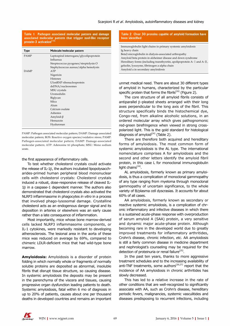
unmet medical need. There are about 30 different types of amyloid in humans, characterized by the particular specific protein that forms the fibrils[24] (Figure 2).
The core structure of all amyloid fibrils consists of antiparallel β-pleated sheets arranged with their long axes perpendicular to the long axis of the fibril. This structure specifically binds the histochemical dye, Congo-red, from alkaline alcoholic solutions, in an ordered molecular array which gives pathognomonic red-green birefringence when viewed in strong cross-polarized light. This is the gold standard for histological diagnosis of amyloid[24] (Table 2).
There are therefore both acquired and hereditary forms of amyloidosis. The most common form of systemic amyloidosis is the AL type. The international nomenclature comprises A for amyloidosis and the second and other letters identify the amyloid fibril protein, in this case L for monoclonal immunoglobulin light chains[24].
AL amyloidosis, formerly known as primary amyloi-dosis, is thus a complication of monoclonal gammopathy of any type ranging from myeloma through monoclonal gammopathy of uncertain significance, to the whole variety of B/plasma cell dyscrasias. It accounts for about 60% of all cases.
AA amyloidosis, formerly known as secondary or reactive systemic amyloidosis, is a complication of chr-onic inflammatory and infective diseases in which there is a sustained acute-phase response with overproduction of serum amyloid A (SAA) protein, a very sensitive and dynamic major acute-phase protein. Although becoming rare in the developed world due to greatly improved treatments for inflammatory arthritides, Crohn’s disease, chronic infection, etc. AA amyloidosis is still a fairly common disease in medicine department and nephrologist’s counseling may be required for the detection of proteinuria or renal failure[25].
In the past ten years, thanks to more aggressive treatment schedules and to the increasing availability of anti-TNF treatments, some authors[26,27] report that the incidence of AA amyloidosis in chronic arthritides has slowly decreased.
This has led to a relative increase in the rate of other conditions that are well-recognized to significantly associate with AA, such as Crohn’s disease, hereditary periodic fevers, malignancies, systemic vasculitides and diseases predisposing to recurrent infections, including
the first appearance of inflammatory cells. To test whether cholesterol crystals could activate
the release of IL-1β, the authors incubated lipopolysacch-arides-primed human peripheral blood mononuclear cells with cholesterol crystals: Cholesterol crystals induced a robust, dose-responsive release of cleaved IL-1β in a caspase-1 dependent manner. The authors also demonstrated that cholesterol crystals also activated the NLRP3 inflammasome in phagocytes in vitro in a process that involved phago-lysosomal damage. Crystalline cholesterol acts as an endogenous danger signal and its deposition in arteries or elsewhere was an early cause rather than a late consequence of inflammation.
Most importantly, mice whose bone marrow-derived cells lacked NLRP3 inflammasome components, or IL-1 cytokines, were markedly resistant to developing atherosclerosis. The lesional area in the aorta of these mice was reduced on average by 69%, compared to chimeric LDLR-deficient mice that had wild-type bone marrow.
Amyloidosis: Amyloidosis is a disorder of protein folding in which normally whole or fragments of normally soluble proteins are deposited as abnormal, insoluble fibrils that disrupt tissue structure, so causing disease. In systemic amyloidosis the deposits may be present in the parenchyma of the viscera and tissues, causing progressive organ dysfunction leading patients to death. Systemic amyloidosis, fatal within 6 mo of diagnosis in up to 20% of patients, causes about one per thousand deaths in developed countries and remains an important
69 January 6, 2016|Volume 5|Issue 1|WJN|www.wjgnet.com
Type Molecule/molecular pattern
PAMP Leptospiral interrogans/glycolipoproteinInfluenzaStreptococcus pyogenes/streptolysin OStaphylococcus aureus/alpha hemolysin
DAMP ATPNigericinHistonesU1snRNP ribonucleoproteindsDNA/nucleosomesMSU crystalsUromodulinBiglycanSilicaAlumCalcium oxalateAsbestosAmyloid-βHemazoinHyaluronan
Table 1 Pathogen associated molecular pattern and damage associated molecular pattern that trigger nod-like receptor protein-3 activation[12]
PAMP: Pathogen associated molecular pattern; DAMP: Damage associated molecular pattern; ROS: Reactive oxygen species/oxidative stress; PAMP: Pathogen-associated molecular pattern; DAMP: Damage-associated molecular pattern; ATP: Adenosine tri phosphate; MSU: Mono sodium urate.
Immunoglobulin light chains in primary systemic amyloidosis Ig heavy chain Beta2-microglobulin in dialysis-associated arthropathy Amyloid beta protein in alzheimer disease and down syndromeHereditary forms (including transthyretin, apolipoprotein A-Ⅰ and A-Ⅱ, gelsolin, lysozyme, fibrinogen a-alpha chainAmyloid a in secondary amyloidosis
Table 2 Over 30 proteins capable of amyloid formation have been identified
Scarpioni R et al . Amyloidosis, autoinflammatory diseases and kidney

cystic fibrosis, bronchiectasis, epidermolysis bullosa, cyclic neutropenia, acquired or inherited immunode-ficiencies, injection-drug use and acne conglobata.
SAA is a major acute-phase protein present in serum but also shown to possess proinflammatory properties, as meaning that it can induce the release of cytokines from different cell types, including THP-1 monocytes, human neutrophils, and mast cells.
SAA is mainly produced in the liver under the regulation of IL-1, IL-6, and TNF-, but its expression has also been demonstrated in other cell types, including macrophages, endothelial cells, and smooth muscle cells.
Moreover, SAA has been shown to induce the secretion of cathepsin B and inflammasome components from human macrophages. As processing of SAA by cathepsin B may result in production of amyloidogenic SAA fragments: Experimentally, SAA has demonstrated to induce a strong expression of IL1β and TNFα in human macrophages[28].
SAA mediates its effect through activation of NLRP3 inflammasome: SAA is a potent activator of the NLRP3 inflammasome via a cathepsin B- and P2X7-dependent manner and is the first physiological proinflammatory mediator that can provide signals needed for expression of pro-IL-1 (as shown in Figure 1) and activation of the inflammasome cascade, resulting in activation of caspase-1 and secretion of mature IL-1β so resulting in formation of amyloidogenic fragments[28]. The conversion of the circulating soluble protein SAA into stable, highly ordered, amyloid fibrils that accumulate extracellularly causing organ damage is a multi-step process.
As an acute phase reactant secreted by the liver under the transcriptional control of IL-1 and IL-6, SAA increases up to 1000 fold following an inflammatory stimulation. If such stimuli persist, as occurs in several chronic diseases, SAA concentration may reach a critical threshold over which it becomes prone to aggregation. Moreover, the estimated ten years’ survival was reported to be much higher in the patients with lower SAA levels, below 10 mg/dL[29].
A β-2m amyloidosis, so-called DIALYSIS-RELATED AMYLOIDOSIS, is a serious complication of long-term dialysis for end-stage renal failure in which β2-microglobulin, normally catabolized by the kidneys, is not adequately cleared and accumulates in the plasma, rising in concentration from its normal value of 1-2 mg/L to up to 70 mg/L[30].
All amyloid deposits have the feature to be largely ignored by the usually very efficient physiological mech-anisms by which abnormal protein debris is cleared from the interstitial space in the tissues. Dead cells, effete matrix and structural proteins, blood cells and plasma proteins extravasated in injury, are normally rapidly cleared with no local or systemic clinical consequences. In contrast, although macrophages and giant cells are occasionally seen, especially around local rather than deposited as amyloid fibrils in and around bones and joints, causing pain, bone cysts and pathological fractures.
Here we will address our description on the involve-ment of the kidney in autoinflammatory disease driven mainly by secondary, or reactive, AA amyloidosis.
AA AMYLOIDOSISA clear example of renal involvement in autoinfla-mmatory disease with amyloid A deposition may be considered the Muckle-Wells (MWS) disease associated with AA-amyloidosis. MWS is inherited as an autosomal dominant condition, meaning each child of a sufferer has a 50% chance of developing the syndrome.
MWS is a rare genetic autoinflammatory syndrome and the intermediate-severity form of cryopyrin-associated periodic syndrome (CAPS). As with other forms of this syndrome, it presents with recurrent episodes of fever, skin rash, joint pain, abdominal pain and conjunctivitis, but in addition sufferers typically develop a progressive sensorineural deafness and amyloidosis[9].
The protein affected in MWS is cryopyrin, produced by the NLRP3 gene located on chromosome 1. The gene is expressed in white blood cells (mainly neutrophils) and chondrocytes (cartilage cells). Cryopyrin is an essential component of the inflammasome, an intracellular protein complex involved in the innate immune system. The abnormal inflammasome in MWS allows unrestricted activation of the enzyme caspase-1, which in turn causes overproduction of active IL-1, switching on the inflammatory cascade in an uncontrolled manner[31].
MWS can have severe consequences due to chronic high levels of inflammation in the body. This can be life-threatening if generalized amyloidosis of the AA type develops, due to long-term buildup of amyloid protein products from the chronic inflammation in MWS. Organ damage results from the extracellular deposition of proteolytic fragments of the acute-phase reactant SAA as amyloid fibrils[9]. A sustained high concentration of SAA is the prerequisite for developing AA amyloidosis.
70 January 6, 2016|Volume 5|Issue 1|WJN|www.wjgnet.com
Amyloidfibrils
Congo red
Figure 2 Structural features of amyloid[25].
Scarpioni R et al . Amyloidosis, autoinflammatory diseases and kidney

The kidneys, liver and spleen are the main target organs of AA amyloid deposits (Figure 3).
In more than 90% of the patients proteinuria, nephrotic syndrome and/or renal dysfunction dominate the clinical picture at onset[9,24,26,27]. If not effectively treated, this disease invariably leads to end stage kidney disease[9] and renal replacement therapy, that are still associated with a poor outcome[27].
Over 25% of MWS patients have elevated se-rum amyloid, and at least 25% have amyloidosis. Serum AA testing is essential to follow, along with C-Reactive Protein (C-RP), Erythrocyte Sedimentation Rate (ESR) and other laboratory tests. Amyloidosis is also a risk to some patients affected by different types of CAPS, a group of autoinflammatory disord-ers characterized by recurrent episodes of systemic inflammation marked by fever, tissue inflammation, particularly of the joints and skin, and other constitutional symptoms, clinically defined by a spectrum of varying severity. Amyloidosis may be associated in familial cold autoinflammatory syndrome and in neonatal-onset multisystem inflammatory disease/chronic infantile neurological cutaneous and articular syndrome, but not so much as in MWS. Generalized amyloidosis is due to a permanent buildup of amyloid in the kidneys, liver and elsewhere, that can be fatal[32].
Clinical AA amyloidosis is typically preceded by many years of active inflammation before presenting, most commonly with renal involvement[33].
In AA amyloidosis renal dysfunction is reported to be the predominant disease manifestation. Mortality, amyloid burden, and renal prognosis all significantly correlated with the SAA concentration during follow-up. The risk of death was reported to be 17.7 times as high among patients with highest SAA concentrations. In the previously reported[25] largest study on AA amyloidosis involving 374 patients, the most frequent underlying disorder was inflammatory arthritis and only rare causes of AA amyloidosis included vasculitis, sickle cell anemia, malignant disease, epidermolysis bullosa, and cyclic neutropenia. Renal involvement was reported to be frequent: In 97% of patients, more than 500 mg
of proteinuria per day were present or the serum creatinine concentration was more than 1.5 mg/dL. The relative risk of progression to end-stage renal failure was also increased among patients whose renal function was relatively worse at baseline, with an increase by a factor of 5 for each doubling of the baseline serum creatinine concentration (P < 0.001).
Fortunately, the cardiac involvement is not so fre-quent in AA amyloidosis and it is reported to be present in only 1 patient, and findings consistent with cardiac infiltration were present in only 2 among 224 patients who underwent echocardiography[34].
A worse renal outcome in patients with chronic sepsis or Crohn’s disease was reported[28], possibly related to the high frequency of surgical intervention and administration of immunosuppressive drugs, probably due to greater severity of disease associated or not at increased risk of infection.
Therapy Further studies need to elucidate whether persistent inflammation serves as a catalyst by sensing and conver-ting the endothelium into a proinflammatory surface that makes the vasculature more vulnerable to the effects of other circulating risk factors. Such a scenario is supported by the strong documented association between inflammatory markers and endothelial dysfun-ction in patients with CKD.
Similarly, effective anti-inflammatory treatment, or whatever is needed to control the acute-phase response and maintain circulating SAA serum levels in the normal range, is life saving in AA amyloidosis[9,25,27]. Rigorous compliance with colchicine therapy for Familiar Mediterranean Fever (FMF) prevents and ameliorates AA amyloidosis even in patients who do not experience complete relief of symptoms. The key is to control SAA production, closely monitoring SAA serum levels in all patients with AA amyloidosis, and tailoring their treatment to keep these as low as possible. However, many patients are already in severe or end-stage organ failure when diagnosed with amyloidosis and new approaches are desperately needed to save them.
Treatment of AA amyloidosis has to be addressed in suppressing underlying inflammatory disease and reducing the SAA concentration as much as possible. If not effectively treated, this disease invariably leads to end stage kidney disease and renal replacement therapy, that are still associated with high mortality rate[25].
In a unpublished experience we observed in a female patient aged 40, affected by Chron disease with nephrotic proteinuria of 14 g/daily and CKD stage 3 secondary to renal AA amyloidosis histologically proven, the control of the baseline chronic bowel inflammatory disease with the monoclonal antibody adalimumab, a TNF-alfa inhibitor, significantly reduced the proteinuria levels up to 6 g/daily, while still remaining in the nephrotic range. The TNF-inhibitor therapy also reduced SAA levels from more than 4 mg/dL (normal values <
71 January 6, 2016|Volume 5|Issue 1|WJN|www.wjgnet.com
Figure 3 Renal biopsy: Amyloid fibrils bind congo red stain, yielding the pathognomonic apple-green birefringence under cross-polarized light microscopy[9].
Scarpioni R et al . Amyloidosis, autoinflammatory diseases and kidney

0.5) up to quite normal values (0.61 mg/dL).Among AA amyloidosis therapy, some years ago
interest was pointed on eprodisate, structurally similar to heparin sulfate, a glycosaminoglycan that is known to promote fibril assembly, inducing amyloid formation. Eprodisate, negatively charged, sulfonated molecule that is structurally similar to heparin sulfate and works by competitively inhibiting the interaction between SAA and glycosaminoglycans.
A RCT was conducted[35] enrolling 180 patients with AA amyloidosis-associated nephropathy; patients were treated with eprodisate or placebo for 24 mo: The authors reported that the treatment was associated with a 42% reduction in the risk of worsening renal disease (as measured by creatinine clearance) or death (CI: 0.37-0.93; P = 0.02), compared with placebo. Surprisingly, there was no significant difference in terms of the overall changes in proteinuria: A second phase Ⅲ trial is now ongoing.
Higher levels of aspecific laboratory inflammatory markers such as C-RP and sTNF are independently associated with faster rates of kidney function loss in CKD[36]. Pravastatin, a HMG-CoA reductase inhibitor, was reported to prevent loss of kidney function to a greater extent in CKD individuals with coronary artery disease (CAD) with greater evidence of inflammation, although this was of borderline significance. These data suggest that inflammation may mediate the loss of kidney function among subjects with CKD and concomitant CAD[37].
Some years ago, other authors experimentally found that inhibition of the isoprenoid pathway by another statin, lovastatin, resulted in a dose-dependent reduction of amyloid formed in mouse recombinant SAA produced in Escherichia coli, hypothesizing the isoprenoid metabolism as a potential target for prevention and treatment of AA amyloidosis[38].
More recently, Luo et al[39] studied the effects of another statin, rosuvastatin (RSV), and observed that, compared with controls, diabetic Sprague-Dawley rats showed severe metabolic disorder, cardiac dysfunction, fibrosis, disorganized ultrastructure, and excessive activation NLRP3 inflammasome, ASC, IL-1β and mitogen-activated protein kinases. The NLRP3 inflammasome was found activated in response to high levels of glucose. RSV was added and continued for 8 wk. The effect and underlying mechanisms of action of RSV in diabetic cardiomyopathy (DCM) and whether NLRP3 was a target for RSV in DCM, was studied. The authors concluded that, compared with diabetics rats alone, RSV experimentally ameliorated the overexpression of NLRP3 inflammasome and silencing NLRP3, ameliorated cardiac remodeling and dysfunction, so identifying RSV as a significant potential therapy via inhibition of NLRP3 inflammasome.
Due to the strong association between proinfla-mmatory cytokines and complications common in ESRD, such as vascular calcification and wasting, the potential role of both general and targeted anticytokine
treatment strategies in ESRD patients needs further evaluation[40]. Inflammation has to be considered an important target for pathogenetic interventions both in AKI and in progression of CKD, as recently suggested[41].
Therapeutic interventions that suppress inflam-mation and oxidative stress may address both short-term (dynamic) and long-term (structural) contributors to a decline in the GFR in patients with CKD and could possibly stabilize or even improve kidney function[9,42].
However, despite major technologic improvements in dialysis techniques, a lot of haemodialysis and perit-oneal dialysis patients show serological evidence of an activated inflammatory response, as clearly indicated by increased circulating levels of non-specific markers of inflammation and proinflammatory cytokines such as IL-6.
Dialysis treatment save the lives of patients with ESRD but it does not cure the burden of clinical consequences related to uremic state, i.e., the marked risk for atherosclerotic cardiovascular disease and inflammation. Renal transplantation (TPX) can be considered in selected patients progressing to ESRD, but unfortunately, it is a choice not offered to all ESRD patients due to the low number of transplants performed in some countries.
Novel treatments to control inflammation processes, and also to prevent progression of renal damage, are under development and anti-cytokine agents are becoming the mainstay of therapy to prevent and treat AA, including patients with FMF that do not respond or do not tolerate adequate colchicine dosages and targeting key molecular events in the fibrillogenesis process[43]; also the role of other drugs are in progress[44].
Unfortunately, control of fibril-protein production is not possible in some forms of amyloidosis and in others it is often slow. There is no therapy that directly targets amyloid deposits for enhanced clearance. However, all amyloid deposits contain the normal, non-fibrillar plasma glycoprotein, serum amyloid P component (SAP).
Other authors[45] showed that administration of anti-human-SAP antibodies to mice with amyloid deposits containing human SAP triggers a potent, complement-dependent, macrophage-derived giant cell reaction that swiftly removes massive visceral amyloid deposits without adverse effects. Interestingly, the authors found that a combination of a drug that depletes circulating SAP and an antibody that targets residual SAP within the deposits results in clearance of amyloid deposits. A humanized version of the anti-SAP antibody has been developed with a view to clinical evaluation of this dual approach, hypothesizing this combined therapy to eliminate amyloid deposits.
IL-1 blockadeClinical observations to date suggest that although IL-1 plays a key role in activation of the innate immune system, blockade of this cytokine appears to have few adverse effects. Anti-IL-1 therapy appears to
72 January 6, 2016|Volume 5|Issue 1|WJN|www.wjgnet.com
Scarpioni R et al . Amyloidosis, autoinflammatory diseases and kidney

increase the risk of infection only marginally, and there is no clear evidence for increased risk of malignancy, despite lymphoma and other types of cancer have been reported in children treated with TNF blockers, often when along with certain other drugs (such as azathioprine or 6-mercaptopurine). Safety block of IL-1 after 12 mo after renal TPX was reported[46] also in a renal transplanted patients affected by MWS with systemic amyloidosis treated with triple immunosuppressive drug regimen and at the same time canakinumab: No flares of MWS was observed during this period.
In our experience we did not observe increased hospitalization rate due to infections or malignancy in two patients affected by Muckle Wells syndrome treated with IL-1 blockers who had been followed for over three years[9].
CONCLUSIONThe release of proinflammatory cytokines during inflam-mation represents an attempt to respond to injury, but it may produce detrimental effects. The best-characterized inflammasome is the NLRP3 that, once activated, leads to the active form of caspase-1, the enzyme required for the maturation of IL-1β. SAA, as acute-phase protein with also proinflammatory properties, is a well recognized potent activator of the NLRP3. Additional mechanisms bringing to renal inflammatory, systemic diseases and fibrotic processes, resulting in kidney insufficiency were recently reported, via the activation of the inflammasome.
Currently, treatment options in amyloidosis rely on reducing the supply of the precursor protein and thus depend absolutely upon accurate typing of the amyloid. Intercalating agents able to induce physical disruption of the fibrillar structure of the native fibrils, once mature fibrils have been deposited, are under study in some types of non-AA amyloidosis, hence producing an intermediate: It so resulting to be more readily available for enzymatic degradation.
The administration of anti-human SAP antibodies[47] to mice with amyloid deposits containing human SAP triggers a potent, complement-dependent, reaction that swiftly removes massive visceral amyloid deposits without adverse effects. These promising results achieved in mouse models based on intermediary metabolism may not be extended to humans, so speci-fical trials are needed to test this hypothesis also in humans.
The role of statins is a new aspect targeted towards NLRP3 and not only in ameliorating dyslipidemic profile: Treatment with statins may represent a promising further test for this well-known class of drugs beyond the CV risk reduction, mediated by the reduction of lipidic profile[39].
Recently, great interest is growing on the role of NLRP3 inflammasome that incorporates several signals of tissue injury, infectious or non-infectious, and
consequently brings, via the activation of caspase-1, to the secretion of the pro-inflammatory cytokines IL-1β and IL-18. Block of the IL-1 system seems to be a fascinating option to counteract caspase activation and reducing IL-1 levels and consequently also SAA levels, that appear to be dramatically reduced within normal values even in patients with border line levels up to thousands of times. The mainway is to control the primary cause of inflammation and IL-1 blockers have demonstrated in rheumatologic field to be really effective and safe, even when associated to important immunosuppressant therapy, such as in kidney transplant patients. Moreover, a possible targeted intervention of IL-1 receptor blockade, even in active vasculitis, was recently suggested[22].
Several question points remain open, such as whether it is right to consider IL-1 block as target for treating CKD. And also if it is really the NLRP3 inflam-masome a gauge of kidney injury damage or if we can specifically target the NLRP3 inflammasome for therapeutic intervention, as recently postulated by other authors[12].
The direct involvement of NLRP3 in kidney disease has not been demonstrated yet, despite recently several manuscripts address a reasonable suspicion about it. Moreover, a deeper knowledge on the role of NLRP3 inflammasome and of reactive AA amyloidosis in renal diseases is requested. Whether blocking IL-1 is really effective in delaying the progression of renal damage has yet to be demonstrated by large trials, despite, at the moment, the high costs severely limit the use of such drugs.
REFERENCES1 Carrero JJ, Stenvinkel P. Persistent inflammation as a catalyst for
other risk factors in chronic kidney disease: a hypothesis proposal. Clin J Am Soc Nephrol 2009; 4 Suppl 1: S49S55 [PMID: 19996005 DOI: 10.2215/CJN.02720409]
2 Pulskens WP, Butter LM, Teske GJ, Claessen N, Dessing MC, Flavell RA, Sutterwala FS, Florquin S, Leemans JC. Nlrp3 prevents early renal interstitial edema and vascular permeability in unilateral ureteral obstruction. PLoS One 2014; 9: e85775 [PMID: 24454932 DOI: 10.1371/journal.pone.0085775]
3 Iyer SS, Pulskens WP, Sadler JJ, Butter LM, Teske GJ, Ulland TK, Eisenbarth SC, Florquin S, Flavell RA, Leemans JC, Sutterwala FS. Necrotic cells trigger a sterile inflammatory response through the Nlrp3 inflammasome. Proc Natl Acad Sci USA 2009; 106: 2038820393 [PMID: 19918053 DOI: 10.1073/pnas.0908698106]
4 Vilaysane A, Chun J, Seamone ME, Wang W, Chin R, Hirota S, Li Y, Clark SA, Tschopp J, Trpkov K, Hemmelgarn BR, Beck PL, Muruve DA. The NLRP3 inflammasome promotes renal inflammation and contributes to CKD. J Am Soc Nephrol 2010; 21: 17321744 [PMID: 20688930 DOI: 10.1681/ASN.2010020143]
5 Vesey DA, Cheung C, Cuttle L, Endre Z, Gobe G, Johnson DW. Interleukin1beta stimulates human renal fibroblast proliferation and matrix protein production by means of a transforming growth factorbetadependent mechanism. J Lab Clin Med 2002; 140: 342350 [PMID: 12434136]
6 Anders HJ, Muruve DA. The inflammasomes in kidney disease. J Am Soc Nephrol 2011; 22: 10071018 [PMID: 21566058 DOI: 10.1681/ASN.2010080798]
7 Mulay SR, Kulkarni OP, Rupanagudi KV, Migliorini A, Darisipudi
73 January 6, 2016|Volume 5|Issue 1|WJN|www.wjgnet.com
Scarpioni R et al . Amyloidosis, autoinflammatory diseases and kidney

MN, Vilaysane A, Muruve D, Shi Y, Munro F, Liapis H, Anders HJ. Calcium oxalate crystals induce renal inflammation by NLRP3-mediated IL-1β secretion. J Clin Invest 2013; 123: 236246 [PMID: 23221343 DOI: 10.1172/JCI63679]
8 Lichtnekert J, Kulkarni OP, Mulay SR, Rupanagudi KV, Ryu M, Allam R, Vielhauer V, Muruve D, Lindenmeyer MT, Cohen CD, Anders HJ. AntiGBM glomerulonephritis involves IL1 but is independent of NLRP3/ASC inflammasomemediated activation of caspase1. PLoS One 2011; 6: e26778 [PMID: 22046355 DOI: 10.1371/journal.pone.0026778]
9 Scarpioni R, Rigante D, Cantarini L, Ricardi M, Albertazzi V, Melfa L, Lazzaro A. Renal involvement in secondary amyloidosis of MuckleWells syndrome: marked improvement of renal function and reduction of proteinuria after therapy with human antiinterleukin1β monoclonal antibody canakinumab. Clin Rheumatol 2015; 34: 13111316 [PMID: 24510061 DOI: 10.1007/s1006701324812]
10 Turner CM, Arulkumaran N, Singer M, Unwin RJ, Tam FW. Is the inflammasome a potential therapeutic target in renal disease? BMC Nephrol 2014; 15: 21 [PMID: 24450291 DOI: 10.1186/147123 691521]
11 Church LD, Savic S, McDermott MF. Long term management of patients with cryopyrinassociated periodic syndromes (CAPS): focus on rilonacept (IL1 Trap). Biologics 2008; 2: 733742 [PMID: 19707454]
12 Lorenz G, Darisipudi MN, Anders HJ. Canonical and noncanonical effects of the NLRP3 inflammasome in kidney inflammation and fibrosis. Nephrol Dial Transplant 2014; 29: 4148 [PMID: 24026244 DOI: 10.1093/ndt/gft332]
13 Faust J, Menke J, Kriegsmann J, Kelley VR, Mayet WJ, Galle PR, Schwarting A. Correlation of renal tubular epithelial cellderived interleukin18 upregulation with disease activity in MRLFaslpr mice with autoimmune lupus nephritis. Arthritis Rheum 2002; 46: 30833095 [PMID: 12428253 DOI: 10.1002/art.10563]
14 VanderBrink BA, Asanuma H, Hile K, Zhang H, Rink RC, Meldrum KK. Interleukin18 stimulates a positive feedback loop during renal obstruction via interleukin18 receptor. J Urol 2011; 186: 15021508 [PMID: 21855933 DOI: 10.1016/j.juro.2011.05.046]
15 Franke EI, Vanderbrink BA, Hile KL, Zhang H, Cain A, Matsui F, Meldrum KK. Renal IL18 production is macrophage independent during obstructive injury. PLoS One 2012; 7: e47417 [PMID: 23077611 DOI: 10.1371/journal.pone.0047417]
16 Zhang C, Boini KM, Xia M, Abais JM, Li X, Liu Q, Li PL. Activation of Nod-like receptor protein 3 inflammasomes turns on podocyte injury and glomerular sclerosis in hyperhomocysteinemia. Hypertension 2012; 60: 154162 [PMID: 22647887 DOI: 10.1161/HYPERTENSIONAHA.111.189688]
17 Mulay SR, Evan A, Anders HJ. Molecular mechanisms of crystalrelated kidney inflammation and injury. Implications for cholesterol embolism, crystalline nephropathies and kidney stone disease. Nephrol Dial Transplant 2014; 29: 507514 [PMID: 24163269 DOI: 10.1093/ndt/gft248]
18 Martinon F, Pétrilli V, Mayor A, Tardivel A, Tschopp J. Goutassociated uric acid crystals activate the NALP3 inflammasome. Nature 2006; 440: 237241 [PMID: 16407889 DOI: 10.1038/nature04516]
19 Scarpioni R. An Innovative Approach to Managing a Patient With Complicated Gouty Arthritis and Chronic Kidney Disease. Joint Spine Bone 2015; In press
20 Schroder K, Tschopp J. The inflammasomes. Cell 2010; 140: 821832 [PMID: 20303873 DOI: 10.1016/j.cell.2010.01.040]
21 Schreiber A, Luft FC, Kettritz R. Phagocyte NADPH oxidase restrains the inflammasome in ANCAinduced GN. J Am Soc Nephrol 2015; 26: 411424 [PMID: 25012177 DOI: 10.1681/ASN.2013111177]
22 Dinarello CA. Interleukin1its multiple biological effects and its association with hemodialysis. Blood Purif 1988; 6: 164172 [PMID: 3293622]
23 Duewell P, Kono H, Rayner KJ, Sirois CM, Vladimer G, Bauernfeind FG, Abela GS, Franchi L, Nuñez G, Schnurr M, Espevik T, Lien E, Fitzgerald KA, Rock KL, Moore KJ, Wright
SD, Hornung V, Latz E. NLRP3 inflammasomes are required for atherogenesis and activated by cholesterol crystals. Nature 2010; 464: 13571361 [PMID: 20428172 DOI: 10.1038/nature08938]
24 Merlini G, Bellotti V. Molecular mechanisms of amyloidosis. N Engl J Med 2003; 349: 583596 [PMID: 12904524 DOI: 10.1056/NEJMra023144]
25 Lachmann HJ, Goodman HJ, Gilbertson JA, Gallimore JR, Sabin CA, Gillmore JD, Hawkins PN. Natural history and outcome in systemic AA amyloidosis. N Engl J Med 2007; 356: 23612371 [PMID: 17554117 DOI: 10.1056/NEJMoa070265]
26 Immonen K, Finne P, GrönhagenRiska C, Pettersson T, Klaukka T, Kautiainen H, Hakala M. A marked decline in the incidence of renal replacement therapy for amyloidosis associated with inflammatory rheumatic diseases data from nationwide registries in Finland. Amyloid 2011; 18: 2528 [PMID: 21284495 DOI: 10.3109/13506129.2010.549252]
27 Obici L, Merlini G. AA amyloidosis: basic knowledge, unmet needs and future treatments. Swiss Med Wkly 2012; 142: w13580 [PMID: 22653707 DOI: 10.4414/smw.2012.13580]
28 Niemi K, Teirilä L, Lappalainen J, Rajamäki K, Baumann MH, Öörni K, Wolff H, Kovanen PT, Matikainen S, Eklund KK. Serum amyloid A activates the NLRP3 inflammasome via P2X7 receptor and a cathepsin Bsensitive pathway. J Immunol 2011; 186: 61196128 [PMID: 21508263 DOI: 10.4049/jimmunol.1002843]
29 Gillmore JD, Lovat LB, Persey MR, Pepys MB, Hawkins PN. Amyloid load and clinical outcome in AA amyloidosis in relation to circulating concentration of serum amyloid A protein. Lancet 2001; 358: 2429 [PMID: 11454373 DOI: 10.1016/S01406736(00)0525 21]
30 Bataille S, Fernandez C, Zink JV, Brunet P, Berland Y, Burtey S. The Case | A hip fracture in a hemodialysis patient. Pathologic righthip fracture from β2-microglobulin amyloidosis. Kidney Int 2013; 83: 12111212 [PMID: 23728017 DOI: 10.1038/ki.2012.436]
31 Kümmerle-Deschner JB, Tyrrell PN, Reess F, Kötter I, Lohse P, Girschick H, Huemer C, Horneff G, Haas JP, Koitschev A, Deuter C, Benseler SM. Risk factors for severe MuckleWells syndrome. Arthritis Rheum 2010; 62: 37833791 [PMID: 20722029 DOI: 10.1002/art.27696]
32 Cantarini L, Rigante D, Brizi MG, Lucherini OM, Sebastiani GD, Vitale A, Gianneramo V, Galeazzi M. Clinical and biochemical landmarks in systemic autoinflammatory diseases. Ann Med 2012; 44: 664673 [PMID: 21972825 DOI: 10.3109/07853890.2011.598546]
33 Simons JP, AlShawi R, Ellmerich S, Speck I, Aslam S, Hutchinson WL, Mangione PP, Disterer P, Gilbertson JA, Hunt T, Millar DJ, Minogue S, Bodin K, Pepys MB, Hawkins PN. Pathogenetic mechanisms of amyloid A amyloidosis. Proc Natl Acad Sci USA 2013; 110: 1611516120 [PMID: 23959890 DOI: 10.1073/pnas.1306621110]
34 Dubrey SW , Cha K, Simms RW, Skinner M, Falk RH. Electrocardiography and Doppler echocardiography in secondary (AA) amyloidosis. Am J Cardiol 1996; 77: 313315 [PMID: 8607418 DOI: 10.1016/S00029149(97)894039]
35 Dember LM, Hawkins PN, Hazenberg BP, Gorevic PD, Merlini G, Butrimiene I, Livneh A, Lesnyak O, Puéchal X, Lachmann HJ, Obici L, Balshaw R, Garceau D, Hauck W, Skinner M. Eprodisate for the treatment of renal disease in AA amyloidosis. N Engl J Med 2007; 356: 23492360 [PMID: 17554116 DOI: 10.1056/NEJMoa065644]
36 Scarpioni R, Ricardi M, Albertazzi V, Melfa L. Treatment of dyslipidemia in chronic kidney disease: Effectiveness and safety of statins. World J Nephrol 2012; 1: 184194 [PMID: 24175258 DOI: 10.5527/wjn.v1.i6.184]
37 Tonelli M, Sacks F, Pfeffer M, Jhangri GS, Curhan G. Biomarkers of inflammation and progression of chronic kidney disease. Kidney Int 2005; 68: 237245 [PMID: 15954913 DOI: 10.1111/j.15231755.2005.00398.x]
38 van der Hilst JC, KluveBeckerman B, Bodar EJ, van der Meer JW, Drenth JP, Simon A. Lovastatin inhibits formation of AA amyloid. J Leukoc Biol 2008; 83: 12951299 [PMID: 18285405
74 January 6, 2016|Volume 5|Issue 1|WJN|www.wjgnet.com
Scarpioni R et al . Amyloidosis, autoinflammatory diseases and kidney
DOI: 10.1189/jlb.1107723]39 Luo B, Li B, Wang W, Liu X, Liu X, Xia Y, Zhang C, Zhang Y,
Zhang M, An F. Rosuvastatin alleviates diabetic cardiomyopathy by inhibiting NLRP3 inflammasome and MAPK pathways in a type 2 diabetes rat model. Cardiovasc Drugs Ther 2014; 28: 3343 [PMID: 24254031 DOI: 10.1007/s1055701364981]
40 Stenvinkel P, Ketteler M, Johnson RJ, Lindholm B, PecoitsFilho R, Riella M, Heimbürger O, Cederholm T, Girndt M. IL10, IL6, and TNFalpha: central factors in the altered cytokine network of uremiathe good, the bad, and the ugly. Kidney Int 2005; 67: 12161233 [PMID: 15780075 DOI: 10.1111/j.15231755.2005.00200.x]
41 Cantaluppi V, Quercia AD, Dellepiane S, Ferrario S, Camussi G, Biancone L. Interaction between systemic inflammation and renal tubular epithelial cells. Nephrol Dial Transplant 2014; 29: 20042011 [PMID: 24589723 DOI: 10.1093/ndt/gfu046]
42 Cachofeiro V, Goicochea M, de Vinuesa SG, Oubiña P, Lahera V, Luño J. Oxidative stress and inflammation, a link between chronic kidney disease and cardiovascular disease. Kidney Int Suppl 2008; (111): S4S9 [PMID: 19034325 DOI: 10.1038/ki.2008.516]
43 Obici L, Merlini G. Amyloidosis in autoinflammatory syndromes. Autoimmun Rev 2012; 12: 1417 [PMID: 22878269 DOI: 10.1016/j.autrev.2012.07.016]
44 Lachmann HJ, Quartier P, So A, Hawkins PN. The emerging role of interleukin-1β in autoinflammatory diseases. Arthritis Rheum 2011; 63: 314324 [PMID: 20967858]
45 Sayed RH, Hawkins PN, Lachmann HJ. Emerging treatments for amyloidosis. Kidney Int 2015; 87: 516526 [PMID: 25469850 DOI: 10.1038/ki.2014.368]
46 Bodin K, Ellmerich S, Kahan MC, Tennent GA, Loesch A, Gilbertson JA, Hutchinson WL, Mangione PP, Gallimore JR, Millar DJ, Minogue S, Dhillon AP, Taylor GW, Bradwell AR, Petrie A, Gillmore JD, Bellotti V, Botto M, Hawkins PN, Pepys MB. Antibodies to human serum amyloid P component eliminate visceral amyloid deposits. Nature 2010; 468: 9397 [PMID: 20962779 DOI: 10.1038/nature09494]
47 Kortus-Götze B, Hoyer J. Successful renal transplantation in Muckle-Wells syndrome treated with anti-IL-1β-monoclonal antibody. NDT Plus 2011; 4: 404405 [PMID: 25984207 DOI: 10.1093/ndtplus/sfr118]
P- Reviewer: Friedman EA, Fujigaki Y, Lehtonen SH, Shou ZF S- Editor: Qiu S L- Editor: A E- Editor: Jiao XK
75 January 6, 2016|Volume 5|Issue 1|WJN|www.wjgnet.com
Scarpioni R et al . Amyloidosis, autoinflammatory diseases and kidney

Role of calcium in polycystic kidney disease: From signaling to pathology
Alessandra Mangolini, Lucia de Stephanis, Gianluca Aguiari
Alessandra Mangolini, Lucia de Stephanis, Gianluca Aguiari, Department of Biomedical and Surgical Specialty Sciences, Section of Biochemistry, Molecular Biology and Medical Genetics, University of Ferrara, 44121 Ferrara, Italy
Alessandra Mangolini, Department of Morphology, Surgery and Experimental Medicine, Section of Pathology, Oncology and Experimental Biology, University of Ferrara, 44121 Ferrara, Italy
Author contributions: Mangolini A and de Stephanis L performed the experiments; Aguiari G generated the figures and wrote the manuscript.
Supported by University of Ferrara local funds: FAR 2012, 2013, 2014 and Regione Emilia Romagna grant (Ricerca Regione-Università) 2007-2009.
Conflict-of-interest statement: The authors declare that they have no commercial, personal, political, intellectual or religious conflict of interests regarding the data or scientific reports included in the present article. There is no conflict of interest associated with the senior author or other coauthors contributed their efforts in this paper.
Open-Access: This article is an open-access article which was selected by an in-house editor and fully peer-reviewed by external reviewers. It is distributed in accordance with the Creative Commons Attribution Non Commercial (CC BY-NC 4.0) license, which permits others to distribute, remix, adapt, build upon this work non-commercially, and license their derivative works on different terms, provided the original work is properly cited and the use is non-commercial. See: http://creativecommons.org/licenses/by-nc/4.0/
Correspondence to: Gianluca Aguiari, PhD, Department of Biomedical and Surgical Specialty Sciences, Section of Biochemistry, Molecular Biology and Medical Genetics, University of Ferrara, via Fossato di Mortara 74, 44121 Ferrara, Italy. [email protected] Telephone: +39-532-974460
Received: September 9, 2015Peer-review started: September 10, 2015First decision: October 8, 2015Revised: November 21, 2015
Accepted: December 8, 2015 Article in press: December 11, 2015Published online: January 6, 2016
AbstractAutosomal dominant polycystic kidney disease (ADPKD) is the most common inherited monogenic kidney disease. Characterized by the development and growth of cysts that cause progressive kidney enlargement, it ultimately leads to end-stage renal disease. Approximately 85% of ADPKD cases are caused by mutations in the PKD1 gene, while mutations in the PKD2 gene account for the remaining 15% of cases. The PKD1 gene encodes for polycystin-1 (PC1), a large multi-functional memb-rane receptor protein able to regulate ion channel complexes, whereas polycystin-2 (PC2), encoded by the PKD2 gene, is an integral membrane protein that functions as a calcium-permeable cation channel, located mainly in the endoplasmic reticulum (ER). In the primary cilia of the epithelial cells, PC1 interacts with PC2 to form a polycystin complex that acts as a mechanosensor, regulating signaling pathways involved in the differentiation of kidney tubular epithelial cells. Despite progress in understanding the function of these proteins, the molecular mechanisms associated with the pathogenesis of ADPKD remain unclear. In this review we discuss how an imbalance between functional PC1 and PC2 proteins may disrupt calcium channel activities in the cilium, plasma membrane and ER, thereby altering intracellular calcium signaling and leading to the aberrant cell proliferation and apoptosis associated with the development and growth of renal cysts. Research in this field could lead to the discovery of new molecules able to rebalance intracellular calcium, thereby normalizing cell proliferation and reducing kidney cyst progression.
Key words: Autosomal dominant polycystic kidney disease; Calcium signaling; cAMP; Cell growth; Non-capacitative calcium entry
MINIREVIEWS
76 January 6, 2016|Volume 5|Issue 1|WJN|www.wjgnet.com
World Journal of NephrologyW J N
Submit a Manuscript: http://www.wjgnet.com/esps/Help Desk: http://www.wjgnet.com/esps/helpdesk.aspxDOI: 10.5527/wjn.v5.i1.76
World J Nephrol 2016 January 6; 5(1): 76-83ISSN 2220-6124 (online)
© 2016 Baishideng Publishing Group Inc. All rights reserved.

© The Author(s) 2016. Published by Baishideng Publishing Group Inc. All rights reserved.
Core tip: In the present article, we discuss: (1) the regulation of calcium signaling in the primary cilia of auto-somal dominant polycystic kidney disease (ADPKD) cells and the downstream processes that lead to cystogenesis; (2) how calcium impairment promotes cell proliferation by activating different signaling pathways; (3) the activity of non-capacitative calcium entry channels, which in PKD1-silenced cells stimulates cell growth by Ca2+ oscillations and nuclear factor of activated T-cells activation, highlighting new findings showing the role of polycystin-2 in calcium oscillations; (4) the impairment of intracellular calcium signaling associated with apoptosis; and (5) the use of calcium channel blockers and calcium modulators in the treatment of ADPKD.
Mangolini A, de Stephanis L, Aguiari G. Role of calcium in polycystic kidney disease: From signaling to pathology. World J Nephrol 2016; 5(1): 76-83 Available from: URL: http://www.wjgnet.com/2220-6124/full/v5/i1/76.htm DOI: http://dx.doi.org/10.5527/wjn.v5.i1.76
INTRODUCTIONAutosomal dominant polycystic kidney disease (ADPKD) is the most common inherited pathology of the kidneys, having an incidence of 1:500-1:1000 individuals. It accounts for roughly 10% of cases of end-stage renal disease[1,2], which results from the progressive bilateral development and expansion of fluid-filled cysts arising from the de-differentiation of renal tubule epithelial cells[1]. ADPKD is caused by the mutation of two genes: PKD1, which accounts for 85% of cases, and PKD2, associated with the remaining 15% of cases[1]. In ADPKD, the focal cyst development in the kidneys seems to be associated with a somatic second hit brought on by either loss of heterozygosity or other mutations in renal cyst lining epithelial cells[3,4]. PKD1 and PKD2 genes encode for polycystin-1 (PC1) and polycystin-2 (PC2) proteins, respectively[1,5]. PC1 is an integral membrane receptor with a large extracellular region consisting of a variety of domains involved in cell-cell and cell-matrix interactions. It also bears 11 transmembrane domains, and a short cytoplasmic segment containing motifs involved in signal transduction[1]. PC2, on the other hand, is an integral transmembrane protein mainly localized to the endoplasmic reticulum (ER); it is anchored to cell membranes by six transmembrane regions, and has two cytoplasmic N- and C-terminal tails. PC2 functions as a nonselective cation channel that transports calcium, and shows significant homology with transient receptor potential (TRP) channels[5-8]. Indeed, the polycystins and their homologous proteins are considered a new subfamily of TRP channels, and accordingly known as TRP polycystic proteins[1].
An interaction between PC1 and PC2 forms the so-
called polycystin complex. This is mainly confined to the primary cilia of kidney epithelial cells, where it acts as a flow sensor, triggering intracellular calcium release via the activation of the PC2 channel in response to fluid-flow changes. Disruption of this complex impairs intracellular calcium influx, and leads to the development and expansion of kidney cysts[1,9,10].
Polycystins are able to regulate calcium channel activity not only in the cilia, but also in other cellular compartments, including the plasma membrane and ER. Indeed, PC1 and PC2 co-assembly has been seen to generate a cation-permeable current through the plasma membrane[11], and PC1 and PC2 are known to regulate intracellular calcium release in the ER through their interaction with the inositol 1,4,5-trisphosphate receptor (IP3R)[12-14]. In this context, PC2 enhances calcium release from the ER by stimulating the activity of the IP3 receptor, while PC1 inhibits this process by reducing PC2-IP3R interaction via a mechanism involving the stromal interaction molecule-1 (STIM1) and the PI3K/Akt pathway[12,15]. PC1 can also regulate other types of calcium channels, including non-capacitative calcium entry (NCCE) channels, which are able to generate intracellular calcium oscillations[16]. PC2, on the other hand, regulates intracellular calcium release by either interacting with the calcium channels TRPC1 and TRPV4 on the plasma membrane and in primary cilia, and/or through an association with ryanodine and IP3 receptors in the ER[8,17-19]. Moreover, PC2 appears to be able to generate a non-specific voltage-dependent cation current in native HEK293 kidney cells. This current is strongly associated with PC2 activity, and is completely abolished by the depletion of PC2 protein[20].
Taken together these findings suggest that PC1 and PC2 may affect calcium influx from different cellular compartments, including cilium boundaries (cilioplasm), plasma membrane and ER. Dysregulation of calcium signaling due to loss of polycystin function causes the aberrant activation of different pathways associated with abnormal cell proliferation and fluid secretion, thereby leading to the development and expansion of kidney cysts. However, the cascade of events that occur between polycystin dysfunction and kidney cyst formation in ADPKD is not yet fully understood.
In this review, we discuss the impact of polycystin loss of function on calcium signaling, which may alter different pathways associated with the cell growth and apoptosis that are a typical hallmark of ADPKD. Moreover, we report the potential effects of calcium dysregulation on kidney cyst formation and progression. Finally, we also discuss the state of the art in calcium channel modulators, able to restore normal calcium release and therefore appealing targets for ADPKD treatment.
ROLE OF THE POLYCYSTIN COMPLEX IN PRIMARY RENAL CILIA It is well known that PC1 and PC2 co-localize in the
Mangolini A et al . Calcium and polycystic kidney disease
77 January 6, 2016|Volume 5|Issue 1|WJN|www.wjgnet.com

primary cilia of kidney epithelial cells, performing a mechano-sensor function by transducing calcium signals in response to changes in tubular fluid flow. Loss or dysfunction of either PC1 or PC2 causes the inability of cells to sense mechanical stimuli due to bending of the cilia, which leads to abnormal cell morphology and polarity, and thereby contributes to renal cyst form-ation[10,21,22]. The calcium signaling triggered by fluid shear stress initiates in the primary cilia through PC2-dependent calcium release. This is initially confined to the cilioplasm, but, through the ryanodine receptor, the same calcium signal subsequently activates a cytosolic calcium response that induces calcium influx from intracellular stores[23].
PC2, as mentioned above, is also able to interact with the transient potential receptor channels TRPC1 and TRPV4. PC2 and TRPC1 assemble to form a hetero-multimeric channel, not associated with PC1, which is activated in response to G-protein-coupled receptor stimulation, and shows a pattern of single-channel con-ductance distinct from that of the individual PC2 and TRPC1 channels[24]. Direct or indirect activation of the PC2/TRPC1 complex, either by cilium bending or through the activation of plasma membrane GPCRs, may affect the mechano-transduction of cilium-associated calcium signals[24]. PC2 can also form a heteromeric channel complex with TRPV4. This complex displays molecular mechano-sensor properties, being able to generate flow-induced calcium influx, which seems to be abolished by the depletion of TRPV4 channel in renal epithelial cells[18]. It is also plausible that polycystins cooperate with other proteins located in the cilium, such as cystin, polaris, inversin, and kinesin-Ⅱ, as defects in these proteins may lead to the formation of kidney cysts[21,25].
In the primary cilia, PC1/PC2, PC2/TRPC1 and PC2/TRPV4 complexes regulate the calcium signaling activated by cilium deflection due to changes in fluid flow. Surprisingly, however, depletion of TRPC1 and TRPV4 is not associated with cyst formation, despite it altering ciliary calcium signaling. Hence, the impairment of ciliary calcium signaling alone is not sufficient to trig-ger kidney cyst development, a process which, instead, seems to be closely linked to the activity/function of PC1 and PC2 proteins. In fact, a recent study has shown that ablation of the cilia in both PC1- and PC2-deficient cells reduces cyst growth, suggesting that the loss of cilia may cause milder cyst progression than in the cilia-equipped ADPKD cells[26]. Moreover, cilia-dependent cyst growth is not associated with the activation of the mitogen-activated protein kinase/extracellular signal-regulated kinase, mammalian target of rapamycin (mTOR) or cyclic adenosinemonophosphate (cAMP) pathways[26]. As a whole, these findings suggest that the polycystins govern ciliary signaling by an unknown mech-anism when the normal kidney epithelial cell phenotype is maintained. In ADPKD, the inactivation of polycystins alters cilia-dependent signaling, thereby promoting the formation of the characteristic kidney cysts.
CALCIUM SIGNALING AND CELL PROLIFERATION IN ADPKD CELLSADPKD is strongly associated with the altered cell prolife-ration of cystic kidney epithelial cells that represents a typical hallmark of the disease. The disruption of calcium signaling associated with PC1 and PC2 deficiency could be the primary event behind the increased cell growth seen in ADPKD. Indeed, we do know that calcium restri-ction in ADPKD cells causes cAMP-dependent activation of the B-Raf/mitogen-activated protein kinase kinase (MEK)/extracellular-signal-regulated kinases (ERK) pathway, which results in increased cell growth[27]. Moreover, this reduction in intracellular calcium levels also inhibits the activity of AKT kinase, a negative regulator of B-Raf[27]. In cystic cells, normal growth can be restored by increasing their cytosolic calcium concentration, which increases AKT activity and inhibits cAMP-dependent B-Raf/ERK activation[28].
Low intracellular calcium levels may also stimulate the activity of the ciliary calcium-sensitive adenylyl cyclases AC5 and AC6, as well as the plasma-membrane-anchored AC6, leading to the elevation of cAMP[29,30]. Therefore, loss of polycystin function may promote the activity of AC5/6 by reducing intracellular Ca2+ release from the cilia, ER and plasma membrane[29,30] (Figure 1). Indeed, it has recently been reported that the double knockout of PKD1 and AC6 genes decreases cystogenesis, improves renal function and increases survival in a mouse model of ADPKD. These improvements in renal function occur through a reduction in cAMP levels and inhibition of the B-Raf/MEK/ERK pathway, suggesting that AC6 could be a key mediator of cyst formation in ADPKD[31]. In addition, cAMP elevation may activate the cAMP-response element-binding protein, which promotes cell proliferation in an epidermal growth factor receptor (EGFR)-activation-dependent manner by stimulating expression of the EGF-like peptide amphiregulin[32]. EGFR signaling is dependent upon a mechanism involving the sequential activation of Ras, Raf-1, MEK and ERK. It can converge on the same pathway activated by cAMP, thereby leading to activation of the ERK kinases that promote cell proliferation in ADPKD cells[33] (Figure 1). Furthermore, the abnormal activity of mTOR kinase has been observed to contribute to increased cell proliferation and cyst formation in ADPKD cyst-lining epithelial cells. In normal kidney epithelial cells mTOR activity is inhibited by PC1, which interacts with TSC1/TSC2, an inhibitory complex of mTOR, preventing its inactivation. Conversely, in ADPKD cells polycystin dysfunction promotes mTOR activation by inhibition of the TSC1/TSC2 complex, through a mechanism involving the cAMP-dependent B-Raf/ERK pathway[34,35] (Figure 1).
The expression of full-length PC1 has been shown to inhibit intracellular calcium release in response to ATP in Madin-Darby canine kidney (MDCK) cells, in a mechanism that involves the interaction of STIM1 with
78 January 6, 2016|Volume 5|Issue 1|WJN|www.wjgnet.com
Mangolini A et al . Calcium and polycystic kidney disease

increase in both basal and intracellular calcium release, leading to the activation of the nuclear factor of activated T-cells (NFAT)[38]. Moreover, NFAT activation, associated with increased cell proliferation in HEK-293 cells, is also observed after the downregulation of PC1 by RNA interference[16]. NFAT activation occurs through a rise in the frequency of intracellular calcium oscillations, caused by the increased activity of NCCE channels[16] (Figure 1). In HEK293 cells, these calcium oscillations can be increased by either reduced or undetectable levels of PC1, but are only induced by the absence of PC2 (Figure 2A). Normal calcium oscillations can be restored in PKD1-deficient cells via the reintroduction of mouse wild-type PC1, and in PKD2 knockout cells via the
IP3R, and reduces the association between PC2 and the IP3 receptor[15]. Moreover, PC1 seems able to regulate intracellular calcium release and PC2-IP3R-STIM1 interaction through the PI3K/Akt signaling pathway[15]. The exogenous expression of the C-terminal fragment of PC1 (PC1-Cter) could function as a dominant neg-ative effector, causing an increased intracellular calcium release in response to ATP treatment, as seen in HEK-293 cells[36]. Furthermore, PC1-Cter-expressing cells not only exhibit increased levels of basal calcium, but also show enhanced cell proliferation, which is associated with the activation of ERK kinases[37]. Consistently, the transfection of HEK-293 cells with the C-terminal tail of PC1 has been observed to cause an
79 January 6, 2016|Volume 5|Issue 1|WJN|www.wjgnet.com
Figure 1 Diagram showing calcium-dependent dysregulated signaling pathways that promote cell proliferation and apoptosis in autosomal dominant polycystic kidney disease cells. Loss of PC1 and/or PC2 function causes a reduction in cytosolic calcium influx from three different cellular compartments: (1) the primary cilium after mechanical stimuli; (2) the endoplasmic reticulum, in an IP3R- and RyR-dependent manner; and (3) the plasma membrane, through a reduction in SOCE channel activity. The reduced concentration of cytosolic calcium may activate Ca2+ sensitive adenylyl cyclases 5 and 6, leading to a rise in cAMP. Increased levels of cAMP cause the activation of B-Raf/MEK/ERK and CREB/AREG/EGFR pathways, as well as stimulating mTOR signaling, through the active form of ERK kinases that inactivate the TSC1/TSC2 complex. Moreover, deficiency of PC1 and/or PC2 enhances the activity of NCCE channels, which, by increasing calcium oscillation frequency, results in the activation of the transcription factor NFAT. The abnormal activation of these signaling pathways promotes cell proliferation and kidney cyst formation. In addition, the reduction in Ca2+ influx from the ER to the cytosol caused by a deficiency in PC2 channel activity brings about an imbalance in ER calcium concentration, resulting in ER Ca2+ overload. The increased ER calcium concentration sensitizes kidney cystic cells to apoptotic stimuli by abnormal ER calcium release, which may induce mitochondrial damage and thereby lead to cytochrome C release and activation of apoptosis. AC 5/6: Adenylyl cyclase 5/6; AR: Amphiregulin; Ca2+ OX: Calcium oscillations; cAMP: Cyclic adenosine monophosphate; CREB: cAMP response element binding transcription factor; EGFR: Epidermal growth factor receptor; ER: Endoplasmic reticulum; ERK: Extracellular-signal-regulated kinases; GRB2: Growth factor receptor-bound protein 2; IP3R: Inositol 1,4,5-trisphosphate receptor; MEK: Mitogen-activated protein kinase kinase; mTOR: Mammalian target of rapamycin; NCCE: Non-capacitative calcium channel entry; NFAT: Nuclear factor of activated T-cells; PKA: Protein kinase A; PC1: Polycystin-1; PC2: Polycystin-2; S6K: Ribosomal S6 kinase; Raf: Rapidly accelerated fibrosarcoma kinase; Ras: Rat sarcoma viral oncogene homolog family; Rheb: Ras homolog enriched in brain; RyR: Ryanodine receptor; SERCA: Sarcoplasmic endoplasmic reticulum calcium ATPase; SOCE: Store-operated calcium channel entry; TRPC1: Transient receptor potential channel 1; TRPV4: Transient receptor potential cation channel subfamily V member 4; TSC: Tuberous sclerosis complex.
DecreasedIncreased
AREGFR
GRB2
CREB
RasCa2+PC1
PC2
Ca2+
NCCE
Ca2+OX
Calcineurin
Raf
MEK
ERK
P-ERK
NFAT
Nucleus
TSC2
TSC1
Rheb
PKA
cAMP
SOCE
Ca2+
p21
AR
p27
Cell growth and proliferation
Mitochondrial calciumProtein synthesis
S6K
mTOR
AC5/6
PC1
Ca2+
Ca2+
PC2
PC2
PC2
PC2
PC2Ca
2+
TRPV
4
Cilium
TRPC1
Ca2+
PC1
AC6
PC2Ca2+
Ca2+
Ca2+
SERCA
Ca2+
IP3R
RyR
ER
ADPKD cell
Ca2+
Apoptosis
Mangolini A et al . Calcium and polycystic kidney disease
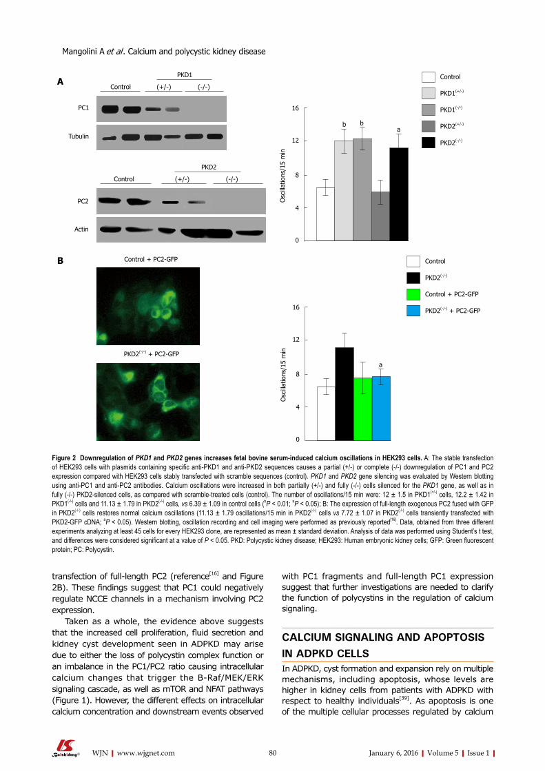
transfection of full-length PC2 (reference[16] and Figure 2B). These findings suggest that PC1 could negatively regulate NCCE channels in a mechanism involving PC2 expression.
Taken as a whole, the evidence above suggests that the increased cell proliferation, fluid secretion and kidney cyst development seen in ADPKD may arise due to either the loss of polycystin complex function or an imbalance in the PC1/PC2 ratio causing intracellular calcium changes that trigger the B-Raf/MEK/ERK signaling cascade, as well as mTOR and NFAT pathways (Figure 1). However, the different effects on intracellular calcium concentration and downstream events observed
with PC1 fragments and full-length PC1 expression suggest that further investigations are needed to clarify the function of polycystins in the regulation of calcium signaling.
CALCIUM SIGNALING AND APOPTOSIS IN ADPKD CELLSIn ADPKD, cyst formation and expansion rely on multiple mechanisms, including apoptosis, whose levels are higher in kidney cells from patients with ADPKD with respect to healthy individuals[39]. As apoptosis is one of the multiple cellular processes regulated by calcium
80 January 6, 2016|Volume 5|Issue 1|WJN|www.wjgnet.com
Figure 2 Downregulation of PKD1 and PKD2 genes increases fetal bovine serum-induced calcium oscillations in HEK293 cells. A: The stable transfection of HEK293 cells with plasmids containing specific anti-PKD1 and anti-PKD2 sequences causes a partial (+/-) or complete (-/-) downregulation of PC1 and PC2 expression compared with HEK293 cells stably transfected with scramble sequences (control). PKD1 and PKD2 gene silencing was evaluated by Western blotting using anti-PC1 and anti-PC2 antibodies. Calcium oscillations were increased in both partially (+/-) and fully (-/-) cells silenced for the PKD1 gene, as well as in fully (-/-) PKD2-silenced cells, as compared with scramble-treated cells (control). The number of oscillations/15 min were: 12 ± 1.5 in PKD1(+/-) cells, 12.2 ± 1.42 in PKD1(-/-) cells and 11.13 ± 1.79 in PKD2(-/-) cells, vs 6.39 ± 1.09 in control cells (bP < 0.01; aP < 0.05); B: The expression of full-length exogenous PC2 fused with GFP in PKD2(-/-) cells restores normal calcium oscillations (11.13 ± 1.79 oscillations/15 min in PKD2(-/-) cells vs 7.72 ± 1.07 in PKD2(-/-) cells transiently transfected with PKD2-GFP cDNA; aP < 0.05). Western blotting, oscillation recording and cell imaging were performed as previously reported[16]. Data, obtained from three different experiments analyzing at least 45 cells for every HEK293 clone, are represented as mean ± standard deviation. Analysis of data was performed using Student’s t test, and differences were considered significant at a value of P < 0.05. PKD: Polycystic kidney disease; HEK293: Human embryonic kidney cells; GFP: Green fluorescent protein; PC: Polycystin.
PKD2
Control (+/-) (-/-)
PC2
Actin
Control + PC2-GFP
PKD2(-/-) + PC2-GFP
Control
PKD2(-/-)
Control + PC2-GFP
PKD2(-/-) + PC2-GFP
Osc
illat
ions
/15
min
16
12
8
4
0
Control
PKD1(+/-)
PKD1(-/-)
PKD2(+/-)
PKD2(-/-)
PKD1
Control (+/-) (-/-)
PC1
Tubulin
Osc
illat
ions
/15
min
16
12
8
4
0
A
B
bba
a
Mangolini A et al . Calcium and polycystic kidney disease

signaling, this increase in apoptosis may be associated with abnormal intracellular calcium influx. Indeed, it has been demonstrated that cell sensitivity to apoptotic stimuli can be enhanced by calcium accumulation in the ER of renal epithelial cells deprived of functional PC2[40]. Conversely, expression of the PC2 protein, which functions as a calcium channel, inhibits apoptosis by lowering ER calcium levels[40]. Therefore, polycystin dysfunction appears to bring about an imbalance in ER calcium concentration through a reduction in the activity of the PC2 channel, causing calcium overload in the ER. This increase in ER calcium concentration, and its subsequent release, sensitizes cystic kidney cells to apoptotic stimuli. The excess calcium released from the ER is absorbed by the mitochondria, potentially causing damage that may lead to the release of cytochrome C, which activates the programmed cell death (Figure 1). In light of these findings, it seems that PC2 may function as an anti-apoptotic calcium channel in kidney epithelial cells[40]. Likewise, programmed cell death in kidney cells may be also regulated by PC1. In fact, apoptosis is prevented in MDCK cells, through the activation of the phosphatidylinositol 3-kinase/Akt signaling pathway, by the expression of full-length PC1[41,42].
CALCIUM CHANNELS AS A TARGET FOR ADPKD THERAPY Drugs able to inhibit mTOR and cAMP-related pathways have already completed clinical trials. In particular, the use of the vasopressin V2 receptor Tolvaptan has led to significant improvements in renal function[43], although treatment with mTOR signaling pathway inhibitors did not yield satisfactory results[44]. Investigation into the calcium modulator molecules as an alternative treatment for ADPKD has also begun. To this end, significant results have already been achieved in preclinical trials of triptolide, an active diterpene that induces intrac-ellular calcium release through a PC2-dependent mechanism. Treatment with this molecule improved renal function in a mouse model for ADPKD, inhibiting cyst expansion by restoring normal calcium signaling and cell proliferation[45,46]. Conversely, retrospective studies have shown that treating ADPKD patients with calcium channel blockers provokes a worsening of renal function, as compared to untreated patients, by reducing the glomerular filtration rate[47]. However, treatment of PKD2(-/WS25) ADPKD mice with R-568, a type-2 calcimimetic molecule that triggers the activation of calcium-sensing receptors, showed no detectable effect on cystogenesis[48]. Nonetheless, despite the unsatisfactory results yielded by current therapeutic interventions relying on calcium channel modulators, it is worthwhile continuing this line of research, as further studies into other calcium regulators may lead to the discovery of more efficient drugs.
CONCLUSIONPolycystin complex, formed by the interaction between
PC1 and PC2, may function as a calcium-permeable receptor-channel complex able to regulate intracellular calcium signaling. As both PC1 and PC2 are mutated in ADPKD, and in light of their effects on cell proliferation and apoptosis, considered typical hallmarks of ADPKD, it is highly plausible that such mutations play a central role in the disease. In complex or alone, PC1 and PC2 can both act in different cellular compartments, inclu-ding the plasma membrane, endoplasmic reticulum and primary cilium, but the downstream effects of their dysfunction in ADPKD have still not been clarified. Nevertheless, it is known that functional loss of either PC1 or PC2 causes calcium signaling disruption, which is considered a primary event for kidney cyst formation in ADPKD. Although intracellular Ca2+ alteration abnor-mally activates several pathways that stimulate cell proliferation in ADPKD cystic cells, including cAMP-dependent B-Raf/MEK/ERK signaling and mTOR, EGFR and NFAT pathways, these are not activated in the cyst formation process associated with ciliary signaling impairment. Further investigation is therefore required to clarify the function of polycystins, and in turn identify new targets for ADPKD treatment.
ACKNOWLEDGMENTSWe greatly thank Professor Harris PC (Mayo Clinic, Rochester, MN, United States) for performing Western blot analyses, and Professor Witzgall R (Institute for Mole-cular and Cellular Anatomy, University of Regensburg, Germany) for providing full-length PKD2-GFP plasmid.
REFERENCES1 Harris PC, Torres VE. Polycystic kidney disease. Annu Rev Med
2009; 60: 321-337 [PMID: 18947299 DOI: 10.1146/annurev.med.60.101707.125712]
2 Cornec-Le Gall E, Audrézet MP, Le Meur Y, Chen JM, Férec C. Genetics and pathogenesis of autosomal dominant polycystic kidney disease: 20 years on. Hum Mutat 2014; 35: 1393-1406 [PMID: 25263802 DOI: 10.1002/humu.22708]
3 Qian F, Watnick TJ, Onuchic LF, Germino GG. The molecular basis of focal cyst formation in human autosomal dominant polycystic kidney disease type I. Cell 1996; 87: 979-987 [PMID: 8978603 DOI: 10.1016/S0092-8674(00)81793-6]
4 Wu G, D’Agati V, Cai Y, Markowitz G, Park JH, Reynolds DM, Maeda Y, Le TC, Hou H, Kucherlapati R, Edelmann W, Somlo S. Somatic inactivation of Pkd2 results in polycystic kidney disease. Cell 1998; 93: 177-188 [PMID: 9568711 DOI: 10.1016/S0092-8674(00)81570-6]
5 Mochizuki T, Wu G, Hayashi T, Xenophontos SL, Veldhuisen B, Saris JJ, Reynolds DM, Cai Y, Gabow PA, Pierides A, Kimberling WJ, Breuning MH, Deltas CC, Peters DJ, Somlo S. PKD2, a gene for polycystic kidney disease that encodes an integral membrane protein. Science 1996; 272: 1339-1342 [PMID: 8650545 DOI: 10.1126/science.272.5266.1339]
6 Koulen P, Cai Y, Geng L, Maeda Y, Nishimura S, Witzgall R, Ehrlich BE, Somlo S. Polycystin-2 is an intracellular calcium release channel. Nat Cell Biol 2002; 4: 191-197 [PMID: 11854751 DOI: 10.1038/ncb754]
7 González-Perrett S, Kim K, Ibarra C, Damiano AE, Zotta E, Batelli M, Harris PC, Reisin IL, Arnaout MA, Cantiello HF. Polycystin-2, the protein mutated in autosomal dominant polycystic kidney disease (ADPKD), is a Ca2+-permeable nonselective cation
81 January 6, 2016|Volume 5|Issue 1|WJN|www.wjgnet.com
Mangolini A et al . Calcium and polycystic kidney disease

channel. Proc Natl Acad Sci USA 2001; 98: 1182-1187 [PMID: 11252306 DOI: 10.1073/pnas.98.3.1182]
8 Tsiokas L, Arnould T, Zhu C, Kim E, Walz G, Sukhatme VP. Specific association of the gene product of PKD2 with the TRPC1 channel. Proc Natl Acad Sci USA 1999; 96: 3934-3939 [PMID: 10097141 DOI: 10.1073/pnas.96.7.3934]
9 Qian F, Germino FJ, Cai Y, Zhang X, Somlo S, Germino GG. PKD1 interacts with PKD2 through a probable coiled-coil domain. Nat Genet 1997; 16: 179-183 [PMID: 9171830 DOI: 10.1038/ng0697-179]
10 Nauli SM, Alenghat FJ, Luo Y, Williams E, Vassilev P, Li X, Elia AE, Lu W, Brown EM, Quinn SJ, Ingber DE, Zhou J. Polycystins 1 and 2 mediate mechanosensation in the primary cilium of kidney cells. Nat Genet 2003; 33: 129-137 [PMID: 12514735 DOI: 10.1038/ng1076]
11 Hanaoka K, Qian F, Boletta A, Bhunia AK, Piontek K, Tsiokas L, Sukhatme VP, Guggino WB, Germino GG. Co-assembly of polycystin-1 and -2 produces unique cation-permeable currents. Nature 2000; 408: 990-994 [PMID: 11140688 DOI: 10.1038/3505 0128]
12 Li Y, Wright JM, Qian F, Germino GG, Guggino WB. Polycystin 2 interacts with type I inositol 1,4,5-trisphosphate receptor to modulate intracellular Ca2+ signaling. J Biol Chem 2005; 280: 41298-41306 [PMID: 16223735 DOI: 10.1074/jbc.M510082200]
13 Sammels E, Devogelaere B, Mekahli D, Bultynck G, Missiaen L, Parys JB, Cai Y, Somlo S, De Smedt H. Polycystin-2 activation by inositol 1,4,5-trisphosphate-induced Ca2+ release requires its direct association with the inositol 1,4,5-trisphosphate receptor in a signaling microdomain. J Biol Chem 2010; 285: 18794-18805 [PMID: 20375013 DOI: 10.1074/jbc.M109.090662]
14 Li Y, Santoso NG, Yu S, Woodward OM, Qian F, Guggino WB. Polycystin-1 interacts with inositol 1,4,5-trisphosphate receptor to modulate intracellular Ca2+ signaling with implications for polycystic kidney disease. J Biol Chem 2009; 284: 36431-36441 [PMID: 19854836 DOI: 10.1074/jbc.M109.068916]
15 Santoso NG, Cebotaru L, Guggino WB. Polycystin-1, 2, and STIM1 interact with IP(3)R to modulate ER Ca release through the PI3K/Akt pathway. Cell Physiol Biochem 2011; 27: 715-726 [PMID: 21691089 DOI: 10.1159/000330080]
16 Aguiari G, Trimi V, Bogo M, Mangolini A, Szabadkai G, Pinton P, Witzgall R, Harris PC, Borea PA, Rizzuto R, del Senno L. Novel role for polycystin-1 in modulating cell proliferation through calcium oscillations in kidney cells. Cell Prolif 2008; 41: 554-573 [PMID: 18422703 DOI: 10.1111/j.1365-2184.2008.00529]
17 Zhang P, Luo Y, Chasan B, González-Perrett S, Montalbetti N, Timpanaro GA, Cantero Mdel R, Ramos AJ, Goldmann WH, Zhou J, Cantiello HF. The multimeric structure of polycystin-2 (TRPP2): structural-functional correlates of homo- and hetero-multimers with TRPC1. Hum Mol Genet 2009; 18: 1238-1251 [PMID: 19193631 DOI: 10.1093/hmg/ddp024]
18 Köttgen M, Buchholz B, Garcia-Gonzalez MA, Kotsis F, Fu X, Doerken M, Boehlke C, Steffl D, Tauber R, Wegierski T, Nitschke R, Suzuki M, Kramer-Zucker A, Germino GG, Watnick T, Prenen J, Nilius B, Kuehn EW, Walz G. TRPP2 and TRPV4 form a polymodal sensory channel complex. J Cell Biol 2008; 182: 437-447 [PMID: 18695040 DOI: 10.1083/jcb.200805124]
19 Anyatonwu GI, Estrada M, Tian X, Somlo S, Ehrlich BE. Regulation of ryanodine receptor-dependent calcium signaling by polycystin-2. Proc Natl Acad Sci USA 2007; 104: 6454-6459 [PMID: 17404231 DOI: 10.1073/pnas.0610324104]
20 Pelucchi B, Aguiari G, Pignatelli A, Manzati E, Witzgall R, Del Senno L, Belluzzi O. Nonspecific cation current associated with native polycystin-2 in HEK-293 cells. J Am Soc Nephrol 2006; 17: 388-397 [PMID: 16396967 DOI: 10.1681/ASN.2004121146]
21 Yoder BK, Hou X, Guay-Woodford LM. The polycystic kidney disease proteins, polycystin-1, polycystin-2, polaris, and cystin, are co-localized in renal cilia. J Am Soc Nephrol 2002; 13: 2508-2516 [PMID: 12239239 DOI: 10.1097/01.ASN.0000029587.47950.25]
22 Nauli SM, Rossetti S, Kolb RJ, Alenghat FJ, Consugar MB, Harris PC, Ingber DE, Loghman-Adham M, Zhou J. Loss of polycystin-1
in human cyst-lining epithelia leads to ciliary dysfunction. J Am Soc Nephrol 2006; 17: 1015-1025 [PMID: 16565258 DOI: 10.1681/ASN.2005080830]
23 Jin X, Mohieldin AM, Muntean BS, Green JA, Shah JV, Mykytyn K, Nauli SM. Cilioplasm is a cellular compartment for calcium signaling in response to mechanical and chemical stimuli. Cell Mol Life Sci 2014; 71: 2165-2178 [PMID: 24104765 DOI: 10.1007/s00018-013-1483-1]
24 Bai CX, Giamarchi A, Rodat-Despoix L, Padilla F, Downs T, Tsiokas L, Delmas P. Formation of a new receptor-operated channel by heteromeric assembly of TRPP2 and TRPC1 subunits. EMBO Rep 2008; 9: 472-479 [PMID: 18323855 DOI: 10.1038/embor.2008.29]
25 Ong AC, Wheatley DN. Polycystic kidney disease--the ciliary connection. Lancet 2003; 361: 774-776 [PMID: 12620752 DOI: 10.1016/S0140-6736(03)12662-1]
26 Ma M, Tian X, Igarashi P, Pazour GJ, Somlo S. Loss of cilia suppresses cyst growth in genetic models of autosomal dominant polycystic kidney disease. Nat Genet 2013; 45: 1004-1012 [PMID: 23892607 DOI: 10.1038/ng.2715]
27 Yamaguchi T, Wallace DP, Magenheimer BS, Hempson SJ, Grantham JJ, Calvet JP. Calcium restriction allows cAMP activation of the B-Raf/ERK pathway, switching cells to a cAMP-dependent growth-stimulated phenotype. J Biol Chem 2004; 279: 40419-40430 [PMID: 15263001 DOI: 10.1074/jbc.M405079200]
28 Yamaguchi T, Hempson SJ, Reif GA, Hedge AM, Wallace DP. Calcium restores a normal proliferation phenotype in human polycystic kidney disease epithelial cells. J Am Soc Nephrol 2006; 17: 178-187 [PMID: 16319189]
29 Choi YH, Suzuki A, Hajarnis S, Ma Z, Chapin HC, Caplan MJ, Pontoglio M, Somlo S, Igarashi P. Polycystin-2 and phospho-diesterase 4C are components of a ciliary A-kinase anchoring protein complex that is disrupted in cystic kidney diseases. Proc Natl Acad Sci USA 2011; 108: 10679-10684 [PMID: 21670265 DOI: 10.1073/pnas.1016214108]
30 Torres VE, Harris PC. Strategies targeting cAMP signaling in the treatment of polycystic kidney disease. J Am Soc Nephrol 2014; 25: 18-32 [PMID: 24335972 DOI: 10.1681/ASN.2013040398]
31 Rees S, Kittikulsuth W, Roos K, Strait KA, Van Hoek A, Kohan DE. Adenylyl cyclase 6 deficiency ameliorates polycystic kidney disease. J Am Soc Nephrol 2014; 25: 232-237 [PMID: 24158982 DOI: 10.1681/ASN.2013010077]
32 Aguiari G, Bizzarri F, Bonon A, Mangolini A, Magri E, Pedriali M, Querzoli P, Somlo S, Harris PC, Catizone L, Del Senno L. Polycystin-1 regulates amphiregulin expression through CREB and AP1 signalling: implications in ADPKD cell proliferation. J Mol Med (Berl) 2012; 90: 1267-1282 [PMID: 22570239 DOI: 10.1007/s00109-012-0902-3]
33 Yamaguchi T, Nagao S, Wallace DP, Belibi FA, Cowley BD, Pelling JC, Grantham JJ. Cyclic AMP activates B-Raf and ERK in cyst epithelial cells from autosomal-dominant polycystic kidneys. Kidney Int 2003; 63: 1983-1994 [PMID: 12753285]
34 Mekahli D, Parys JB, Bultynck G, Missiaen L, De Smedt H. Polycystins and cellular Ca2+ signaling. Cell Mol Life Sci 2013; 70: 2697-2712 [PMID: 23076254 DOI: 10.1007/s00018-012-1188-x]
35 Aguiari G, Catizone L, Del Senno L. Multidrug therapy for polycystic kidney disease: a review and perspective. Am J Nephrol 2013; 37: 175-182 [PMID: 23428809 DOI: 10.1159/000346812]
36 Aguiari G, Campanella M, Manzati E, Pinton P, Banzi M, Moretti S, Piva R, Rizzuto R, del Senno L. Expression of polycystin-1 C-terminal fragment enhances the ATP-induced Ca2+ release in human kidney cells. Biochem Biophys Res Commun 2003; 301: 657-664 [PMID: 12565830]
37 Manzati E, Aguiari G, Banzi M, Manzati M, Selvatici R, Falzarano S, Maestri I, Pinton P, Rizzuto R, del Senno L. The cytoplasmic C-terminus of polycystin-1 increases cell proliferation in kidney epithelial cells through serum-activated and Ca(2+)-dependent pathway(s). Exp Cell Res 2005; 304: 391-406 [PMID: 15748886]
38 Puri S, Magenheimer BS, Maser RL, Ryan EM, Zien CA, Walker DD, Wallace DP, Hempson SJ, Calvet JP. Polycystin-1 activates
82 January 6, 2016|Volume 5|Issue 1|WJN|www.wjgnet.com
Mangolini A et al . Calcium and polycystic kidney disease

the calcineurin/NFAT (nuclear factor of activated T-cells) signaling pathway. J Biol Chem 2004; 279: 55455-55464 [PMID: 15466861]
39 Goilav B. Apoptosis in polycystic kidney disease. Biochim Biophys Acta 2011; 1812: 1272-1280 [PMID: 21241798 DOI: 10.1016/j.bbadis.2011.01.006]
40 Wegierski T, Steffl D, Kopp C, Tauber R, Buchholz B, Nitschke R, Kuehn EW, Walz G, Köttgen M. TRPP2 channels regulate apoptosis through the Ca2+ concentration in the endoplasmic reticulum. EMBO J 2009; 28: 490-499 [PMID: 19153608 DOI: 10.1038/emboj.2008.307]
41 Boletta A, Qian F, Onuchic LF, Bhunia AK, Phakdeekitcharoen B, Hanaoka K, Guggino W, Monaco L, Germino GG. Polycystin-1, the gene product of PKD1, induces resistance to apoptosis and spontan-eous tubulogenesis in MDCK cells. Mol Cell 2000; 6: 1267-1273 [PMID: 11106764 DOI: 10.1016/S1097-2765(00)00123-4]
42 Boca M, Distefano G, Qian F, Bhunia AK, Germino GG, Boletta A. Polycystin-1 induces resistance to apoptosis through the phosphatidylinositol 3-kinase/Akt signaling pathway. J Am Soc Nephrol 2006; 17: 637-647 [PMID: 16452497]
43 Torres VE, Chapman AB, Devuyst O, Gansevoort RT, Grantham JJ, Higashihara E, Perrone RD, Krasa HB, Ouyang J, Czerwiec FS. Tolvaptan in patients with autosomal dominant polycystic kidney disease. N Engl J Med 2012; 367: 2407-2418 [PMID: 23121377 DOI: 10.1056/NEJMoa1205511]
44 Xue C, Dai B, Mei C. Long-term treatment with mammalian target of rapamycin inhibitor does not benefit patients with autosomal dominant polycystic kidney disease: a meta-analysis. Nephron Clin Pract 2013; 124: 10-16 [PMID: 24022660 DOI: 10.1159/000354398]
45 Leuenroth SJ, Okuhara D, Shotwell JD, Markowitz GS, Yu Z, Somlo S, Crews CM. Triptolide is a traditional Chinese medicine-derived inhibitor of polycystic kidney disease. Proc Natl Acad Sci USA 2007; 104: 4389-4394 [PMID: 17360534]
46 Leuenroth SJ, Bencivenga N, Igarashi P, Somlo S, Crews CM. Triptolide reduces cystogenesis in a model of ADPKD. J Am Soc Nephrol 2008; 19: 1659-1662 [PMID: 18650476 DOI: 10.1681/ASN.2008030259]
47 Mitobe M, Yoshida T, Sugiura H, Shiohira S, Shimada K, Nitta K, Tsuchiya K. Clinical effects of calcium channel blockers and renin-angiotensin-aldosterone system inhibitors on changes in the estimated glomerular filtration rate in patients with polycystic kidney disease. Clin Exp Nephrol 2010; 14: 573-577 [PMID: 20700620 DOI: 10.1007/s10157-010-0329-5]
48 Wang X, Harris PC, Somlo S, Batlle D, Torres VE. Effect of calcium-sensing receptor activation in models of autosomal recessive or dominant polycystic kidney disease. Nephrol Dial Transplant 2009; 24: 526-534 [PMID: 18826972 DOI: 10.1093/ndt/gfn527]
P- Reviewer: Saleem M, Sands JM, Yorioka N S- Editor: Gong XM L- Editor: A E- Editor: Jiao XK
83 January 6, 2016|Volume 5|Issue 1|WJN|www.wjgnet.com
Mangolini A et al . Calcium and polycystic kidney disease

Percutaneous nephrolithotomy in pediatric age group: Assessment of effectiveness and complications
Ender Ozden, Mehmet Necmettin Mercimek
Ender Ozden, Department of Urology, Faculty of Medicine, Ondokuz Mayis University, 55210 Samsun, Turkey
Mehmet Necmettin Mercimek, Department of Urology, Faculty of Medicine, Sanko University, 27090 Gaziantep, Turkey
Author contributions: Ozden E and Mercimek MN contributed equally to this paper, generated the tables and wrote the manuscript; and Ozden E designed the aim of the editorial and provided necessary corrections.
Conflict-of-interest statement: The authors have no relation with the companies and products mentioned in this study and authors declare nothing to disclose.
Open-Access: This article is an open-access article which was selected by an inhouse editor and fully peer-reviewed by external reviewers. It is distributed in accordance with the Creative Commons Attribution Non Commercial (CC BY-NC 4.0) license, which permits others to distribute, remix, adapt, build upon this work non-commercially, and license their derivative works on different terms, provided the original work is properly cited and the use is non-commercial. See: http://creativecommons.org/licenses/by-nc/4.0/
Correspondence to: Ender Ozden, MD, FEBU, Associate Professor, Department of Urology, Faculty of Medicine, Ondokuz Mayis University, Kurupelit Campus, Atakum, 55210 Samsun, Turkey. [email protected]: +90-532-4467976
Received: August 28, 2015Peer-review started: September 6, 2015First decision: October 27, 2015Revised: November 14, 2015Accepted: December 13, 2015 Article in press: December 14, 2015Published online: January 6, 2016
AbstractManagement of kidney stone disease in pediatric
population is a challenging condition in urology practice. While the incidence of kidney stone is increasing in those group, technological innovations have conrtibuted to the development of minimally invasive treatment of urinary stone disease such as mini-percutenous nephrolitotomy (mini-PCNL), micro-PCNL, ultra mini-PCNL. In this review we tried to evaluate the effect of new teratment techni-ques on pediatric kidney stones.
Key words: Percutaneous nephrolithotomy; Pediatric; Kidney stone; Urolithiasis
© The Author(s) 2016. Published by Baishideng Publishing Group Inc. All rights reserved.
Core tip: In this article, minimally invasive treatment options of pediatric kidney stone disease are examined. Also, the effectiveness and complication rates of these techniques were reviewed in the light of recent publications.
Ozden E, Mercimek MN. Percutaneous nephrolithotomy in pediatric age group: Assessment of effectiveness and compli-cations. World J Nephrol 2016; 5(1): 84-89 Available from: URL: http://www.wjgnet.com/2220-6124/full/v5/i1/84.htm DOI: http://dx.doi.org/10.5527/wjn.v5.i1.84
INTRODUCTIONThe incidence of kidney stones in pediatric population is increasing and is reported that 50 cases per 100000 children[1]. The majority of kidney stones contain calcium. Most consist of calcium-oxalate but to a lesser extent calcium phosphate. Much less commonly kidney stones consist of urate, cysteine or struvite. Unlike adults, urinary stone disease in pediatric population is associated with genetic, metabolic and anatomical causes. Children with urolithiasis are considered high
MINIREVIEWS
84 January 6, 2016|Volume 5|Issue 1|WJN|www.wjgnet.com
World Journal of NephrologyW J N
Submit a Manuscript: http://www.wjgnet.com/esps/Help Desk: http://www.wjgnet.com/esps/helpdesk.aspxDOI: 10.5527/wjn.v5.i1.84
World J Nephrol 2016 January 6; 5(1): 84-89ISSN 2220-6124 (online)
© 2016 Baishideng Publishing Group Inc. All rights reserved.

risk for recurrent stone formation, and it is crucial for children to receive a treatment method that will provide them stone free[2].
Most pediatric urinary stones can be managed effectively by minimaly invasive treatment modalities such as extracoporeal shock wave lithotripsy (SWL), percutaneous nephrolithotomy (PCNL), retrograde intrarenal surgery (RIRS)[3]. However, PCNL can have a significant role in cases involving large and/or SWL resistant stones. Acording to the European Association of Urology guidelines, PCNL is recommended as primary treatment option for large renal stones (> 20 mm) and also for > 10 mm stones of the lower renal pole[4].
The surgical management of pediatric kidney stones with PCNL has been developed due to improvement of endoruologic devices and acquired experiences. Standard PCNL required 24-30 F nephrostomy sheath for renal access. But this method is associated with complications such as hemoglobin drop, blood transfusion, damage of renal parenchyma, and postoperative analgesic requirement. In order to decrease morbidity associated with PCNL in pediatric patients small size instruments have been used. Thus, PCNL is performed with small size endoscopes via smaller percutaneous tract in dia-meters ranging from 11 F to 20 F and this was named as Miniperc or Mini-PCNL[5]. Recently, Micro-PCNL or microperc has been described as another minimally inva-zive PCNL technique that is performed through a 4.8 F all-seeing needle[6].
The literature was reviewed for success and complication rates regarding recent PCNL techniques in pediatric age group.
MINI-PCNL: SURGICAL TECHNİQUE, SUCCESS AND COMPLICATION RATESThe first pediatric PCNL was described using a 15 F peel-away sheat and 10 F pediatric cystoscope by Helal et al[7] in 1977. Yet, this technique was developed using an 11 F access sheat by Jackman et al[8] in pediaric patients. Since then, the new form of PCNL has become a treatment option for adults as well[9,10]. The first 12 F nephroscope was presented to perform mini-PCNL in 2001[9]. The new device consisted of 15 F and 18 F sheats, a system of continuous low pressure irrigation, and a 6 F working channel. In time, this technique has developed and also accumulated in the pediatric patients for the treatment of renal stones regardless of the size of the stone. There is no common consensus as to exact size that is used for mini-PCNL, but usually access sheats below 20 F is accepted[11].
Mini-PCNL is performed under general anesthesia. After introduction of anethesia with the patient in the lithotomy position, retrograde ureteral catheterization is performed with 3-5 F ureteral catheter to fill the collecting system during percutaneous access. Then, the patient is repositioned in the prone posistion with a 30°-45° upward tilt of the affected site. Adequate
padding of the pressure points should be done to prevent pressure induced injuries and neuropraxias[12,13]. Prone position is the most preferred technique but it has been reported that supine position vs prone position has equal safety and effectiveness[14]. Percutaneous renal access is achieved under the fluoroscopic and/or ultrasonic guidance. A lower pole posterior calyx access is preferred, but site of renal puncture may vary depending on localization and burden of stone and renal anatomy. Puncture tract dilatation is performed with dilators, followed by placement of the sheath. According to the endsocopic equipment used in mini-PCNL different sheath size has been reported in literature. Although most preferred one is 16 F sheath, 15 F, 16 F, 18 F or 20 F sheaths have been used. Also, the most common endoscopes used are 9 F, 5 F ureteroscope, 12 F and 15 F mini-nephroscopes[15,16]. Acording to the localization of the stone 7 F, 9 F and 14 F flexible ureteroscopes can be used. Stone disintegration is usually performed with laser and/or pneumotic lithotripsy that vary according to the surgeon preference[17].
PCNL is a challenging procedure in pediatric popu-lation because of the small kidney and the low tolerance to blood loss. The use of the mini-PCNL technique is becomig increasinly popular in the teratment of kindey stones in pediatric patients.
In the first publications, standart PCNL technique was performed for the treatment of kidney stone in children and stone-free rate (SFR) has been reported to be 47%-98%[18,19]. Adult instruments were used with minimal complications. Badway et al[19] reported their results of 60 children using a 26 F and 28 F Amplatz sheat. SFR was reported approximately 84% with PCNL monotherapy, with only one procedure being abandoned due to intraoperative bleeding. Samad et al[18] performed 188 PCNLs using a 17 F or 26 F nephroscope in children aged 6-16 years. SFR was reported 47% after PCNL momotherapy and transfusion rate was 3%. Bilen et al[20] compared the use of 26 F, 20 F and 14 F Mini-PCNL. The mean patient age of the children in each group was 13.2 years, 5.9 years and 6.3 years, respectively. The stone burden, previous surgery and the mean hae-moglobin drop postoperatively did not change between the groups; however, the blood transfusion rate was higher in the 26 F and 20 F Amplatz sheath groups. The SFR was highest in the Mini-PCNL group, at 90%, compared to 69.5% in the 26 F and 80% in the 20 F group.
There is no consensus on definition of SFR. It is usually considered as stone fragments smaller than 3 or 4 mm. But untreated residual fragments can cause a stone related events. Due to the fact that pediatric patients have a risk for stone recurrence. It is important to achieve complete stone clearance by selected treatment methods in the treatment of kidney stones in pediatrics[21].
Wang et al[22] reported their results of 247 renal units with calculi in 234 patients who underwent mini-PCNL aged under 3 years. All procedure were performed by
Ozden E et al . PCNL in pediatric age group
85 January 6, 2016|Volume 5|Issue 1|WJN|www.wjgnet.com

single tract, including 245 14 F tracts, 1 16 F tract and 1 12 F tract, respectively. 191 cases had stone burden 1-2 cm² and 30 cases stone burden > 2 cm², 26 cases < 1 cm². Mean operating time was 32.5 min (range 21-62 min). Complete stone free rate has been reported as 240 renal unit (97.2%). In another mini-PCNL study SFR rates has been reported as 90.8% in stone burden < 20 mm, but 76.3% in stone burden > 20 mm[23]. In Table 1, there is an overwiew of the recent published data of mini-PCNL.
Due to the minimally invasive nature of mini-PCNL in the case of providing complete stone clearance and a clear nephrostomy tract makes the procedure in tube-less manner. Bilen et al[32] evaluated result of tubeless (ureteral catheter but no nephrostomy drainage tube) vs conventinal mini-PCNL (nephrostomy drainage tube) in infants and preschool children. In this study with 28 renal unit in 26 patients, the tubeles mini-PCNL group had significantly shorter surgery and fluoroscopy times. Complications rates were higher and duration of hospitalization were longer in the nephrostomy group. Stone-free rates were reported as 91.6% and 78.5% in tubeless and nephrostomy group, respectively.
The aim of the minimally invasive PCNL is to reduce complications such as blood loss, intraoperative -posto-perative pain and hospital stay. On the other hand it is believed that a smal calibre tract is less injurious to nephrons. But many authors have reported that 24-26 F dilataion does not cause significant morbidity in children, it has been reported that there is no advantage in using a small access based on renal scaring alone[33].
The caliber and number of tracts are associated with intraoperative hemorrhage during PCNL in children[34]. Complication rates have significantly reduced with the development of the smallest and least traumatic endos-copic appliances. Moreover, it is reported that there is a significant correlation of intraoperative bleeding with duration of surgery, stone burden and sheath size[35]. In additon that it is stated that operative time, sheat size, mid calyceal puncture and partial staghorn formation are independet predictors of complications[29].
It is important that using a common definition in the expression of complication to determine the risk factors for complications. Recently, the modified Clavien system for classifying surgical complications has been used for this purpose[36]. But complications are not always reported according to this system in recent publications (Table 2). Modified Clavien Classification has been shown.
The fist time, Ozden et al[24] indicated perioperative complications of PCNL in pediatric patients using the modified Clavien grading system. Transient fever (grade I) is one of the most frequent complication. But it is not always microbial in origin[37]. It is determined that transient fever rate is 31% in 188 PCNLs. However, postoperative infection is reported in approximately 6% of pediatric patients[20,38].
Bleeding is a serious complication during intraope-rative and postoperative period in pediatric patients which is associated with sheath size, stone burden, number of tracts and operative time. Hemoglobin drop requiring transfusion (gare Ⅱ) is reported in 0.4%-24% of patients[39,40]. In another study higher hemoglobin drop
86 January 6, 2016|Volume 5|Issue 1|WJN|www.wjgnet.com
Ref. Year Renal unıt Mean age Stone size (mean) Tract Mean operative time (min)
Initial SFR % Complications (%, overall)
Ozden et al[24] 2010 100 9.5 yr 507.5 mm² 20.8 F (mean) 79.1 85 25Zeng et al[25] 2012 20 20.6 mo 2.2 cm 14-16 F 77.5 95 NRResorlu et al[26] 2012 106 9.6 yr 23.7 mm 12-22 F 76.3 85.8 17Yan et al[27] 2012 27 42.6 mo 1.85 cm 14-16 F 86.5 85.2 15Wah et al[28] 2013 23 4.76 yr 3.44 cm² 16 F 109.4 83.6 14Onal et al[29] 2013 1205 8.8 yr 4.09 cm² Cutoff size 20 F 93.5 81.6 27.7Elderwy et al[30] 2014 47 8 (median) yr 2.3 cm (median) 20-24 F 90 91.4 10.6Desoky et al[31] 2015 22 9.5 yr 2.4 cm 20 F 65.1 90.9 36.3Brodie et al[15] 2015 46 7.3 yr NM 16 F NR 76 NR
Table 1 Mini- percutenous nephrolitotomy
NR: Non reported; NM: Not measured; SFR: Stone-free rate.
Grade Ⅰ Any deviation from the normal postoperative course without the need for treatmentGrade Ⅱ Requiring pharmacological treatment with drugs Blood transfusions and total parenteral nutrition are also includedGrade Ⅲ Requiring surgical, endoscopic or radiological intervention Grade Ⅲa Intervention not under general anesthesia Grade Ⅲb Intervention under general anesthesiaGrade Ⅳ Life-threatening complication requiring IC/ICU management Grade Ⅳa Single organ dysfunction (including dialysis) Grade Ⅳb Multiorgan dysfunctionGrade Ⅴ Death of a patient
Table 2 Modified clavien classification
Ozden E et al . PCNL in pediatric age group
ICU: Intensive care unit.

saline irrigation tube, 0.9 or a 0.6 mm-diameter micro-optic, a 272 μm laser fiber. The outher diameter of this modified needle is 1.6 mm (4.85 F). The first time this new technique were used in 15 adults. Mean stone size, operation time was 30.4 mm, 101.4 min, respectively. Postoperative complete stone clearance achieved in 11 patients[45]. Since then this method has addopted to the tretamnet of pediatric kidney stones. In a study, 24 infant treated with micro-PCNL. The mean age, stone size, operation time were 15.8 mo, 13.5 mm, 53.7 min, respectively. There is no major complication and hemo-globine drop requiring blood transfusion reported[46]. More experience and more knowledge is needed for the effectiveness of this method.
CONCLUSIONTechnological innovations have contributed to the deve-lopment of minimally invasive treatment of urinary stone disease. It can be said that to increase the efficacy and reduce complications is the main objective of physcians. In this manner new teratment methods which use for minimally invasive management of kindey stones in pediatric population has offered various treatment alternatives to the surgens. However, level of experience and new publications can contribte us to provide com-plete stone clearance and to reduce complication rates.
REFERENCES1 Sas DJ, Hulsey TC, Shatat IF, Orak JK. Increasing incidence of
kidney stones in children evaluated in the emergency department. J Pediatr 2010; 157: 132-137 [PMID: 20362300 DOI: 10.1016/j.jpeds.2010.02.004]
2 Tasian GE, Copelovitch L. Evaluation and medical management of kidney stones in children. J Urol 2014; 192: 1329-1336 [PMID: 24960469 DOI: 10.1016/j.juro.2014.04.108]
3 Tekgül S. Ureteroscopy versus shock wave lithotripsy for renal calculi in children. J Urol 2011; 185: 1188-1189 [PMID: 21334638 DOI: 10.1016/j.juro.2011.01.047]
4 Tekgul S, Dogan HS`, Erdem E. Urinary stone disease, guidelines on pediatric urology. EAU Urol Guidel 2015; 56-58
5 Mishra S, Sharma R, Garg C, Kurien A, Sabnis R, Desai M. Prospective comparative study of miniperc and standard PNL for treatment of 1 to 2 cm size renal stone. BJU Int 2011; 108: 896-899; discussion 899-900 [PMID: 21477212 DOI: 10.1111/j.1464-410x.2010.09936.x]
6 Ramón de Fata F, García-Tello A, Andrés G, Redondo C, Meilán E, Gimbernat H, Angulo JC. Comparative study of retrograde intrarenal surgery and micropercutaneous nephrolithotomy in the
has been determined in pediatric patients performed PCNL when size of the tract dilataion exceeded 22 F[34].
There is a debate on the classification of grade Ⅲ complication, is that auxillary procedures such as RIRS, SWL and second look PCNL. It is recommended to considered them as part of treatment strategy. However, such as hydrothorax requiring chest tube or urine leakage requiring urinary diversion can be classified as Clavien grade Ⅲ complication[24]. It is said that grade Ⅲ, Ⅳ, Ⅴ complications should be quite rare and more likely associated with surgical techniques and expereince[41]. Complication rates have been shown in literature in Table 3.
ULTRA-MINI PCNL: SURGICAL TECHNIQUE AND NEW REPORTSIn 2013, the new PCNL technique was described by Desai et al[34] using of a novel 6 F mini nephroscope through an 11-13 F metal sheath to perform holmium: YAG laser lithotripsy. The new procedure was performed in 36 patients with a mean stone size 14.9 mm. Two patient were preschool children. It was reported that mean operative time, stone free rate at postoperative 1st day and 1st month were 59.8%, 88.9%, and 97.2%, respectively. Complication rate were reported as 16.% in 6 patients, according to Clavien classification, including 2 sepsis, 1 urinary extravasation, and 3 fever. The authors determined that there was no needed blood trans-fusion[43]. In another study results of 62 patients were reported using a 3.5 F nephroscope. Nephrostomy tract was dilatated up to 13 F. Only four of the 62 patients were children. Mean stone size was 16.8 mm, stone free rate at the 1st month was reported approximately 87%[44]. There is no sufficient data available to compare this new technique with other methods which use for the treatment of pediatric urinary stones. The new technique’s effectiviness and safety remain to be seeen in larger prospective studies in pediatric patients.
MICRO-PCNL: SRUGICAL TECHNIQUESRecently, Micro-PCNL or microperc has been described as another minimally invasive PCNL technique that is performed through a 4.85 F all-seeing needle. A three-way connector is attached to the latter, which admits a
87 January 6, 2016|Volume 5|Issue 1|WJN|www.wjgnet.com
Ozden E et al . PCNL in pediatric age group
Ref. Year Renal unit Overall complication rate (%) Grade Ⅰ-Ⅱ (%) Grade Ⅲ (%) Grade Ⅳ-Ⅴ (%)
Ozden et al[24] 2010 100 25 21 4 -Resorlu et al[26] 2012 106 17 17 - -Yan et al[27] 2012 27 15 15 - -Wah et al[28] 2013 23 14 13.6 0.4 -Onal et al[29] 2013 1205 27.7 23.04 3.46 1.2Pan et al[42] 2013 59 11.9 11.9 - -Elderwy et al[30] 2014 47 10.6 8.5 2.1 -Desoky et al[31] 2015 22 36.3 22.7 13.6
Table 3 Complication rates of mini- percutenous nephrolitotomy accoring to modified clavien classification

treatment of intermediate-sized kidney stones. Actas Urol Esp 2014; 38: 576-583 [PMID: 24934458 DOI: 10.1016/j.acuroe.2014.09.007]
7 Helal M, Black T, Lockhart J, Figueroa TE. The Hickman peel-away sheath: alternative for pediatric percutaneous nephrolithotomy. J Endourol 1997; 11: 171-172 [PMID: 9181444 DOI: 10.1089/end.1997.11.171]
8 Jackman SV, Hedican SP, Peters CA, Docimo SG. Percutaneous nephrolithotomy in infants and preschool age children: experience with a new technique. Urology 1998; 52: 697-701 [PMID: 9763096 DOI: 10.1016/S0090-4295(98)00315-X]
9 Lahme S, Bichler KH, Strohmaier WL, Götz T. Minimally invasive PCNL in patients with renal pelvic and calyceal stones. Eur Urol 2001; 40: 619-624 [PMID: 11805407 DOI: 10.1159/000049847]
10 Jackman SV, Docimo SG, Cadeddu JA, Bishoff JT, Kavoussi LR, Jarrett TW. The “mini-perc” technique: a less invasive alternative to percutaneous nephrolithotomy. World J Urol 1998; 16: 371-374 [PMID: 9870281 DOI: 10.1007/s003450050083]
11 Sabnis RB, Jagtap J, Mishra S, Desai M. Treating renal calculi 1-2 cm in diameter with minipercutaneous or retrograde intrarenal surgery: a prospective comparative study. BJU Int 2012; 110: E346-E349 [PMID: 22487401 DOI: 10.1111/j.1464-410X.2012.110 89.x]
12 Xiao B, Zhang X, Hu WG, Chen S, Li YH, Tang YZ, Liu YB, Li JX. Mini-percutaneous Nephrolithotomy Under Total Ultrasonography in Patients Aged Less Than 3 Years: A Single-center Initial Experience from China. Chin Med J (Engl) 2015; 128: 1596-1600 [PMID: 26063360 DOI: 10.4103/0366-6999.158312]
13 Jeong CW, Jung JW, Cha WH, Lee BK, Lee S, Jeong SJ, Hong SK, Byun SS, Lee SE. Seoul National University Renal Stone Complexity Score for Predicting Stone-Free Rate after Percutaneous Nephrolithotomy. PLoS One 2013; 8: e65888 [PMID: 23824752 DOI: 10.1371/journal.pone.0065888]
14 Zhan HL, Li ZC, Zhou XF, Yang F, Huang JF, Lu MH. Supine lithotomy versus prone position in minimally invasive percutaneous nephrolithotomy for upper urinary tract calculi. Urol Int 2013; 91: 320-325 [PMID: 24089026 DOI: 10.1159/000351337]
15 Brodie KE, Lane VA, Lee TW, Roberts JP, Raghavan A, Hughes D, Godbole PP. Outcomes following ‘mini’ percutaneous nephrolitho-tomy for renal calculi in children. A single-centre study. J Pediatr Urol 2015; 11: 120.e1-120.e5 [PMID: 26048706]
16 Kumar A, Kumar N, Vasudeva P, Kumar Jha S, Kumar R, Singh H. A prospective, randomized comparison of shock wave lithotripsy, retrograde intrarenal surgery and miniperc for treatment of 1 to 2 cm radiolucent lower calyceal renal calculi: a single center experience. J Urol 2015; 193: 160-164 [PMID: 25066869 DOI: 10.1016/j.juro.2014.07.088]
17 Guven S, Frattini A, Onal B, Desai M, Montanari E, Kums J, Garofalo M, de la Rosette J. Percutaneous nephrolithotomy in children in different age groups: data from the Clinical Research Office of the Endourological Society (CROES) Percutaneous Nephrolithotomy Global Study. BJU Int 2013; 111: 148-156 [PMID: 22578216 DOI: 10.1111/j.1464-410X.2012.11239.x]
18 Samad L, Aquil S, Zaidi Z. Paediatric percutaneous nephrolit-hotomy: setting new frontiers. BJU Int 2006; 97: 359-363 [PMID: 16430647 DOI: 10.1111/j.1464-410X.2006.05932.x]
19 Smaldone MC, Docimo SG, Ost MC. Contemporary surgical management of pediatric urolithiasis. Urol Clin North Am 2010; 37: 253-267 [PMID: 20569803 DOI: 10.1016/j.ucl.2010.03.006]
20 Bilen CY, Koçak B, Kitirci G, Ozkaya O, Sarikaya S. Percutaneous nephrolithotomy in children: lessons learned in 5 years at a single institution. J Urol 2007; 177: 1867-1871 [PMID: 17437838 DOI: 10.1016/j.juro.2007.01.052]
21 Skolarikos A, Papatsoris AG. Diagnosis and management of postpercutaneous nephrolithotomy residual stone fragments. J Endourol 2009; 23: 1751-1755 [PMID: 19747041 DOI: 10.1089/end.2009.1546]
22 Wang F, An HQ, Li J, Tian CY, Wang YJ. Minimally invasive percutaneous nephrolithotomy in children less than three years of age: five-year experience in 234 cases. Urol Int 2014; 92: 433-439 [PMID: 24732755 DOI: 10.1159/000355573]
23 Abdelhafez MF, Amend B, Bedke J, Kruck S, Nagele U, Stenzl A, Schilling D. Minimally invasive percutaneous nephrolithotomy: a comparative study of the management of small and large renal stones. Urology 2013; 81: 241-245 [PMID: 23374768 DOI: 10.1016/j.urology.2012.09.030]
24 Ozden E, Mercimek MN, Yakupoglu YK, Ozkaya O, Sarikaya S. Modified Clavien classification in percutaneous nephrolithotomy: assessment of complications in children. J Urol 2011; 185: 264-268 [PMID: 21074805 DOI: 10.1016/j.juro.2010.09.023]
25 Zeng G, Zhao Z, Zhao Z, Yuan J, Wu W, Zhong W. Percutaneous nephrolithotomy in infants: evaluation of a single-center experience. Urology 2012; 80: 408-411 [PMID: 22743259 DOI: 10.1016/j.urology.2012.04.058]
26 Resorlu B, Unsal A, Tepeler A, Atis G, Tokatli Z, Oztuna D, Armagan A, Gurbuz C, Caskurlu T, Saglam R. Comparison of retrograde intrarenal surgery and mini-percutaneous nephrolitho-tomy in children with moderate-size kidney stones: results of multi-institutional analysis. Urology 2012; 80: 519-523 [PMID: 22673546 DOI: 10.1016/j.urology.2012.04.018]
27 Yan X, Al-Hayek S, Gan W, Zhu W, Li X, Guo H. Minimally invasive percutaneous nephrolithotomy in preschool age children with kidney calculi (including stones induced by melamine-contaminated milk powder). Pediatr Surg Int 2012; 28: 1021-1024 [PMID: 22729672 DOI: 10.1007/s00383-012-3112-8]
28 Wah TM, Kidger L, Kennish S, Irving H, Najmaldin A. MINI PCNL in a pediatric population. Cardiovasc Intervent Radiol 2013; 36: 249-254 [PMID: 22918446]
29 Onal B, Dogan HS, Satar N, Bilen CY, Güneş A, Ozden E, Ozturk A, Demirci D, Istanbulluoğlu O, Gurocak S, Nazli O, Tanriverdi O, Kefi A, Korgali E, Silay MS, Inci K, Izol V, Altintas R, Kilicarslan H, Sarikaya S, Yalcin V, Aygun C, Gevher F, Aridogan IA, Tekgul S. Factors affecting complication rates of percutaneous nephrolithotomy in children: results of a multi-institutional retrospective analysis by the Turkish pediatric urology society. J Urol 2014; 191: 777-782 [PMID: 24095906 DOI: 10.1016/j.juro.2013.09.061]
30 Elderwy AA, Gadelmoula M, Elgammal MA, Osama E, Al-Hazmi H, Hammouda H, Osman E, Abdullah MA, Neel KF. Percutaneous nephrolithotomy in children: A preliminary report. Urol Ann 2014; 6: 187-191 [PMID: 25125889 DOI: 10.4103/0974-7796.134255]
31 Desoky EA, ElSayed ER, Eliwa A, Sleem M, Shabana W, Dawood T, Teleb M, Khalil S. Flank-free Modified Supine Percutaneous Nephrolithotomy in Pediatric Age Group. Urology 2015; 85: 1162-1165 [PMID: 25794427 DOI: 10.1016/j.urology.2015.01.030]
32 Bilen CY, Gunay M, Ozden E, Inci K, Sarikaya S, Tekgul S. Tubeless mini percutaneous nephrolithotomy in infants and preschool children: a preliminary report. J Urol 2010; 184: 2498-2502 [PMID: 20961572 DOI: 10.1016/j.juro.2010.08.039]
33 Traxer O, Smith TG, Pearle MS, Corwin TS, Saboorian H, Cadeddu JA. Renal parenchymal injury after standard and mini percutaneous nephrostolithotomy. J Urol 2001; 165: 1693-1695 [PMID: 11342957 DOI: 10.1016/S0022-5347(05)66395-1]
34 Desai MR, Kukreja RA, Patel SH, Bapat SD. Percutaneous nephrolithotomy for complex pediatric renal calculus disease. J Endourol 2004; 18: 23-27 [PMID: 15006048 DOI: 10.1089/089277904322836613]
35 Zeren S, Satar N, Bayazit Y, Bayazit AK, Payasli K, Ozkeçeli R. Percutaneous nephrolithotomy in the management of pediatric renal calculi. J Endourol 2002; 16: 75-78 [PMID: 11962558 DOI: 10.1089/089277902753619546]
36 Tefekli A, Ali Karadag M, Tepeler K, Sari E, Berberoglu Y, Baykal M, Sarilar O, Muslumanoglu AY. Classification of percutaneous nephrolithotomy complications using the modified clavien grading system: looking for a standard. Eur Urol 2008; 53: 184-190 [PMID: 17651892 DOI: 10.1016/j.eururo.2007.06.049]
37 Xiao B, Hu W, Zhang X, Chen S, Li Y, Li J. Ultrasound-guided mini-percutaneous nephrolithotomy in patients aged less than 3 years: the largest reported single-center experience in China. Urolithiasis 2015; Epub ahead of print [PMID: 26232329 DOI: 10.1007/s00240-015-0809-x]
88 January 6, 2016|Volume 5|Issue 1|WJN|www.wjgnet.com
Ozden E et al . PCNL in pediatric age group

38 Samad L, Qureshi S, Zaidi Z. Does percutaneous nephrolithotomy in children cause significant renal scarring? J Pediatr Urol 2007; 3: 36-39 [PMID: 18947696 DOI: 10.1016/j.jpurol.2006.02.001]
39 Bogris S, Papatsoris AG. Status quo of percutaneous nephrolitho-tomy in children. Urol Res 2010; 38: 1-5 [PMID: 19921165 DOI: 10.1007/s00240-009-0240-2]
40 Zeng G, Zhao Z, Wan S, Zhong W, Wu W. Comparison of children versus adults undergoing mini-percutaneous nephrolithotomy: large-scale analysis of a single institution. PLoS One 2013; 8: e66850 [PMID: 23826158 DOI: 10.1371/journal.pone.0066850]
41 Zeng G, Zhao Z, Wan S, Mai Z, Wu W, Zhong W, Yuan J. Minimally invasive percutaneous nephrolithotomy for simple and complex renal caliceal stones: a comparative analysis of more than 10,000 cases. J Endourol 2013; 27: 1203-1208 [PMID: 23924320 DOI: 10.1089/end.2013.0061]
42 Pan J, Chen Q, Xue W, Chen Y, Xia L, Chen H, Huang Y. RIRS versus mPCNL for single renal stone of 2-3 cm: clinical outcome and cost-effective analysis in Chinese medical setting. Urolithiasis 2013;
41: 73-78 [PMID: 23532427 DOI: 10.1007/s00240-012-053 3-8]43 Desai J, Zeng G, Zhao Z, Zhong W, Chen W, Wu W. A novel
technique of ultra-mini-percutaneous nephrolithotomy: introduction and an initial experience for treatment of upper urinary calculi less than 2 cm. Biomed Res Int 2013; 2013: 490793 [PMID: 23984372]
44 Desai J, Solanki R. Ultra-mini percutaneous nephrolithotomy (UMP): one more armamentarium. BJU Int 2013; 112: 1046-1049 [PMID: 23841665 DOI: 10.1111/bju.12193]
45 Bader MJ, Gratzke C, Seitz M, Sharma R, Stief CG, Desai M. The “all-seeing needle”: initial results of an optical puncture system confirming access in percutaneous nephrolithotomy. Eur Urol 2011; 59: 1054-1059 [PMID: 21477921 DOI: 10.1016/j.eururo.2011.03.0 26]
46 Dede O, Sancaktutar AA, Baş O, Dağgüllu M, Utangaç M, Penbegul N, Soylemez H, Hatipoglu NK, Bodakci MN, Bozkurt Y, Atar M, Dede G. Micro-percutaneous nephrolithotomy in infants: a single-center experience. Urolithiasis 2015; Epub ahead of print [PMID: 26209010 DOI: 10.1007/s00240-015-0807-z]
P- Reviewer: Watanabe T S- Editor: Qi Y L- Editor: A E- Editor: Jiao XK
89 January 6, 2016|Volume 5|Issue 1|WJN|www.wjgnet.com
Ozden E et al . PCNL in pediatric age group

Central blood pressure and chronic kidney disease
Yoichi Ohno, Yoshihiko Kanno, Tsuneo Takenaka
Yoichi Ohno, Department of Nephrology, Saitama Medical University, Saitama 350-0495, Japan
Yoshihiko Kanno, Department of Nephrology, Tokyo Medical University, Tokyo 160-0023, Japan
Tsuneo Takenaka, Department of Medicine, International University of Health and Welfare, Clinical Research Center, Sanno Hospital, Tokyo 107-0052, Japan
Author contributions: All authors equally contributed to this paper with conception and design of the study, acquisition and analysis of the data, drafting and critical revision and editing, and final approval of the final version.
Conflict-of-interest statement: No potential conflicts of interest. No financial support.
Open-Access: This article is an open-access article which was selected by an in-house editor and fully peer-reviewed by external reviewers. It is distributed in accordance with the Creative Commons Attribution Non Commercial (CC BY-NC 4.0) license, which permits others to distribute, remix, adapt, build upon this work non-commercially, and license their derivative works on different terms, provided the original work is properly cited and the use is non-commercial. See: http://creativecommons.org/licenses/by-nc/4.0/
Correspondence to: Tsuneo Takenaka, MD, PhD, Department of Medicine, International University of Health and Welfare, Clinical Research Center, Sanno Hospital, 8-10-16 Akasaka Minato, Tokyo 107-0052, Japan. [email protected]: +81-3-34023151 Fax: +81-3-34043652
Received: August 30, 2015Peer-review started: August 31, 2015First decision: October 27, 2015Revised: November 17, 2015Accepted: December 13, 2015Article in press: December 14, 2015Published online: January 6, 2016
AbstractIn this review, we focused on the relationship between
MINIREVIEWS
90 January 6, 2016|Volume 5|Issue 1|WJN|www.wjgnet.com
World Journal of NephrologyW J N
Submit a Manuscript: http://www.wjgnet.com/esps/Help Desk: http://www.wjgnet.com/esps/helpdesk.aspxDOI: 10.5527/wjn.v5.i1.90
World J Nephrol 2016 January 6; 5(1): 90-100ISSN 2220-6124 (online)
© 2016 Baishideng Publishing Group Inc. All rights reserved.
central blood pressure and chronic kidney diseases (CKD). Wave reflection is a major mechanism that determines central blood pressure in patients with CKD. Recent medical technology advances have enabled non-invasive central blood pressure measurements. Clinical trials have demonstrated that compared with brachial blood pressure, central blood pressure is a stronger risk factor for cardiovascular (CV) and renal diseases. CKD is characterized by a diminished renal autoregulatory ability, an augmented direct transmission of systemic blood pressure to glomeruli, and an increase in proteinuria. Any elevation in central blood pressure accelerates CKD progression. In the kidney, interstitial inflammation induces oxidative stress to handle pro-teinuria. Oxidative stress facilitates atherogenesis, increases arterial stiffness and central blood pressure, and worsens the CV prognosis in patients with CKD. A vicious cycle exists between CKD and central blood pressure. To stop this cycle, vasodilator antihypertensive drugs and statins can reduce central blood pressure and oxidative stress. Even in early-stage CKD, mineral and bone disorders (MBD) may develop. MBD promotes oxidative stress, arteriosclerosis, and elevated central blood pressure in patients with CKD. Early intervention or prevention seems necessary to maintain vascular health in patients with CKD.
Key words: Atherosclerosis; Mineral and bone disorder; Oxidative stress; Proteinuria; Renal autoregulation
© The Author(s) 2016. Published by Baishideng Publishing Group Inc. All rights reserved.
Core tip: Wave reflection is a major mechanism that determines central blood pressure in chronic kidney disease (CKD). Diminished renal autoregulatory ability characterizes CKD, allowing an increase in proteinuria. Thus, any elevations of central blood pressure acce-lerate the progression of CKD. The kidney produces oxidative stress compounds due to proteinuria handling and secondary interstitial inflammation. Oxidative stress facilitates atherogenesis, increases arterial stiffness and central blood pressure. Furthermore, even in early stages of CKD, mineral and bone disorder (MBD) is

developed. CKD-MBD facilitates to induce oxidative stress and elevation of central blood pressure. To keep vascular health in CKD, early intervention or prevention seems mandatory.
Ohno Y, Kanno Y, Takenaka T. Central blood pressure and chronic kidney disease. World J Nephrol 2016; 5(1): 90-100 Available from: URL: http://www.wjgnet.com/2220-6124/full/v5/i1/90.htm DOI: http://dx.doi.org/10.5527/wjn.v5.i1.90
INTRODUCTIONBlood pressure is the product of the cardiac output and total peripheral vascular resistance. In turn, cardiac output is the product of the stroke volume and heart rate. The diastolic and mean blood pressures remain similar along the systemic arterial tree[1]. Therefore, the aortic and brachial mean blood pressures are comparable[1]. However, systolic blood pressure differs significantly between the central and peripheral arteries, even within a single cardiac beat. Specifically, the central systolic blood pressure is lower than the brachial systolic blood pressure, which itself is lower than the systolic blood pressure at the dorsal foot artery. At any given site within the arterial tree, the systolic blood pressure increases as the distance from the heart increases[2].
CENTRAL HEMODYNAMIC MECHANISMSHow does a single heart stroke cause variations in blood pressure from the aorta to the peripheral arteries? Two mechanisms have been proposed (Figure 1): Wave reflection and amplification[2]. Wave reflection occurs at all levels of the arterial tree[3]. Reflection occurs in areas where the arterial caliber is decreasing, or at areas where a single artery divides into two or three branches. Each wave reflection causes a backward wave in arterial system. If all backward waves could be integrated, the single wave would ascend approximately from the aortic bifurcation. Backward and forward waves yield summation effects, resulting in augmented systolic blood pressure. As shown in Figure 2, the augmentation index (AI) is defined as the augmented pressure/forwarding pulse pressure. The summation effects are affected by many factors[4], including the degree of wave reflection, heart rate, height, and pulse wave velocity (PWV). The reflection magnitude is modulated by the stroke volume and arterial stiffness. A greater stroke volume enlarges the reflection. Increased arterial stiffness, such as that in elderly individuals, also increases reflection. A short height and fast PWV also allow the backward wave to reach the ascending aorta during systole, resulting in a very high central systolic blood pressure and significant stress on the left ventricle[5,6]. Normally, the backward wave arrives at the ascending aorta in diastole, facili
tating coronary perfusion. A slower heart rate lengthens the ejection period and allows the backward wave to reach the ascending aorta at late systole. Thus, the early arrival of a large reflection wave at the ascending aorta increases both the central systolic blood pressure and cardiovascular (CV) risk.
Pressure amplification is a physiological phenomenon that is evident in young people with supple, flexible, elastic arteries[2]. In such individuals, the arterial wall flexes like a whip with each heart stroke. We will attempt the difficult process of explaining pressure amplification without a mathematical analysis. During the systolic phase, the pulse wave arrives at the aorta and proceeds at the speed of the PWV. The PWV speed is well known to increase along with systolic blood pressure. One can divide the forwarding pulse into three parts: Initial, middle, and last. In the initial part, blood travels from the heart to the aorta, which is very soft and becomes distended. Accordingly, the PWV is slow in the initial part and functionally increases the aortic root stiffness with a small increase in blood pressure. During the middle part, the forwarding pulse emerges from the heart. As the aortic root stiffness is higher in the middle part than in the initial part, the PWV also increases. Consequently, some of the middle part catches up with the initial part, leading to a moderate amplification of systolic pressure. Finally, the last part enters the aorta at the fastest PWV, further amplifying the systolic blood pressure. Closure of the aortic valve ends this escalation of systolic pressure. Importantly, this pressure amplification continues along the length of the aorta as the pulse wave travels. Thus, in young people, the central systolic blood pressure remains low, compared with the brachial systolic blood pressure[7].
Indeed, isolated systolic hypertension is a CV risk in elderly patients, not in young subjects[7]. The above description may explain why pressure amplification is a major cause of isolated systolic hypertension in young individuals, whereas wave reflection causes central systolic blood pressure elevation in the elderly. The Framingham Study focused attention toward pulse pressure as the best measure of CV risk, at least in older subjects[7]. Since pulse pressure is a surrogate measure of arterial stiffness, such data indicate that arterial stiffness is a key determinant of CV risk in older subjects. Although there is a debate, the data from Framingham study suggest that diastolic pressure remains the best predictor of coronary heart disease risk in younger subjects. As chronic kidney disease (CKD) is rather common in elderly populations whose artery is stiff due to the remodeling[2,5,6], wave reflection, rather than pressure amplification, determines the central blood pressure in this patient population.
CENTRAL BLOOD PRESSURE MEASUREMENT METHODSThe central systolic blood pressure places a direct
Ohno Y et al . Pathophysiology of cardio-renal syndrome
91 January 6, 2016|Volume 5|Issue 1|WJN|www.wjgnet.com

burden on the left ventricle and is a better predictor of CV prognosis than the brachial blood pressure. Central blood pressure correlates better with real blood pressure for heart and great vessels than brachial blood pressure. A lower central blood pressure is associated with a better CV outcome, regardless of brachial blood pressure. Until recently, intravascular catheterization was only the method to measure central blood pressure efficiently. This method is direct and accurate, and therefore remains the gold standard for central blood pressure assessment. However, it is so invasive that only selected patients can undergo such an evaluation. Recent progress in medical technologies has enabled noninvasive assessments of central blood pressure.
Currently, two devices that provide consistent central blood pressure readings are available on the market[8]. First, Karamanoglu et al[9] performed invasive simultaneous measurements of both the brachial and aortic pulse waveforms and used a Fourier analysis to generate a generalized transfer function. This transfer function allows the estimation of an aortic waveform from a brachial waveform. The transfer function was later used to develop a device that uses a tonometer to access the radial pulse waveform and estimate an aortic pulse waveform (Figure 3). This device is able to calculate the aortic blood pressure through calibration with indirect brachial blood pressure measurements obtained via the cuff method. Second, Takazawa et al[10] independently developed a new device to access the central blood pressure. The authors invasively measured the aortic blood pressure during cardiac catheterization,
while simultaneously indirectly measuring both the radial pulse waveform and brachial blood pressure. They found that the second peak of radial pulse waveform correlated with the aortic waveform peak, thus enabling an indirect estimation of the aortic systolic blood pressure without using the generalized transfer function (Figure 4).
Notably, cuff measurements of brachial blood pressure via oscillometric methods have such large errors that invasive measurements of the brachial blood pressure are approximately 10 mmHg higher than noninvasive measurements[8]. Both devices have been described as calibrating the central blood pressure through the indirect measurement of brachial blood pressure. Thus, invasive measurement yields central blood pressure values approximately 10 mmHg higher than deviceassisted indirect central blood pressure values. Although we are very familiar with the indirect measurement of brachial blood pressure, great cautions are required when discussing the accuracy of the method to assess the exact blood pressure.
INCREASED CENTRAL BLOOD PRESSURE IS AN IMPORTANT CV RISKRecent clinical studies have shown that the increase in central blood pressure is a stronger CV risk than the brachial blood pressure. Williams et al[11] divided a cohort of enrolled hypertensive AngloSaxon and Scandinavian patients into two groups: Those treated with calcium channel blockerbased medications, and treated with betablockerbased regimens. During the followup period, both groups exhibited similar brachial blood pressure control. However, fewer CV events occurred in patients receiving calcium channel blockerbased therapy. Importantly, the central blood pressure was significantly lower in those treated with calcium channel blockers than in those treated with betablockers (Figure 5). The authors also demonstrated that central blood pressure contributed to the number of total CV events and the development of renal impairment, suggesting that a correct central blood pressure measurement is a more accurate parameter than brachial blood pressure in preventing CV and renal events. Roman et al[12] performed a populationbased longitudinal study of prevalent and incident CV disease in 3502 American Indians; 319 of these subjects suffered fatal and nonfatal CV events during a 5year followup. The authors concluded that the measurement of central blood pressure more strongly predicts CV events than does brachial blood pressure. However, Chirinos et al[13] enrolled 2606 patients with CKD patients and observed the incidence of hospitalization for newonset heart failure over a 3.5year period. These authors concluded that a fast aortic PWV, but not a high central blood pressure, predicted heart failure. It is difficult to distinguish heart failure from fluid retention in patients with CKD partly due to vascular remodeling including calcification. In contrast, our previous study indicated
92 January 6, 2016|Volume 5|Issue 1|WJN|www.wjgnet.com
Figure 1 Representative pulse waveforms along the aorta in young, middle-aged and elderly persons. In younger subjects (age: 24 yr), the rate of propagation is relatively low in arterial vessels, which become progressively narrower and less distensible. Because of the summation of the forward and the backward wave at each point of the arterial tree, peak systolic blood pressure increases markedly from central to peripheral arteries, while end-diastolic blood pressure tends to be reduced and mean arterial pressureremains unchanged. In older subjects (age: 68 yr), because of the more rapid propagation of pressure wave with resulting changes in wave reflections, the amplification of PP disappears, making that central and peripheral BP become identical. At 54 yr of age, the situation is intermediate between younger and older subjects[2].
Ohno Y et al . Pathophysiology of cardio-renal syndrome
Age and PP amplification
Age 68 yr
Age 54 yr
Age 24 yr
(mm
Hg)
(mm
Hg)
(mm
Hg)
150
100
50150
100
50150
100
50Renal artery Femoral
arteryLliac artery
Abdominal aortaThoracic aorta
Ascending aorta Renal artery

along the aorta during cardiac catheterization in patients with an average age of 65 years, and reported that the aortic systolic pressure was approximately 10 mmHg higher at the level of the kidney than in the ascending aorta.
CENTRAL BLOOD PRESSURE AS A CAUSE OF CKDWhen exposed to high blood pressure, the arteries and arterioles constrict and increase their vascular resistance to buffer the direct transmission of systemic pressure to capillary beds in the terminal organs, in a process called autoregulatory or myogenic vasoconstriction[16]. In addition to myogenic constriction, tubuloglomerular feedback (TGF) affects renal autoregulation[17]. TGF
that AI predicted CV events in hemodialysis patients[14]. Collectively, these data suggest that the blood pressure in the ascending aorta is a significant CV risk for the development of atherosclerotic CV diseases.
What about abdominal aortic blood pressure, to which the kidney is exposed? The backward wave travels for a shorter distance and meets the forwarding wave sooner in the aorta at the renal artery level, compared to the ascending aorta. Thus, the backward wave augments the forwarding wave in midsystole, leading to greater summation effects[5]. The pressure amplification at this level is also greater than in the ascending aorta because the forwarding wave travels a longer distance. Collectively, the renal arterial pressure should fall between the aortic root and brachial blood pressures. Indeed, Hope et al[15] examined blood pressure profiles
93 January 6, 2016|Volume 5|Issue 1|WJN|www.wjgnet.com
Pluse pressure
Systolic pressure (P2)
End systolic pressure
Augmentedpressure
Ejection duration
Bloo
d pr
essu
re (
mm
Hg)
Time (ms)
0 100 200 300 400 500 600 700 800
130
120
110
100
90
80
70
60TR
Figure 2 Example central pressure waveform. TR indicates timing of the reflected pressure wave; P1 and P2 represent the first and second systolic peaks, respectively[3]. Augmentation index is defined as augmented pressure/forwarding pulse pressure (P1).
0 200 400 600 800 1000
140
130
120
110
100
90
Bloo
d pr
essu
re (
mm
Hg)
Time (ms)
Radial
140
130
120
110
100
90
0 200 400 600 800 1000
Time (ms)
Aortic
Bloo
d pr
essu
re (
mm
Hg)
Figure 3 Estimation of central pulse waveform. Radial tonometry detects radial pulse waveform with high systolic blood pressure over 140 mmHg (left panel). Using this radial waveform, generalized transfer function calculates aortic pulse waveform (right panel). Please note that aortic systolic blood pressure is 120 mmHg (Available from: URL: http://hogimed.fr/?q=sphyg%20p1).
Ohno Y et al . Pathophysiology of cardio-renal syndrome
P1

is the mechanism specific for the kidney to maintain glomerular filtration rate constant. Elevations of blood pressure temporally increase both glomerular capillary pressure and filtration rate. This elicits an increase in tubular flow that reaches macula densa. Then, the reabsorption by macula densa is increased. Macula densa cells release the mediator to constrict afferent arteriole, thereby returning both glomerular capillary pressure and filtration rate to the baseline. Many studies have repeatedly demonstrated that the dysregulation of these autoregulatory responses in CKD[18]. Nephritic patients commonly exhibit mesangial changes, which damage the effectiveness of TGF[19]. In nephrosclerosis, afferent arteriolar changes such as hyalinosis (benign nephrosclerosis) and fibrinoid necrosis (malignant nephrosclerosis) preclude the normal autoregulatory behavior of the afferent arteriole[20]. In diabetes, hyperglycemia facilitates the reuptake of NaCl through sodiumglucose cotransporters at the proximal tubules. NaCl delivery to the macula densa is reduced in diabetes, thereby reducing the TGF[21]. CKD is characterized by the diminished TGF, activating reninangiotensin system, causing secondary hyperaldosteronism, volume expansion and hypertension. Consequently, in patients with CKD, systemic blood pressure is transmitted rather directly to the glomeruli, partly because of inadequate renal autoregulatory adjustments. Thus, glomerular hyperfiltration and hypertension are commonly observed in this patient population.
Proteinuria is a clinical marker of glomerular hypertension[21]. We performed clinical studies to determine the role of central hemodynamics in CKD progression. As the aortic blood pressures at the renal artery and aortic root differ, we focused on central hemodynamic parameters such as AI and the time for reflection (TR), rather than the central blood pressure itself. TR indicates the time required for the reflection pressure to arrive
at the ascending aorta (Figure 2). As discussed above, an inappropriate activation of renin angiotensin system is common in CKD. Our previous data indicated that AI correlated positively with proteinuria in 99 nondiabetic patients with CKD[5]. Among 44 patients with angiotensin inhibition, a higher basal AI led to a greater annual decrease in creatinine clearance (Figure 6), suggesting that in addition to angiotensin, AI is a risk factor for the progression of nondiabetic CKDs. We further performed an observational study of 42 nondiabetic patients with CKD[22]. A multivariate regression analysis revealed a correlation between annual increases in serum creatinine and the TR, suggesting that the TR predicts the progression of renal dysfunction in patients with CKD. Finally, we performed a randomized controlled trial of 59 hypertensive CKD patients to assess the longterm effects of calcium antagonists on AI[23]. All patients received an angiotensin receptor blocker and amlodipine or azelnidipine. Compared to amlodipine, azelnidipine reduced proteinuria and AI to a greater extent. Consequently, these data support the notion that any reductions in the abdominal aortic blood pressure would decrease proteinuria, thus slowing the progression of CKD.
Recent findings indicate an increase in blood pressure variability causes kidney damage[24]. Blood pressure exhibits beattobeat, daytoday, and visittovisit variations. Renal autoregulatory adjustments occur with some delay following changes in blood pressure[16,17]. The myogenic mechanism requires a few second to initiate, and the TGF requires a slightly longer time to complete its final adjustment. If the blood pressure suddenly decreases, the low blood pressure must perfuse the kidney, which exhibits high vascular resistance due to the remaining autoregulatory vasoconstriction; this situation presumably leads to renal ischemia. If the blood pressure increases abruptly, this high systemic blood pressure is transmitted to the glomeruli rather directly before an adequate autoregulatory increase in renal vascular resistance can occur. Thus, marked blood pressure variability may induce ischemiareperfusion type renal damage[25]. Of interest, a high aortic pulse pressure was found to correlate with an increase in beattobeat blood pressure variability[26]. Collectively, central hemodynamic abnormality hastens the progression of CKD presumably by causing renal ischemia in addition to glomerular hypertension.
INCREASED CENTRAL BLOOD PRESSURE AS A CONSEQUENCE OF CKDThe glomeruli continuously leak proteins into the ultrafiltrate[27]. These filtered proteins are largely taken up by proximal tubular cells and handled in one of two ways. Under physiological conditions, the glomerulus leaks a small amount of proteins that are absorbed by proximal tubular cells and subjected to acid hydrolysis. Under pathological conditions, such as proteinuric CKD, each
94 January 6, 2016|Volume 5|Issue 1|WJN|www.wjgnet.com
180
160
140
120
100
80
60
40
Y = 0.91X-1.5r = 0.95P < 0.001
r-SB
P2 (
mm
Hg)
40 60 80 100 120 140 160 180
a-SBP (mmHg)
Figure 4 Relationship between radial second peak of systolic blood pressure and aortic systolic blood pressure. There is a strong positive relation between two[10]. r-SBP2: Radial second peak of systolic blood pressure; a-SBP: Aortic systolic blood pressure.
Ohno Y et al . Pathophysiology of cardio-renal syndrome

glomerulus leaks a large amount of proteins. Because the capacity of the proximal tubules to hydrolyze proteins is limited, oxidative degradation begins to break down the absorbed proteins, thus triggering an atherogenic chain reaction. Reactive oxidative species (ROS) diffuse into the peritubular capillary and oxidize circulating molecules such as lowdensity lipoprotein cholesterol (LDLC). Oxidized LDLC consequently induces inflammation in the arterial wall to initiate an atheroma[28]. Subsequently, these atheromas become focal points for de novo oxidative stress and promote progression to systemic atherosclerosis. In addition to generating oxidative stress, the proximal tubular cells secrete various chemokines that recruit inflammatory cells into the renal interstitium. In turn, interstitial inflammation accelerates oxidative stress. Nonproteinuric CKDs, such as hydronephrosis, cystic kidney disease, and ischemiareperfusion, also increase
oxidative stress[25]. Cystic expansion or increased intratubular pressure causes tubular cell damage and can induce apoptosis or necrosis. In a manner similar to that observed in proteinuric CKDs, inflammatory cells accumulate in the renal interstitium to remove the debris and replace it with fibrotic material. Epithelialmesenchymal transition may contribute to fibrosis process. Interstitial inflammation also triggers an atherogenic chain reaction in nonproteinuric CKDs.
Atherosclerosis is characterized by arterial stiffness, for which PWV is a good index. The carotidfemoral (cf) PWV has been used in many studies. We previously conducted an observational study of 102 hypertensive patients with CKD[29]. These patients were divided into two groups according to the use or nonuse of an angiotensin converting enzyme inhibitor or angiotensin receptor blocker and observed for 4 years. The heartfemoral (hf) PWV was measured repeatedly. Compared with cfPWV, which measures arterial stiffness below the aortic arch, hfPWV assesses arterial stiffness across the total aorta and iliac artery. Brachial blood pressure was similarly controlled in both groups. However, although gradual hfPWV elevation was observed in the group without angiotensin inhibition, this value remained unchanged in the patients under angiotensin inhibition. As expected from the lack of a progressive increase in PWV[30], angiotensin inhibition reduced both CV and renal deaths. In addition, PWV correlated positively with AI in patients with CKD[22]. In other words, a rapid PWV indicates a high AI and central blood pressure. Maintenance of the central blood pressure within a normal range is a mandatory step in maintaining renal blood flow and glomerular filtration without inducing oxidative stress. It would be advantageous to slow the progression of renal dysfunction in CKD. From these results, we propose a working hypothesis in which a vicious cycle exists between CKD and increased central
95 January 6, 2016|Volume 5|Issue 1|WJN|www.wjgnet.com
140
135
130
125
120
115
mm
Hg
Central SBPDiff mean (AUC) = 4.3 (3.3, 5.4) mmHg
133.9133.2
125.5
121.2
Peripheral SBPDiff mean (AUC) = 0.7 (-0.4, 1.7) mmHg
0 0.5 1 1.5 2 2.5 3 3.5 4 4.5 5 5.5 6 AUC
Time (yr)
Figure 5 Principal results of ASCOT-CAFÉ study. Brachial blood pressure was similar between the patients treated with beta-blocker-based medication (close circles) and calcium channel blocker-based treatment (closed triangles). However, central blood pressure was higher in the former (open circles) than the latter group (open triangles)[11]. SBP: Systolic blood pressure.
0 20 40 60 80 100 120 140
Al (%)
20
10
0
-10
-20
-30
-40
-50
Chan
ges
in C
cr (
mL/
min
per
yea
r)
b
Figure 6 Relationship between annual changes in creatinine clearance and augmentation index. There is an inverse relation between two[5].
Ohno Y et al . Pathophysiology of cardio-renal syndrome

blood pressure (Figure 7).Mineral and bone disorders (MBDs) also underlie the
development of CV diseases in CKD[31]. Notably, MBDs are initiated at an early stage of CKD. For example, renal klotho expression is already reduced at CKD stage 2. This situation induces an increase in FGF23 expression. In CKD stage 3, a decreased calcitriol level and secondary hyperparathyroidism are common observations. In CKD stage 45, hyperphosphatemia and hypocalcemia become evident. FGF23 increases ROS production in vascular smooth muscle cells and induces cardiac hypertrophy[32,33]. In vascular smooth muscle cells, the excessive uptake of phosphate through Pit1 induces the expression of osteocytespecific genes, thus changing the cellular phenotype from vascular to bone[34]. Arterial calcification is a significant CV risk, especially in patients with advanced CKD[35]. Aortic root calcification is common in this population. Lam et al[36] demonstrated that aortic root remodeling is a significant CV risk. We performed a crosssectional study to characterize the central hemodynamics in 1392 CKD patients. As shown in Figure 8, the AI was lower in stage 5 than in stage 1[37]. Because of the marked increase in aortic root stiffness, the forwarding wave cannot adequately stretch the aortic root; subsequently, the forwarding pressure increases to the extent that the reflection pressure contributes slightly to the peak aortic
pressure, thus reducing the AI. Collectively, our results provide functional evidence that aortic root stiffness is markedly increased in stage 5 CKD, which would account for the high CV risk faced by advanced CKD patients. In addition, our data indicated that diastolic blood pressures were lower in CKD stages 35 were lower than stage 1. Under physiological conditions, aorta stores approximately half of the stroke volume during systole. The pooled blood keeps the organ well perfused during diastole. Increased aortic stiffness not only decreases this storage capacity and thus reduces coronary perfusion, but also elicits central high blood pressure and resultant left ventricular hypertrophy[38,39]. Thus, increased aortic stiffness exacerbates myocardial ischemia, worsening CV prognosis.
POSSIBLE THERAPIES FOR CENTRAL HIGH BLOOD PRESSURE IN CKDMost patients with CKD manifest hypertension, and the selection of antihypertensive agents might determine their central blood pressure[40]. Vasodilating antihypertensive agents, including calcium channel blockers, angiotensin receptor blockers, converting enzyme inhibitors, and alphaadrenergic blockers, preferentially reduce the central blood pressure rather than the brachial blood pressure (Figure 9). Aliskiren was not available in the market when this study was performed. In contrast, nonvasodilating antihypertensive medications, such as diuretics and betaadrenergic blockers, similarly reduce the central and brachial blood pressures. Thus, the administration of vasodilator antihypertensive agents to hypertensive patients with CKD more efficiently lowers the central blood pressure, compared with nonvasodilator antihypertensive medications, thereby ameliorating proteinuria and preventing the development of atherosclerotic CV diseases. In this regard, patients whose blood pressure had not reached goal values, despite treatment with an angiotensin receptor blocker, were evaluated in a retrospective study[41]. Patients treated with additional calcium channel blockers or additional diuretics were compared. Both calcium channel blocker and diuretic treatment considerably reduced the brachial blood pressure. However, although both agents reduced the AI, calcium channel blockers yielded greater improvements in this parameter. Compared with those using diuretics, patients using calcium channel blockers exhibited a greater decrease in protein excretion. Interestingly, decreases in proteinuria correlated with reductions in AI. Similarly, Bakris et al[42] demonstrated that combined treatment with converting enzyme inhibitors and calcium channel blockers provided better renal protection than combined treatment with both converting enzyme inhibitors and diuretics. Although angiotensin receptor blockers, converting enzyme inhibitors, and direct renin inhibitors have been established as firstline antihypertensive drugs for proteinuric patients with CKD[43], calcium channel
96 January 6, 2016|Volume 5|Issue 1|WJN|www.wjgnet.com
Primary insult
Chronic kidney diseases(renal dysfunction ↑)
Glomerularpressure ↑
Renin-angiotesin
Renalautoregulation ↓
Arterialthick ness ↑
Centralpressure ↑
Arterialstiffness ↑
Cardiovasculardiseases
Figure 7 Working hypothesis underlying cardiovascular disease and chronic kidney disease. Lowering central blood pressure could cut vicious cycle between CVD and CKD[29]. CKD: Chronic kidney disease; CVD: Cardiovascular disease.
Ohno Y et al . Pathophysiology of cardio-renal syndrome

blockers appears more suitable than diuretics for secondline antihypertensive treatment. In addition, calcium channel blockers flatten intraindividual variations in blood pressure[44]. Therefore, calcium channel blockers appear to retard the progression of hypertensive nonproteinuric CKD by preventing additional ischemiareperfusion renal damage.
Endothelial cells secrete nitric oxide in response to shear stress, thus relaxing the arteries[45]. Alternatively, oxidative stress reduces the bioavailability of nitric oxide, which elicits vasoconstriction and arterial remodeling[46]. Clinically, flowmediated vasodilation (FMD) can be used to assess endothelial function[47]. Blood flow stimulates the endothelium to release vasodilators such as nitric oxide, and the effects of the vasodilators can be assessed by monitoring arterial diameter with ultrasound device. We enrolled 36 CKD patients with dyslipidemia to evaluate the effects of statin on FMD[27]. Although FMD is was reduced in patients with CKD, this parameter correlated inversely with the magnitude of proteinuria. Furthermore, atorvastatin treatment improved both FMD, as well as LDLC levels. In addition, our previous data suggest that combined treatment with statins and angiotensin inhibitors attenuated the progressive increases in PWV observed in hemodialysis patients[48]. Statins exert pleiotrophic actions, including immunomodulation, antiinflammation, and oxidative stress reduction[49]. Statins also inhibit both podocyte injury and protein reuptake by proximal tubules (Figure 10)[27]. As discussed, oxidative stress is a mediator of atherosclerosis development in CKD. In patients with CKD, the judicious use of statins might help to end the vicious cycle between the progression of renal dysfunction and central high blood pressure.
A recent study demonstrated that when compared with calcium carbonate treatment, sevelamer hydrochloride treatment for the control of hyperphosphatemia slowed coronary artery calcification and suppressed advanced glycation end products (AGEs) in hemodialysis patients[50]. Similarly, our previous data indicated
97 January 6, 2016|Volume 5|Issue 1|WJN|www.wjgnet.com
Figure 8 Comparison of augmentation index among all chronic kidney disease stages. AI was adjusted with confounding factors including age, blood pressure, pulse rate, and vasodilator antihypertensive drugs. 1Indicated significant difference from stage 1[37]. CKD: Chronic kidney disease.
P < 0.001(G-H)
1.4
0
-10
-20
Δ SBP
2 (m
mH
g)
ba
(N) (1.032) (62) (848) (1.649) (679) (617)
VD o
nly
Non
-VD
onl
y
Non
-HT
VD (
est)
Non
-VD
(es
t)
Mix
ed
-3.1
-3.1 -2.5
0.0
-1.1
-3.6
-2.2
P < 0.001 (K-H)
P < 0.001
P < 0.001
P < 0.001
P = 0.023
P < 0.0011.1
NS
Figure 9 Disparate effects of antihypertensive drugs on central blood pressure. Differences between brachial systolic blood pressure and central systolic blood pressure (ΔSBP2) were compared between vasodilator (VD) and non-vasosilator (non-VD) antihypertensive medications. ΔSBP2 was adjusted by age, gender, height, BMI, diastolic blood pressure and use of nitrate. The comparison between actual VD and non-VD only regimen was shown in left panel (A). Right panel (B) depicted comparisons among VD(est), non-VD(est) and Mixed combination of VD and non-VD. “(est)” indicated including data derived from mixed combination, for which the effects of VD and non-VD alone on ΔSBP2 were estimated. Non-hypertensive population (non-HT) was used as physiological reference for ΔSBP2. VD antihypertensive drugs included angiotensin receptor blocker, calcium channel blocker, converting enzyme inhibitor and alpha-blocker. Non-VD group includes beta-blocker and diuretics. Mann-Whitney U test was used to compare the means, unless otherwise specified. K-W and G-H described Kruskal-Wallis and Games-Howell multiple comparison test[40].
1
120
100
80
60
40
20
0
Adju
sted
Al (
%)
1 2 3 4 5
CKD stage
Ohno Y et al . Pathophysiology of cardio-renal syndrome

that switching from calcium carbonate to sevelamer hydrochloride reduced LDLC levels and attenuated progressive increases in PWV in hemodialysis patients[51]. Another vicious cycle appears to link oxidative stress and AGEs[52]. AGEs induce ROS in vascular cells, leading to ongoing AGE formation and atherogenesis. Therefore, a blockade of ROS or AGE formation might interrupt this vicious cycle. In contrast to the inverse association between 25hydroxyvitamin D and hypertension risk, 1,25dihydroxyvitamin D was positively associated with risk of hypertension[53]. Thus, careful supplementation of vitamin D is mandatory for CKD patients. These observations suggest that an appropriate treatment for hyperphosphatemia would cut this vicious cycle and arrest further increases in arterial stiffness (especially aortic root stiffness) and central hemodynamic deteriorations in patients with stage 5 CKD.
CONCLUSIONIt is not possible to determine the exact central blood pressure from brachial blood pressure. However, central blood pressure is a stronger predictor of CV and renal diseases, compared with brachial blood pressure, and should therefore be used to guide antihypertensive therapy. In CKD patients, the arteries, including the aorta, become stiff even at early stages of disease[54]. As the proportions of elderly citizens are increasing within
populations, the prevalence of CKD might also increase. For these patients, central blood pressure measurements and subsequent therapeutic interventions could improve their renal and CV prognoses. However, even Western medicine remains far from meeting this goal. We hope that this review will enlighten all individuals with an interest in medical care, including medical staff members, nephrologists and cardiologists, to the details of this issue.
ACKNOWLEDGMENTSAll authors have no conflicts of interest to declare. We thank Ms. Mami Dogi for technical assistance in acquiring the data, and appreciate Ms. Sachiko Nakazato for secretary help during preparation of the manuscript.
REFERENCES1 Takenaka T, Kikuta T, Watanabe Y, Inoue T, Takane H, Ohno
Y, Suzuki H. Validation of carotid blood pressure assessment by tonometry. J Hypertens 2012; 30: 429-432; author reply 432 [PMID: 22236973 DOI: 10.1097/HJH.0b013e32834c7af9]
2 Nichols WW, O’Rourke M, Vlachopoulos C. McDonald’s blood flow in arteries. Theoretical, Experimental and Clinical Principles 6th ed. Auckland: Arnold, 2011: 54-113, 201-222, 284-292, 347-401
3 Sharman JE, Davies JE, Jenkins C, Marwick TH. Augmentation index, left ventricular contractility, and wave reflection. Hypertension 2009; 54: 1099-1105 [PMID: 19720955 DOI: 10.1161/HYPERTEN
98 January 6, 2016|Volume 5|Issue 1|WJN|www.wjgnet.com
Stain (-)
Podocyteinjury
Stain (+)
Podocyteinjury ↓↓
Glomerularprotein leak
Proximaltubule
Proteinuptake
Proximaltubule
Glomerularprotein leak ↓↓
Oxidativestress
Proteinuptake ↓
Oxidativestress ↓
Peritubularcapillarybed
Peritubularcapillarybed
Proteinuria Proteinuria
Oxidized LDLin peritubularcapillary bed
Oxidized LDL ↓in peritubularcapillary bed
Figure 10 Multiple actions of statin on chronic kidney diseases: Statin decreases proximal tubular uptake of protein leaked from glomeruli, reducing oxidative stress. Statin also improve podocyte injury, reducing glomerular protein leak[27]. LDL: Low-density lipoprotein.
Ohno Y et al . Pathophysiology of cardio-renal syndrome

SIONAHA.109.133066]4 Wilkinson IB, MacCallum H, Flint L, Cockcroft JR, Newby
DE, Webb DJ. The influence of heart rate on augmentation index and central arterial pressure in humans. J Physiol 2000; 525 Pt 1: 263-270 [PMID: 10811742 DOI: 10.1111/j.1469-7793.2000.t01-1-00263.x]
5 Takenaka T, Mimura T, Kanno Y, Suzuki H. Qualification of arterial stiffness as a risk factor to the progression of chronic kidney diseases. Am J Nephrol 2005; 25: 417-424 [PMID: 16110203 DOI: 10.1159/000087605]
6 London G, Guerin A, Pannier B, Marchais S, Benetos A, Safar M. Increased systolic pressure in chronic uremia. Role of arterial wave reflections. Hypertension 1992; 20: 10-19 [PMID: 1618545 DOI: 10.1161/01.HYP.20.1.10]
7 Wilkinson IB, Franklin SS, Hall IR, Tyrrell S, Cockcroft JR. Pressure amplification explains why pulse pressure is unrelated to risk in young subjects. Hypertension 2001; 38: 1461-1466 [PMID: 11751736 DOI: 10.1161/hy1201.097723]
8 Herbert A, Cruickshank JK, Laurent S, Boutouyrie P. Establishing reference values for central blood pressure and its amplification in a general healthy population and according to cardiovascular risk factors. Eur Heart J 2014; 35: 3122-3133 [PMID: 25112663 DOI: 10.1093/eurheartj/ehu293]
9 Karamanoglu M, O’Rourke MF, Avolio AP, Kelly RP. An analysis of the relationship between central aortic and peripheral upper limb pressure waves in man. Eur Heart J 1993; 14: 160-167 [PMID: 8449191 DOI: 10.1093/eurheartj/14.2.160]
10 Takazawa K, Kobayashi H, Shindo N, Tanaka N, Yamashina A. Relationship between radial and central arterial pulse wave and evaluation of central aortic pressure using the radial arterial pulse wave. Hypertens Res 2007; 30: 219-228 [PMID: 17510503 DOI: 10.1291/hypres.30.219]
11 Williams B, Lacy PS, Thom SM, Cruickshank K, Stanton A, Collier D, Hughes AD, Thurston H, O’Rourke M. Differential impact of blood pressure-lowering drugs on central aortic pressure and clinical outcomes: principal results of the Conduit Artery Function Evaluation (CAFE) study. Circulation 2006; 113: 1213-1225 [PMID: 16476843 DOI: 10.1161/CIRCULATIONAHA.105.5954 96]
12 Roman MJ, Devereux RB, Kizer JR, Lee ET, Galloway JM, Ali T, Umans JG, Howard BV. Central pressure more strongly relates to vascular disease and outcome than does brachial pressure: the Strong Heart Study. Hypertension 2007; 50: 197-203 [PMID: 17485598 DOI: 10.1161/HYPERTENSIONAHA.107.089078]
13 Chirinos JA, Khan A, Bansal N, Dries DL, Feldman HI, Ford V, Anderson AH, Kallem R, Lash JP, Ojo A, Schreiber M, Sheridan A, Strelsin J, Teal V, Roy J, Pan Q, Go AS, Townsend RR. Arterial stiffness, central pressures, and incident hospitalized heart failure in the chronic renal insufficiency cohort study. Circ Heart Fail 2014; 7: 709-716 [PMID: 25059422 DOI: 10.1161/CIRCHEARTFAILURE.113.001041]
14 Takenaka T, Sato T, Hoshi H, Kato N, Sueyoshi K, Tsuda M, Watanabe Y, Takane H, Ohno Y, Suzuki H. Height constitutes an important predictor of mortality in end-stage renal disease. Cardiol Res Pract 2011; 2011: 242353 [PMID: 21113297 DOI: 10.4061/2011/242353]
15 Hope SA, Tay DB, Meredith IT, Cameron JD. Waveform dispersion, not reflection, may be the major determinant of aortic pressure wave morphology. Am J Physiol Heart Circ Physiol 2005; 289: H2497-H2502 [PMID: 16024572 DOI: 10.1152/ajpheart.00411.2005]
16 Loutzenhiser R, Bidani A, Chilton L. Renal myogenic response: kinetic attributes and physiological role. Circ Res 2002; 90: 1316-1324 [PMID: 12089070 DOI: 10.1161/01.RES.0000024262.11 534.18]
17 Takenaka T, Harrison-Bernard LM, Inscho EW, Carmines PK, Navar LG. Autoregulation of afferent arteriolar blood flow in juxtamedullary nephrons. Am J Physiol 1994; 267: F879-F887 [PMID: 7977792]
18 Bidani AK, Griffin KA. Pathophysiology of hypertensive renal
damage: implications for therapy. Hypertension 2004; 44: 595-601 [PMID: 15452024 DOI: 10.1161/01.HYP.0000145180.38707.84]
19 Takenaka T, Okada H, Kanno Y, Inoue T, Ryuzaki M, Nakamoto H, Kawachi H, Shimizu F, Suzuki H. Exogenous 5’-nucleotidase improves glomerular autoregulation in Thy-1 nephritic rats. Am J Physiol Renal Physiol 2006; 290: F844-F853 [PMID: 16189293 DOI: 10.1152/ajprenal.00112.2005]
20 Hida K, Wada J, Odawara M, Kunitomi M, Hayakawa N, Kashihara N, Makino H. Malignant hypertension with a rare complication of pulmonary alveolar hemorrhage. Am J Nephrol 2000; 20: 64-67 [PMID: 10644871 DOI: 10.1159/000013558]
21 Takenaka T, Inoue T, Ohno Y, Miyazaki T, Nishiyama A, Ishii N, Suzuki H. Elucidating mechanisms underlying altered renal autoregulation in diabetes. Am J Physiol Regul Integr Comp Physiol 2012; 303: R495-R504 [PMID: 22739351 DOI: 10.1152/ajpregu.00217.2012]
22 Takenaka T, Mimura T, Kikuta T, Kato N, Inoue T, Kanno Y, Ohno Y, Kobayashi T, Miyawaki Y, Suzuki H. Time for reflection predicts the progression of renal dysfunction in patients with nondiabetic chronic kidney disease. Clin Exp Hypertens 2009; 31: 220-230 [PMID: 19387898 DOI: 10.1080/10641960902822476]
23 Takenaka T, Seto T, Okayama M, Kojima E, Nodaira Y, Sueyoshi K, Kikuta T, Watanabe Y, Inoue T, Takane H, Ohno Y, Suzuki H. Long-term effects of calcium antagonists on augmentation index in hypertensive patients with chronic kidney disease: a randomized controlled study. Am J Nephrol 2012; 35: 416-423 [PMID: 22517217 DOI: 10.1159/000338132]
24 Yano Y, Fujimoto S, Kramer H, Sato Y, Konta T, Iseki K, Iseki C, Moriyama T, Yamagata K, Tsuruya K, Narita I, Kondo M, Kimura K, Asahi K, Kurahashi I, Ohashi Y, Watanabe T. Long-Term Blood Pressure Variability, New-Onset Diabetes Mellitus, and New-Onset Chronic Kidney Disease in the Japanese General Population. Hypertension 2015; 66: 30-36 [PMID: 25987664 DOI: 10.1161/HYPERTENSIONAHA.115.05472]
25 Ying Y, Kim J, Westphal SN, Long KE, Padanilam BJ. Targeted deletion of p53 in the proximal tubule prevents ischemic renal injury. J Am Soc Nephrol 2014; 25: 2707-2716 [PMID: 24854277 DOI: 10.1681/ASN.2013121270]
26 Webb AJ, Rothwell PM. Physiological correlates of beat-to-beat, ambulatory, and day-to-day home blood pressure variability after transient ischemic attack or minor stroke. Stroke 2014; 45: 533-538 [PMID: 24407950 DOI: 10.1161/STROKEAHA.113.003321]
27 Takenaka T, Takane H, Kikuta T, Watanabe Y, Suzuki H. Statin improves flow-mediated vasodilation in chronic kidney diseases. Int J Hypertens 2013; 2013: 876865 [PMID: 24396591 DOI: 10.1155/2013/876865]
28 Takenaka T, Takahashi K, Kobayashi T, Oshima E, Iwasaki S, Suzuki H. Oxidized low density lipoprotein (Ox-LDL) as a marker of atherosclerosis in hemodialysis (HD) patients. Clin Nephrol 2002; 58: 33-37 [PMID: 12141404]
29 Mimura T, Takenaka T, Kanno Y, Moriwaki K, Okada H, Suzuki H. Vascular compliance is secured under angiotensin inhibition in non-diabetic chronic kidney diseases. J Hum Hypertens 2008; 22: 38-47 [PMID: 17653243 DOI: 10.1038/sj.jhh.1002264]
30 O'Rourke MF, Safar ME. Relationship between aortic stiffening and microvascular disease in brain and kidney: cause and logic of therapy. Hypertension 2005; 46: 200-204 [PMID: 15911742 DOI: 10.1161/01.HYP.0000168052.00426.65]
31 Jia G, Aroor AR, Sowers JR. Arterial Stiffness: A Nexus between Cardiac and Renal Disease. Cardiorenal Med 2014; 4: 60-71 [PMID: 24847335 DOI: 10.1159/000360867]
32 Faul C, Amaral AP, Oskouei B, Hu MC, Sloan A, Isakova T, Gutiérrez OM, Aguillon-Prada R, Lincoln J, Hare JM, Mundel P, Morales A, Scialla J, Fischer M, Soliman EZ, Chen J, Go AS, Rosas SE, Nessel L, Townsend RR, Feldman HI, St John Sutton M, Ojo A, Gadegbeku C, Di Marco GS, Reuter S, Kentrup D, Tiemann K, Brand M, Hill JA, Moe OW, Kuro-O M, Kusek JW, Keane MG, Wolf M. FGF23 induces left ventricular hypertrophy. J Clin Invest 2011; 121: 4393-4408 [PMID: 21985788 DOI: 10.1172/JCI46122]
33 Six I, Okazaki H, Gross P, Cagnard J, Boudot C, Maizel J, Drueke
99 January 6, 2016|Volume 5|Issue 1|WJN|www.wjgnet.com
Ohno Y et al . Pathophysiology of cardio-renal syndrome

TB, Massy ZA. Direct, acute effects of Klotho and FGF23 on vascular smooth muscle and endothelium. PLoS One 2014; 9: e93423 [PMID: 24695641 DOI: 10.1371/journal.pone.0093423]
34 Voelkl J, Alesutan I, Leibrock CB, Quintanilla-Martinez L, Kuhn V, Feger M, Mia S, Ahmed MS, Rosenblatt KP, Kuro-O M, Lang F. Spironolactone ameliorates PIT1-dependent vascular osteoinduction in klotho-hypomorphic mice. J Clin Invest 2013; 123: 812-822 [PMID: 23298834 DOI: 10.1172/JCI64093]
35 Ortiz A, Covic A, Fliser D, Fouque D, Goldsmith D, Kanbay M, Mallamaci F, Massy ZA, Rossignol P, Vanholder R, Wiecek A, Zoccali C, London GM. Epidemiology, contributors to, and clinical trials of mortality risk in chronic kidney failure. Lancet 2014; 383: 1831-1843 [PMID: 24856028 DOI: 10.1016/S0140-6736(14)6038 4-6]
36 Lam CS, Xanthakis V, Sullivan LM, Lieb W, Aragam J, Redfield MM, Mitchell GF, Benjamin EJ, Vasan RS. Aortic root remodeling over the adult life course: longitudinal data from the Framingham Heart Study. Circulation 2010; 122: 884-890 [PMID: 20713896 DOI: 10.1161/CIRCULATIONAHA.110.937839]
37 Kanno Y, Takenaka T, Watanabe Y, Inoue T, Takane H, Ohno Y, Hayashi M, Suzuki H. Paradoxical distribution of augmentation index level in chronic kidney diseases. Nephrol Rev 2012; 4: e19 [DOI: 10.4081/nr.2012.e19]
38 Tomiyama H, Yamashina A. Vascular Dysfunction: A Key Player in Chronic Cardio-renal Syndrome. Intern Med 2015; 54: 1465-1472 [PMID: 26073234 DOI: 10.2169/internalmedicine.54.4502]
39 Hashimoto J, Ito S. Aortic stiffness determines diastolic blood flow reversal in the descending thoracic aorta: potential implication for retrograde embolic stroke in hypertension. Hypertension 2013; 62: 542-549 [PMID: 23798349 DOI: 10.1161/HYPERTENSIONAHA.113.01318]
40 Miyashita H, Aizawa A, Hashimoto J, Hirooka Y, Imai Y, Kawano Y, Kohara K, Sunagawa K, Suzuki H, Tabara Y, Takazawa K, Takenaka T, Yasuda H, Shimada K. Cross-sectional characterization of all classes of antihypertensives in terms of central blood pressure in Japanese hypertensive patients. Am J Hypertens 2010; 23: 260-268 [PMID: 20044738 DOI: 10.1038/ajh.2009.255]
41 Takenaka T, Uchida K, Kojima E, Gen S, Nodaira Y, Hoshi H, Kato N, Takane H, Ohno Y, Suzuki H. Amlodipine and loop diuretics as the second anti-hypertensive medication for the treatment of hypertension with chronic kidney diseases. Clin Exp Hypertens 2011; 33: 210-215 [PMID: 21699446 DOI: 10.3109/10641963.2011.583965]
42 Bakris GL, Sarafidis PA, Weir MR, Dahlöf B, Pitt B, Jamerson K, Velazquez EJ, Staikos-Byrne L, Kelly RY, Shi V, Chiang YT, Weber MA. Renal outcomes with different fixed-dose combination therapies in patients with hypertension at high risk for cardiovascular events (ACCOMPLISH): a prespecified secondary analysis of a randomised controlled trial. Lancet 2010; 375: 1173-1181 [PMID: 20170948 DOI: 10.1016/S0140-6736(09)62100-0]
43 Takenaka T, Nobe K, Okayama M, Kojima E, Nodaira Y, Sueyoshi K, Hoshi H, Watanabe Y, Takane H, Suzuki H. Aliskiren reduces morning blood pressure in hypertensive patients with diabetic nephropathy. Clin Exp Hypertens 2012; 34: 243-248 [PMID: 22559034 DOI: 10.3109/10641963.2012.681080]
44 Rothwell PM, Howard SC, Dolan E, O’Brien E, Dobson JE, Dahlöf B, Poulter NR, Sever PS. Effects of beta blockers and calcium-channel blockers on within-individual variability in blood pressure and risk of stroke. Lancet Neurol 2010; 9: 469-480 [PMID: 20227347 DOI: 10.1016/S1474-4422(10)70066-1]
45 Griffith TM, Edwards DH, Lewis MJ, Newby AC, Henderson AH. The nature of endothelium-derived vascular relaxant factor. Nature 1984; 308: 645-647 [PMID: 6424031]
46 Vanhoutte PM. How We Learned to Say NO. Arterioscler Thromb Vasc Biol 2009; 29: 1156-1160 [PMID: 19605779 DOI: 10.1161/ATVBAHA.109.190215]
47 Thorne S, Mullen MJ, Clarkson P, Donald AE, Deanfield JE. Early endothelial dysfunction in adults at risk from atherosclerosis: different responses to L-arginine. J Am Coll Cardiol 1998; 32: 110-116 [PMID: 9669257 DOI: 10.1016/S0735-1097(98)00211-3]
48 Takenaka T, Kobayashi K, Suzuki H. Pulse wave velocity as an indicator of arteriosclerosis in hemodialysis patients. Atherosclerosis 2004; 176: 405-409 [PMID: 15380466 DOI: 10.1016/j.atherosclerosis.2004.05.024]
49 Davignon J. Beneficial cardiovascular pleiotropic effects of statins. Circulation 2004; 109: III39-III43 [PMID: 15198965 DOI: 10.1161/01.CIR.0000131517.20177.5a]
50 Kakuta T, Tanaka R, Hyodo T, Suzuki H, Kanai G, Nagaoka M, Takahashi H, Hirawa N, Oogushi Y, Miyata T, Kobayashi H, Fukagawa M, Saito A. Effect of sevelamer and calcium-based phosphate binders on coronary artery calcification and accumulation of circulating advanced glycation end products in hemodialysis patients. Am J Kidney Dis 2011; 57: 422-431 [PMID: 21239096 DOI: 10.1053/j.ajkd.2010.10.055]
51 Takenaka T, Suzuki H. New strategy to attenuate pulse wave velocity in haemodialysis patients. Nephrol Dial Transplant 2005; 20: 811-816 [PMID: 15772266 DOI: 10.1093/ndt/gfh656]
52 Schleicher E, Friess U. Oxidative stress, AGE, and atherosclerosis. Kidney Int Suppl 2007; (106): S17-S26 [PMID: 17653206 DOI: 10.1038/sj.ki.5002382]
53 van Ballegooijen AJ, Gansevoort RT, Lambers-Heerspink HJ, de Zeeuw D, Visser M, Brouwer IA, Kema IP, de Borst MH, Bakker SJ, Joosten MM. Plasma 1,25-Dihydroxyvitamin D and the Risk of Developing Hypertension: The Prevention of Renal and Vascular End-Stage Disease Study. Hypertension 2015; 66: 563-570 [PMID: 26195480 DOI: 10.1161/HYPERTENSIONAHA.115.05837]
54 Wang MC, Tsai WC, Chen JY, Huang JJ. Stepwise increase in arterial stiffness corresponding with the stages of chronic kidney disease. Am J Kidney Dis 2005; 45: 494-501 [PMID: 15754271 DOI: 10.1053/j.ajkd.2004.11.011]
P- Reviewer: Trimarchi H S- Editor: Qi Y L- Editor: A E- Editor: Jiao XK
100 January 6, 2016|Volume 5|Issue 1|WJN|www.wjgnet.com
Ohno Y et al . Pathophysiology of cardio-renal syndrome

Incidence and prevalence of hepatitis B and hepatitis C viruses in hemodialysis patients in Lebanon
Antoine Abou Rached, Lara El Khoury, Talal El Imad, Abdallah S Geara, Josette Jreijiry, Walid Ammar
Antoine Abou Rached, National Hepatitis Program, Ministry of Public Health, Beirut 1106 9800, Lebanon
Antoine Abou Rached, Department of Internal Medicine, Division of Gastroenterology, Lebanese University, Beirut 2903 1308, Lebanon
Lara El Khoury, Talal El Imad, Josette Jreijiry, Faculty of Medical Sciences, Lebanese University, Beirut 2903 1308, Lebanon
Abdallah S Geara, School of Medicine, West Virginia University, Morgantown, WV 26506-9100, United States
WalidAmmar, Ministry of Public Health, Beirut 1106 9800, Lebanon
Author contributions: Abou Rached A was the major investigator who designed the study and contributed to data analysis and paper writing; El Khoury L, El Imad T and Jreijiry J collected the data and contributed to data analysis and paper writing; Geara AS performed the statistical analysis; Ammar W supervised the study and contributed to results analysis.
Institutional review board statement: This study is retros-pective, involving anonymous clinical data without affecting the patient’s rights and welfare. These data were obtained directly from the ministry of public health registries in collaboration with its general director. No IRB approval was required.
Informed consent statement: We performed a retrospective study using anonymous patients data collected through the ministry of public health which routinely compiles all HBsAG and HCV serology results from the affiliated HD centers across Lebanon on a monthly basis. Since then, no informed consent was required.
Conflict-of-interest statement: No funding has been received for the conduct of this study. There were no financial or personal interests to be disclosed.
Data sharing statement: All data will be made available alongside the article when published. No additional data are available.
Open-Access: This article is an open-access article which was selected by an in-house editor and fully peer-reviewed by external reviewers. It is distributed in accordance with the Creative Commons Attribution Non Commercial (CC BY-NC 4.0) license, which permits others to distribute, remix, adapt, build upon this work non-commercially, and license their derivative works on different terms, provided the original work is properly cited and the use is non-commercial. See: http://creativecommons.org/licenses/by-nc/4.0/
Correspondence to: Antoine Abou Rached, MD, Department of Internal Medicine, Division of Gastroenterology, Lebanese University, Hadath Campus, PO Box #3, Beirut 2903 1308, Lebanon. [email protected]: +961-5-451100Fax: +961-5-455131
Received: April 5, 2015 Peer-review started: April 7, 2015 First decision: July 10, 2015Revised: September 23, 2015 Accepted: October 23, 2015 Article in press: October 27, 2015Published online: January 6, 2016
AbstractAIM: To determine the incidence and the prevalence of hepatitis B and C viral infections in patients on hemodialysis (HD) across Lebanon.
METHODS: We reviewed the data registry at the Lebanese Ministry of Public Health where records of monthly hepatitis B surface antigen (HBsAg) and hepatitis C virus (HCV) serology are reported from 60 affiliated HD centers across Lebanon. All patients who were on HD or who started HD between October 2010 and July 2012 were included in the study. Patients from seven HD centers were excluded due to inadequate and incomplete results reporting. During the selected
101 January 6, 2016|Volume 5|Issue 1|WJN|www.wjgnet.com
World Journal of NephrologyW J N
Submit a Manuscript: http://www.wjgnet.com/esps/Help Desk: http://www.wjgnet.com/esps/helpdesk.aspxDOI: 10.5527/wjn.v5.i1.101
World J Nephrol 2016 January 6; 5(1): 101-107ISSN 2220-6124 (online)
© 2016 Baishideng Publishing Group Inc. All rights reserved.
Retrospective Study
ORIGINAL ARTICLE

period, HBsAg and HCV serology were available for 3769 patients from 53 HD centers distributed at all Lebanese governorates. The prevalence was calculated by dividing the number of patients with positive HBsAg or HCV serology to the total number of patients. The Incidence was calculated by dividing the number of newly acquired infection to number of patients-years (p-y). Incidence rates at different governorates were compared to each other using two tailed Z test and a P value of < 0.05 was considered significant.
RESULTS: Sixty out of 3769 HD patients were found to have positive HBS Ag and 177 out of 3769 were positive for HCV Antibodies. The prevalence of hepatitis B virus (HBV) and HCV in HD patients across Lebanon was 1.6%, and 4.7%, respectively. The comparison of prevalence according to geographic distribution could not be done accurately due to the frequent shift of patients between dialysis centers at different governorates. The incidence rate was 0.27 per 100 p-y for HBV and 0.37 per 100 p-y for HCV. There was no significant difference concerning the incidence of HBV between HD centers at different governorates (all P values > 0.1), but this difference was highly significant concerning the incidence rates of HCV which occurred predominantly in the southern centers (1.47 per 100 p-y) with a P value of 0.00068 and 0.00374 when compared to Mount Lebanon (0.21 per 100 p-y) and the Northern centers (0.19 per 100 p-y), respectively.
CONCLUSION: The incidence rate of HBV and HCV is very low in the Lebanese HD centers and their prevalence is decreasing over the last two decades.
Key words: Hemodialysis; Prevalence; Hepatitis C virus; Incidence; Hepatitis B virus; Lebanon
© The Author(s) 2016. Published by Baishideng Publishing Group Inc. All rights reserved.
Core tip: This is the largest and most statistically significant study addressing the prevalence and the incidence of hepatitis B virus (HBV) and hepatitis C virus (HCV) in 3769 patients on hemodialysis (HD) through 88% of all HD centers across Lebanon over a period of 22 mo. The prevalence of HBV and HCV in the studied population was 1.6%, and 4.7%, respectively. The incidence rate was 0.27 per 100 p-y for HBV, and 0.37 per 100 p-y for HCV. These values are amongst the lowest rates reported in other countries, which is most probably related to good adherence to infection control standards in the Lebanese HD centers.
Abou Rached A, El Khoury L, El Imad T, Geara AS, Jreijiry J, Ammar W. Incidence and prevalence of hepatitis B and hepatitis C viruses in hemodialysis patients in Lebanon. World J Nephrol 2016; 5(1): 101-107 Available from: URL: http://www.wjgnet.com/2220-6124/full/v5/i1/101.htm DOI: http://dx.doi.org/10.5527/wjn.v5.i1.101
INTRODUCTIONThe susceptibility to acquire viral hepatitis during hemodialysis (HD) has several potential underlying reasons related to both the patient and the HD procedure. First, although the rate of blood products transfusions has decreased since the introduction of erythropoietin stimulating agents, HD dependent patients still subjects of recurrent transfusions. Second, HD machines and membranes are shared between different patients which increases the risk of direct blood cross contamination within one HD unit. Vaccination does not offer the same level of protection against HBV transmission in HD patients as in the general population and finally and once exposed to HBV or HCV, End stage renal disease (ESRD) patients are more prone to become chronic carriers compared to the general population[1]. Acquiring an HBV and/or HCV infection has long-term impact on morbidity and mortality of HD patients. It has been suggested that HCV seropositivity is associated with all-cause as well as cardiovascular mortality in HD patients[2]. In addition, HBV and/or HCV infection changes the clinical course and the prognosis after kidney transplantation. In Lebanon, HBV and HCV prevalence in HD patients has not been widely studied previously. The available studies were limited to few dialysis centers and date back to the late 1990’s[3-5]. Reassessing the extent of the problem for both viruses establishes the infection control protocols and the general means to prevent transmission of hepatitis infection in ESRD patients on HD.
The main objective of this study is to determine the incidence and the prevalence of HBV and HCV infections in ESRD patients on HD across Lebanon. The current common practice is that ESRD patients on HD should be screened for HBV and HCV infection before the initiation of HD and monitored monthly thereafter. This serology is reported to the ministry of public health (MOPH) on a monthly basis. Our goal is that by establishing the annual incidence of HBV and HCV infection in HD patients in the different centers in Lebanon, we will be able to document how extensive is the viral hepatitis in the Lebanese HD centers. In addition, a secondary objective is to compare the incidence and the prevalence between the different Lebanese regions in order to localize a potentially high risk center.
MATERIALS AND METHODSEach dialysis center in Lebanon is required to report monthly serology for both HBV (HBsAg) and HCV (HCV antibody) for all its HD patients.
We reviewed the MOPH registry, which compiles all Lebanese dialysis centers data, for the period extending from October 2010 to July 2012. Using this data; we conducted an assessment of the prevalence and incidence of HBV and HCV in the HD population in Lebanon.
The study population included all the patients’ who
Abou Rached A et al . Hepatitis B and C in hemodialysis patients in Lebanon
102 January 6, 2016|Volume 5|Issue 1|WJN|www.wjgnet.com
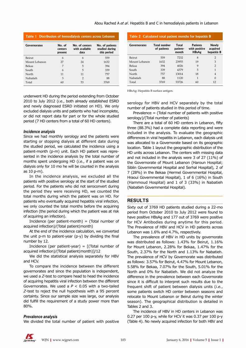
underwent HD during the period extending from October 2010 to July 2012 (i.e., both already established ESRD and newly diagnosed ESRD initiated on HD). We only excluded dialysis units and patients who had incomplete, or did not report data for part or for the whole studied period (7 HD centers from a total of 60 HD centers).
Incidence analysisSince we had monthly serology and the patients were starting or stopping dialysis at different date during the studied period, we calculated the incidence using a patient-month (p-m) unit. Each HD patient was repre-sented in the incidence analysis by the total number of months spent undergoing HD (i.e., if a patient was on dialysis only for 10 mo he will be counted in the analysis as 10 p-m).
In the incidence analysis, we excluded all the patients with positive serology at the start of the studied period. For the patients who did not seroconvert during the period they were receiving HD, we counted the total months during which the patient was on HD. For patients who eventually acquired hepatitis viral infection, we only counted the total months before the acquiring infection (the period during which the patient was at risk of acquiring an infection).
Incidence (per patient-month) = (Total number of acquired infection)/(Total patient|month)
At the end of the incidence calculation, we converted the unit p-m to patient-year (p-y) by dividing the final number by 12.
Incidence (per patient-year) = [(Total number of acquired infection)/(Total patient|month)]/12
We did the statistical analysis separately for HBV and HCV.
To compare the incidence between the different governorates and since the population is independent, we used a Z-test to compare head to head the incidence of acquiring hepatitis viral infection between the different Governorates. We used a P < 0.05 with a two-tailed Z-test to reject the null hypothesis with a 95 percent certainty. Since our sample size was large, our analysis did fulfill the requirement of a study power more than 80%.
Prevalence analysisWe divided the total number of patient with positive
serology for HBV and HCV separately by the total number of patients studied in this period of time.
Prevalence = (Total number of patients with positive serology)/(Total number of patients)
There are a total of 60 HD centers in Lebanon, fifty three (88.3%) had a complete data reporting and were included in the analysis. To evaluate the geographic differences in viral hepatitis in Lebanon, each dialysis unit was allocated to a Governorate based on its geographic location. Table 1 layout the geographic distribution of the HD units across Lebanon. The centers with missing data and not included in the analysis were 3 of 27 (11%) of the Governorate of Mount Lebanon (Haroun Hospital; Siblin Governmental Hospital and Serhal Hospital), 2 of 7 (28%) in the Bekaa (Hermel Governmental Hospital, Hraoui Governmental Hospital), 1 of 6 (16%) in South (Hammoud Hospital) and 1 of 3 (33%) in Nabatieh (Nabatieh Governmental Hospital).
RESULTSSixty out of 3769 HD patients studied during a 22-mo period from October 2010 to July 2012 were found to have positive HBsAg and 177 out of 3769 were positive for HCV Antibodies during anytime for this period. The Prevalence of HBV and HCV in HD patients across Lebanon was 1.6% and 4.7%, respectively.
The prevalence of HBV in HD units by governorate was distributed as follows: 1.43% for Beirut, 1.16% for Mount Lebanon, 2.28% for Bekaa, 1.47% for the South, 2.37% for the North and 1.13% for Nabatieh. The prevalence of HCV by Governorate was distributed as follows: 3.57% for Beirut, 4.47% for Mount Lebanon, 5.58% for Bekaa, 7.07% for the South, 5.01% for the North and 0% for Nabatieh. We did not analyze the difference in the prevalence between each Governorate since it is difficult to interpret such results due to the frequent shift of patient between dialysis units (i.e., some patients switch HD center between seasons and relocate to Mount Lebanon or Beirut during the winter season). The geographical distribution is detailed in Tables 2 and 3.
The incidence of HBV in HD centers in Lebanon was 0.27 per 100 p-y, while for HCV it was 0.37 per 100 p-y (Table 4). No newly acquired infection for both HBV and
103 January 6, 2016|Volume 5|Issue 1|WJN|www.wjgnet.com
Governorates No. of centers present
No. of centers with available
data
No. of patients studied during
this period
Beirut 6 6 559Mount Lebanon 27 24 1632Bekaa 7 5 394South 6 5 339North 11 11 757Nabatieh 3 2 88Total 60 53 3769
Table 1 Distribution of hemodialysis centers across Lebanon
Governorates Total number of patients
Total patient-month
Patients with positive
HBsAg
Newly acquired
hepatitis B
Beirut 559 7232 8 2Mount Lebanon 1632 23955 19 3Bekaa 394 4026 9 2South 339 4379 5 1North 757 13014 18 4Nabatieh 88 1120 1 0Total 3769 53726 60 12
Table 2 Calculated total patient months for hepatitis B
HBsAg: Hepatitis B surface antigen.
Abou Rached A et al . Hepatitis B and C in hemodialysis patients in Lebanon

randomly selected from 308 representative dialysis facilities in France, Germany, Italy, Japan, Spain, the United Kingdom, and United States, an overall HCV prevalence of 13% was found in 8615 patients[6].
Globally the prevalence of HCV among patients undergoing HD varies from as low as 6.1% in Germany in 2002[7] to as high as 76% in Casablanca in 2005[8]. In general, North Africa and the Middle East were cited as high prevalence areas, both in the general population and in HD patients, by the WHO in 1999[9]. Previous studies from the region have reported a prevalence of anti-HCV antibodies in HD patients of 50% in Saudi Arabia in 2000[10], 19.1% in Tunisia in 1994[11], 20.2% in Turkey in 2006[12] and 34.6% in Jordan in 2007[13].
Concerning the prevalence of HBV, Abourached et al[5] reported on 2007 a prevalence of 2.62% (0%-6.5%) of HBsAg in 17 HD centers in Lebanon, our study showed a decrease of the prevalence to 1.6%, ranging from 1.4% in Nabatieh and Bekaa, to 2.4% in the North. This prevalence is slightly elevated than that reported in different study In Lebanon concerning the general population[14]. We observed a high variability among the 53 different centers, ranging from as low as 0% to as high as 15%.This observed prevalence is lower than that reported in the 2008 by the Saudi Centre for Organ Transplantation (SCOT) report, where HBV seropositivity was 4.6% in the Saudi HD population while among Jordanian HD patients it was 5.9%[15]. It is also lower than that reported in HD patients in other regions including Europe (4.1%), Japan (2.2%) and the United States (2.4%) during the period extending from 1996 to 2002[16]. A study sample from the DOPPS, which included 8615 adult HD patients from 308 dialysis facilities in Western Europe and the United States, reported prevalence rates for HBV infection ranging from 0% to 6.6%[17]. Studies from less developed countries estimated that the proportion of HBsAg carriers in the HD population varies from 2% to 20%[18,19].
Prospective follow up of seronegative HD patients enabled us to observe 12 newly acquired infection for HBV and 16 newly acquired infection for HCV during a twenty two months period. We observed a 0.37 per 100 p-y incidence of new HCV infections during the 22-mo observation period. The reported incidence of new HCV infections varies considerably between countries. A rate
HCV were observed in Nabatieh.While comparing the incidence of HBV in HD units
between different governorates in Lebanon, no statis-tically significant difference was found (with a P-value always higher than 0.05) (Table 5). In Contrast, a statistically significant difference was found between the incidence of HCV in the South (1.47 per 100 p-y) compared to Mount Lebanon (0.21 per 100 p-y) and the North (0.19 per 100 p-y) showing a higher incidence in the South with a P-value of 0.00068 and 0.00374 respectively (Tables 5 and 6 for a list of the different calculated P-values).
DISCUSSIONIt is well known that HD patients are at high risk for HCV and HBV infections. In Lebanon three small studies were done concerning the prevalence of HCV in HD patients. Naman et al[4] reported in 1996 that the prevalence of HCV among HD in Lebanon was 27% with a high variety between the 5 centers studied (10%-39%), Abdelnour et al[3] reported in 1997 in various hospitals a prevalence of 16%, Abourached et al[5] reported in 2006, a prevalence of 13% (2.2%-38%) in 17 HD centers in Lebanon.
In this epidemiologic study covering more than 88% of the HD centers in Lebanon, the prevalence of anti-HCV antibodies in ESRD patients undergoing HD was 4.7%, showing a decrease in the prevalence of HCV among HD patients in Lebanon over the last two decades. The lowest prevalence was in Beirut (3.5%) and the highest in the South (7%). We observed a high variability among the 53 different centers studied, ranging from as low as 0% to as high as 20%.
The reduction in HCV prevalence in HD patients is a common trend across several countries and it was mainly related to the reduction in the number of transfusions in HD patients and the improvement of the laboratory screening techniques for detection of anti-HCV antibodies in blood donors. The prevalence of HCV infection in patients on HD is highly variable but clearly much higher than in the general population of the respective countries. In phase one of the Dialysis Outcomes and Practice Patterns Study (DOPPS), a prospective observational study of adult HD patients
104 January 6, 2016|Volume 5|Issue 1|WJN|www.wjgnet.com
Governorates Total number of patients
Total patient-month
Patients with positive HCV
Abs
Newly acquired
hepatitis C
Beirut 559 7055 20 3Mount Lebanon 1632 22970 73 4Bekaa 394 3797 22 2South 339 4078 24 5North 757 12597 38 2Nabatieh 88 1142 0 0Total 3769 51639 177 16
Table 3 Total patient-months distribution for hepatitis C
HCV: Hepatitis C virus.
Governorates Incidence of HBV (per 100 p-y)
Incidence of HCV (per 100 p-y)
Beirut 0.33 0.51Mount Lebanon 0.15 0.21Bekaa 0.59 0.63South 0.27 1.47North 0.37 0.19Nabatieh 0 0Across Lebanon 0.27 0.37
Table 4 Incidence of hepatitis B and hepatitis C among hemodialysis centers in Lebanon
HCV: Hepatitis C virus; HBV: Hepatitis B virus.
Abou Rached A et al . Hepatitis B and C in hemodialysis patients in Lebanon

as low as 0.4% was observed in France from 1997 to 2000[20] but higher rates have been reported from the Mediterranean region. According to the 2008 SCOT report, the annual rate of HCV sero-conversion in Saudi HD patients was 7%-9% while in Jordan it was 2.6%[14]. The incidence rate of 0.27 per 100 p-y for HBsAg is slightly less than that reported in Europe, Japan and the United States (0.4-1.8 per 100 p-y)[17]. While comparing the incidence of HBV infection among the different Governorates, no statistically significant difference was found. In contrast, a statistically significant difference in the incidence of HCV infection was found between the South and both Mount Lebanon and the North. This interestingly high incidence of HCV infection of 1.47 per 100 p-y found in the South need to be assessed further to be able to find a potential reason for this higher incidence.
In this study we collected data from 53 centers distributed across all the six Governorates of Lebanon from the total 60 centers reporting monthly serology to the MOPH, giving us a total of 3769 patients studied over a 22-mo period. This significantly increased the statistical power and the validity of the results.
In general, the prevalence and incidence of HBV and HCV infections in HD patients are directly related to the prevalence of these infections in the general population, the quality of healthcare services in a community and
the standards of infection control practices in HD units. In Lebanon, patients on maintenance HD were found to have a higher prevalence of HCV infection of 4.7% when compared to the general population since, according to available data, anti-HCV prevalence rate of 0.2% to 0.4% of the general population in Lebanon[21], but we noted a significant decrease of this prevalence in this group of patients during the last 20 years. Also the prevalence of HBV was slightly higher than the general population 2.62% vs 2.2%.
This higher prevalence of HBV and HCV infection in HD compared to the general population has been confirmed by several reports from different countries[22].
The incidence of HBV and HCV in HD centers in Lebanon is very low compared to others countries in the region, this can be due to the good applications of the standards of infection control practices in HD centers and the strict surveillance by the MOPH.
A notable result of this study was the significantly higher incidence of HCV found mainly in the South that may be due to variation in the degree of implementation of the universal precautions to prevent nosocomial transmission. In order to evaluate the reason for the variation between the different HD units, a more detailed evaluation of each dialysis patient should be done especially in the units with the lowest and the highest incidence for HBV or HCV. Such studies would assist in
105 January 6, 2016|Volume 5|Issue 1|WJN|www.wjgnet.com
HBV Beirut Mount Lebanon Bekaa South North Nabatieh
Beirut P = 0.3759 P = 0.5562 P = 0.8702 P = 0.8948 P = 0.579NS NS NS NS
Mount Lebanon P = 0.1059 P = 0.6073 P = 0.2208 P = 0.7086NS NS NS NS
Bekaa P = 0.5038 P = 0.5709 P = 0.4578NS NS NS
South P = 0.778 P = 0.6159NS NS
North P = 0.5547NS
Nabatieh
Table 5 P -values for hepatitis B
NS: Non-significant; HBV: Hepatitis B virus.
HCV Beirut Mount Lebanon Bekaa South North Nabatieh
Beirut P = 0.229 P = 0.8164 P = 0.1274 P = 0.2598 P = 0.4854NS NS NS NS NS
Mount Lebanon P = 0.1822 P = 0.00068 P = 0.9079 P = 0.6548NS NS NS
Bekaa P = 0.2951 P = 0.2036 P = 0.438NS NS NS
South P = 0.00374 P = 0.2346NS
North P = 0.6707NS
Nabatieh
Table 6 P -values for hepatitis C
NS: Nonsignificant; HCV: Hepatitis C virus.
Abou Rached A et al . Hepatitis B and C in hemodialysis patients in Lebanon

guiding interventions aiming to reduce the occurrence of these infections and thus reduce the morbidity and mortality of the HD population in Lebanon.
Finally this study demonstrated a reduce in the prevalence of HBV and HCV infections in HD centers during the last 2 decades and a low incidence rate due to the good applications of the standards of infection controls practice.
COMMENTSBackgroundEnd stage renal disease (ESRD) patients on hemodialysis (HD) are particularly at higher risk for acquiring hepatitis C virus (HCV) and hepatitis B virus (HBV) than the general population, due to the sharing of contaminated machines within the same center and the higher rates of blood transfusions. Such infections have a negative impact on the clinical course of ESRD causing higher rates of morbidity and mortality. Since then, it is essential to determine the prevalence and the incidence of HBV and HCV infections in HD patients in each country, then to decide accordingly about further interventions to control such infections.
Research frontiersThe prevalence of HCV and HBV in patients on HD in Lebanon was only addressed by small studies including few HD centers. This study will determine the prevalence and incidence of HCV and HBV in a significantly larger population including 3769 HD patients through 88% of all HD centers across Lebanon between October 2010 and July 2012.
Innovations and breakthroughsPrevious studies done on smaller sample sizes limited to some Lebanese HD centers reported a prevalence of 2.6% and 13%-27%, for HBV and HCV, respectively. No reports are yet available about their incidence rate. Although accurate comparison could not be done due to the different sample sizes, this study showed a reduction in HBV and HCV prevalence through HD patients in Lebanon (1.6% and 4.7%, respectively). On the other hand, their calculated prevalence and incidence rates were found to be among the lowest values reported in other countries, either in the Middle East, or in Europe and United States. Another notable result of this study was the significantly higher incidence rate of HCV in the Southern Lebanese HD centers.
ApplicationsThis study reflects an appropriate adherence to standards of infection control in the Lebanese HD centers limiting the spread of HBV and HCV between HD patients. However, it emphasizes the need for further investigations to reestablish those standards in some centers having significantly higher incidence rate of HCV, located mainly in the South of Lebanon.
Peer-reviewIn this manuscript, the authors report on the prevalence and incidence of hepatitis B and hepatitis C among HD patients in Lebanon. This paper is clinically interesting.
REFERENCES1 Ribot S, Rothstein M, Goldblat M, Grasso M. Duration of hepatitis
B surface antigenemia (HBs Ag) in hemodialysis patients. Arch Intern Med 1979; 139: 178-180 [PMID: 434972 DOI: 10.1001/archinte.1979.03630390036015]
2 Kato A, Takita T, Furuhashi M, Fujimoto T, Suzuki H, Maruyama Y, Sakao Y, Miyajima H. Association of HCV core antigen seropositivity with long-term mortality in patients on regular hemo-dialysis. Nephron Extra 2012; 2: 76-86 [PMID: 22619670 DOI: 10.1159/000337333]
3 Abdelnour GE, Matar GM, Sharara HM, Abdelnoor AM.
Detection of anti-hepatitis C-virus antibodies and hepatitis C-virus RNA in Lebanese hemodialysis patients. Eur J Epidemiol 1997; 13: 863-867 [PMID: 9476813 DOI: 10.1023/A:1007468322940]
4 Naman RE, Mansour I, Klayme S, Khalil G. [Hepatitis C virus in hemodialysis patients and blood donors in Lebanon]. J Med Liban 1996; 44: 4-9 [PMID: 8965318]
5 Abourached A, Saniour P, Bou Jaoude J. prevalence des hepatities B et C chez les donneurs de sang, les hémodialysés et les toxico-manes au Liban. Available from: URL: http://www.snfge.org/content/P12. 2007
6 Fissell RB, Bragg-Gresham JL, Woods JD, Jadoul M, Gillespie B, Hedderwick SA, Rayner HC, Greenwood RN, Akiba T, Young EW. Patterns of hepatitis C prevalence and seroconversion in hemodialysis units from three continents: the DOPPS. Kidney Int 2004; 65: 2335-2342 [PMID: 15149347 DOI: 10.1111/j.1523-1755.2004.00649.x]
7 Hinrichsen H, Leimenstoll G, Stegen G, Schrader H, Fölsch UR, Schmidt WE. Prevalence and risk factors of hepatitis C virus infection in haemodialysis patients: a multicentre study in 2796 patients. Gut 2002; 51: 429-433 [PMID: 12171969 DOI: 10.1136/gut.51.3.429]
8 Boulaajaj K, Elomari Y, Elmaliki B, Madkouri B, Zaid D, Benchemsi N. [Prevalence of hepatitis C, hepatitis B and HIV infection among haemodialysis patients in Ibn-Rochd university hospital, Casablanca]. Nephrol Ther 2005; 1: 274-284 [PMID: 16895696 DOI: 10.1016/j.nephro.2005.06.012]
9 Global surveillance and control of hepatitis C. Report of a WHO Consultation organized in collaboration with the Viral Hepatitis Prevention Board, Antwerp, Belgium. J Viral Hepat 1999; 6: 35-47 [PMID: 10847128 DOI: 10.1046/j.1365-2893.1999.6120139.x]
10 Souqiyyeh MZ, Al-Attar MB, Zakaria H, Shaheen FA. Dialysis centers in the kingdom of saudi arabia. Saudi J Kidney Dis Transpl 2001; 12: 293-304 [PMID: 18209376]
11 Jemni S, Ikbel K, Kortas M, Mahjoub J, Ghachem L, Bidet JM, Boukef K. Seropositivity to hepatitis C virus in Tunisian haemo-dialysis patients. Nouv Rev Fr Hematol 1994; 36: 349-351 [PMID: 7534398]
12 Yakaryilmaz F, Gurbuz OA, Guliter S, Mert A, Songur Y, Karakan T, Keles H. Prevalence of occult hepatitis B and hepatitis C virus infections in Turkish hemodialysis patients. Ren Fail 2006; 28: 729-735 [PMID: 17162434 DOI: 10.1080/08860220600925602]
13 Nabulsi MM, El Saleeby CM, Araj GF. The current status of hepatitis B in Lebanon. J Med Liban 2003; 51: 64-70 [PMID: 15298159]
14 Batieha A, Abdallah S, Maghaireh M, Awad Z, Al-Akash N, Batieneh A, Ajlouni KA. Epidemiology and cost of haemodialysis in Jordan. East Mediterr Health J 2007; 13: 654-663 [PMID: 17687839]
15 Al Hijazat M, Ajlouni YM. Hepatitis B infection among patients receiving chronic hemodialysis at the Royal Medical Services in Jordan. Saudi J Kidney Dis Transpl 2008; 19: 260-267 [PMID: 18310881]
16 Goodkin DA, Young EW, Kurokawa K, Prütz KG, Levin NW. Mortality among hemodialysis patients in Europe, Japan, and the United States: case-mix effects. Am J Kidney Dis 2004; 44: 16-21 [PMID: 15486869 DOI: 10.1016/S0272-6386(04)01100-X]
17 Burdick RA, Bragg-Gresham JL, Woods JD, Hedderwick SA, Kurokawa K, Combe C, Saito A, LaBrecque J, Port FK, Young EW. Patterns of hepatitis B prevalence and seroconversion in hemodialysis units from three continents: the DOPPS. Kidney Int 2003; 63: 2222-2229 [PMID: 12753311 DOI: 10.1046/j.1523-1755.2003.00017.x]
18 Ferreira RC, Teles SA, Dias MA, Tavares VR, Silva SA, Gomes SA, Yoshida CF, Martins RM. Hepatitis B virus infection profile in hemodialysis patients in Central Brazil: prevalence, risk factors, and genotypes. Mem Inst Oswaldo Cruz 2006; 101: 689-692 [PMID: 17072485 DOI: 10.1590/S0074-02762006000600019]
19 Telaku S, Fejza H, Elezi Y, Bicaj T. Hepatitis B and C in dialysis units in Kosova. Virol J 2009; 6: 72 [PMID: 19497116 DOI: 10.1186/1743-422X-6-72]
20 Izopet J, Sandres-Sauné K, Kamar N, Salama G, Dubois M,
106 January 6, 2016|Volume 5|Issue 1|WJN|www.wjgnet.com
COMMENTS
Abou Rached A et al . Hepatitis B and C in hemodialysis patients in Lebanon

Pasquier C, Rostaing L. Incidence of HCV infection in French hemodialysis units: a prospective study. J Med Virol 2005; 77: 70-76 [PMID: 16032714 DOI: 10.1002/jmv.20415]
21 Araj GF, Kfoury-Baz EE, Barada KA, Nassif RE, Alami SY. Hepatitis C virus: prevalence in Lebanese blood donors and brief
overview of the disease. J Med Liban 1995; 43: 11-16 [PMID: 8676356]
22 Fabrizi F, Poordad FF, Martin P. Hepatitis C infection and the patient with end-stage renal disease. Hepatology 2002; 36: 3-10 [PMID: 12085342 DOI: 10.1053/jhep.2002.34613]
P- Reviewer: Kelesidis T, Lazarevic I, Ohashi N, Yorioka N S- Editor: Ji FF L- Editor: A E- Editor: Jiao XK
107 January 6, 2016|Volume 5|Issue 1|WJN|www.wjgnet.com
Abou Rached A et al . Hepatitis B and C in hemodialysis patients in Lebanon

Renal and perinephric abscesses in West China Hospital: 10-year retrospective-descriptive study
Xiao-Qin Liu, Cheng-Cheng Wang, Yan-Bin Liu, Kai Liu
Xiao-Qin Liu, Cheng-Cheng Wang, Yan-Bin Liu, Kai Liu, Center of Infectious Diseases, West China Hospital of Sichuan University, Chengdu 610041, Sichuan Province, China
Author contributions: Liu XQ analyzed the data and wrote the paper; Wang CC collected the data; Liu YB and Liu K designed the research.
Institutional review board statement: This study was reviewed and approved by West China Hospital Sichuan University Clinical Trials and Biomedical Ethics Committee.
Informed consent statement: This analysis used anonymous clinical data that were obtained from patients who provided informed written consent before treatment.
Conflict-of-interest statement: The authors declare that there is not conflict of interest related to this study.
Data sharing statement: No additional data are available.
Open-Access: This article is an open-access article which was selected by an in-house editor and fully peer-reviewed by external reviewers. It is distributed in accordance with the Creative Commons Attribution Non Commercial (CC BY-NC 4.0) license, which permits others to distribute, remix, adapt, build upon this work non-commercially, and license their derivative works on different terms, provided the original work is properly cited and the use is non-commercial. See: http://creativecommons.org/licenses/by-nc/4.0/
Correspondence to: Dr. Kai Liu, Center of Infectious Diseases, West China Hospital of Sichuan University, 37 Guoxuexiang, Wuhou District, Chengdu 610041, Sichuan Province, China. [email protected] Telephone: +86-28-85423597Fax: +86-28-85423597
Received: June 29, 2015 Peer-review started: July 4, 2015 First decision: September 17, 2015 Revised: September 28, 2015 Accepted: November 23, 2015Article in press: November 25, 2015Published online: January 6, 2016
AbstractAIM: To elucidate the clinical, radiological and laboratory profiles of renal abscess (RA) and perinephric abscess (PNA), along with related treatment and outcome.
METHODS: Ninety-eight patients diagnosed with RA or PNA using the primary discharge diagnoses identified from the International Statistical Classification of Diseases and Related Health Problems Tenth Edition (ICD-10) codes (RA: N15.101, PNA: N15.102) between September 2004 and December 2014 in West China Hospital were selected. Medical records including patients’ characteristics, symptoms and signs, high-risk factors, radiological features, causative microorganisms and antibiotic-resistance profiles, treatment approaches, and clinical outcomes were collected and analyzed.
RESULTS: The mean age of the patients was 46.49 years with a male to female ratio of 41:57. Lumbar pain (76.5%) and fever (53.1%) were the most common symptoms. Other symptoms and signs included chills (28.6%), anorexia and vomiting (25.5%), lethargy (10.2%), abdominal pain (11.2%), flank mass (12.2%), flank fistula (2.0%), gross hematuria (7.1%), frequency (14.3%), dysuria (9.2%), pyuria (5.1%) and weight loss (1.0%). Painful percussion of the costovertebral angle (87.8%) was the most common physical finding. The main predisposing factors were lithiasis (48.0%), diabetes mellitus (33.7%) followed by history of urological surgery (16.3%), urinary tract infections (14.3%), renal function impairment (13.3%), liver cirrhosis (2.0%), neurogenic bladder (1.0%), renal cyst (1.0%), hydronephrosis (1.0%), chronic hepatitis B (1.0%), post-discectomy (1.0%) and post-colectomy (1.0%). Ultrasound (US) and computed tomography were the most valuable diagnostic tools and US was recommended as the initial diagnostic imaging choice. Escherichia coli (51.4%), Staphylococcus aureus (10.0%) and Klebsiella pneumoniae (8.6%) were the main causative microorganisms. Intravenous antibiotic
108 January 6, 2016|Volume 5|Issue 1|WJN|www.wjgnet.com
World Journal of NephrologyW J N
Submit a Manuscript: http://www.wjgnet.com/esps/Help Desk: http://www.wjgnet.com/esps/helpdesk.aspxDOI: 10.5527/wjn.v5.i1.108
World J Nephrol 2016 January 6; 5(1): 108-114ISSN 2220-6124 (online)
© 2016 Baishideng Publishing Group Inc. All rights reserved.
Retrospective Study
ORIGINAL ARTICLE

therapy was necessary while intervention including surgical and nonsurgical approaches were reserved for larger abscesses, multiple abscesses, PNAs and non-responders.
CONCLUSION: Heightened alertness, prompt diagnosis, and especially proper antibiotics in conjunction with interventional approaches allow a promising clinical outcome of renal and perinephric abscesses.
Key words: Renal abscess; Causative pathogens; Perinephric abscess; Diagnosis; Antibiotic resistance; Interventional treatment; Conservative treatment
© The Author(s) 2016. Published by Baishideng Publishing Group Inc. All rights reserved.
Core tip: Renal and perinephric abscesses are uncom-mon but potentially lethal infectious diseases and the case-fatality rates most frequently cited in previous studies are still high. However, the previous case-fatality rates need to be updated, since prompt diagnosis and appropriate therapeutic strategies have contributed to lower mortality. This article reports the characteristics of patients identified with renal or perinephric abscesses and shares the management experience and outcome in West China Hospital during the last decade.
Liu XQ, Wang CC, Liu YB, Liu K. Renal and perinephric abscesses in West China Hospital: 10-year retrospective-descriptive study. World J Nephrol 2016; 5(1): 108-114 Available from: URL: http://www.wjgnet.com/2220-6124/full/v5/i1/108.htm DOI: http://dx.doi.org/10.5527/wjn.v5.i1.108
INTRODUCTIONRenal abscess (RA) is defined as encapsulated pus confined to the renal parenchyma and is further divided into renal cortical or corticomedullary abscess[1]. Perinephric abscess (PNA) is a collection of suppurative material located between Gerota’s fascia and the renal capsule[2]. Complications of urinary tract infections (UTIs) and hematogenous seeding from primary infected sites are the common source of infection[2,3]. Additionally, rupture of renal cortical abscess or renal carbuncle can result in the formation of PNA[2].
As a result of its anatomical location and potential to spread, RA is potentially lethal and the prognosis can be poor, especially in immunosuppressed and cachectic patients[1,4]. PNA originates from hematogenous dissemination, and often has an acute presentation with pain and high spiking temperatures[2], while in most cases, PNA is notoriously silent clinically[2,3], thereby the diagnosis can be challenging[2]. It is reported that only 35%38% of patients with PNA are correctly diagnosed at the time of admission[5,6]. The mortality rates of RA and PNA in recent series are reported to range from 1% to 14%[3,712], while complicated abscess may carry
a higher mortality[1]. Due to the above situation, a retrospectivedescriptive study was conducted with 98 relevant cases identified with RA and PNA between September 2004 and December 2014 in West China Hospital, in an attempt to recognize the disease and describe our experience with it over the past 10 years.
MATERIALS AND METHODSThe data presented in this study were obtained from medical records of patients selected using the primary discharge diagnoses identified from the International Statistical Classification of Diseases and Related Health Problems Tenth Edition (ICD10) codes (RA: N15.101, PNA: N15.102) during the last decade in our hospital. Suspected patients were diagnosed based on both clinical and radiological criteria. Abscesses ≤ 3 cm were defined as small, medium 35 cm, and > 5 cm large. The rule that culture findings guide selection of an antibiotic regimen was firmly followed, while before the culture results were obtained, initial empirical antibiotics were provided once a clinical diagnosis of RA and PNA was made. Antibiotics typically included piperacillin plus amikacin and metronidazole, piperacillin/tazobactam or thirdgeneration cephalosporin plus metronidazole, or quinolones plus metronidazole. When patients had a severe condition such as sepsis or were prone to infection with extendedspectrum βlactamase (ESBL)producing organisms, carbapenem antibiotics (e.g., imipenem/cilastatin) were also included in prescription.
Treatment modes were subdivided into two groups: Conservative treatment and interventional treatment. The latter comprised five categories: Antibiotics plus percutaneous drainage; antibiotics plus double J tube insertion; antibiotics plus nephrostomy; antibiotics plus surgical drainage; and antibiotics plus nephrectomy. Since improvement in clinical manifestations usually precedes that in radiological imaging findings, patients were mainly assessed by their clinical improvement. The clinical outcome was classified as cure, clinical improvement (mainly including remission or disappearance of initial symptoms, shrinkage of the abscess cavity upon imaging, recovery of white blood cell and neutrophil counts, and negative results for blood and urine culture), or death.
BiostatisticsThe statistical methods of this study were reviewed by Liang Huang from Center of Infectious Diseases, West China Hospital of Sichuan University, Chengdu, Sichuan Province, China.
RESULTSPatient characteristicsAmong the 98 patients, there were 41 (41.8%) men and 57 (58.2%) women. The age ranged from 18 to 75 years with a mean of 46.49 ± 15.07 years. RA was observed in 68 (69.4%) patients and PNA in 30 (30.6%).
Liu XQ et al . Renal and perinephric abscesses
109 January 6, 2016|Volume 5|Issue 1|WJN|www.wjgnet.com

Fifteen patients (15.3%) had no identifiable systemic or urological disorder that might have been involved in abscess formation, whereas for other patients, the spectrum of predisposing factors remained consistent with conventional predisposing factors: Diabetes mellitus (n = 33, 33.7%); lithiasis (n = 47, 48.0%) which included renal calculi (n = 32, 32.7%), ureteric calculi (n = 5, 5.1%), renal and ureteric calculi (n = 10, 10.2%); history of urological surgery (n = 16, 16.3%); UTIs (n = 14, 14.3%); renal function impairment (n = 13, 13.3%); liver cirrhosis (n = 2, 2.0%); neurogenic bladder (n = 1, 1.0%); and other diseases (n = 5, 5.1%) including one each with renal cyst, hydronephrosis, and chronic hepatitis B, postdiscectomy and postcolectomy.
The most common initial symptoms were lumbar pain (n = 75, 76.5%) and fever (n = 52, 53.1%). Each grade of fever was observed: 38 ℃39 ℃ in approximately 31.6%, 39.1 ℃41 ℃ in approximately 20.4%, and absent or lowgrade fever in 48.0%. Painful percussion of the costovertebral angle (n = 86, 87.8%) was the most common physical finding (Table 1). Patients with RA in this study were more inclined to experience lethargy than PNA (P < 0.05) and there was no statistical significance in other symptoms when compared RA with PNA (P > 0.05).
Laboratory data and abscess characteristicsThere was no significant difference between RA and PNA in white blood cell count (W = 996.5, P > 0.05), neutrophil count (W = 947, P > 0.05), hemoglobin (W = 0.9773, P > 0.05), blood urea nitrogen (W = 992, P > 0.05) and serum creatinine (W = 1038, P > 0.05). Hematuria and leukocyturia were most common findings in urine test (Table 2). Of the 98 patients, 77 (78.6%) patients had a solitary abscess and 10 (10.2%) had multiple abscesses. The right side (55.1%) remained the predominant anatomical site and bilateral abscesses were found in two (2.0%) cases. The average size of
RA and PNA was 6.25(range: 0.5017.00)cm and 8.35 (range: 4.5020.00) cm, respectively. The average size of abscess was 4.00 (range: 1.8010.50) cm in the conservative group, and 7.65 (range: 0.5020.00) cm in the interventional group (Table 3).
Microbiological dataThe results of blood, abscess and urine culture were available for 92, 54 and 91 patients, respectively. Blood and urine cultures were positive in 13 (14.1%) and 23 (25.3%) patients, respectively, and pathogenic organisms were isolated from pus in 33 (61.1%) cases. Of all the positive cultures (n = 69), the most frequently isolated pathogen was Escherichia coli (n = 35, 50.7%) followed by Staphylococcus aureus (S. aureus) (n = 7, 10.1%), Klebsiella pneumoniae (K. pneumoniae) (n = 6, 8.7%), Pseudomonas aeruginosa (n = 3, 4.3%), Candida spp. (n = 7, 10.1%), Enterobacteriaceae (n = 6, 8.7%), Entercoccus faecium (n = 2, 2.9%), Enterococcus faecalis (n = 1, 1.4%) and Aspergillus spp. (n = 2, 2.9%). E. coli was more frequently found in patients with RA than those with PNA (χ2 = 6.832, P < 0.01), while there was no significant difference in the distribution of K. pneumoniae (P > 0.05), S. aureus (P > 0.05) and Candida spp. (P > 0.05) (Table 4). We detected ESBL in the isolated strains of E. coli in 12 (17.4%) cases and K. pneumoniae in one (1.4%) case. We analyzed the antibiotic resistance of E. coli, S. aureus and K. pneumoniae isolated from blood, abscess and urine culture (Table 5).
Imaging studiesImaging results were available for 97 patients. Ultrasound (US) was applied in 80 cases and alone in 31 (31.6%) cases. Computed tomography (CT) was performed in 63 cases and alone in 16 (16.3%) cases. Magnetic resonance imaging (MRI) was applied in three cases and alone in one (1.0%) case. The imaging results are shown in Table 6.
110 January 6, 2016|Volume 5|Issue 1|WJN|www.wjgnet.com
Variables ValueTotal (n = 98)
Lumbar pain 75 (76.5) Fever 52 (53.1)Chills 28 (28.6)Anorexia and vomiting 25 (25.5)Lethargy 10 (10.2) Abdominal pain 11 (11.2) Flank mass 12 (12.2) Flank fistula 2 (2.0) Gross hematuria 7 (7.1)Frequency 14 (14.3)Dysuria 9 (9.2)Pyuria 5 (5.1)Loss of weight 1 (1.0)Painful percussion of the CVA 86 (87.8)
Table 1 Characteristics of patients with renal or perinephric abscesses n (%)
CVA: Costovertebral angle.
Variables Value
RA PNABlood analysis 64 30WBC (109/L) 10.82 (range: 2.42-29.95) 12.40 (range: 2.68-25.45)NEUT (%) 81.00 (range: 48.30-96.00) 79.00 (range: 49.30-94.70)HGB (g/L) 105.75 ± 22.52 101.66 ± 20.13BUN (mmol/L) 5.26 (range: 1.10-20.30) 5.10 (range: 2.80-23.18)Serum creatinine (umol/L)
82.00 (range: 25.10-346.00) 86.60 (range: 49.00-560.0)
Urine analysis 64 30No finding (%) 13 (13.3) 3 (3.1)Hematuria (%) 47 (48.0) 23 (23.5)Pyuria (%) 16 (16.3) 8 (8.2)Proteinuria (%) 32 (32.7) 16 (16.3)Leukocyturia (%) 41 (41.8) 23 (23.5)
Table 2 Blood and urine analysis
RA: Renal abscess; PNA: Perinephric abscess; WBC: White blood cell; NEUT: Neutrophil count; HGB: Hemoglobin; BUN: Blood urea nitrogen.
Liu XQ et al . Renal and perinephric abscesses

women was noted, while in previous studies, the male: Female ratio was reported as 1:31:7[7,18,20], and a female predominance as high as 91.8% was also observed[16].
Diagnosis of RA or PNA remains challenging because the symptoms can be insidious and obscure[1,2]. Patients with RA may present with fever, chills, flank or abdominal pain, fatigue, nausea, decreased appetite, weight loss and even persistent hiccups[7,21,22]. In our study, fever was not always accompanied by chills and the high percentage of absent/lowgrade fever might be explained by prior antibiotic therapy. Patients with PNA often present with anorexia, nausea and vomiting, flank
Treatment and outcomeThe average hospitalization duration was 17 d (range 592 d). Of the 98 patients, 23 (23.5%) received conservative treatment and 75 (76.5%) received an interventional procedure. Fiftyseven (58.2%) patients were cured, 40 (40.8%) showed clinical improvement by the time of hospital discharge, and one (1.0%) died of multiple organ dysfunction syndrome. Interventional treatment contributed to a better clinical outcome than conservative treatment (Z = 3.897, P < 0.01). The outcome tended to be better in patients with RA than in those with PNA irrespective of the therapeutic mode (Z = 8.027, P < 0.01) (Table 3).
DISCUSSION RA refers to a collection of purulent material within the kidney[13]. PNA represents an extensive infection in the perinephric space[2]. It is reported that approximately 30% of PNAs come from hematogenous dissemination[2], whereas in most cases, they result from rupture of RA[12,14]. Previous data show that > 80% of PNAs occur secondary to renal tract calculi with ascending UTIs[15].
RAs and PNAs are seen in all age groups and those aged 42.371.62 years were previously reported to be the dominant population[1012,1619]. A similar result was found in the present study. A slight predominance in
111 January 6, 2016|Volume 5|Issue 1|WJN|www.wjgnet.com
Variables Abscess size (cm) Hospital stay (d)
No. of Patients (RA/PNA)
Cure (RA/PNA ) Clinical improvement (RA/PNA )
Death (RA/PNA)
Ab 4.00 (range: 1.80-10.50) 20.7 23 (19/4) 2 (2/0) 21 (17/4) 0 (0/0)Intervention 7.65 (range: 0.50-20.00) 15.9 75 (49/26) 54 (38/16) 20 (10/10) 1 (1/0)Ab + PCD 8 3 4 1Ab + pigtails 2 1 1 0Ab + nephrostomy 4 0 4 0Ab + SD 29 21 8 0Ab + NC 32 29 3 0RA 6.25 (range: 0.50-17.00) 68 40 27 1PNA 8.35 (range: 4.50-20.00) 30 16 14 0
Table 3 Treatment and outcome
Ab: Antibiotic; PCD: Percutaneous drainage; SD: Surgical drainage; NC: Nephrectomy.
Variables Culture Blood Pus Urine
(Total = 92) (Total = 54) (Total = 91)No finding 79 21 68Escherichia coli 8 17 10Staphylococcus aureus 2 4 1Klebsiella pneumoniaee 1 3 2Pseudomonas aeruginosa 1 1 1Other Enterobacteriaceae 1 3 2Entercoccus faecium 0 0 2Enterococcus faecalis 0 1 0Candida 0 2 5Aspergillus 0 2 0
Table 4 Causative microorganisms isolated from blood, abscess and urine culture
Results Imaging tool US CT MRI
(Total = 80) (Total = 63) (Total = 3)Negative (%) 2 (2.0) 1 (1.0)Abscess (%) 23 (23.5) 37 (37.8) 1 (1.0)Hydronephrosis (%) 26 (26.5) 8 (8.2)Mass (%) 13 (13.3) 11 (11.2) 2 (2.0)Echogenic alteration (%) 11 (11.2)Cyst (%) 4 (4.1) 4 (4.1)Hematoma (%) 1 (1.0) 2 (2.0)
Table 6 Imaging results
US: Ultrasound; CT: Computerized tomography; MRI: Magnetic resonance imaging.
ST: Trimethoprim-sulfamethoxazole.
Variables Causative pathogens isolatedEscherichia
coli(n = 35)
Klebsiellapneumoniae
(n = 6)
Staphylococcusaureus
(n = 7)
Penicillin (%) 85.0 100.0 62.5Levofloxacin (%) 46.2 0 50.0Gentamicin (%) 53.3 0 50.0Amikacin (%) 5.0 0 -Cefotaxime (%) 61.1 50.0 50.0Ceftriaxone (%) 63.2 50.0 -Imipenem/cilastatin (%) 0 0 60.0Vancomycin (%) - - 0ST (%) 70.0 0 37.5
Table 5 The antibiotic resistance rate of causative pathogens isolated
Liu XQ et al . Renal and perinephric abscesses

pain, flank mass, signs of sepsis, weight loss, fistula formation and urinary tract complaints[2,4,15,22]. However, patients with RA were more likely subjected to lethargy in this study.
Consistent with previous studies, lithiasis (48.0%) and diabetes mellitus (33.7%) remained the predominant risk factors in the present study[3,8,9,12,16,22]. Diabetes mellitus accounts for 33.3%62.5% of all PNAs[2,7,23] 43.5%47% of RAs[7,16,17] and 28%50% of RAs and PNAs[3,7,10,11]. Anatomical malformation of the urinary tract, vesicoureteral reflux and obstructive tumors in renal polycystic disease are other previously described risk factors[1,18]. There was no significant difference between RA and PNA in white blood cell count, neutrophil count, hemoglobin, blood urea nitrogen and serum creatinine, which suggested that patients with RA and PNA shared similar inflammation reaction level and risk of renal impairment in this study.
US, CT and MRI were necessary to establish reliable preoperative diagnosis. US as the initial and classical imaging modality is utilized to measure renal size, discern focal lesions, and detect the true nature of a fluidcontaining mass and obstruction of the collecting system. US is not affected by poor renal function or allergy to contrast material[24,25]. The accuracy of US in the diagnosis of RA is reported to be 70%93%[3,23]
with sensitivity and specificity of 78.2% and 88.8%, respectively[23].
CT has been documented to diagnose RA or PNA with an accuracy of 92%96.4%[3,6], with specificity of 88%[23]. In our study, the accuracy of US and CT was 23.7% and 38.1%, respectively. However, when we combined the imaging results with clinical and laboratory data, the final diagnostic accuracy was 52.0%, and the average duration between admission and diagnosis was 2.16 d. With its convenience, accuracy, availability and low cost, US has made a major contribution to accurate and early diagnosis at our unit.
Since ascending dissemination of UTI has surpassed hematogenous dissemination and become the dominant predisposing factor[6,18], Gramnegative bacteria, especially E. coli, Proteus spp. and K. pneumoniae have been the most common pathogens in recent years[3,10,16,18]. In this study, E. coli was most frequently isolated from patients with RA than those with PNA. Polymicrobial abscesses have been increasingly frequently observed, ranging from 19.2% to 33.3% in incidence[7,9]. Two (2.0%) polymicrobial cases were found in the present study. There has been an increasing incidence of abscesses caused by fungi, especially Candida, particularly in immunosuppressed patients[7,12,18], and similar cases were observed in this study.
The selection of antimicrobial therapy ideally should be based on culture findings, however, there is an inevitable delay in obtaining results[2]. We recommend that empirical broadspectrum intravenous antibiotics should be initiated for critically ill patients after admission. Once the blood or abscess fluid cultures and bacterial isolation tests are confirmed, targeted antibiotic
regimens should be prescribed accordingly.There is a consensus that small RAs may resolve
with antibiotic treatment alone, and percutaneous or surgical drainage may be suitable for large RAs and PNAs. However there is a continuing argument about the proper treatment of middlesized abscesses[3,5,7,16].
Although the option of conservative management of RAs and PNAs seems attractive and feasible and successful cases have been reported[4,16], those cases were selected and limitations in size, location and number of abscesses were obvious and in Iwamoto’s case, the patient had received percutaneous drainage prior to conservative treatment[4].
In the present study, interventional approaches helped to detect the cause of disease and confirm the diagnosis, and culture of pus/aspirate/debris helped guide selection of an antibiotic regimen. The diagnostic and therapeutic value of percutaneous drainage has been confirmed since early years[26]. The application of interventional procedures contributed to a lower casefatality rate and lower risk for intensive care unit (ICU) admission[6,9]. Patients subjected to interventional treatment achieved a better clinical outcome than those received conservative treatment and the cure rate of interventional treatment was 27 times higher than that of conservative treatment. On the other hand, patients with RA were more likely to achieve a better prognosis than those with PNA irrespective of the therapeutic regimens.
The mean duration of hospitalization in the interventional treatment group was 15.9 d compared with 20.7 d in the conservative treatment group. In 20112014, the bed turnover time in the urological department of our hospital was 9.687, 9.623, 8.92 and 8.62 d, respectively. In consideration of the pressure relating to bed turnover time, interventional treatment could be a better alternative mode to meet the social needs.
Several limitations should be noted in the present study. First, the number of patients selected was not large enough, and exclusive reliance on the claims data might have resulted in potential bias. A larger populationbased retrospectivedescriptive study is needed to extrapolate better and confirm our results. Second, the imaging tools were not manipulated by the same technician, thus there is inevitable error in the imaging results obtained. Finally, the number of causative microorganisms isolated from pus/blood/urine was small. In fact, the isolation rates of ESBLs of E. coli and K. pneumoniae (excluding ICU) in our hospital in 2013 were 59.8% and 29.7%, respectively. The antibiotic resistance rate of E. coli to penicillin, cefotaxime, gentamicin, amikacin, trimethoprimsulfamethoxazole and imipenem/cilastatin was 88.9%, 60.7%, 44.8%, 3.0%, 56.9% and 0.7%, respectively. The antibiotic resistance rate of K. pneumoniae was 78.8%, 26.2%, 16.6%, 4.9%, 25.2% and 1.5%, respectively. The antibiotic resistance rate of S. aureus to penicillin, gentamicin, sulfamethoxazole and vancomycin was 94.1%, 29.3%, 20.5% and 0%, respectively.
112 January 6, 2016|Volume 5|Issue 1|WJN|www.wjgnet.com
Liu XQ et al . Renal and perinephric abscesses

Since RA and PNA can be lethal[3,6], to reduce the fatality rate when clinical suspicion is around, we recommend that physicians use US for primary evaluation and proceed to CT for confirmation. For small abscesses, intravenous antibiotics alone seem efficient. Interventional regimens are the firstline treatment for larger RAs, multiple abscesses, PNAs, and nonresponders. The mode of therapy for mediumsized abscesses should depend on an individual basis, with due consideration of the clinical scenario and risk factors. However, interventional treatment is more capable of offering a promising clinical outcome and better bed turnover time and social benefits.
ACKNOWLEDGMENTSThe authors thank Dr. Lei Zhang for revising the manuscript.
COMMENTSBackgroundRenal and perinephric abscesses are severe complications of urinary tract infections. Since their symptoms are insidious, the diagnosis can be challenging. Awareness combined with efficient imaging and laboratory results contribute to timely diagnosis, and appropriate treatments can lead to a good outcome and low mortality.
Research frontiersConservative treatment is currently reported to be practical for perinephric abscess (PNA) or larger renal abscess (RA) in certain cases, but in most cases, interventional treatment remains the classical therapy.
Innovations and breakthroughsThis study collected 98 patients diagnosed with RA or PNA in West China Hospital during the past decade. The clinical and laboratory profiles of these patients were described and analyzed. The study revealed the local epidemiological features of RA and PNA, and advocated interventional treatment for PNA and large or medium-sized RA.
ApplicationsThis study sorted the clinical and laboratory data of RA and PNA, aiming to help strengthen the awareness of physicians and share the management experience.
TerminologyExtended-spectrum β-lactamase is an enzyme that can hydrolyze β-lactam antibiotics, including penicillins and cephalosporins. Bacteria that can produce extended-spectrum β-lactamase are resistant to β-lactam antibiotics.
Peer-reviewThe manuscript presents interesting data regarding the renal and perirenal abscesses for a 10-year period.
REFERENCES1 Jaik NP, Sajuitha K, Mathew M, Sekar U, Kuruvilla S, Abraham
G, Shroff S. Renal abscess. J Assoc Physicians India 2006; 54: 241-243 [PMID: 16800353]
2 Gardiner RA, Gwynne RA, Roberts SA. Perinephric abscess. BJU Int 2011; 107 Suppl 3: 20-23 [PMID: 21492371 DOI: 10.1111/j.1464-410X.2011.10050.x]
3 Coelho RF, Schneider-Monteiro ED, Mesquita JL, Mazzucchi E, Marmo Lucon A, Srougi M. Renal and perinephric abscesses:
analysis of 65 consecutive cases. World J Surg 2007; 31: 431-436 [PMID: 17219288 DOI: 10.1007/s00268-006-0162-x]
4 Iwamoto Y, Kato M. A case with fistula formation between a perinephric retroperitoneal abscess, a ureter and a descending colon: Successful outcome after conservative management. Can Urol Assoc J 2014; 8: E644-E646 [PMID: 25295138 DOI: 10.5489/cuaj.1789]
5 Shu T, Green JM, Orihuela E. Renal and perirenal abscesses in patients with otherwise anatomically normal urinary tracts. J Urol 2004; 172: 148-150 [PMID: 15201757 DOI: 10.1097/01.ju.0000132140.48587.b8]
6 Meng MV, Mario LA, McAninch JW. Current treatment and outcomes of perinephric abscesses. J Urol 2002; 168: 1337-1340 [PMID: 12352387 DOI: 10.1016/S0022-5347(05)64443-6]
7 Lin HS, Ye JJ, Huang TY, Huang PY, Wu TS, Lee MH. Characteristics and factors influencing treatment outcome of renal and perinephric abscess--a 5-year experience at a tertiary teaching hospital in Taiwan. J Microbiol Immunol Infect 2008; 41: 342-350 [PMID: 18787743]
8 Ko MC, Liu CC, Liu CK, Woung LC, Chen HF, Su HF, Li CY. Incidence of renal and perinephric abscess in diabetic patients: a population-based national study. Epidemiol Infect 2011; 139: 229-235 [PMID: 20478081 DOI: 10.1017/S095026881000107X]
9 Fullá J, Storme O, Fica A, Varas MA, Flores J, Marchant F, Varas D. [Renal and perinephric abscesses: a series of 44 cases]. Rev Chilena Infectol 2009; 26: 445-451 [PMID: 19915755]
10 Lee BE, Seol HY, Kim TK, Seong EY, Song SH, Lee DW, Lee SB, Kwak IS. Recent clinical overview of renal and perirenal abscesses in 56 consecutive cases. Korean J Intern Med 2008; 23: 140-148 [PMID: 18787367 DOI: 10.3904/kjim.2008.23.3.140]
11 Rai RS, Karan SC, Kayastha A. Renal and perinephric abscesses revisited. Med J Armed Forces India 2007; 63: 223-225 [DOI: 10.1016/S0377-1237(07)80139-0]
12 El-Nahas AR, Faisal R, Mohsen T, Al-Marhoon MS, Abol-Enein H. What is the best drainage method for a perinephric abscess? Int Braz J Urol 2010; 36: 29-37 [PMID: 20202232 DOI: 10.1590/S1677-55382010000100005]
13 Srinivasan K, Seguias L. Fever and renal mass in a young child. Renal abscess. Pediatr Ann 2011; 40: 421-423 [PMID: 21902116 DOI: 10.3928/00904481-20110815-03]
14 Dembry LM, Andriole VT. Renal and perirenal abscesses. Infect Dis Clin North Am 1997; 11: 663-680 [PMID: 9378929 DOI: 10.1016/S0891-5520(05)70379-2]
15 Tsukagoshi D, Dinkovski B, Dasan S, Jethwa J. Perinephric abscess secondary to a staghorn calculus presenting as a subcutaneous abscess. CJEM 2006; 8: 285-288 [PMID: 17324312 DOI: 10.1017/S1481803500013889]
16 Lee SH, Jung HJ, Mah SY, Chung BH. Renal abscesses measuring 5 cm or less: outcome of medical treatment without therapeutic drainage. Yonsei Med J 2010; 51: 569-573 [PMID: 20499424 DOI: 10.3349/ymj.2010.51.4.569]
17 Yen DH, Hu SC, Tsai J, Kao WF, Chern CH, Wang LM, Lee CH. Renal abscess: early diagnosis and treatment. Am J Emerg Med 1999; 17: 192-197 [PMID: 10102326 DOI: 10.1016/S0735-6757(99)90060-8]
18 Velciov S, Gluhovschi G, Trandafirescu V, Petrica L, Bozdog G, Gluhovschi C, Bob F, Gădălean F, Bobu M. Specifics of the renal abscess in nephrology: observations of a clinic from a county hospital in Western Romania. Rom J Intern Med 2011; 49: 59-66 [PMID: 22026254]
19 Hung CH, Liou JD, Yan MY, Chang CC. Immediate percutaneous drainage compared with surgical drainage of renal abscess. Int Urol Nephrol 2007; 39: 51-55 [PMID: 17043919 DOI: 10.1007/s11255-006-9033-5]
20 Hamzah AA, Adnan AS, Ahmed R, Rahman MNG, Khan AH. Clinical Characteristics and Treatment Outcome of Perinephric Abscess Treated in a Teaching Hospital. Risk 2013; 14: 67 [DOI: 10.5923/j.cmd.20130304.05]
21 Flanagan M, Jennings K, Krywko D. Renal abscess in a patient presenting with persistent hiccups. Case Rep Emerg Med 2013;
113 January 6, 2016|Volume 5|Issue 1|WJN|www.wjgnet.com
COMMENTS
Liu XQ et al . Renal and perinephric abscesses

2013: 459453 [PMID: 23431479 DOI: 10.1155/2013/459453]22 Mnif MF, Kamoun M, Kacem FH, Bouaziz Z, Charfi N, Mnif F,
Naceur BB, Rekik N, Abid M. Complicated urinary tract infections associated with diabetes mellitus: Pathogenesis, diagnosis and management. Indian J Endocrinol Metab 2013; 17: 442-445 [PMID: 23869299 DOI: 10.4103/2230-8210.111637]
23 Stojadinović M, Mićić S, Milovanović D. Ultrasonographic and computed tomography findings in renal suppurations: performance indicators and risks for diagnostic failure. Urol Int 2008; 80: 389-397 [PMID: 18587250 DOI: 10.1159/000132697]
24 Claudon M, Cosgrove D, Albrecht T, Bolondi L, Bosio M, Calliada F, Correas JM, Darge K, Dietrich C, D’Onofrio M, Evans DH, Filice C, Greiner L, Jäger K, Jong Nd, Leen E, Lencioni R,
Lindsell D, Martegani A, Meairs S, Nolsøe C, Piscaglia F, Ricci P, Seidel G, Skjoldbye B, Solbiati L, Thorelius L, Tranquart F, Weskott HP, Whittingham T. Guidelines and good clinical practice recommendations for contrast enhanced ultrasound (CEUS) - update 2008. Ultraschall Med 2008; 29: 28-44 [PMID: 18270887 DOI: 10.1055/s-2007-963785]
25 Shimizu M, Katayama K, Kato E, Miyayama S, Sugata T, Ohta K. Evolution of acute focal bacterial nephritis into a renal abscess. Pediatr Nephrol 2005; 20: 93-95 [PMID: 15503174 DOI: 10.1007/s00467-004-1646-2]
26 Lang EK. Renal, perirenal, and pararenal abscesses: percutaneous drainage. Radiology 1990; 174: 109-113 [PMID: 2294535 DOI: 10.1148/radiology.174.1.2294535]
P- Reviewer: Katsiari M, Roilides E, Takebayashi K S- Editor: Ji FF L- Editor: A E- Editor: Jiao XK
114 January 6, 2016|Volume 5|Issue 1|WJN|www.wjgnet.com
Liu XQ et al . Renal and perinephric abscesses

Sex bias in response to hepatitis B vaccination in end-stage renal disease patients: Meta-analysis
Hossein Khedmat, Aghdas Aghaei, Mohammad Ebrahim Ghamar-Chehreh, Shahram Agah
Hossein Khedmat, Aghdas Aghaei, Mohammad Ebrahim Ghamar-Chehreh, Baqiyatallah Research Center for Gastroenterology and Liver Disease, Baqiyatallah University of Medical Sciences, Tehran 141553651, Iran
Shahram Agah, Colorectal Research Center, Iran University of Medical Sciences, Tehran 1997997653, Iran
Author contributions: Khedmat H prepared the manuscript; Aghaei A helped with database preparation; GhamarChehreh ME helped with literature search; Agah S designed the study; all the authors helped with the finalization of the manuscript and wherever else their help was needed.
Supported by Grant from the Baqiyatallah University of Medical Sciences.
Conflict-of-interest statement: None.
Data sharing statement: No additional data are available.
Open-Access: This article is an openaccess article which was selected by an inhouse editor and fully peerreviewed by external reviewers. It is distributed in accordance with the Creative Commons Attribution Non Commercial (CC BYNC 4.0) license, which permits others to distribute, remix, adapt, build upon this work noncommercially, and license their derivative works on different terms, provided the original work is properly cited and the use is noncommercial. See: http://creativecommons.org/licenses/bync/4.0/
Correspondence to: Mohammad Ebrahim Ghamar-Chehreh, MD, Assistant Professor, Baqiyatallah Research Center for Gastroenterology and Liver Disease, Baqiyatallah University of Medical Sciences, MullaSadra Street, Tehran 141553651, Iran. [email protected]: +982188934125Fax: +982188934125
Received: February 3, 2015 Peer-review started: February 4, 2015 First decision: March 11, 2015Revised: October 30, 2015Accepted: December 4, 2015
Article in press: December 8, 2015Published online: January 6, 2016
AbstractAIM: To systematically review the literature for studies investigating the potential effect of gender of dialysis patients on the immunogenicity of hepatitis B virus vaccines.
METHODS: Literature searches were conducted by the MEDLINE and Google Scholar. The key words used included “hepatitis B (HB)”, “vaccine”, “dialysis”, “hemodialysis”, “sex”, “male” and “female”. Data of seroresponse to HB vaccine in clinical trials regarding sex of the recipients have been achieved and analyzed. Finally data from 19 clinical trials have been pooled and analyzed.
RESULTS: Analysis of response to HB vaccination in our dialysis population showed males significantly res-pond less to hepatitis B vaccination (P = 0.002, Z = 3.08) with no significant heterogeneity detected [P = 0.766; heterogeneity χ 2 = 14.30 (df = 19); I 2 = 0%]. A reanalysis of the pooled data was conducted regarding the dialysis mode to evaluate potential differential impact of sex on HB vaccine response. Hemodialysis was the only subgroup that showed a significant difference regarding dialysis mode in response to HB vaccination regarding sex (P = 0.042, Z = 2.03).
CONCLUSION: This Meta-analysis showed significant effect for the sex of chronic kidney disease and dialysis patients on the immunogenicity of HB vaccine. This sex discrimination was most prominent among hemodialysis patients.
Key words: Hepatitis B virus vaccination; Hepatitis B virus; Immunogenicity; Dialysis patients; Gender; Sex
META-ANALYSIS
115 January 6, 2016|Volume 5|Issue 1|WJN|www.wjgnet.com
World Journal of NephrologyW J N
Submit a Manuscript: http://www.wjgnet.com/esps/Help Desk: http://www.wjgnet.com/esps/helpdesk.aspxDOI: 10.5527/wjn.v5.i1.115
World J Nephrol 2016 January 6; 5(1): 115-124ISSN 2220-6124 (online)
© 2016 Baishideng Publishing Group Inc. All rights reserved.

© The Author(s) 2016. Published by Baishideng Publishing Group Inc. All rights reserved.
Core tip: This study showed that gender of the dialysis patients is a significant factor affecting serresponse to hepatitis B vaccination (HBV) in the immunocompromised population of hemodialysis population. This gender bias was most significantly prominent when patients were under hemodialysis (vs other renal replacement thera-pies including peritoneal dialysis). The relevance of such a finding is to enable the practitioners to be alerted on the effects of HBV vaccinations in dialysis patients and give them clues to individualize vaccination protocols for patients with specific epidemiological characters.
Khedmat H, Aghaei A, GhamarChehreh ME, Agah S. Sex bias in response to hepatitis B vaccination in endstage renal disease patients: Metaanalysis. World J Nephrol 2016; 5(1): 115124 Available from: URL: http://www.wjgnet.com/22206124/full/v5/i1/115.htm DOI: http://dx.doi.org/10.5527/wjn.v5.i1.115
INTRODUCTIONHepatitis B virus (HBV) infection is one of the most widespread chronic viral infections in the world with two billion people infected worldwide, and a matter of substantial amounts of financial and health burden throughout the world[1]. The significance of HBV infection in dialysis setting is even higher, because of the high rate of infection due to contaminations, transfusions and injections, and also the high rate of associated survival disadvantage[2]. To tackle this problem in this population, hygienic precautions have been developed whose effectiveness has been very well established[3]. Nevertheless, despite all the precautions, there are still a relatively large proportion of dialysis patients who develop the infection[4]. For the same reason, hepatitis B vaccination is an inevitable part of any preventive protocol that has been developed and proposed by health societies for the dialysis setting[5].
As mentioned, vaccination against HBV infection, though very effective, has not thoroughly eradicated the infection in the dialysis patients[6]. It has been shown that seroconversion due to HBV vaccination in dialysis patients is not perfect; and systematic reviews have shown that there are a number of factors adversely affecting response rate to HBV vaccination in dialysis patients. Erythropoietin use, diabetes mellitus, dialysis mode, vaccine administration mode, adjuvant use, vaccine type (recombinant vs plasma-derived), and the effect of age and nutritional status of dialysis patients on the immunogenicity of HBV vaccine are among them. Considering these factors, in a previous paper we pro-posed individualization of HBV vaccination in dialysis patients based on the epidemiology of the associated factors in their patient population. In the current paper, we systematically review the existing literature for
studies investigating the potential effect of sex of dialysis patients on the immunogenicity of HBV vaccines in their patient population.
MATERIALS AND METHODSSearch strategy and data acquisition The literature has been searches through the National Library of Medicine’s (MEDLINE) database, and Google Scholar; the latter database has been particularly used to find relevant citations of the trials of interest; as well, specific journals have been searched to identify all the associated evidence. The key words used included “hepatitis B”, “vaccine”, “dialysis”, “hemodialysis”, “haemodialysis”, “peritoneal dialysis”, “gender”, “sex”, “male” and “female”. The search has also been repeated using the reference lists of the associated systematic reviews and meta-analyses. There was no restriction in regard to the time of publication for our searches; and all the studies fulfilling the inclusion criteria were included into the analysis, irrespective of their publication year.
Inclusion and exclusion criteriaWe used a number of inclusion criteria for the found studies in this systematic review: (1) they had to be available as full text (wherever the full text was not available, we contacted the corresponding author with a kind requests for the full text papers); and (2) their data is presented in a form that could be used construct a database for meta analysis were considered eligible for inclusion. There was no restriction regarding the type of vaccines employed in the trials and they were included into the meta-analysis if their vaccine was either plasma-derived or recombinant DNA preparations. The administered dosages or follow up times or vaccination routs were also not subjects to any preferable inclusion or exclusion. Studies were excluded if: (1) they reported not data on response to HBV vaccination separately for either gender in term of epidemiology of seroconversion for either gender groups; and (2) trials were published as abstracts with no enough methodology description.
End pointThe association of the gender of dialysis patients has been associated with seroresponse to HB vaccine in the included trials. In cases both seroprotection and seroconversion had been reported by the included trials, seroconversion has been used as the end-point.
Source of supportThis meta-analysis was not supported by any pharma-ceutical company. The source of support in this study is a grant from Baqiyatallah University of Medical Sciences, Tehran, Iran.
Literature reviewAfter excluding studies not fulfilling inclusion criteria, 19 clinical trials[7-25] have been remained whose demo-
Khedmat H et al . Sex and HBV vaccine immunogenicity
116 January 6, 2016|Volume 5|Issue 1|WJN|www.wjgnet.com

graphic data is summarized in Table 1. Demographic data of the 1709 dialysis patients reported in the 19 published papers included in this meta-analysis is presented in Table 2. Details of the vaccination appro-aches employed in the studies is summarized in Table 3.
Statistical analysisThe Meta analysis has been performed using a random-effects approach. Test of heterogeneity between the studies has been assessed using the I2 statistics, which describes the proportion of total variation across studies
that is the result of heterogeneity rather than chance. Statistical heterogeneity was present, defined as P ≤ 0.05 or I2 > 50%. All statistical analyses was conducted using “metan” user-written commands. The meta-analysis has been performed using software Stata v.9.0 (Stata corp, TX, United States).
RESULTSPatient characteristicsDemographic and clinical characteristics of the included
117 January 6, 2016|Volume 5|Issue 1|WJN|www.wjgnet.com
Study ID First author Ref. Year of publish Country of origin Participant number Dialysis mode
1 Abdul N Khan [7] 1996 United States 97 HD and CAPD 2 Kai Ming Chow [8] 2010 China 87 CAPD 3 Ismail Hamdi Kara [9] 2004 Turkey 34 HD4 Baris Afsar [10] 2009 Turkey 188 HD5 (ID) 6 (IM) Andre F Charest [11] 2000 Canada 97 HD7 Yao-Lung Liu [12] 2005 Taiwan 69 HD and CAPD8 Nancy M Waite [13] 1995 Canada 77 HD9 Salwa Ibrahim [14] 2006 Egypt 29 HD10 Shih-Yi Lin [15] 2012 Taiwan 156 HD and CAPD11 Dede sit [16] 2007 Turkey 64 HD12 Gerald DaRoza [17] 2003 Canada 165 CKD13 Jamshid Roozbeh [18] 2005 Iran 62 HD14 Khalid Al Saran [19] 2014 Saudi Arabia 144 HD15 Kevin S Eardley [20] 2002 United Kingdom 105 HD16 Sabahattin Ocak [21] 2008 Turkey 49 HD17 EO Morais [22] 2007 Brazil 70 CKD18 Sh Taheri [23] 2005 Iran 125 CKD (32), HD (93)19 Carol Dacko [24] 1996 United States 32 CAPD 20 Gerald M Fraser [25] 1994 United States 59 HD and CAPD
Table 1 Basic demographic data of the included clinical trials
CAPD: Continuous ambulatory peritoneal dialysis; HD: Hemodialysis; ID: Intra-dermal; IM: Intramuscular.
Author Ref. Age (mean ± SD) Gender male (%) Duration of dialysis (mo)
Abdul N Khan [7] 47 ± 14 (CAPD) 26(55%; CAPD) 18 ± 23 (CAPD)51 ± 18 (HD) 26 (52%; HD) 56 ± 73 (HD)
Kai Ming Chow [8] 60 ± 11 51/87 (59) 5.8 (median)Ismail Hamdi Kara [9] 44 ± 15 19 (56) 27 ± 15Baris Afsar [10] NA (for total) 66 (35) NA (for total)Andre F Charest [11] 52 ± 2 (ID) 73 (75) 3.4 ± 1.0 (ID)
46 ± 2 (IM) 4.8 ± 2.0 (IM)Yao-Lung Liu [12] 52 ± 16 (CAPD) 28 (41) 43 ± 33 (CAPD)
61 ± 11 (HD) 60 ± 49 (HD)Nancy M Waite [13] NA (for total) 49 (64) NA (for total)Salwa Ibrahim [14] 46 ± 11 19 (66) 80 ± 59Shih-Yi Lin [15] NA(for total) 64/156(41) NADede sit [16] NA (for total) 31 (48) NA (for total)Gerald DaRoza [17] 60 ± 15 106 (46) NAJamshid Roozbeh [18] NA(for total) 37/62 (60) NAKhalid Al Saran [19] 51 ± 15 78/66 (54) 40Kevin S Eardley [20] 61 ± 13 58/47 (55) 18Sabahattin Ocak [21] 54 ± 13 56/30 (65) 30 ± 18EO Morais [22] 54.5 (median) 40 (57) 26Sh Taheri [23] 50 ± 17 77 (62) NACarol Dacko [24] NA (for total) 19 (59) NA (for total)Gerald M Fraser [25] NA (for total) 117 (58) NA
Table 2 Demography of the participants in the studies included in the meta-analysis
SD: Standard deviation; CAPD: Continuous ambulatory peritoneal dialysis; HD: Hemodialysis; NA: Not available; ID: Intra-dermal; IM: Intramuscular.
Khedmat H et al . Sex and HBV vaccine immunogenicity

2, 6). In the remaining one trial, patients either received a 3 or 4 times vaccine administration schedule.
Summary of outcomeAnalysis of response to HB vaccination in our dialysis population showed a significant relation to their gender with females significantly responding a better response to vaccination (P = 0.002, Z = 3.08; Figure 1). As well no significant heterogeneity has been detected in the analysis of the included studies [P = 0.766; heterogeneity χ 2 = 14.30 (df = 19); I2 = 0%].
Reanalysis regarding dialysis modeThen, a reanalysis of the pooled data was conducted regarding the dialysis mode to evaluate potential differential impact of gender on HB vaccine response. Hemodialysis was the only subgroup that showed a significant difference regarding dialysis mode in response to HB vaccination regarding gender and in other subgroups, gender was not discriminatory factor in vaccine response (Figure 2; HD group: P = 0.042, Z = 2.03; CAPD group: P = 0.136, Z = 1.49; HD/CAPD group: P = 0.618, Z = 0.5; CKD group: P = 0.302, Z = 1.03; CKD/HD group: P = 0.448, Z = 0.76).
Reanalysis regarding vaccination scheduleAgain, the data had been reanalyzed regarding potential effect of vaccination schedule between the patient groups on the differential vaccine response regarding gender of the patients. Despite a relatively lower p value achieved for schedule “4 times vaccination”, none of the subgroups showed any significant difference (Figure 3; “4 times vaccination” group: P = 0.055, Z = 1.92; “3 times vaccination” group: P = 0.088, Z = 1.71; “others” group: P = 0.393, Z = 0.86).
trials have been summarized in Table 1. All of the included clinical trials were published in English and the date of publication ranged from 1994 to 2014. Eight out of the nineteen studies (42%) were from the Middle East [Turkey (4), Iran (2), Saudi Arabia and Egypt each one study] and the remaining were from Canada (3 studies), United States (3 studies), China and Taiwan (3 studies), and United Kingdom and Brazil (1 study, each). In 10 (52.6%) studies, all patients were under hemodialysis while in two (10.5%) only patients under continuous ambulatory peritoneal dialysis (CAPD) was investigated, in 2 (10.5%) patients were chronic kidney disease (CKD) not on renal replacement therapy, in one study patients were either on maintenance hemodialysis or CKD not on dialysis, and in the remaining 4 (21%) studies, both of the dialysis modes were used.
Mean age of the participants in the included cohorts ranged from 44 to 61 years, mean duration of dialysis also ranged from 3.4 to over 80 mo and gender distribution ranged from 35% to 75% in favor of males (Table 2). In two of the studies intradermal mode of vaccination has been used besides the intramuscular mode, and in one study only intradermal mode of vaccine administration had been used. In only one study, some of the patients received plasma-derived vaccines, while in all others, the vaccine was recombinant productions. In 13 trials with intramuscular administration of the vaccine, 40 mcg had been prescribed in all patients, in one study either 40 or 80 mcg was used, and in one another 20, 40 or 80 mcg were used for vaccination. Intradermal administration of vaccine was used in doses ranging from 5 mcg to 20 mcg in different trials. One study had not declared mode of vaccine administration. Schedule of vaccination in four of the studies was 3 times (with different time intervals) and in the others but one, were a 4-times schedule (0, 1,
118 January 6, 2016|Volume 5|Issue 1|WJN|www.wjgnet.com
Author Ref. Vaccination mode Vaccine type Vaccine dose Schedule (mo)
Abdul N Khan [7] IM Recombinant (Engerix-B) 40 mcg 0, 1, (2), 6Kai Ming Chow [8] IM Recombinant (Engerix-B) 40 mcg and 80 mcg 0, 1, 6Ismail Hamdi Kara [9] IM Recombinant (Engerix-B) 40 mcg 0, 1, 2, 6Baris Afsar [10] IM Recombinant - 0, 1, 2, 6Andre F Charest [11] ID and IM Recombinant (Engerix-B) 40 mcg (IM); 5 mcg (ID) 0, 1, 2, 6Yao-Lung Liu [12] IM Recombinant (Engerix-B) 40 mcg 0, 1, 2, 6Nancy M Waite [13] IM Recombinant (Engerix-B) 40 mcg 0,1,2,6Salwa Ibrahim [14] IM Recombinant (Engerix-B) 40 mcg 0, 1, 2, 6Shih-Yi Lin [15] IM Recombinant (Engerix-B) 40 mcg 0, 1, 2, 6Dede sit [16] IM Recombinant (Hepavax) 40 mcg 0, 1, 2, 6Gerald DaRoza [17] IM Recombinant and plasma derived 20, 40 and 80 mcg 0, 1, 6Jamshid Roozbeh [18] IM and ID Recombinant (Herberbiovac-HB) 40 mcg (IM); 20 mcg (ID) 0, 1, 4Khalid Al Saran [19] IM Recombinant (Engerix-B) 40 mcg 0, 1, 2, 6Kevin S Eardley [20] IM Recombinant (Aventis MSD) 40 mcg 0, 1, 2, 12Sabahattin Ocak [21] IM Recombinant (Euvax-B) 40 mcg 0, 1, 2, 6EO Morais [22] ID Recombinant (Greencross) 2 × 5 mcg 16 injection within 8 wkSh Taheri [23] IM Recombinant (Havana) 40 mcg 0, 1, 6Carol Dacko [24] IM Recombinant (Engerix) 40 mcg 0, 1, 2, 6Gerald M Fraser [25] NA Recombinant (Engerix-B) 20 mcg 0, 1, 2, 6
Table 3 Vaccination information details in the included clinical trials
ID: Intra-dermal; IM: Intramuscular.
Khedmat H et al . Sex and HBV vaccine immunogenicity

Reanalysis regarding vaccine typeThe data then had been reanalyzed after removing the only trial in which a plasma-derived vaccine had been used, in order to censor potential effects of vaccine type on the study results. Nonetheless, the findings didn’t change significantly (“Recombinant vaccine” group: P = 0.014, Z = 2.47; “Recombinant or plasma-derived vaccines” group: P = 0.288, Z = 1.06).
DISCUSSIONIn the dialysis setting, HBV vaccination has been confirmed as an essential part of immunization, and guidelines proposed by several experts as well as health organizations almost universally recommended this procedure for this patient population[5,26,27]. These recommendations are despite the fact that patients with advanced kidney diseases have compromised immune system function, and cannot well respond to any immunization attempt made through vaccination.
The impaired immunogenicity in renal disease
patients has been explained by different mechanisms, most notably impaired cellular immunity system in this population[28-30]. However, clinical trials have also proposed several other factors having predictive values in this era; but due to the controversial evidence pro-vided by different reports, systematic reviews and meta-analyses have been conducted to pool data of all the published trials to provide a thorough conclusion from the cumulative data. Most of the published systematic reviews on this subject have been performed by Fabrizi et al[31] investigating potential effects of a large number of factors on HBV vaccination in dialysis patients. For example they found no significant effects for using erythropoetin (Epo)[31] and some other adjuvants[32] on the immunogeneity of HB vaccination in kidney disease patients; while several other factors significantly associated with seroconversion have also been reported by the same authors that included use of levamisole[33], granulocyte macrophage-colony stimulating factor[32] and thymopentin use[34]. Seroresponse of patients on maintenance hemodialysis vs peritoneal dialysis
119 January 6, 2016|Volume 5|Issue 1|WJN|www.wjgnet.com
Study %
ID RR (95%CI) Weight
Abdul N Khan 0.85 (0.63, 1.14) 5.80
Ismail Hamdi Kara 0.27 (0.07, 0.95) 1.03
Baris Afsar 0.89 (0.75, 1.04) 12.34
Charest ID 0.72 (0.38, 1.38) 1.55
Charest IM 0.87 (0.39, 1.94) 1.09
Kevin S Eardley 1.09 (0.84, 1.43) 5.01
Ocak et al [21] 0.86 (0.46, 1.59) 2.49
Yao-Lung Liu 0.53 (0.19, 1.50) 1.54
E O MORAIS 0.97 (0.78, 1.22) 4.85
Nancy M Waite 0.79 (0.61, 1.01) 5.26
Sh Taheri 0.94 (0.79, 1.11) 8.49
Shih-Yi Lin 1.07 (0.88, 1.31) 8.91
Kai Ming Chow 0.83 (0.64, 1.09) 5.66
Carol Dacko 0.82 (0.44, 1.09) 1.66
Gerald DaRoza 0.93 (0.81, 1.07) 11.29
Jamshid Roozbeh 0.97 (0.61, 1.52) 2.88
Gerald M Fraser 0.71 (0.38, 1.35) 2.43
Salwa Ibrahim 0.92 (0.74, 1.13) 2.34
Dede Sit 0.94 (0.70, 1.26) 4.17
Khalid Al Saran 0.97 (0.87, 1.09) 11.20
Overall (I 2 = 0.0%, P = 0.766) 0.91 (0.86, 0.97) 100.00
-1 1 10
Figure 1 Forest plot: Meta-analysis of the association between gender of the end-stage renal disease patients and seroresponse to hepatitis B vaccination.
Khedmat H et al . Sex and HBV vaccine immunogenicity

showed no significant difference[35]; whereas intradermal (vs intramuscular) administration of HB vaccine had
been associated with a significantly higher vaccine response[36]. Diabetes mellitus[37] and older age[38] were
120 January 6, 2016|Volume 5|Issue 1|WJN|www.wjgnet.com
%
Weight
Trialnam RR (95%CI) (Ⅰ-Ⅴ)
Hemodialysis/CAPD
Abdul N Khan 1996 Hemodialysis/CAPD 0.85 (0.63, 1.14) 3.14
Yao-Lung Liu 2005 Hemodialysis/CAPD 0.53 (0.19, 1.50) 0.26
Shin-Yi Lin 2012 Hemodialysis/CAPD 1.07 (0.88, 1.31) 6.84
Gerald M Fraser 1994 Hemodialysis/CAPD 0.71 (0.38, 1.35) 0.70
Ⅰ-Ⅴ Subtotal (I 2 = 23.4%, P = 0.271) 0.96 (0.82, 1.13) 10.94
Hemodialysis
Ismail Hamdi Kara 2004 Hemodialysis 0.27 (0.07, 0.95) 0.17
Baris Afsar 2009 Hemodialysis 0.89 (0.75, 1.04) 10.63
Charest ID 2000 Hemodialysis 0.72 (0.38, 1.38) 0.66
Charest IM 2000 Hemodialysis 0.87 (0.39, 1.94) 0.43
Kevin S Eardley 2002 Hemodialysis 1.09 (0.84, 1.43) 3.93
Ocak et al [21] 2008 Hemodialysis 0.86 (0.46, 1.59) 0.74
Nancy M Waite 1995 Hemodialysis 0.79 (0.61, 1.01) 4.60
Jamshid Roozbeh 2005 Hemodialysis 0.97 (0.61, 1.52) 1.34
Salwa Ibrahim 2006 Hemodialysis 0.92 (0.74, 1.13) 6.36
Dede Sit 2007 Hemodialysis 0.94 (0.70, 1.26) 3.19
Khalid Al Saran 2014 Hemodialysis 0.97 (0.87, 1.09) 22.88
Ⅰ-Ⅴ Subtotal (I 2 = 0.0%, P = 0.571) 0.93 (0.86, 1.00) 54.93
Chronic kidney disease
E O MORAIS 2007 Chronic kidney disease 0.97 (0.78, 1.22) 5.57
Gerald DaRoza 2003 Chronic kidney disease 0.93 (0.81, 1.07) 14.62
Ⅰ-Ⅴ Subtotal (I 2 = 0.0%, P = 0.725) 0.94 (0.84, 1.06) 20.19
CKD/hemodialysis
Sh Taheri 2005 CKD/hemodialysis 0.94 (0.79, 1.11) 9.30
Ⅰ-Ⅴ Subtotal (I 2 = .%, P = .) 0.94 (0.79, 1.11) 9.30
CAPD
Kai Ming Chow 2010 CAPD 0.83 (0.64, 1.09) 3.91
Carol Dacko 1996 CAPD 0.82 (0.44, 1.52) 0.73
Ⅰ-Ⅴ Subtotal (I 2 = 0.0%, P = 0.961) 0.83 (0.65, 1.06) 4.65
Heterogeneity between groups: P = 0.907
Ⅰ-Ⅴ Overall (I 2 = 0.0%, P = 0.804) 0.93 (0.88, 0.98) 100.00
D + L Overall 0.93 (0.88, 0.98)
-1 1
Figure 2 Forest plot: Meta-analysis of the association between gender of the end-stage renal disease patients and seroresponse to hepatitis B vaccination in patients with different therapy modality.
Khedmat H et al . Sex and HBV vaccine immunogenicity

also significantly associated with poorer response to HB vaccination.
Very limited data coming from the previous clinical trials proposes that gender is a major interfering factor in the context of HB vaccine immunogenicity[9]. On the other hand, most of the existing clinical trials represent no significant role for gender on response to HB vaccination, either in kidney disease patients[7,10] or other end-stage organ disease patients[39]. However, the patient population in each of the clinical trials was
limited, and in case there is a delicate difference in seroresponse to HB vaccine between the two genders, it can be easily lost. In fact, looking to most of the included clinical trials, males had relatively but not statistically significantly less percentages of response rate to HB vaccination[10,13]. This urged us to conduct this meta-analysis to pool the existing data to represent a universal outlook to the issue.
This meta-analysis showed that in the kidney disease setting, males significantly represent lower
121 January 6, 2016|Volume 5|Issue 1|WJN|www.wjgnet.com
%
Vaccination Weight
Trialnam year schedule RR (95%CI) (Ⅰ-Ⅴ)
Others
Abdul N Khan 1996 Others 0.85 (0.63, 1.14) 3.14
E O MORAIS 2007 Others 0.97 (0.78, 1.22) 5.57
Ⅰ-Ⅴ Subtotal (I 2 = 0.0%, P = 0.463) 0.92 (0.77, 1.11) 8.71
4 times vaccination schedule
Ismail Hamdi Kara 2004 4 times vaccination schedule 0.27 (0.07, 0.95) 0.17
Baris Afsar 2009 4 times vaccination schedule 0.89 (0.75, 1.04) 10.63
Charest ID 2000 4 times vaccination schedule 0.72 (0.38, 1.38) 0.66
Charest IM 2000 4 times vaccination schedule 0.87 (0.39, 1.94) 0.43
Kevin S Eardley 2002 4 times vaccination schedule 1.09 (0.84, 1.43) 3.93
Ocak et al [21] 2008 4 times vaccination schedule 0.86 (0.46, 1.59) 0.74
Yao-Lung Liu 2005 4 times vaccination schedule 0.53 (0.19, 1.50) 0.26
Nancy M Waite 1995 4 times vaccination schedule 0.79 (0.61, 1.01) 4.60
Shin-Yi Lin 2012 4 times vaccination schedule 1.07 (0.88, 1.31) 6.84
Carol Dacko 1996 4 times vaccination schedule 0.82 (0.44, 1.52) 0.73
Gerald M Fraser 1994 4 times vaccination schedule 0.71 (0.38, 1.35) 0.70
Salwa lbrah 2006 4 times vaccination schedule 0.92 (0.74, 1.13) 6.36
Dede Sit 2007 4 times vaccination schedule 0.94 (0.70, 1.26) 3.19
Khalid Al Saran 2014 4 times vaccination schedule 0.97 (0.87, 1.09) 22.88
Ⅰ-Ⅴ Subtotal (I 2 = 0.0%, P = 0.498) 0.94 (0.88, 1.00) 62.12
3 times vaccination schedule
Sh Taheri 2005 3 times vaccination schedule 0.94 (0.79, 1.11) 9.30
Kai Ming Chow 2010 3 times vaccination schedule 0.83 (0.64, 1.09) 3.91
Gerald DaRoza 2003 3 times vaccination schedule 0.93 (0.81, 1.07) 14.62
Jamshid Roozbeh 2005 3 times vaccination schedule 0.97 (0.61, 1.52) 1.34
Ⅰ-Ⅴ Subtotal (I 2 = 0.0%, P = 0.889) 0.92 (0.83, 1.01) 29.17
Heterogeneity between groups: P = 0.947
Ⅰ-Ⅴ Overall (I 2 = 0.0%, P = 0.804) 0.93 (0.88, 0.98) 100
D + L Overall 0.93 (0.88, 0.98)
-1 1
Figure 3 Forest plot: Meta-analysis of the association between gender of the end-stage renal disease patients and seroresponse to hepatitis B vaccination in patients with different vaccination schedules.
Khedmat H et al . Sex and HBV vaccine immunogenicity

seroconversion due to HB vaccination than females. This finding is of clinical relevance. In a previous study, it had been proposed that immunization against HB in dialysis patients should be individualized based on factors that significantly affect seroresponse in these patients[6]. So, according to the data derived from the current meta-analysis, male patients should be more rigorously surveyed after HB vaccination in dialysis setting. Moreover, future studies are recommended to find more potent immunization programs especially in this vulnerable population.
For having a more precise view on the subject, the data has been reanalyzed after stratifying the included trials based on their patients’ dialysis mode, and found that the observed sex bias in the seroconversion due to HB vaccine was only significant in hemodialysis patients, and no significant difference has been observed for patients on peritoneal dialysis or CKD patients not on dialysis. Although on one hand this finding may urge us to pay more attention in men under maintenance hemodialysis therapy, we should have in mind that lack of detecting any sex discrimination in other study groups may be simply due to the comparatively limited sample size in the latter groups.
Once again, the data has been stratified based on their vaccination schedule, mainly in patients receiving 3 or 4 doses of vaccination. Although in none of the two schedules any significant difference in the seroresponse to HB vaccination has been detected regarding patients’ sex, those on 4 times vaccination schedule represented a P value of 0.055 for sex; which might be of some value for some investigators.
Although this study is of some limitations, we believe that the findings of this study add significantly to the literature, and helps specialists to monitor their kidney disease patients more effectively and protect them against HBV infection attainment. This systematic review represents the strongest evidence on the significance of sex on the seroresponse to HB vaccination in kidney disease patients with males having more impaired immune response to the vaccination. Moreover, this sex bias was significantly more prominent among hemodialysis (vs other therapeutic procedures) patients, and in those on 4 times vaccination schedule (vs 3 times), although the latter failed to reach the significance level. It should also be mentioned that the age range of the included patients in the current meta-analysis (44-61 years) is much younger than the general age of the dialysis population, which might put some limitations in the globalization of our study results. In conclusion, this Meta analysis showed significant effect for the sex of CKD and dialysis patients on the immunogenicity of HB vaccine, with a better response for females. This sex discrimination was most prominent among hemodialysis patients. This finding suggests us to specify a sex-dependent vaccine dosage administration for patients with kidney disease. Future studies directing to find strategies with more efficacy, as well as surveys directing to find other interfering factors in this regard
are recommended.
COMMENTSBackgroundDialysis patients are substantially at higher risk of developing hepatitis B virus (HBV) infection, so preventive measures are of extreme importance in this population. Anti-HBV vaccination has been the most popular preventive strategy in this population for a long time; nonetheless, its feasibility in this population has been under serious doubt. Several factors have been documented as players of significant roles in the seroresponse to HBV vaccination.
Research frontiersDuring the past decades, several surveys have been performed to unveil the potential associations between dialysis patients demographic data and their seroresponse to HBV vaccination. Moreover, several systematic reviews as well as meta analyses were published to investigate these associations using pooled data of the randomized trials. To the authors’ knowledge, this is the first meta-analysis that have ever investigated an citation between dialysis patents gender and their seroconversion rate after HBV vaccination.
Innovations and breakthroughsBased on the current meta-analysis, gender is a significant factor determining response to HBV vaccination in kidney disease patients, with females significantly better responding to the vaccination. This may led future scientists to develop some individualized vaccination protocols that improve the response rate of the males to the vaccination.
Applications Sex is a significant factor predicting seroresponse to HBV vaccination. Cumulation of data of different factors playing roles in this context can help authors to develop specific vaccination protocols for specific groups that maximizes immunization rate in this population.
TerminologyHemodialysis is a type of renal replacement therapy which purifies the blood from unwanted materials in a way similar to kidney function. Peritoneal dialysis is a type of renal replacement therapy that uses peritoneal space for purification of the blood contents using dialysates getting injected into it. Chronic kidney disease patients are those who have significant renal function disturbance without a need to renal replacement therapy.
Peer-reviewThe paper is well-written and the results have potential clinical applications.
REFERENCES1 Lavanchy D. Hepatitis B virus epidemiology, disease burden,
treatment, and current and emerging prevention and control measures. J Viral Hepat 2004; 11: 97107 [PMID: 14996343 DOI: 10.1046/j.13652893.2003.00487.x]
2 Kunisaki KM, Janoff EN. Influenza in immunosuppressed populations: a review of infection frequency, morbidity, mortality, and vaccine responses. Lancet Infect Dis 2009; 9: 493504 [PMID: 19628174 DOI: 10.1016/S14733099(09)701756]
3 Najem GR, Louria DB, Thind IS, Lavenhar MA, Gocke DJ, Baskin SE, Miller AM, Frankel HJ, Notkin J, Jacobs MG, Weiner B. Control of hepatitis B infection. The role of surveillance and an isolation hemodialysis center. JAMA 1981; 245: 153157 [PMID: 7452830 DOI: 10.1001/jama.245.2.153]
4 Harnett JD, Zeldis JB, Parfrey PS, Kennedy M, Sircar R, Steinmann TI, Guttmann RD. Hepatitis B disease in dialysis and transplant patients. Further epidemiologic and serologic studies. Transplantation 1987; 44: 369376 [PMID: 2820093 DOI: 10.1097/0000789019870900000009]
5 Fabrizi F, Lunghi G, Martin P. Hepatitis B virus infection in hemodialysis: recent discoveries. J Nephrol 2002; 15: 463468
122 January 6, 2016|Volume 5|Issue 1|WJN|www.wjgnet.com
COMMENTS
Khedmat H et al . Sex and HBV vaccine immunogenicity

[PMID: 12455711]6 Khedmat H, Izadi M, Alavian SM. Hepatitis B Virus Vaccination
in Hemodialysis Patients: A Necessity for Individualizing of Immunization? Int J Travel Med Glob Health 2013; 1: 3738
7 Khan AN, Bernardini J, Rault RM, Piraino B. Low seroconversion with hepatitis B vaccination in peritoneal dialysis patients. Perit Dial Int 1996; 16: 370373 [PMID: 8863329]
8 Chow KM, Law MC, Leung CB, Szeto CC, Li PK. Antibody response to hepatitis B vaccine in endstage renal disease patients. Nephron Clin Pract 2006; 103: c89c93 [PMID: 16534237 DOI: 10.1159/000092016]
9 Kara IH, Yilmaz ME, Suner A, Kadiroglu AK, Isikoglu B. The evaluation of immune responses that occur after HBV infection and HBV vaccination in hemodialysis patients. Vaccine 2004; 22: 39633967 [PMID: 15364445 DOI: 10.1016/j.vaccine.2004.04.001]
10 Afsar B, Elsurer R, Eyileten T, Yilmaz MI, Caglar K. Antibody response following hepatitis B vaccination in dialysis patients: does depression and life quality matter? Vaccine 2009; 27: 58655869 [PMID: 19654067 DOI: 10.1016/j.vaccine.2009.07.055]
11 Charest AF, McDougall J, Goldstein MB. A randomized comparison of intradermal and intramuscular vaccination against hepatitis B virus in incident chronic hemodialysis patients. Am J Kidney Dis 2000; 36: 976982 [PMID: 11054354 DOI: 10.1053/ajkd.2000.19099]
12 Liu YL, Kao MT, Huang CC. A comparison of responsiveness to hepatitis B vaccination in patients on hemodialysis and peritoneal dialysis. Vaccine 2005; 23: 39573960 [PMID: 15917117 DOI: 10.1016/j.vaccine.2005.02.033]
13 Waite NM, Thomson LG, Goldstein MB. Successful vaccination with intradermal hepatitis B vaccine in hemodialysis patients previously nonresponsive to intramuscular hepatitis B vaccine. J Am Soc Nephrol 1995; 5: 19301934 [PMID: 7620090]
14 Ibrahim S, el-Din S, Bazzal I. Antibody level after hepatitis-B vaccination in hemodialysis patients: impact of dialysis adequacy, chronic inflammation, local endemicity and nutritional status. J Natl Med Assoc 2006; 98: 19531957 [PMID: 17225840]
15 Lin SY, Liu JH, Wang SM, Wang IK, Tsai CA, Liu YL, Lin HH, Chang CC, Huang CC. Association of response to hepatitis B vaccination and survival in dialysis patients. BMC Nephrol 2012; 13: 97 [PMID: 22935561 DOI: 10.1186/147123691397]
16 Sit D, Kadiroglu AK, Kayabasi H, Yilmaz Z, Yilmaz ME. The effect of inflammatory reactions on antibody unresponsiveness to hepatitis B vaccine in hemodialysis patients. Hepat Mon 2007; 7: 1519
17 DaRoza G, Loewen A, Djurdjev O, Love J, Kempston C, Burnett S, Kiaii M, Taylor PA, Levin A. Stage of chronic kidney disease predicts seroconversion after hepatitis B immunization: earlier is better. Am J Kidney Dis 2003; 42: 11841192 [PMID: 14655190 DOI: 10.1053/j.ajkd.2003.08.019]
18 Roozbeh J, Moini M, Lankarani KB, Sagheb MM, Shahpoori S, Bastani B. Low dose intradermal versus high dose intramuscular hepatitis B vaccination in patients on chronic hemodialysis. ASAIO J 2005; 51: 242245 [PMID: 15968954 DOI: 10.1097/01.MAT.0000161935.66158.97]
19 Al Saran K, Sabry A, Al Halawany Z, Ismail M. Factors affecting response to hepatitis B vaccine among hemodialysis patients in a large Saudi Hemodialysis Center. Saudi J Kidney Dis Transpl 2014; 25: 185191 [PMID: 24434410 DOI: 10.4103/13192442.124572]
20 Eardley KS, Jones HE, Osman H, Smith SA. Efficacy of the accelerated hepatitis B vaccination schedule used in haemodialysis patients postexposure to virus: a singlecentre experience. Nephrol Dial Transplant 2002; 17: 19821987 [PMID: 12401857 DOI: 10.1093/ndt/17.11.1982]
21 Ocak S, Eskiocak AF. The evaluation of immune responses to hepatitis B vaccination in diabetic and nondiabetic haemodialysis patients and the use of tetanus toxoid. Nephrology (Carlton) 2008; 13: 487491 [PMID: 18331434 DOI: 10.1111/j.14401797.2008.0093 6.x]
22 Morais EO, Resende MR, Oliveira AM, Sinkoc VM, Garcia MT, Angerami RN, da Silva LJ. Intradermal hepatitis B vaccination in patients with advanced chronic renal failure: immunogenicity and
followup. Aliment Pharmacol Ther 2007; 25: 849855 [PMID: 17373924 DOI: 10.1111/j.13652036.2007.03210.x]
23 Taheri S, Shahidi S, Moghtaderi J, Seirafian S, Emami A, Eftekhari SM. Response rate to hepatitis B vaccination in patients with chronic renal failure and endstagerenaldisease: Influence of diabetes mellitus. J Res Med Sci 2005; 10: 384390
24 Dacko C, Holley JL. The influence of nutritional status, dialysis adequacy, and residual renal function on the response to hepatitis B vaccination in peritoneal dialysis patients. Adv Perit Dial 1996; 12: 315317 [PMID: 8865927]
25 Fraser GM, Ochana N, Fenyves D, Neumann L, Chazan R, Niv Y, Chaimovitch S. Increasing serum creatinine and age reduce the response to hepatitis B vaccine in renal failure patients. J Hepatol 1994; 21: 450454 [PMID: 7836717 DOI: 10.1016/S01688278(05)803277]
26 Miller ER, Alter MJ, Tokars JI. Protective effect of hepatitis B vaccine in chronic hemodialysis patients. Am J Kidney Dis 1999; 33: 356360 [PMID: 10023650 DOI: 10.1016/S02726386(99)703124]
27 Rangel MC, Coronado VG, Euler GL, Strikas RA. Vaccine recommendations for patients on chronic dialysis. The Advisory Committee on Immunization Practices and the American Academy of Pediatrics. Semin Dial 2000; 13: 101107 [PMID: 10795113 DOI: 10.1046/j.1525139x.2000.00029.x]
28 Kurz P, Köhler H, Meuer S, Hütteroth T, Meyer zum Büschenfelde KH. Impaired cellular immune responses in chronic renal failure: evidence for a T cell defect. Kidney Int 1986; 29: 12091214 [PMID: 3489122 DOI: 10.1038/ki.1986.129]
29 Girndt M, Sester U, Sester M, Kaul H, Köhler H. Impaired cellular immune function in patients with endstage renal failure. Nephrol Dial Transplant 1999; 14: 28072810 [PMID: 10570074 DOI: 10.1093/ndt/14.12.2807]
30 Pesanti EL. Immunologic defects and vaccination in patients with chronic renal failure. Infect Dis Clin North Am 2001; 15: 813832 [PMID: 11570143 DOI: 10.1016/S08915520(05)701744]
31 Fabrizi F, Dixit V, Martin P, Messa P. Erythropoietin use and immunogenicity of hepatitis B virus vaccine in chronic kidney disease patients: a metaanalysis. Kidney Blood Press Res 2012; 35: 504510 [PMID: 22813903 DOI: 10.1159/000335956]
32 Fabrizi F, Ganeshan SV, Dixit V, Martin P. Meta-analysis: the adjuvant role of granulocyte macrophagecolony stimulating factor on immunological response to hepatitis B virus vaccine in endstage renal disease. Aliment Pharmacol Ther 2006; 24: 789796 [PMID: 16918882 DOI: 10.1111/j.13652036.2006.03035.x]
33 Fabrizi F, Dixit V, Messa P, Martin P. Meta-analysis: levamisole improves the immune response to hepatitis B vaccine in dialysis patients. Aliment Pharmacol Ther 2010; 32: 756762 [PMID: 20662784 DOI: 10.1111/j.13652036.2010.04410.x]
34 Fabrizi F, Dixit V, Martin P. Meta-analysis: the adjuvant role of thymopentin on immunological response to hepatitis B virus vaccine in endstage renal disease. Aliment Pharmacol Ther 2006; 23: 15591566 [PMID: 16696803 DOI: 10.1111/j.13652036.2006.02923.x]
35 Fabrizi F, Dixit V, Bunnapradist S, Martin P. Meta-analysis: the dialysis mode and immunological response to hepatitis B virus vaccine in dialysis population. Aliment Pharmacol Ther 2006; 23: 11051112 [PMID: 16611270 DOI: 10.1111/j.13652036.2006.02877.x]
36 Fabrizi F, Dixit V, Messa P, Martin P. Intradermal vs intramuscular vaccine against hepatitis B infection in dialysis patients: a metaanalysis of randomized trials. J Viral Hepat 2011; 18: 730737 [PMID: 20819147 DOI: 10.1111/j.13652893.2010.01354.x]
37 Fabrizi F, Dixit V, Martin P, Messa P. Meta-analysis: the impact of diabetes mellitus on the immunological response to hepatitis B virus vaccine in dialysis patients. Aliment Pharmacol Ther 2011; 33: 815821 [PMID: 21281319 DOI: 10.1111/j.13652036.2011.0458 9.x]
38 Fabrizi F, Martin P, Dixit V, Bunnapradist S, Dulai G. Meta-analysis: the effect of age on immunological response to hepatitis B vaccine in endstage renal disease. Aliment Pharmacol Ther 2004; 20: 10531062 [PMID: 15569107 DOI: 10.1111/j.13652036.2004.
123 January 6, 2016|Volume 5|Issue 1|WJN|www.wjgnet.com
Khedmat H et al . Sex and HBV vaccine immunogenicity

02264.x]39 Bonazzi PR, Bacchella T, Freitas AC, Osaki KT, Lopes MH, Freire
MP, Machado MC, Abdala E. Doubledose hepatitis B vaccination
in cirrhotic patients on a liver transplant waiting list. Braz J Infect Dis 2008; 12: 306309 [PMID: 19030730 DOI: 10.1590/S141386702008000400009]
P- Reviewer: Bhimma R, Chang CC, Jaroszynski A S- Editor: Ji FF L- Editor: A E- Editor: Jiao XK
124 January 6, 2016|Volume 5|Issue 1|WJN|www.wjgnet.com
Khedmat H et al . Sex and HBV vaccine immunogenicity

© 2015 Baishideng Publishing Group Inc. All rights reserved.
Published by Baishideng Publishing Group Inc8226 Regency Drive, Pleasanton, CA 94588, USA
Telephone: +1-925-223-8242Fax: +1-925-223-8243
E-mail: [email protected] Desk: http://www.wjgnet.com/esps/helpdesk.aspx
http://www.wjgnet.com