wound assessment & management: wound care fundamentals … · assessment to capture clues that...
TRANSCRIPT
vol. 29 • no. 4 • April 2011 Home Healthcare Nurse 233
Accurate documentation in home health-care has always been of utmost impor-tance, and OASIS-C has taken documenta-
tion of wound-related questions to the next level. Our perspective as Certified Wound Ostomy and Continence Nurses (CWOCNs) practicing in home healthcare is that to develop the staff’s expertise in answering OASIS-C wound-related questions accurately, a fresh approach was needed related to the initial home visit, a review of the fundamentals of wound assessment, mech-anisms of healing, and wound terminology. This article describes an educational initiative to
strengthen skills and documentation related to the OASIS-C M items and seeks to integrate and translate assessment and evaluation findings into accurate OASIS-C documentation. It is not meant to replace the OASIS Guidance Manual. The focus of Part I of this article is laying the founda-tion for developing staff expertise for answering wound M items. Believing the adage that “a picture is worth a thousand words” and to add clarity, an M Item “Tip Sheet” was created to help clinicians better visualize and understand the intent of the M items. Several images are intentionally repeated to align with the M item
BY MARY FARREN, RN, MSN, CWOCN, AND YANICK MARTELLY-KEBREAU, MSN, RN, CWOCN
One Home Health Agency’s Educational Initiative
Wound Assessment & Management:
Wound Care Fundamentals and OASIS-C
Copyright © 2011 Lippincott Williams & Wilkins. Unauthorized reproduction of this article is prohibited.

234 Home Healthcare Nurse www.homehealthcarenurseonline.com
terminology and definitions based on wound eti-ology, the stage of the pressure ulcer, or healing status (Figures 1-8).
* See http://links.lww.com/HHN/A2 for larger images of figures.
M1306—Does this patient have at least one unhealed pressure ulcer at Stage II or higher or designated as “unstageable”?
Yes for figures 1-6:
Figure 1:
Stage II pressure ulcers. (Image
on right courtesy of Monica
Warhaftig.)
or
or
All unstageable pressure ulcers:
Figure 2:
Open
Stage III
or IV
pressure
ulcer.
Figure 3: Closed Stage III or IV
pressure ulcer.
Figure 4:
d.1 non-
removable
dressing (e.g.,
negative
pressure
wound
therapy).
M1307—The oldest nonepithelialized Stage II pressure ulcer that is present at discharge.
Stage II Definition
M1320 Status of Most Problematic (Observable) Pressure Ulcer
Stage I is excluded. Based on clinical judgment, the most problematic can be the largest, most advanced stage, most difficult to treat, etc.
Figure 1:
Partial-thickness loss of dermis
presents as a shallow open ulcer
with red/pink tissue without
slough or eschar or intact or
open/ruptured serum-filled blister.
(Image on right courtesy of Monica
Warhaftig.)
Figure 1:
If the most problematic observable
pressure ulcer is a stage II, the only
correct response is “not healing.”
(Image on right courtesy of Monica
Warhaftig.)
Figure 5: d.2 covered by slough
and/or eschar.
Figure 6:
d.3
suspected
DTI.
Copyright © 2011 Lippincott Williams & Wilkins. Unauthorized reproduction of this article is prohibited.

vol. 29 • no. 4 • April 2011 Home Healthcare Nurse 235
scab on a surgical incision indicates that full epi-thelial resurfacing has not occurred (CMS, 2010).
Figure 8: Surgical wound closed by
primary intention (approximated
incision): Not healing.
OverviewThe groundwork was laid as staff had been edu-cated about the revisions of the Outcome and Assessment Information Set (OASIS) documenta-tion for the latest version of OASIS, the OASIS-C. The OASIS-C Guidance Manual was used as the foundation for the education as the source docu-ment and readers are reminded to keep abreast of changes and clarifications through the CMS Q & A at http://www.qtso.com/hhadownload.html.
The Centers for Medicare and Medicaid Ser-vices (CMS) mandates the collection of OASIS-C data for all home health patients over the age of 18 requiring skilled services whose care is reim-bursed by Medicare and Medicaid with the ex-ception of patients receiving pre- and postnatal services only (CMS, 2009c). OASIS-C collects patient- and agency-specific data that have many purposes, including identification of a particular patient’s service needs, tracking the change over time for patient outcomes, determining payment algorithms, and generating reports that evaluate the home health agency (HHA)’s performance. These reports are used to guide HHAs in their quality improvement efforts and select measures are publicly reported on Medicare.gov/Home-HealthCompare, http://www.medicare.gov/Home-HealthCompare/search.aspx, to help consumers in their selection of home healthcare providers. The reports are generated from OASIS-C docu-mentation completed by staff at required data collection time points. The ability of staff to un-derstand OASIS-C M items and select the most accurate response directly impacts the quality of the reports.
The OASIS-C Integument Assessment data items and terminology can be confusing unless one has been well grounded or “re-introduced” to
Status Terminology: Newly Epithelialized
Figure 3: Closed Stage III and IV
ulcers covered by scar tissue can
be the most problematic. Their
status is always “newly
epithelialized.”
Status Terminology: Not Healing“Not healing” for pressure ulcers, venous stasis ulcers, and surgical wounds: See Table 1, “WOCN Wound Status Definitions.” Per OASIS-C Guid-ance, “Not healing” also includes a surgical wound healing by primary intention that is not completely epithelialized. For example: an inci-sion approximated and closed with sutures, sta-ples, or other adhesive bonding agents that is not completely epithelialized is statused as “not healing.” Also for consideration, observation of a
Figure 3: Closed Stage III and IV
pressure ulcers-regardless of how
long they have been closed.
Figure 7: Surgical wound fully
epithelialized 30 days or less.
Copyright © 2011 Lippincott Williams & Wilkins. Unauthorized reproduction of this article is prohibited.

236 Home Healthcare Nurse www.homehealthcarenurseonline.com
“fully healed,” yet always “newly epithelialized” (see Figure 3). The terminology can be daunting but once put into context it can be less so. This article seeks to clarify these important wound and documentation-related details.
Figure 3: Stage III
pressure ulcer:
“closed” but never
“fully healed” yet
always “newly
epithelialized.”
Pressure Ulcer PreventionHelping clinicians to understand the connection between their clinical assessments, implemen-tation of best practice interventions, and the OASIS-C wound process measure items provides perspective into how the collection of these data paints a picture of the specific patient’s skilled needs and the eventual patient outcomes. This was illustrated by referring to the pressure ulcer M items. This HHA had already incorporated best practice interventions to guide clinical practice in its computerized documentation system and facilitated this effort. We empha-sized how OASIS-C brought a needed focus on the importance of pressure ulcer risk assess-ment and prevention, and further supports the clinical efforts of various healthcare providers to reduce pressure ulcer incidence.
OASIS-C items M1300 Pressure Ulcer Assess-ment and M1302 Risk of Developing Pressure Ulcers identify patients at risk “with or without a formal pressure ulcer screening tool.” M2250 Plan of Care Synopsis indicates the physician’s agreement with planned interventions, and M2400 Intervention Synopsis tracks the imple-mentation of the interventions within a defined quality episode and should ultimately lead to improvement in prevention efforts. M1308 Current Number of Unhealed Pressure Ulcers reports to some degree the success of those interventions. The OASIS items guide the clini-cians to focus on pressure ulcer prevention in their care planning, and reduced incidence be-comes a shared responsibility between clinical care providers, HHAs, patients, and caregivers.
the science or expertise of wound healing. For example, looking for evidence of “closed” but “not healed” Stage III and IV pressure ulcers may sound puzzling but does make sense and is based on the science of wound healing. In this case, full-thickness pressure ulcers (Stage III and/or Stage IV) heal through scar tissue formation, never heal to full tensile strength, and therefore always remain at risk for breakdown. Stage III and IV pressure ulcers that fully granulate and the sur-face is covered with a layer of epidermis (Black et al., 2010) can be said to be “closed” but never
Table 1. Wound Ostomy Continence Nurses Society Guidance on OASIS-C Integumentary Items
Definitions:
Newly epithelialized
■ Wound bed completely covered with new epithelium
■ No exudate■ No avascular tissue (eschar and/or slough)■ No signs or symptoms of infection
Fully granulating
■ Wound bed filled with granulation tissue to the level of the surrounding skin
■ No dead space■ No avascular tissue (eschar and/or slough)■ No signs or symptoms of infection■ Wound edges are open
Early/partial granulation
■ ≥25% of the wound bed is covered with granulation tissue
■ <25% of the wound bed is covered with avascular tissue (eschar and/or slough)
■ No signs or symptoms of infection■ Wound edges open
Not healing
■ Wound with ≥25% avascular tissue (eschar and/or slough) OR
■ Signs/symptoms of infection OR■ Clean but nongranulating wound bed OR■ Closed/hyperkeratotic wound edges OR■ Persistent failure to improve despite appro-
priate comprehensive wound management
Note. Data from WOCN (2009).
Copyright © 2011 Lippincott Williams & Wilkins. Unauthorized reproduction of this article is prohibited.
vol. 29 • no. 4 • April 2011 Home Healthcare Nurse 237
shearing injury can be included as part of the functional activity assessment when the patient is observed in transfer activities by himself, such as from bed to chair and/or while the pa-tient is being transferred by a caregiver. Take the opportunity to teach the patient and all caregiv-ers about the risks and interventions for preven-tion of pressure ulcers, what to look for, and the need to communicate to the nurse and physician any signs of skin breakdown and additional risk. Communicating and interpreting the pressure ulcer risk score with the patient and caregivers further alerts them to the potential for skin breakdown. Share the findings and risk score with the entire home healthcare team providing care, including the physician. Use the phrase “If you see something say something” when teach-ing about skin inspections. This promotes shared responsibility between the patient and all care providers and keeps attention directed toward prevention and early treatment of pressure ulcers and other skin lesions.
When pressure ulcers are observed, follow the 2007 National Pressure Ulcer Advisory Panel rec-ommendations for staging them, see http://www.npuap.org. Failure to capture any unhealed wound or evidence of scar tissue from closed pressure ulcers on the initial OASIS-C assessment that are identified on subsequent assessments will signify that these wounds either developed or deteriorated “under care” of the agency, which may be accurate or not. Wounds present but not observed at the Start of Care have a big impact on the patient, agency, and care provided. Not reporting these wounds in the relevant OASIS-C data items will impact the reported process and outcome measures and may affect the clinical
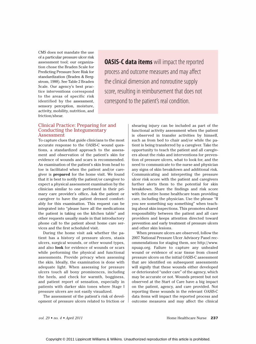
CMS does not mandate the use of a particular pressure ulcer risk assessment tool; our organiza-tion chose the Braden Scale for Predicting Pressure Sore Risk for standardization (Braden & Berg-strom, 1988). See Table 2 Braden Scale. Our agency’s best prac-tice interventions correspond to the areas of specific risk identified by the assessment, sensory perception, moisture, activity, mobility, nutrition, and friction/shear.
Clinical Practice: Preparing for and Conducting the Integumentary AssessmentTo capture clues that guide clinicians to the most accurate response to the OASIS-C wound ques-tions, a standardized approach to the assess-ment and observation of the patient’s skin for evidence of wounds and scars is recommended. An examination of the patient’s skin from head to toe is facilitated when the patient and/or care-giver is prepared for the home visit. We found that it is best to notify the patient/or caregiver to expect a physical assessment examination by the clinician similar to one performed in their pri-mary care provider’s office. Ask the patient or caregiver to have the patient dressed comfort-ably for this examination. This request can be integrated into “please have all the medications the patient is taking on the kitchen table” and other requests usually made in that introductory phone call to the patient about home care ser-vices and the first scheduled visit.
During the home visit ask whether the pa-tient has a history of pressure ulcers, stasis ulcers, surgical wounds, or other wound types, and also look for evidence of wounds or scars while performing the physical and functional assessments. Provide privacy when assessing the skin. Ideally, the examination is done with adequate light. When assessing for pressure ulcers touch all bony prominences, including the heels, and check for warmth, bogginess, and patient report of sensation, especially in patients with darker skin tones where Stage I pressure ulcers are not easily visualized.
The assessment of the patient’s risk of devel-opment of pressure ulcers related to friction or
OASIS-C data items will impact the reported process and outcome measures and may affect the clinical dimension and nonroutine supply score, resulting in reimbursement that does not correspond to the patient’s real condition.
Copyright © 2011 Lippincott Williams & Wilkins. Unauthorized reproduction of this article is prohibited.

238 Home Healthcare Nurse www.homehealthcarenurseonline.com
SENSORY PERCEPTION
ability to respond mean-ingfully to pressure-related discomfort
1. Completely Limited Unresponsive (does not moan, flinch, or grasp) to painful stimuli, due to diminished level of con-sci-ousness or sedation
ORlimited ability to feel pain over most of body.
2. Very LimitedResponds only to painful stimuli. Cannot communicate discomfort except by moan-ing or restlessness
OR has a sensory impairment which limits the ability to feel pain or discomfort over 1/2 of body.
MOISTURE
degree to which skin is exposed to moisture
1. Constantly MoistSkin is kept moist almost constantly by perspira-tion, urine, etc. Dampness is detected every time patient is moved or turned.
2. Very Moist Skin is often, but not always moist. Linen must be changed at least once a shift.
ACTIVITY
degree of physical activity
1. Bedfast
Confi ned to bed.
2. Chairfast Ability to walk severely limited or non-existent. Cannot bear own weight and/or must be assisted into chair or wheelchair.
MOBILITY
ability to change and control body position
1. Completely ImmobileDoes not make even slight changes in body or extremity position without assistance.
2. Very Limited Makes occasional slight changes in body or extremity position but unable to make fre-quent or signifi cant changes independently.
NUTRITION
usual food intake pattern
1. Very PoorNever eats a complete meal. Rarely eats more than 1/3 of any food offered. Eats 2 servings or less of protein (meat or dairy products) per day. Takes fl uids poorly. Does not take a liquid dietary supple-ment
ORis NPO and/or maintained on clear liquids or IVs for more than 5 days.
2. Probably InadequateRarely eats a complete meal and generally eats only about of any food offered. Protein intake includes only 3 servings of meat or dairy products per day. Occasionally will take a dietary supplement
ORreceives less than optimum amount of liquid diet or tube feeding.
FRICTION & SHEAR 1. ProblemRequires moderate to maximum assistance in moving. Complete lifting without sliding against sheets is impossible. Frequently slides down in bed or chair, requiring frequent repositioning with maximum assistance. Spasticity, contractures or agitation leads to almost constant friction.
2. Potential ProblemMoves feebly or requires minimum assis-tance. During a move skin probably slides to some extent against sheets, chair, restraints or other devices. Maintains relatively good position in chair or bed most of the time but occasionally slides down.
© Copyright Barbara Braden and Nancy Bergstrom, 1988. All rights reserved. Reprinted with permission.
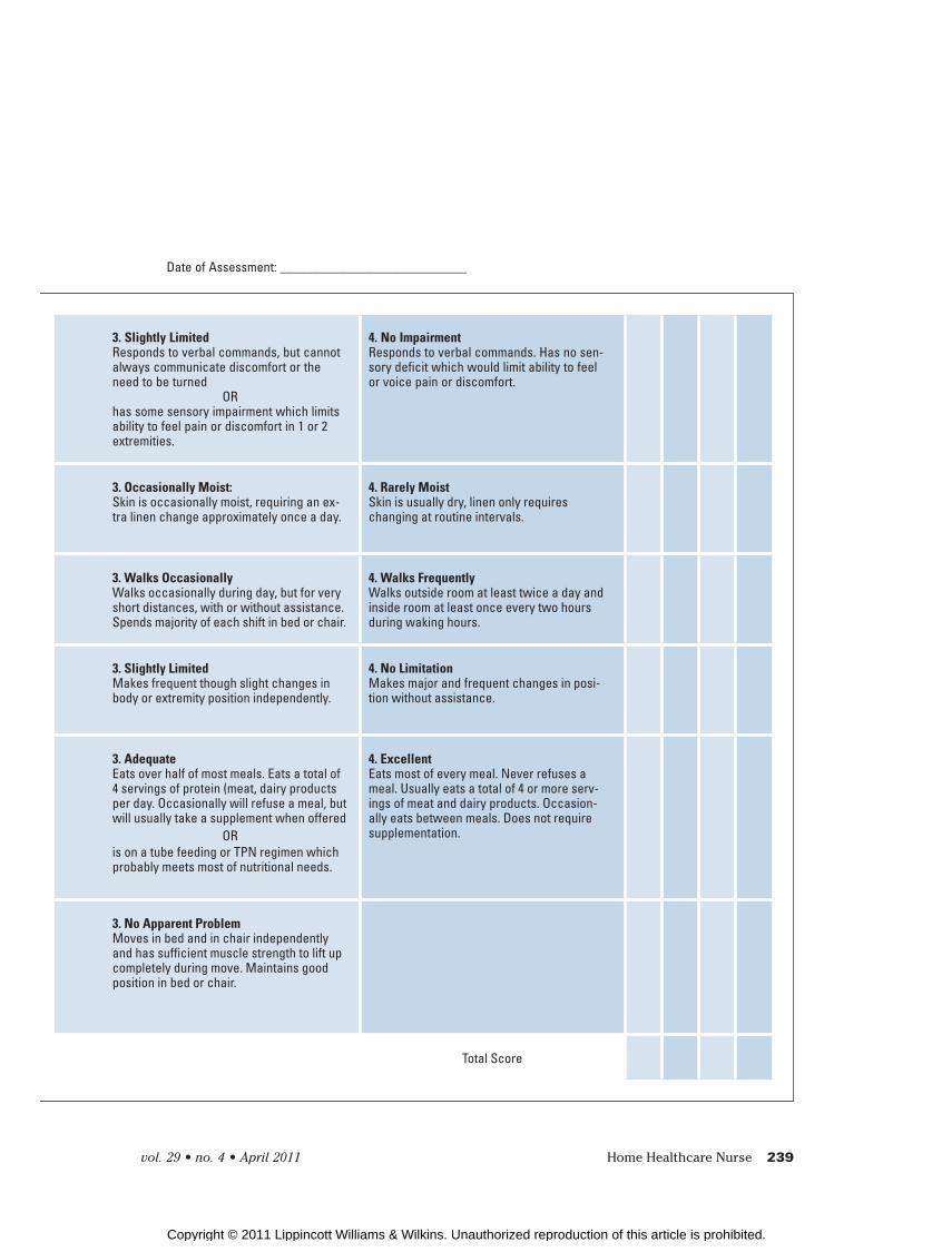
Table 2.
Braden Scale for Predicting Pressure Sore Risk
Patient’s Name: ____________________________ Evaluator’s Name: ____________________________
Copyright © 2011 Lippincott Williams & Wilkins. Unauthorized reproduction of this article is prohibited.
vol. 29 • no. 4 • April 2011 Home Healthcare Nurse 239
3. Slightly LimitedResponds to verbal commands, but cannot always communicate discomfort or the need to be turned
ORhas some sensory impairment which limits ability to feel pain or discomfort in 1 or 2 extremities.
4. No ImpairmentResponds to verbal commands. Has no sen-sory deficit which would limit ability to feel or voice pain or discomfort.
3. Occasionally Moist: Skin is occasionally moist, requiring an ex-tra linen change approximately once a day.
4. Rarely Moist Skin is usually dry, linen only requires changing at routine intervals.
3. Walks OccasionallyWalks occasionally during day, but for very short distances, with or without assistance. Spends majority of each shift in bed or chair.
4. Walks Frequently Walks outside room at least twice a day and inside room at least once every two hours during waking hours.
3. Slightly Limited Makes frequent though slight changes in body or extremity position independently.
4. No Limitation Makes major and frequent changes in posi-tion without assistance.
3. AdequateEats over half of most meals. Eats a total of 4 servings of protein (meat, dairy products per day. Occasionally will refuse a meal, but will usually take a supplement when offered
ORis on a tube feeding or TPN regimen which probably meets most of nutritional needs.
4. Excellent Eats most of every meal. Never refuses a meal. Usually eats a total of 4 or more serv-ings of meat and dairy products. Occasion-ally eats between meals. Does not require supplementation.
3. No Apparent Problem Moves in bed and in chair independently and has suffi cient muscle strength to lift up completely during move. Maintains good position in bed or chair.
Total Score
Date of Assessment: ____________________________
Copyright © 2011 Lippincott Williams & Wilkins. Unauthorized reproduction of this article is prohibited.

240 Home Healthcare Nurse www.homehealthcarenurseonline.com
pressure-related. The presence of scar tissue is a clue that the ulcer was origi-nally a Stage III or IV pressure ulcer and is reported as such on OASIS-C.
• Wounds/scars on the lower legs can result from several possible etiologies. Among them are venous or arterial diseases or mixed venous arterial disease or a surgical procedure. The Wound Ostomy Conti-nence Society offers clinical fact sheets that are very helpful in differentiating wound types of the lower extremities (WOCN, n.d.; http://www.wocn.org/pdfs/WOCN_Library/Fact_Sheets/C_QUICK1.pdf).
• Wounds/scars on the feet can be the result of pressure, arterial disease, and may be neuropathic or diabetes-related, surgical, trauma, or from other etiology.
Consult the OASIS-C Guidance Manual and CMS Q & A instructions to determine when an open or closed (epithelialized) wound or lesion is reportable in OASIS-C data items. For example, from that guidance we know that wounds of sur-gical etiology that are fully reepithelialized longer than 30 days from complete closure become “scars” and are not reported on OASIS-C. There are some skin openings that for OASIS-C pur-poses are not considered surgical wounds. These include, but are not limited to, ostomies, chest tube exit sites, and nephrostomy sites.
2. How will this wound heal?
Depending on the wound type, there are several mechanisms for healing. Determine the answers to these questions: if the wound is a partial- or full-thickness wound, will it heal by regeneration or scar tissue formation? If it is a surgical wound, will it heal by primary or second-ary intention? Should the wound be included in the OASIS-C data items?
• Key to selecting the correct OASIS data wound responses is observing what the wound bed reveals through a detailed as-sessment. The assessment includes identi-fying tissue type and appearance. Look for visible structures such as subcutaneous tis-sue, muscle, tendon, bone, and/or ligaments. The identification of these structures will help to distinguish partial-thickness from
dimension and nonroutine supply score, result-ing in reimbursement that does not correspond to the patient’s real condition.
To assist the clinician in the skin and wound assessment, four “critical questions” were devel-oped to guide the assessment.
1. What is the etiology of this wound or scar?
The patient’s medical and surgical history are important to consider when initially trying to determine the etiology; to confirm observations ask the patient/caregiver and/or physician about the etiology and history of the wound or scar. Determine whether the wound is acute or chronic. Baranoski and Ayello (2008) describe traumatic and surgical wounds that close by primary intention as typical “acute wounds.” Chronic wounds are those healing by second-ary intention. These wounds do not progress through the normal phases of healing in an or-derly and timely manner because the molecular environment is altered (Bryant & Nix, 2007). Therefore, knowing the length of time a wound has remained open is important when deter-mining the “status” for OASIS-C M items and is also a consideration to establish whether the goal of wound treatment and the Plan of Care (POC) is healing, maintenance, or palliation. The goal for wound management and interventions may also influence wound treatment choices.
When determining the etiology of wounds, clinicians can use several rules of thumb:
• Wounds/scars located over a bony promi-nence can suggest the etiology may be
Wound Assessment Critical Analysis Questions
What is the etiology of this wound or scar? Is it acute or chronic?
How will this wound heal… by regeneration or scar tissue formation?
What is the healing status definition that best fits this wound or scar?
Which evidence-based interventions should be implemented, communicated and documented?
Copyright © 2011 Lippincott Williams & Wilkins. Unauthorized reproduction of this article is prohibited.

vol. 29 • no. 4 • April 2011 Home Healthcare Nurse 241
regeneration. If a superficial wound has slough or eschar, it is no longer a partial-thickness wound, but is a full-thickness wound regardless of the depth. Slough is soft, moist avascular (devital-ized) tissue; it may be white, yellow, tan, gray, or green; it may be loose or firmly adherent (WOCN, 2009). Eschar is black or brown necrotic, devital-ized tissue; it can be loose or firmly adherent, hard, soft, dry, or wet (WOCN, 2009). Therefore, a pressure ulcer with slough or eschar is at least a Stage III and may be a Stage IV.
Strength and FunctionThe tissue of a partial-thickness wound regains its full tensile strength and function. With com-plete reepithelialization, the wound is healed.
Full-Thickness WoundsFull-thickness wounds penetrate deeper tissue layers. Full-thickness wounds are observed to have tissue damage involving the total loss of epidermis and dermis, extending into the subcu-taneous tissue and possibly into the muscle or bone (WOCN, 2009).
A full-thickness wound, if pressure-related, is a Stage III or Stage IV pressure ulcer. A Stage III pres-sure ulcer involves tissue loss extending through the dermis and into the subcutaneous tissue. A Stage IV pressure ulcer involves the same tissue loss and, in addition, has muscle or bone exposure. The loss of these original tissue structures is per-manent, and for that reason, Stage III and IV pres-sure ulcers are incapable of “fully healing.” Once
full-thickness wounds and/or provide informa-tion about the stage of a pressure ulcer.
• Wound healing mecha-nisms and times to heal differ depending upon the depth of tissue in-jury; presence of scar signifies that the closed pressure ulcer had full-thickness damage (Bry-ant & Nix, 2007).
Partial-Thickness WoundsPartial-thickness wounds are confined to the skin layers; skin damage does not penetrate below the dermis and may be limited to the epidermal layers only (WOCN, 2009).
Mechanism of Healing: RegenerationThe mechanism for healing of a partial-thickness wound is regeneration. Because the tissue injury of a partial-thickness wound involves only the epidermis and possibly the superficial dermal layers of the skin, the body is able to replace or regenerate that lost or damaged tissue with more of the same tissue. Epithelial tissue usually starts to appear as pink, purple tissue at the edge of a wound, but may also appear as islands in the center of a partial-thickness wound. The newly resurfaced epithelium appears pale pink, dry, and gradually repigments when com-pletely healed (Bryant & Nix, 2007). It is impor-tant to note that Stage II pressure ulcers heal by
Recommended Web Sites
Websites
■ http://www.qtso.com/hhadownload.html
■ http://www.medicare.gov/HomeHealthCompare/search.aspx
■ http://www.npuap.org/pr2.htm
(WOCN) Quick Assessioment of Leg Ulcers at
■ http://www.wocn.org.pdfs/WOCN Library/Fact_Sheets/C_QUICK1.pdf (WOCN) Guid-ance on OASIS-C
■ http://www.wocn.org/pdfs/GuidanceOASIS-C.pdf
To capture clues that guide clinicians to the most accurate response to the OASIS-C wound questions, a standardized approach to the assessment and observation of the patient’s skin for evidence of wounds and scars is recommended. An examination of the patient’s skin from head to toe is facilitated when the patient and/or caregiver is prepared for the home visit.
Copyright © 2011 Lippincott Williams & Wilkins. Unauthorized reproduction of this article is prohibited.
242 Home Healthcare Nurse www.homehealthcarenurseonline.com
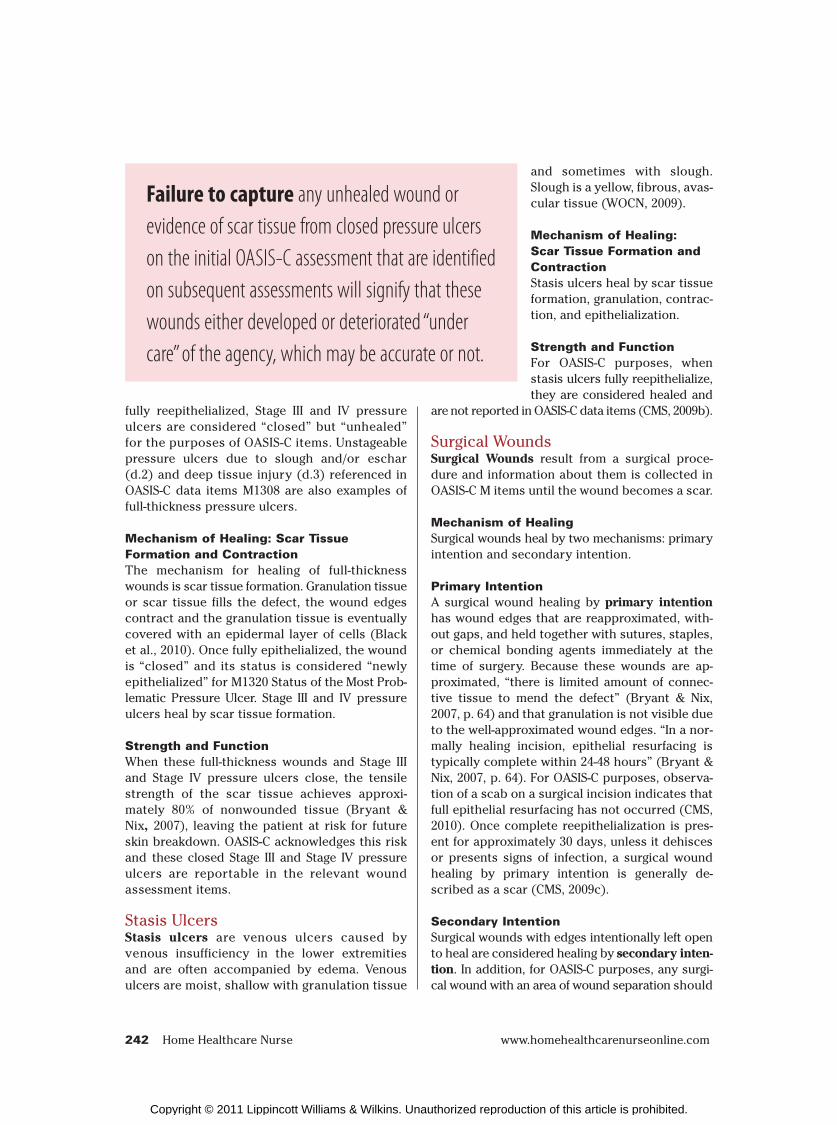
and sometimes with slough. Slough is a yellow, fibrous, avas-cular tissue (WOCN, 2009).
Mechanism of Healing: Scar Tissue Formation and ContractionStasis ulcers heal by scar tissue formation, granulation, contrac-tion, and epithelialization.
Strength and FunctionFor OASIS-C purposes, when stasis ulcers fully reepithelialize, they are considered healed and
are not reported in OASIS-C data items (CMS, 2009b).
Surgical WoundsSurgical Wounds result from a surgical proce-dure and information about them is collected in OASIS-C M items until the wound becomes a scar.
Mechanism of HealingSurgical wounds heal by two mechanisms: primary intention and secondary intention.
Primary IntentionA surgical wound healing by primary intention has wound edges that are reapproximated, with-out gaps, and held together with sutures, staples, or chemical bonding agents immediately at the time of surgery. Because these wounds are ap-proximated, “there is limited amount of connec-tive tissue to mend the defect” (Bryant & Nix, 2007, p. 64) and that granulation is not visible due to the well-approximated wound edges. “In a nor-mally healing incision, epithelial resurfacing is typically complete within 24-48 hours” (Bryant & Nix, 2007, p. 64). For OASIS-C purposes, observa-tion of a scab on a surgical incision indicates that full epithelial resurfacing has not occurred (CMS, 2010). Once complete reepithelialization is pres-ent for approximately 30 days, unless it dehisces or presents signs of infection, a surgical wound healing by primary intention is generally de-scribed as a scar (CMS, 2009c).
Secondary IntentionSurgical wounds with edges intentionally left open to heal are considered healing by secondary inten-tion. In addition, for OASIS-C purposes, any surgi-cal wound with an area of wound separation should
fully reepithelialized, Stage III and IV pressure ulcers are considered “closed” but “unhealed” for the purposes of OASIS-C items. Unstageable pressure ulcers due to slough and/or eschar (d.2) and deep tissue injury (d.3) referenced in OASIS-C data items M1308 are also examples of full-thickness pressure ulcers.
Mechanism of Healing: Scar Tissue Formation and ContractionThe mechanism for healing of full-thickness wounds is scar tissue formation. Granulation tissue or scar tissue fills the defect, the wound edges contract and the granulation tissue is eventually covered with an epidermal layer of cells (Black et al., 2010). Once fully epithelialized, the wound is “closed” and its status is considered “newly epithelialized” for M1320 Status of the Most Prob-lematic Pressure Ulcer. Stage III and IV pressure ulcers heal by scar tissue formation.
Strength and FunctionWhen these full-thickness wounds and Stage III and Stage IV pressure ulcers close, the tensile strength of the scar tissue achieves approxi-mately 80% of nonwounded tissue (Bryant & Nix, 2007), leaving the patient at risk for future skin breakdown. OASIS-C acknowledges this risk and these closed Stage III and Stage IV pressure ulcers are reportable in the relevant wound assessment items.
Stasis UlcersStasis ulcers are venous ulcers caused by venous insufficiency in the lower extremities and are often accompanied by edema. Venous ulcers are moist, shallow with granulation tissue
Failure to capture any unhealed wound or evidence of scar tissue from closed pressure ulcers on the initial OASIS-C assessment that are identified on subsequent assessments will signify that these wounds either developed or deteriorated “under care” of the agency, which may be accurate or not.
Copyright © 2011 Lippincott Williams & Wilkins. Unauthorized reproduction of this article is prohibited.
vol. 29 • no. 4 • April 2011 Home Healthcare Nurse 243
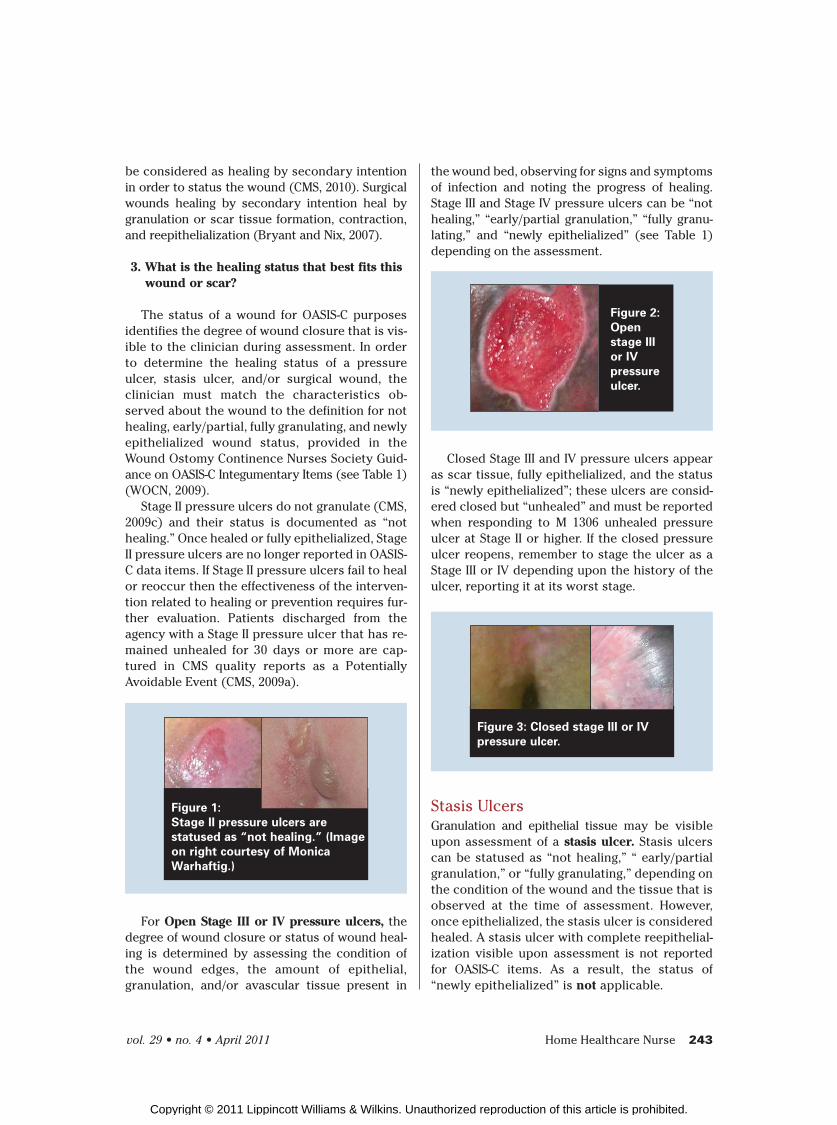
the wound bed, observing for signs and symptoms of infection and noting the progress of healing. Stage III and Stage IV pressure ulcers can be “not healing,” “early/partial granulation,” “fully granu-lating,” and “newly epithelialized” (see Table 1) depending on the assessment.
Figure 2:
Open
stage III
or IV
pressure
ulcer.
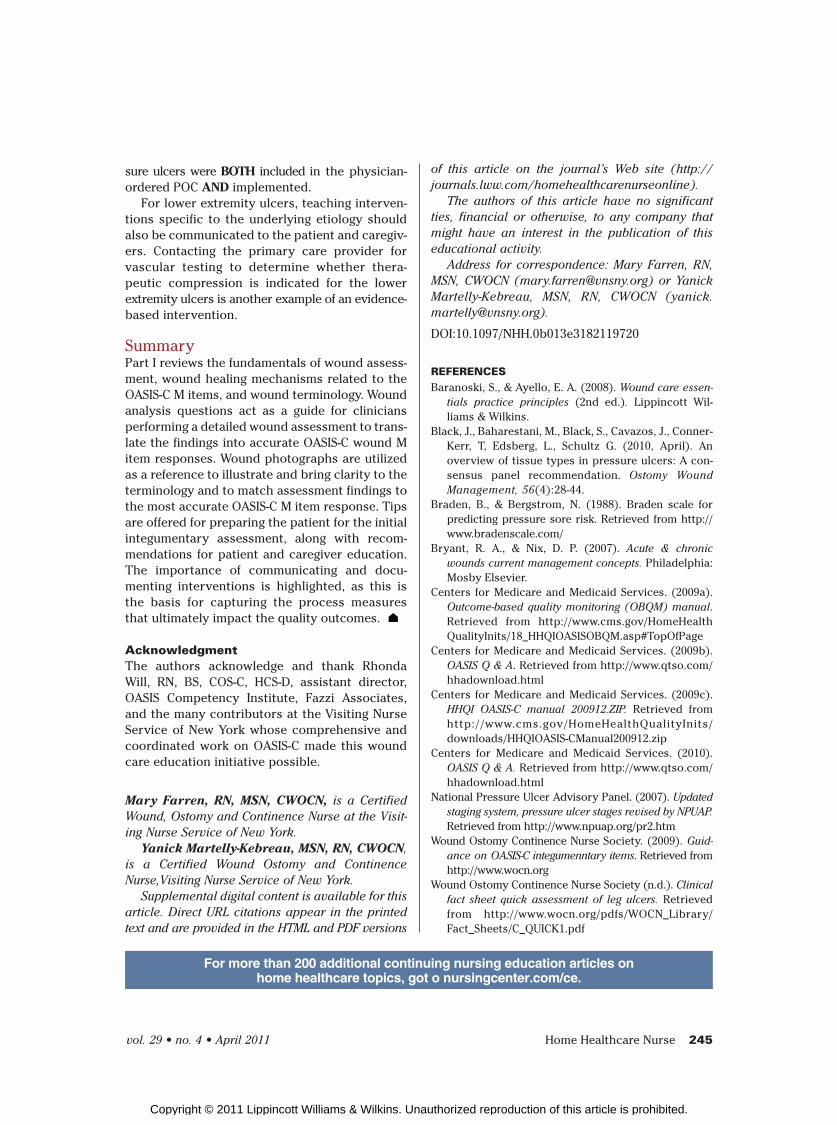
Closed Stage III and IV pressure ulcers appear as scar tissue, fully epithelialized, and the status is “newly epithelialized”; these ulcers are consid-ered closed but “unhealed” and must be reported when responding to M 1306 unhealed pressure ulcer at Stage II or higher. If the closed pressure ulcer reopens, remember to stage the ulcer as a Stage III or IV depending upon the history of the ulcer, reporting it at its worst stage.
Figure 3: Closed stage III or IV
pressure ulcer.
Stasis UlcersGranulation and epithelial tissue may be visible upon assessment of a stasis ulcer. Stasis ulcers can be statused as “not healing,” “ early/partial granulation,” or “fully granulating,” depending on the condition of the wound and the tissue that is observed at the time of assessment. However, once epithelialized, the stasis ulcer is considered healed. A stasis ulcer with complete reepithelial-ization visible upon assessment is not reported for OASIS-C items. As a result, the status of “newly epithelialized” is not applicable.
be considered as healing by secondary intention in order to status the wound (CMS, 2010). Surgical wounds healing by secondary intention heal by granulation or scar tissue formation, contraction, and reepithelialization (Bryant and Nix, 2007).
3. What is the healing status that best fits this wound or scar?
The status of a wound for OASIS-C purposes identifies the degree of wound closure that is vis-ible to the clinician during assessment. In order to determine the healing status of a pressure ulcer, stasis ulcer, and/or surgical wound, the clinician must match the characteristics ob-served about the wound to the definition for not healing, early/partial, fully granulating, and newly epithelialized wound status, provided in the Wound Ostomy Continence Nurses Society Guid-ance on OASIS-C Integumentary Items (see Table 1) (WOCN, 2009).
Stage II pressure ulcers do not granulate (CMS, 2009c) and their status is documented as “not healing.” Once healed or fully epithelialized, Stage II pressure ulcers are no longer reported in OASIS-C data items. If Stage II pressure ulcers fail to heal or reoccur then the effectiveness of the interven-tion related to healing or prevention requires fur-ther evaluation. Patients discharged from the agency with a Stage II pressure ulcer that has re-mained unhealed for 30 days or more are cap-tured in CMS quality reports as a Potentially Avoidable Event (CMS, 2009a).
Figure 1:
Stage II pressure ulcers are
statused as “not healing.” (Image
on right courtesy of Monica
Warhaftig.)
For Open Stage III or IV pressure ulcers, the degree of wound closure or status of wound heal-ing is determined by assessing the condition of the wound edges, the amount of epithelial, granulation, and/or avascular tissue present in
Copyright © 2011 Lippincott Williams & Wilkins. Unauthorized reproduction of this article is prohibited.

244 Home Healthcare Nurse www.homehealthcarenurseonline.com
4. Which evidence-based intervention(s) should be implemented, communicated, and documented based on the etiology of the wound?
Determining the causative factors and the wound etiology is key to identifying interven-tions that facilitate wound healing of all types and the prevention of pressure ulcers. Evidence-based interventions should be applied through-out the process for effective wound management. “Pressure ulcers rarely can be managed to com-plete closure with only one form of topical treatment” (Black et al., 2010).
The use of an agency-wide standardized risk assessment tool helps to identify areas of risk. Our organization chose the Braden Scale for pre-dicting Pressure Sore Risk because it is a stan-dardized tool validated for use on community dwelling individuals (see Table 2—Braden Scale). The risk factors of sensory perception, moisture, activity, mobility, friction, and shear are scored and identify which of these factors or combi-nation of factors can put a patient at risk for pressure ulcer development. Interventions to ad-dress the specific risk for a particular patient can be implemented and taught to the patient and all caregivers. The clinician spends a limited amount of time with the patient as compared with institu-tional settings so that the responsibility to imple-ment the interventions rests with the patient and all caregivers. Documentation of the response to this teaching should include the patient’s and caregivers’ ability and willingness to comply with implementing the interventions. Support sur-faces, mattresses and chair cushion and other offloading pressure devices are examples of in-terventions and should be included in the POC. The clinician should also communicate the risk score and its meaning to the patient, family, care-givers, and physician.
In addition, these interventions are also trans-lated into the OASIS-C process measures. For ex-ample, at start of care (SOC) and resumption of care (ROC), M1300 asks whether the patient was screened for risk of pressure ulcer, M1302 asks whether the patient is at risk for developing pres-sure ulcers, and the POC synopsis M 2250 asks whether the physician-ordered POC includes in-terventions to prevent pressure ulcers. At trans-fer and discharge, the intervention synopsis M 2400 asks whether interventions to prevent pres-
Surgical wounds healing by primary inten-tion have well-approximated wound edges. As a result, granulation tissue is not visible and only the regeneration of epithelial cells is observable on the skin surface. Therefore, it is incorrect to associate early/partial or fully granulating with surgical wounds healing by primary intention. Surgical wounds healing by primary intention with visible signs of infection are classified as “not healing” (see Table 1). Once the incision site is observed to be fully covered with epithelium, the surgical wound healing by primary intention is statused as “newly epithelialized” (see Table 1) and will remain so for 30 days unless it dehisces or presents signs of infection. After 30 days, it is considered a scar and is not reported in any of the OASIS-C data items.
Figure 8: A surgical wound closed
by primary intention (approximated
incisions) statused as “not healing.”
Figure 7: A surgical wound
epithelialized for 30 days or less
is “newly epithelialized.”
For surgical wounds healing by secondary intention, the degree of wound closure and sta-tus of wound healing are determined by assess-ing the condition of the wound edges, the amount of epithelial, granulation, and/or avascular tissue present in the wound bed, observing for signs and symptoms of infection, and noting the prog-ress of healing. These wounds can be statused as “not-healing,” “early/partial granulation,” “fully granulating,” and “newly epithelialized,” (see Table 1) depending on the assessment.
Copyright © 2011 Lippincott Williams & Wilkins. Unauthorized reproduction of this article is prohibited.
vol. 29 • no. 4 • April 2011 Home Healthcare Nurse 245
of this article on the journal’s Web site (http://journals.lww.com/homehealthcarenurseonline).
The authors of this article have no significant ties, financial or otherwise, to any company that might have an interest in the publication of this educational activity.
Address for correspondence: Mary Farren, RN, MSN, CWOCN ([email protected]) or Yanick Martelly-Kebreau, MSN, RN, CWOCN ([email protected]).
DOI:10.1097/NHH.0b013e3182119720
REFERENCESBaranoski, S., & Ayello, E. A. (2008). Wound care essen-
tials practice principles (2nd ed.). Lippincott Wil-liams & Wilkins.
Black, J., Baharestani, M., Black, S., Cavazos, J., Conner-Kerr, T, Edsberg, L., Schultz G. (2010, April). An overview of tissue types in pressure ulcers: A con-sensus panel recommendation. Ostomy Wound Management, 56(4):28-44.
Braden, B., & Bergstrom, N. (1988). Braden scale for predicting pressure sore risk. Retrieved from http://www.bradenscale.com/
Bryant, R. A., & Nix, D. P. (2007). Acute & chronic wounds current management concepts. Philadelphia: Mosby Elsevier.
Centers for Medicare and Medicaid Services. (2009a). Outcome-based quality monitoring (OBQM) manual. Retrieved from http://www.cms.gov/HomeHealthQualityInits/18_HHQIOASISOBQM.asp#TopOfPage
Centers for Medicare and Medicaid Services. (2009b). OASIS Q & A. Retrieved from http://www.qtso.com/hhadownload.html
Centers for Medicare and Medicaid Services. (2009c). HHQI OASIS-C manual 200912.ZIP. Retrieved from http://www.cms.gov/HomeHealthQualityInits/downloads/HHQIOASIS-CManual200912.zip
Centers for Medicare and Medicaid Services. (2010). OASIS Q & A. Retrieved from http://www.qtso.com/hhadownload.html
National Pressure Ulcer Advisory Panel. (2007). Updated staging system, pressure ulcer stages revised by NPUAP. Retrieved from http://www.npuap.org/pr2.htm
Wound Ostomy Continence Nurse Society. (2009). Guid-ance on OASIS-C integumenntary items. Retrieved from http://www.wocn.org
Wound Ostomy Continence Nurse Society (n.d.). Clinical fact sheet quick assessment of leg ulcers. Retrieved from http://www.wocn.org/pdfs/WOCN_Library/Fact_Sheets/C_QUICK1.pdf
sure ulcers were BOTH included in the physician-ordered POC AND implemented.
For lower extremity ulcers, teaching interven-tions specific to the underlying etiology should also be communicated to the patient and caregiv-ers. Contacting the primary care provider for vascular testing to determine whether thera-peutic compression is indicated for the lower extremity ulcers is another example of an evidence-based intervention.
SummaryPart I reviews the fundamentals of wound assess-ment, wound healing mechanisms related to the OASIS-C M items, and wound terminology. Wound analysis questions act as a guide for clinicians performing a detailed wound assessment to trans-late the findings into accurate OASIS-C wound M item responses. Wound photographs are utilized as a reference to illustrate and bring clarity to the terminology and to match assessment findings to the most accurate OASIS-C M item response. Tips are offered for preparing the patient for the initial integumentary assessment, along with recom-mendations for patient and caregiver education. The importance of communicating and docu-menting interventions is highlighted, as this is the basis for capturing the process measures that ultimately impact the quality outcomes.
AcknowledgmentThe authors acknowledge and thank Rhonda Will, RN, BS, COS-C, HCS-D, assistant director, OASIS Competency Institute, Fazzi Associates, and the many contributors at the Visiting Nurse Service of New York whose comprehensive and coordinated work on OASIS-C made this wound care education initiative possible.
Mary Farren, RN, MSN, CWOCN, is a Certified Wound, Ostomy and Continence Nurse at the Visit-ing Nurse Service of New York.
Yanick Martelly-Kebreau, MSN, RN, CWOCN, is a Certified Wound Ostomy and Continence Nurse,Visiting Nurse Service of New York.
Supplemental digital content is available for this article. Direct URL citations appear in the printed text and are provided in the HTML and PDF versions
For more than 200 additional continuing nursing education articles on home healthcare topics, got o nursingcenter.com/ce.
Copyright © 2011 Lippincott Williams & Wilkins. Unauthorized reproduction of this article is prohibited.