www.lightsource.ca cls status update e. matias canadian light source
TRANSCRIPT

www.lightsource.ca
CLS Status Update
E. MatiasCanadian Light Source

www.lightsource.ca
Layout
170.88 m circumference 2.9 GeV ~ 200-300 mADBA lattice with 12-fold period
www.lightsource.ca
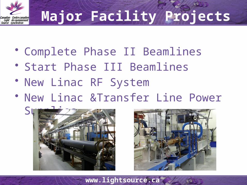
Major Facility Projects
• Complete Phase II Beamlines• Start Phase III Beamlines• New Linac RF System• New Linac &Transfer Line Power Supplies
www.lightsource.ca
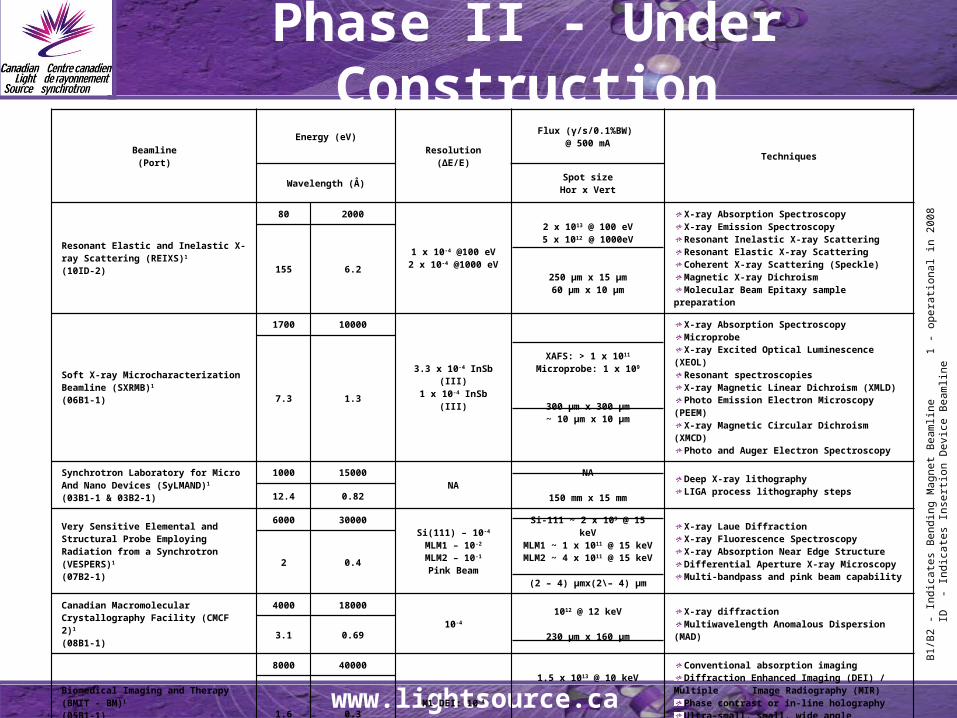
Phase II - Under Construction
Beamline(Port)
Energy (eV)
Resolution(ΔE/E)
Flux (γ/s/0.1%BW) @ 500 mA
Techniques
Wavelength (Å) Spot sizeHor x Vert
Resonant Elastic and Inelastic X-ray Scattering (REIXS)1
(10ID-2)
80 2000
1 x 10⁻⁴ @100 eV2 x 10-4 @1000 eV
2 x 10¹³ @ 100 eV5 x 1012 @ 1000eV
250 μm x 15 μm60 μm x 10 μm
X-ray Absorption Spectroscopy X-ray Emission Spectroscopy Resonant Inelastic X-ray Scattering Resonant Elastic X-ray Scattering Coherent X-ray Scattering (Speckle) Magnetic X-ray Dichroism Molecular Beam Epitaxy sample preparation
155 6.2
Soft X-ray Microcharacterization Beamline (SXRMB)1
(06B1-1)
1700 10000
3.3 x 10⁻⁴ InSb (III)1 x 10⁻⁴ InSb (III)
XAFS: > 1 x 10¹¹
Microprobe: 1 x 10⁹
300 μm x 300 μm~ 10 μm x 10 μm
X-ray Absorption Spectroscopy Microprobe X-ray Excited Optical Luminescence (XEOL) Resonant spectroscopies X-ray Magnetic Linear Dichroism (XMLD) Photo Emission Electron Microscopy (PEEM) X-ray Magnetic Circular Dichroism (XMCD) Photo and Auger Electron Spectroscopy
7.3 1.3
Synchrotron Laboratory for Micro And Nano Devices (SyLMAND)1
(03B1-1 & 03B2-1)
1000 15000NA
NA
150 mm x 15 mm
Deep X-ray lithography LIGA process lithography steps 12.4 0.82
Very Sensitive Elemental and Structural Probe Employing Radiation from a Synchrotron (VESPERS)1
(07B2-1)
6000 30000Si(111) – 10-4
MLM1 – 10-2
MLM2 – 10-1
Pink Beam
Si-111 ~ 2 x 10⁹ @ 15 keVMLM1 ~ 1 x 10¹¹ @ 15 keVMLM2 ~ 4 x 10¹¹ @ 15 keV
(2 – 4) μmx(2\– 4) μm
X-ray Laue Diffraction X-ray Fluorescence Spectroscopy X-ray Absorption Near Edge Structure Differential Aperture X-ray Microscopy Multi-bandpass and pink beam capability
2 0.4
Canadian Macromolecular Crystallography Facility (CMCF 2)1
(08B1-1)
4000 1800010-4
1012 @ 12 keV
230 μm x 160 μm
X-ray diffraction Multiwavelength Anomalous Dispersion (MAD) 3.1 0.69
Biomedical Imaging and Therapy(BMIT - BM)1
(05B1-1)
8000 40000
M1 DEI: 10-4
1.5 x 10¹³ @ 10 keV
231 mm x 4.6 mm @ 23 m
Conventional absorption imaging Diffraction Enhanced Imaging (DEI) / Multiple
Image Radiography (MIR) Phase contrast or in-line holography Ultra-small, small, wide angle scattering imaging Computer Tomography (CT)
1.6 0.3
Biomedical Imaging and Therapy (BMIT - ID)1
(05ID-1 & 05ID-2)
20000 100000 M1 CT:10-3
M2 CT:10-3
M3 DEI:10–5
M4 KES:10-3
4 x 10¹⁴ @ 40 keV
224 mm x 11.2 mm @ 56 m
Imaging – conventional absorption imaging, DEI / MIR, CT, K-edge Subtraction (KES)
Therapy – Microbeam Radiation Therapy, CT Therapy 0.6 0.1
B1/
B2
- In
dica
tes
Ben
ding
Mag
net
Bea
mlin
e
1 -
oper
atio
nal i
n 20
08
ID
- In
dica
tes
Inse
rtio
n D
evic
e B
eam
line
www.lightsource.ca
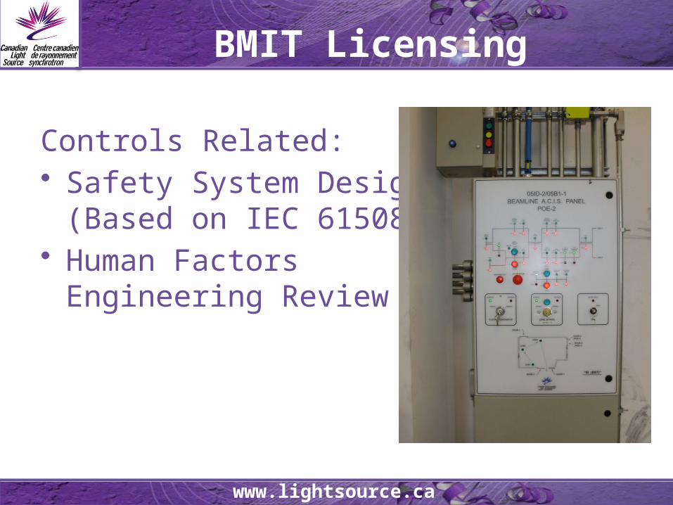
BMIT Licensing
Controls Related:• Safety System Design
(Based on IEC 61508)• Human Factors
Engineering Review
www.lightsource.ca
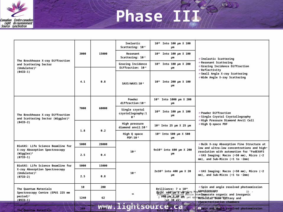
Phase III
The Brockhouse X-ray Diffraction and Scattering Sector (Undulator)2
(04ID-1)
3000 15000
Inelastic Scattering: 10-6 1010 Into 100 μm X 100 μm
Inelastic ScatteringResonant ScatteringGrazing Incidence DiffractionReflectivitySmall Angle X-ray ScatteringWide Angle X-ray Scattering
Resonant Scattering: 10-4 1012 Into 100 μm X 100 μm
Grazing Incidence Diffraction: 10-4 1012 Into 100 μm X 200 μm
4.1 0.8
SAXS/WAXS:10-4 1011 Into 200 μm X 100 μm
The Brockhouse X-ray Diffraction and Scattering Sector (Wiggler)2
(04ID-2)
7000 60000
Powder diffraction:10-
4 1011 Into 1000 μm X 200 μm
Powder DiffractionSingle Crystal CrystallographyHigh Pressure Diamond Anvil CellHigh Q-space PDF
Single crystal crystallography:10-3 1010 Into 100 μm X 100 μm
1.8 0.2
High pressure diamond anvil:10-4 1011 Into 25 μm X 25 μm
High Q space PDF:10-
3 1011 Into 500 μm X 500 μm
BioXAS: Life Science Beamline for X-ray Absorption Spectroscopy (Wiggler)2
(07ID-1)
5000 28000
10-4 9x10¹² into 600 μm X 200 μm
Bulk X-ray Absorption Fine Structure at low and ultra-low concentrations and high resolution with automation for “FedEXAFS”
XAS Imaging: Macro (>50 mm), Micro (~2 mm), and Sub-Micro (<1 to ~2mm)
2.5 0.4
BioXAS: Life Science Beamline for X-ray Absorption Spectroscopy (Undulator)2
(07ID-2)
5000 15000
10-4 2x10¹³ into 400 μm X 20 μmXAS Imaging: Macro (>50 mm), Micro (~2 mm),
and Sub-Micro (<1 to ~2mm)2.5 0.8
The Quantum Materials Spectroscopy Centre (EPU1 225 mm period)2
(09ID-1)
10 200NA
Brilliance: 7 x 10¹⁶ Spin: 400 μm X 40 μm
400 μm X 40 μm(at 30 eV)
Spin and angle resolved photoemission spectroscopy
Separate organic and inorganic Molecular Beam Epitaxy and characterization chambers1240 62
The Quantum Materials Spectroscopy Centre (EPU2 54 mm period)2
(09ID-2)
200 1000NA
Brilliance: 7 x 10¹⁷
Spin: 700 μm X 40 μmARPES: 300 μm X 40 μm
(at 200 eV)
Spin and angle resolved photoemission spectroscopy
Separate organic and inorganic Molecular Beam Epitaxy and characterization chambers62 12
B1/B2 - Indicates Bending Magnet Beamline ID - Indicates Insertion Device Beamline 2 – CFI funded; not yet approved for construction.
www.lightsource.ca
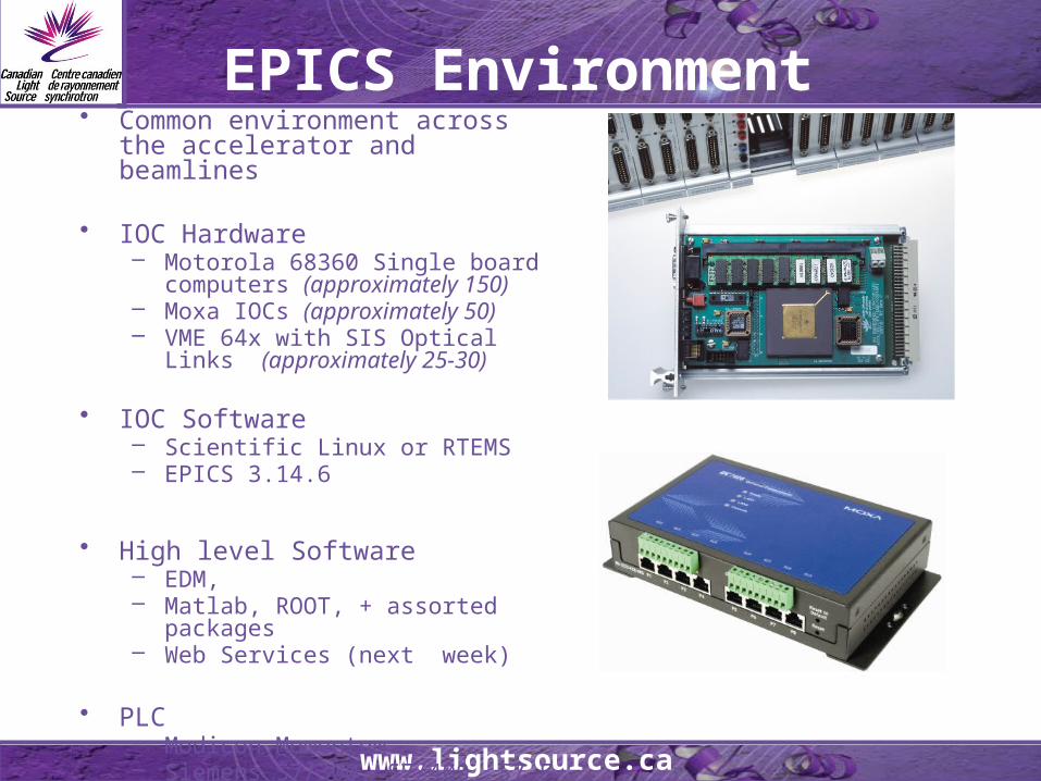
• Common environment across the accelerator and beamlines
• IOC Hardware – Motorola 68360 Single board
computers (approximately 150)– Moxa IOCs (approximately 50)– VME 64x with SIS Optical Links
(approximately 25-30)
• IOC Software– Scientific Linux or RTEMS– EPICS 3.14.6
• High level Software– EDM, – Matlab, ROOT, + assorted packages– Web Services (next week)
• PLC– Modicon Momentum– Siemens S7/300, S7/400, S7 F
EPICS Environment

www.lightsource.ca
Controls and Instrumentation Development Specific Projects
• Time-resolve/Single Bunch– Fill-pattern monitor (J. Vogt/J. Bergstrom)
• EPICS Software (D. Beauregard)
– Transverse Feedback System (S. Hu)– Upgraded timing system
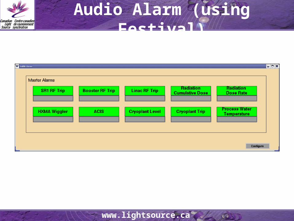
• Alarm Annunciation (D. Beauregard)• Beam profile analysis ……• Beamline Java Interface …..• ScienceStudio/Remote Access …..

www.lightsource.ca
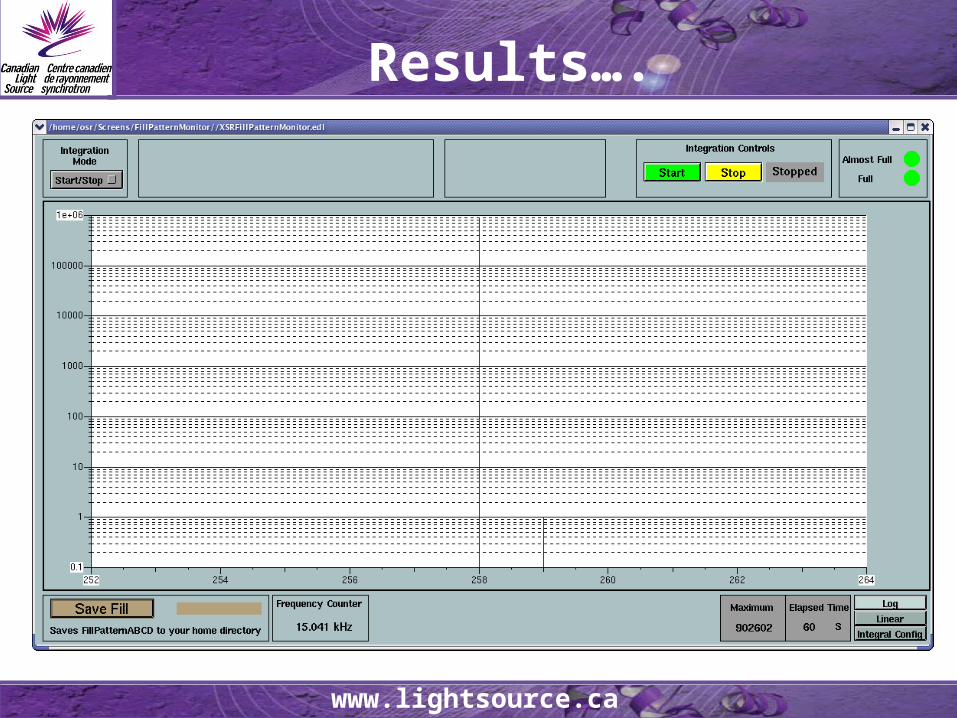
Fill Pattern Monitor
• Complete Phase II Beamlines• Start Phase III Beamlines• New Linac RF System• New Linac Power Supplies

www.lightsource.ca
Results….
www.lightsource.ca
Results….
www.lightsource.ca
Audio Alarm (using Festival)
www.lightsource.ca
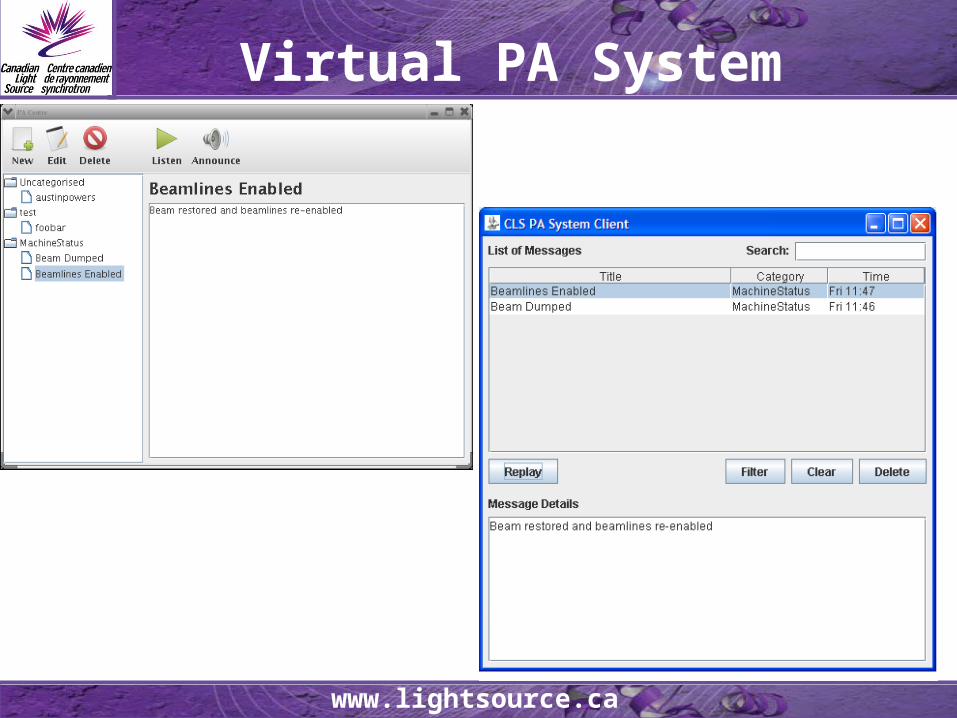
Virtual PA System
www.lightsource.ca
Funding Partners
38 supporting University Partners and growing…