x -ray analysis and the structure of vitamin...
TRANSCRIPT

X -ray Analysis and the Structure of Vitamin B12 •
By DOROTHY CROWFOOT HODGKIN, Oxford.
With 26 Figures.
Contents. Page
I. Introduction..................................................... 167
II. Some Characteristics of the X-ray Crystallographic Methods Applied to the Analysis of Vitamin BI2 . . . . . . . . . . . . . . . . . . . . . . . . . . . . . . . . . . . . . .. 169
III. The Determination of the Structure of Vitamin BI2 ................. 173
1. Preliminary Crystallographic Measurements and Observations ...... 173
2. The Stages of Atom Identification ............................ " 176 a) The Cyanide Group ......................................... 179 b) The "Nucleotide" Group .................................... 179 c) The Corrin Nucleus ......................................... 183 d) The Side-chains Attached to the Nucleus ..................... 185 e) The Propanolamine Groups .................................. 189
3. The Problem of Right and Wrong Atoms ....................... 194
4. The Refinement of the Atomic Positions ........................ 195
5. The Absolute Configuration of the Molecule. . . . . . . . . . . . . . . . . . . . .. 196
6. The Chemical Formulae of Vitamin BI2 and the Hexacarboxylic Acid 196
IV. The Chemical Reactions of Vitamin BI2 ............................ 202
V. Biochemical Problems Connected with the BI2 Structure............. 207
VI. The Crystal Structures of Vitamin BI2 and the Hexacarboxylic Acid .. 212
VII. Conclusion ....................................................... 218
References .......................................................... 218
I. Introduction. The structure of vitamin B12, as we think of it today, is based on
a fascinating complex of evidence obtained by X-ray analysis and by more traditional chemical means. We have reached a position in which we can almost say we "see" the molecule-if not quite as clearly, perhaps, as we should like. We can assign positions in space to the atoms of this very large molecule within less than half an Angstrom unit in twv
J. L. Hartwell et al., Fortschritte der Chemie organischer Naturstoffe / Progress in the Chemistry of Organic Natural Products / Progrès dans la Chimie des Substances Organiques Naturelles© Wien · Springer-Verlag 1958
168 DOROTHY CROWFOOT HODGKIN:
different crystal structures. Vve know its absolute configuration and the exact stereochemistry of all the different asymmetric centres present. Yet most of this knowledge rests on a way of using X-ray diffraction effects which is very far from rigid in its application. Part, at least, of our evidence that our method works at all is the character of the structure it has given us for B12-a structure that fits in an extraordinarily reasonable way with such a variety of observations, chemical and stereochemical and biogenetic, that it is impossible not to believe it is essentially correct.
The nature of our evidence leaves us still asking a number of questions, sometimes of a rather unusual character, particular questions about details of the structural formula of the vitamin itself and its chemical reactions, of its mode of formation in nature and its biological reactivity, and general questions about the stereochemical organisation of different chemical units within a large molecule, and the possible application of X-ray analysis to structural problems of still greater magnitude. It is the purpose of the present review to consider some of these questions against the background of the actual investigation of the structure of B12.
The vitamin was first sought for as a factor occurring in liver, which MINOT and MURPHY recognised in 1926 was highly effective in treating patients suffering from pernicious anaemia (33). It was first isolated crystalline in 1948, by FOLKERS and his colleagues of the Merck Laboratories (36) and shortly afterwards by LESTER SMITH and PARKER (Glaxo Laboratories) (40, I8), and ELLIS, PETROW and SNOOK (British Drug Houses Ltd.) (I7). Since that time, a number of reviews have appeared to which it is useful to refer for detailed references to papers on the chemical behaviour of the vitamin (4I, I9, 39, 23). At first progress was rapid. By 1951 it was known that the vitamin had an approximate formula C61-64Hs3-92013-20N14PCO and contained a nucleotide-like group, 5: 6-dimethyl-1-(<x-D-ribofuranosyl) benziminazole-2' or 3'-phosphate, a cyanide group, one or two propanolamine groups, a number of amide groups-all of which, added together, made up nearly half its molecular weight. The structure of the remaining half of the molecule was discovered very largely by X-ray analysis. This also started in 1948 but was at first very slow. It depended on the study of four different crystal structures, wet and air dry B12, a selenocyanide derivative of B12 and a hexacarboxylic acid obtained by degradation of the vitamin by CANNON, JOHNSON and TODD (II). SO far a general account only of the investigation of all four crystals has been published (25); further detailed structure analytical papers dealing with the individual crystals are in course of preparation, some details from which will be given in this account.

X-ray Analysis and the Structure of Vitamin B 12• 169
The methods of chemical degradation and characterisation used to establish the presence of a large part of the B12 structure are well known and need no detailed description in the present review. X-ray analysis on the other hand is less generally familiar and in its mode of application to B12 followed largely hitherto untried paths, associated with serious dangers and difficulties. For this reason, it seems best to precede any account of the evidence for the structure of the vitamin by a short discussion of the X-ray analytical procedures adopted.
II. Some Characteristics of the X-ray Crystallographic Methods Applied to the Analysis of Vitamin B12•
The theoretical background of the X-ray analytical approach used in the investigation of vitamin B12 has been very fully described by MATHIESON in a recent review, which gives a number of good examples of structure analyses of organic compounds (32). Here, therefore, only a brief outline is necessary, sufficient to indicate the character of the evidence we have acquired for the B12 crystal structures.
From the intensities of the X-rays diffracted by a crystal it is possible to calculate the electron density, (l, at any position in space, xyz, within the crystal unit cell. The formal equations may be summarised as follows:
+00 erellz = ~ 171717 [Fhk
170 DOROTHY CROWFOOT HODGKIN:
In the initial process of finding the atomic positions it is clear that use can be made of the electron density equation (1) provided some direct evidence can be obtained of the phase angles. Such evidence has been given in various researches by the use of isomorphous crystals, crystals containing an anomalous scatterer or crystals containing a particularly heavy atom. It is the third case which is important here.
From the form of the equations (1), (2), (3), it can be seen that one very heavy atom in a crystal containing many light atoms may effectively dominate the X-ray scattering and determine to a large part by its contribution the magnitudes and phase angles of the individual structure factors. Its position in the crystal in such a case can usually be recognised by considering the magnitudes of the observed F values alone, using the calculation of the Patterson or F2 series
co
P XYZ = ~ III P hkl cos fJ -co
over the volume of the unit cell. Peaks in this series correspond with vector distances between atoms and those due to the heavy atoms are prominent and consequently easily detectable.
The analysis of vitamin B12 has shown very clearly that equations (1), (2) and (3) can be applied as in the "heavy atom" method even when there is no dominatingly heavy atom present. We may imagine that if some only of the atomic positions can be found and inserted in equations (2), (3), they will, to a first approximation, determine the phase angles of some of the terms in the equation and a correct component of others. For still other terms, the phase angles derived may be almost wholly wrong. If we calculate an electron density map, using the observed structure factors and phase angles calculated on part of the structure only, we may imagine ourselves laying down in space a pattern of electron density, partly correct, partly 'wildly incorrect. Atoms postulated in the calculation appear as high peaks; in addition the partly correctly phased terms give peaks at other actual atomic positions, rather low in density in proportion to the relatively small correct electron density contribution. The incorrect components may give maxima just anywhere and, of course, confuse the appearance of maxima at the actual atomic positions.
Our hope of recognising the true atomic positions depends on the very rigid conditions that we know determine permissible interatomic distances in crystal structures. A trivalent cobalt atom, for example, we would expect to find surrounded by six others at about 1.9 A away from it in an octahedron and so on. If, after one electron density calculation, we can add to the number of atomic positions we know, these may be introduced into new structure factor and phase angle

X-ray Analysis and the Structure of Vitamin B12. I7 1
calculations and used to derive new electron density patterns which should, on statistical grounds (3I) gradually improve the definition of the structure. In practice most of the atomic distribution seems to be very easily recognisable once the phasing contributions are roughly 50% of the total, though progress may be made long before this point and difficulties may remain long after it.
There are certain problems attached to this process for finding the actual electron density in crystals. The first and most serious is that it is very easy to "make" atoms and so to invent a completely unreal chemical structure. The phase angles have a dominating effect on the appearance of peaks in the electron density patterns. If the structure factor calculation is carried out assuming the presence of an atom at a particular position, then the following Fourier synthesis will show a peak at the assumed position, whether there is really an atom there or no. Falsely introduced peaks are generally a little lower than peaks due to genuine atoms but may easily be mistaken for real atoms-and indeed often were so mistaken in the early stages of the B12 analysis. In problems involving smaller structures it has been found possible to eradicate wrong atoms by a large number of rounds of least squares calculations. But imagination boggles at the number of rounds of calculation that would have to be undertaken to remove, by formal automatic methods, all the wrong atoms inserted at early stages in the B12 crystal structures.
A second problem is that the appearance of peaks in an electron density pattern depends on the quality and quantity of the observed X-ray diffraction spectra which provide figures for the F terms of the series. Formally the limits over which the summation in equation (I) (p. 169) is to be carried out are set at infinity and minus infinity. In practice, they depend on experimental conditions-the limits in spacing out to which X-ray reflections can be observed and recorded. The atomic definition obtainable with reflections from crystal planes having different spacing limits is illustrated by Fig. I, p. 17z.
This shows the same molecule-4: S-diamino-z-chloropyrimidine-as seen in an electron density projection, calculated with phase angles derived from the atomic positions shown and different numbers of observed F values-the series being terminated at different spacing limits. With the B12 crystals reflections were observed out to spacings of I A, corresponding with the series in Fig. I b. From almost the first X-ray photographs taken of B12, it was therefore obvious that it should be possible to calculate an electron density distribution which would show the atoms of B12 as clearly as those in Fig. I b. Here the peak densities are a little low, the peak centres not exactly at the calculated sites but the atoms are shown as clearly separated electron density

172 DOROTHY CROWFOOT HODGKIN:
maxima, differing in height according to their chemical nature, chlorine, nitrogen and carbon. Later on, the hexacarboxylic acid was found to give X-ray reflections to the limit of copper KiX radiation-spacings of 0.78 A-formally capable of giving as good evidence of atomic positions as the series shown in Fig. I a. With good experimental data extending
d> O'l7l .... d> !'!!J)
d> ,NO)' .....
.. ' ( .......
Fig. I. Electron density projections calculated by N. E. WHITE for 4: 5-diamino-2-cbloropyrimidine using different numbers of observed reflections. The measured structure factors have been rnodified to correspond with those from atoms at rest, and the nwnbers used cut off at the spacing limits shown. Hence the patterns, particularly Cal, show marked diffraction effects, as well as the effect of the termination of the series.
to this limit, one might hope to observe individual hydrogen atom positions; in fact, with the B12 crystals and the hexacarboxylic acid, as with the pyrimidine illustrated in Fig. I, the intensity measurements were not of any great accuracy. In these conditions, the electron density due to hydrogen atoms tends to be confused with the heavier atoms and background fluctuations; their positions have to be inferred from other evidence.
The third problem of this type of analysis concerns the actual magnitude of the task of measuring the intensities of the X-ray reflections and

X-ray Analysis and the Structure of Vitamin B 12 . I73
carrying out the calculations hidden in the brief equations (r) and (2). J n order to obtain evidence of the separate positions of atoms in a molecule as large as B12, all calculations have to be done in three dimensions. An initial outlay must be made of the measurement of three-dimensional intensity data-for each B12 crystal studied in detail some 2,500 Fhk!
values were measured several times over, for the hexacarboxylic acid, 3,351. In the circumstances, high accuracy of individual measurement was not attempted. Rapid methods of visual intensity estimation were used throughout the B12 research, and absorption corrections, which are difficult to calculate, were not applied. Then, for anyone crystal, each electron density calculation involved a summation over all the .observed F hk! values at some 54,000 points within the unit cell-points separated by distances of OA A or less, sufficiently small to avoid missing details of the pattern. And each electron density calculation had to have behind it a structure factor calculation to find phases for all the ·observed values of F hk!' anything from one to the maximum of about r20 atomic positions being specified in such calculations. These figures were rather intimidating at the outset of the investigation-when at best, punched card machines were available, for carrying out the computations and a single three-dimensional Fourier summation took ·several weeks to complete. But by the end, all such computing had been transferred to electronic machines-So W. A. C. (the National Bureau of Standards Western Automatic Computer) in Los Angeles, the University automatic computer at Manchester, or Deuce at the National Physical Laboratory, Teddington. As a result computing had .ceased to be a problem; r21/ 2 hours for a round of calculations (even if some hero had to stay up all night to use the machine) seemed a small .expenditure of time to improve even by a little, the definition of the atomic positions in B12.
III. The Determination of the Structure of Vitamin B 12 •
I. Preliminary Crystallographic Measurements and Observations.
Vitamin B12 crystallises from water or from aqueous acetone in beautiful red needles or prisms, elongated along the c axis (Fig. 2) (28). The crystals are quite transparent if kept in their mother liquor but on removal to the air they crack a little all over and become rather opaque, still preserving their sharp edges and birefringence. The refractive indices of the air dried crystals were among the properties quoted by the Merck chemists, who first crystallised the vitamin, as a diagnostic property of B12 ; they are, in fact, a little variable, depending on the rate of drying of the crystals. All three refractive indices are rather high, suggesting the presence of variously oriented aromatic systems,

.~~~~~~~~--~~~~~-----~ .. ~~~~~~-
I74 DOROTHY CROWFOOT HODGKIN:
X (a) = 1.6r6, Y (b) = 1.652, Z (c) = 1.6645, according to the early measurements. Further the crystals are markedly pleochroic with Z and Y red, X almost colourless; this indicated that there was a highly absorbing planar group of some kind in the BI2 molecule, oriented roughly parallel with the crystallographic a plane.
X-ray photographs showed that the wet and dry crystals were very closely related; the unit cell volume of the wet crystal is a little the
17l
I I I I 1m I I I I
/~ ./ \
v f1 \
17l
larger, corresponding to the presence in it of about six more molecules of water per molecule of B12 ; these pass out when the crystals are picked out into the air, leaving behind a slightly reorganised, slightly disordered crystal structure which still gives good X-ray reflections. Measurements of the crystal unit cells and densities showed that the BI2 molecule was large~though less than half the size originally suggested by diffusion
Fig. 2. Crystals of air dried vitamin Bl2> drawn measurements. The molecular weight by M. W. PORTER. [From: Proc. Roy. Soc.
(London) I36B, 609 (!950).J of the asymmetric unit in the wet crystals is r807, in the dry crystals
r676. These units can now be seen to consist of the BI2 molecule of weight probably r346 and about 25 and r8 molecules of water of crystallisation respectively. At first the exact division of unit cell weight between water and vitamin was not known; it was found by the detailed determination of the molecular structure. But estimates fairly close to present figures could be made from chemical analytical data, and particularly from the proportion of cobalt present, once this had been discovered. The variation in formulae suggested at this time, particularly in oxygen content, was due very largely to differences in the degree of drying the crystals for different analyses. These differences are quite interesting in retrospect, in relation to the actual positions of water molecules in the crystals (p. 2T7).
Once the presence of cobalt in the crystals had been recognised it was natural to consider its use for phase determining purposes. A rough calculation based on equation (2) (p. r(9), comparing the contribution at () = 0 for cobalt, fco 2, against that of all the atoms, .I.f2, was very
all
disheartening-the ratio is 0.09 for wet BIZ, O.TO for air dried BI2. Such low ratios suggested that derivatives of BI2 should be sought where the X-ray analytical situation would be more favourable, e. g. degradation products or compounds containing additional heavy atoms. Both types

X-ray Analysis and the Structure of Vitamin B12. I75
of derivative were, in time, found and used-the cobalt-containing degradation product, the hexacarboxylic acid in 1954 and other heavy atom compounds, of which the selenocyanide was the most important, from 1951 on. In the interval the analysis started on the unmodified B12
crystals.
Table 1. Preliminary X-ray Data on BI2 and Related Crystals.
a in A ................................ b ................................ c ................................ e ................................ Mol. wt. of asymmetric unit ............. Approx. no. of atoms in asymmetric unit. Approx. no. of solvent atoms in asymmetric
unit (not counting hydrogen) ..........
Wet B,o Dry B12
25·33 24·35 22.32 21.29 15.92 16.02
1.333 I.338 1807 1673 II8 III
25 18
Dry Bn SeeN
23.98 21.46 16.02
1.37 1701
109
16
I Hexa-
carboxylic acid
24.58 15.52 13.32
I.396
1068
72
5
All four crystals are orthorhombic, space group P2I 2I 2 1, with 4 molecules in the unit cell.
Table I summarises the preliminary X-ray data on the four crystals used for the structure determination of B12• Fig. 3 shows the relation of heavy to total atomic scattering as a function of sin e for each of these four. It will be seen that, even in the B12 crystals, the situation
(l".J
0'2
(l"T lic=:::a:::o--
0'2
_-'7"'--""-t- 812 fJ-<lgmel7t
. ..,.;+::::~=~x:.::_= drY' 8 12 IYet 812
J'O Sin & (CtlA'a)
Fig. 3. The relative scattering contributions of the heavy atoms, fH" compared with the total scattering
power in the Bn crystals as a function ofsin 8, i. e. Ic02/ .2.' f' for wet and air dried B,2, and the hexaall
carboxylic acid, fco2 + fse2/ ,.Ij2 for the selenocyanide of B12 . The arrows show the limit in sin (j to all which reflections were observed for each crystal.

DOROTHY CROWFOOT HODGKIN :
for phase determination is a little more favourable than the calculation at e = 0 suggests; in none of these crystals, however, does the ratio reach that of 0.5 : I considered desirable at the start of the investigation.
2. The Stages of Atom Identification.
The X-ray analysis of the B12 crystals began with the calculation of the three-dimensional Patterson function. The distributions obtained
Fig. 4. Section at z = 1/2 in the three-dimensional Patterson distribution, calculated for air-dried B12 crystals. Four strong, symmetry related peaks indicate cobalt·cobalt vectors_ The two lines drawn in show the orientation of the molecule and particularly of part of the octalledron of atoms surrounding the cobalt atoms.
[From: Proc. Roy. Soc. (London) 242 A, 234 (1957).]
showed clearly heavy peaks due to vectors between the cobalt atoms which unambiguously fixed their positions in the crystal unit cells. In the Patterson distributions also other peaks could be recognised which suggested additional atomic positions (Fig. 4); the evidence derived here was so closely similar to that obtained from the first

X-ray Analysis and the Structure of Vitamin B12. 177
approximate electron density distributions that it is simplest to consider these next.
The first three-dimensional electron density distributions, el' for both wet and air dry B12, were calculated with phase angles based on the cobalt atom contributions only. They show heavy peaks at the postulated cobalt atom positions and, apart from these, space scattered with small, low, irregular maxima, far too many for all to represent actual atoms. Among them some, however, were in stereochemically extremely reasonable positionsfor example, six"-'I.9 A away from the cobalt atom and in a nearly regular octahedron around it. Along one arm of the octahedron a second peak appeared indicating a linear group of two atoms which might be the cyanide group recognised through chemical experi-ments. At right angles to this line, a whole pattern of peaks surrounded the cobalt atom, suggesting the presence of something like a porphyrin group in the crystal. The plane of this group correlated well with the character of the observed pleochroic effects. As a result of these observations a very
! ,
Fig.5. Approximate picture of the crystal structure of air dried vitamin B,a' The drawing shows the positions found for the cobalt and cyanide atoms and the general orientation of a possible porphyrinlike planar group. [From: Proe. Roy. Soc. (London)
242 A, 236 (r957).]
approximate picture could be formed of each crystal whole; that for air dried BI2 is shown in Fig. 5.
structure as a
From this point on, the structure analysis became for a time very confused. Among the maze of peaks appearing in ev it was possible to recognise some which had the geometrical relation to one another expected for certain of the chemically identified fragments of the molecule 4: S-dimethylbenziminazole, for example. In other regions it was possible to trace arrangements of atoms which looked stereo chemically reasonable but for which there was no other supporting evidence. The separate investigations made in this period of el for air dried BI2 at Princeton and at Oxford showed all too clearly that the observed peak patterns could be interpreted in more than one way. And the following rephased calculations gave the first warning that we could all too easily cause wrong atoms to appear as if they were right. All the same, many atomi.c
Fortschritte d. Chern. org. Naturst. XV. r2
a.

DOROTHY CROWFOOT HODGKIN:
positions were, in fact, correctly derived from these first very approximate electron density distributions.
The examination of B12 SeeN and later of the hexacarboxylic acid was tak~n up to provide better phased 1?1 series. And these showed clearly that the "planar" group surrounding the cobalt atom had not got the regular porphyrin form but represented a new nucleus, the "corrin" nucleus. From the point at which this nucleus was recognised,
o
0 0
o o
G o ()
C\~ I I 0 1.
Fig.6a.
o o
() ~ 0
I 0
3A
Fig. 6. (a) A section, at y ~ 10/60, of the electron density distribution, e" calculated for air dried B, •. (b) Peaks in Q" showing the atoms of the cyanide and "nucleotide" groups. As (a) shows, the field in Q,
is scattered with peaks, many of them spurious. Among these some can be found in chemically reasonable situations . Those in (b) are taken from a number of sections; only those near y = 10/60 Lorrespond with
the peaks shown in (a). [From: Proe. Roy. Soc. (London) 242 A, 237 (1957).]

X-ray Analysis and the Structure of Vitamin B12.
the refinement of the different crystal structures became more nearly automatic in character; series followed series of structure factor and electron density calculations, first to place the atoms in the crystals and then to refine their positions.
Both in the chemical degradation experiments and in the X-ray analysis the structure of the molecule tended to become clear, bit by bit. It therefore seems most interesting in the account which follows, to consider separately the evidence bearing on each part of the molecular structure.
a) The Cyanide Group.
Fig.6b.
179
A cyanide group was shown to be present in vitamin B12 in I950 by BRINK, KUEHL and FOLKERS (8) and by WIJMENGA, VEER and LENS (45), partly through mild hydrochloric acid hydrolysis of the vitamin which releases hydrogen cyanide, partly through spectroscopic measurements on vitamin B12 itself, and the cyanide free compound, vitamin B12b. In the crystal structures the position of this group was actually recognised first in the three dimensional Patterson section Fig. 4 (d), (p. I76). It appears as two low peaks, about I A apart in Ih for wet and dry B12 (compare Fig. 6). The general direction of the C=N bond in the crystal was confirmed in an interesting observation of CALLOMAN using polarised infra-red radiation and single crystals of the vitamin (IO). And still more definite evidence that its position had been correctly identified in el was provided by the selenocyanate derivative of B12. As Fig. 7 shows, the selenium atom was found to occupy the same site as the cyanide group in B12 in the octahedron surrounding the cobalt atom (p. ISo).
The actual geometry of the SeCN group, illustrated in Fig. 8, is itself of some interest-and particularly the fact that it is the selenium atom that is here directly attached to cobalt, not the nitrogen as in some selenocyanate complexes. The interatomic distances in the group have not so far been found with any great accuracy (7).
b) The "Nucleotide" Group.
Among the products of the acid hydrolysis of vitamin B12, 5: 6-dimethylbenziminazole, D-ribose and phosphoric acid were early shown
I2*

180 DOROTHY CROWFOOT HODGKIN:
to be present. By degradation and synthesis it was established that, as first released from the vitamin, these were combined in a "nucleotide" like group, 5: 6-dimethylbenziminazole-r-L\-D-ribofuranoside 2', or (more probably) 3' -phosphate (I).
I I
I I
"
Fig. 7- Diagram to illust.rate the relative positions found for the cobalt, cyanide and selenium atoms in wet and air dried B12 crystals and the selenocyanide. The observed positions are shown projected on the c planes and lines showing the orientation of the octahedron in the different crystals are sketched in. Filled circles: Co, eN in wet B12 crystals; open circles: Co, eN in air dry B12 crystals; squares: Co, Se in
B'2 SeeN derivative. [From: Proc. Roy. Soc. (London) 242 A, 240 (1957).]
The ultra-violet absorption spectrum of vitamin B12 is very similar in one region to that of 5: 6-dimethylbenziminazole itself with certain small differences, for example the absence of a characteristic "notch" at about A = 2885 A. BEAVEN, HOLIDAY et al. (2) found that these

X-ray Analysis and the Structure of Vitamin B12. lSI
c~ h CH N c~ 'C- ~
I " CH _C~ /C_ri 0-13 CH \
CH.... ,.OH / CH err o I p
\CH'"C~/ \ ........ 0/ I 0_
HQ-CH2 (I. )
differences were also shown by the platinum complex of 5: 6-dimethylbenziminazole-I-arabopyranoside. They therefore suggested that N(3)
of the benziminazole ring might be directly co-ordinated to the cobalt atom of BIz.
Fig. 8. Sections in the second electron density distribution (12 Se, calculated for the selenocyanide derivative of B12, and showing the appearance of the cobalt atom, selenocyanide and "nucleotide" group, viewed paralh}
to b. [From: Nature (London) I74, II69 (I954).]

DOROTHY CROWFOOT HODGKIN:
This observation suggested that the dimethylbenzirninazole group should be found in fll for each crystal in a position along the opposite arm of the cobalt octahedron to that occupied .by the cyanide group. Here peaks could in fact be found corresponding with all the atoms of the dimethylbenziminazole system, leading onto regions of peaks of
Fig. g. The electron density projected along the c axis calculated for a chlorine substituted vit.amin B,2. Some of the atomic positions found in air dried B12 are shown drawn over the electron density contours.
(From: Nature (London) I76, 551 (195 5).]
high density among which it was possible to place both the atoms of the sugar ring and phosphate group, provided the phosphate was attached to the sugar at the 3' position (Fig. 6, p. 179).
The peak identification was by no means quite straightforward. Particularly in the region chosen for the sugar ring there are alternative ways of fitting atoms to peaks in fll in the BI2 crystals alone. The selection finally made was strengthened by comparison with evidence on the selenocyanate. The peak identified as phosphorus was the next highest to cobalt in two of the three fll distributions. It is, however, 9 A away

X-ray Analysis and the Structure of Vitamin B12.
from the cobalt atom in space, and from time to time hesitations were felt about this identification. It had been observed that removal of the nucleotide-like fragment from B1z left a molecule, Factor B, which was positively charged, a fact which suggested some intimate relation between the phosphate group and cobalt atom (39).
In this early period of uncertainty, it seemed very desirable to obtain some quite direct evidence of the whereabouts or even orientation in the crystal, of the dimethylbenziminazole group. Various possibilities were considered, e. g. the application of polarised infra-red or ultraviolet absorption spectra as in the case of the cyanide group or the preparation of marked chemical derivatives, but these possibilities were not adequately explored at the time. Very much later (1955) chemical substitution in the benziminazole group was achieved by biosynthetic means. 5: 6-Dichlorobenziminazole was incorporated into the vitamin molecule instead of 5 :6-dimethylbenziminazole by Dr. K. H. FANTEs and Mrs. o 'CALLAGHAN. As the electron density projection in Fig. 9 shows (29), the additional electron density appears in the chlorine containing crystal at exactly the sites chosen for the methyl groups in 121 of the vitamin. The position selected for this group in the B1z crystals can thus be regarded as very well established.
c) The Corrin Nucleus.
In a plane roughly at right angles to the line benziminazoleN(3)-Co-CN, a pattern of peaks appeared for which there was no chemical degradative evidence. In this pattern could be traced very roughly four five-membered rings. It was very natural at first to suppose that these might represent a porphyrin-like molecule as shown in Fig. IO.
(Il.) Corrin nucleus.
First attempts to improve the definition of the electron density pattern in this region were based on this idea, but achieved no real success. It was not until the calculations were repeated on the selenocyanate of B1z that it was realised how closely the peak pattern indicated, not

DOROTHY CROWFOOT HODGKIN:
a regular porphyrin nucleus but one that, though similar, differed in an important detail. This is the corrin nucleus (II), in which two of the four five-membered rings are directly linked together (p. r83).
It is difficult now to realise the extreme hesitation we felt about the identification of this nucleus in B12, a natural consequence of our realisation of the imperfection of the electron density patterns, (J1' for each crystal, weak evidence on which to postulate a new, unu"ual type

X-ray Analysis and the Stmcture of Vitamin B 12• I8S
of chemical structure. Almost overnight our attitude to this identification changed when exactly the same distribution of peaks in space was recognised in the first three-dimensional electron density distribution, (h acid, calculated for the hexacarboxylic acid obtained from vitamin BI2
by alkaline hydrolysis. The hexacarboxylic acid was prepared by CANNON, JOHNSON and
TODD in 1953 (II, 6), and from chemical analysis corresponded approximately with the vitamin BI2 molecule, less the easily removable groups, dimethylbenziminazole, sugar, phosphate, propanolamine and amide nitrogen. It crystallised from a mixture of solvents, including ethyl acetate and acetone, in small plate-like crystals grown together in rocks. One of these, an irregular frag-ment chipped out of the rock, and having ~ -+- '--l the dimensions shown in Fig. II, was \ ...... ______ ---.l~ used for the whole of the X-ray analysis of the compound. This crystal gave, as mentioned earlier, very many more X-ray reflections than did the BI2 crystals themselves.
The deduction of the form of the corrin nucleus is illustrated in Figs. I2
and I3. These show single sections in the first electron density distributions +
t
jO'7mm I
calculated for BI2 SeCN and for the hexacarboxylic acid, which include some of the peaks due to atoms of the nucleus. Composite drawings below show the full
Fig. II. Single crystal of the hexa~
peak patterns over the nuclear atoms carboxylic acid used for X-ray analysis.
obtained from the two crystals. The atoms are seen in quite different projected directions and at very different resolutions, but essentially their geometrical distribution in three dimensions is the same (7) (pp.186-I87).
d) The Side-chains Attached to the Nucleus.
Once the corrin nucleus was recognised, calculations were carried out on all four crystals studied in which positions were assigned to the nucleus atoms in the crystal in addition to the cobalt atom. In the BI2
crystals, sites were allotted to most, if not all, of the atoms of the benziminazole, sugar, phosphate and cyanide groups in addition to those of the nucleus. In the hexacarboxylic acid, the sites of the cyanide group and a chlorine atom were recognised in the first electron density distribution calculated and these were included in the subsequent calculations; one atom of the corrin nucleus itself was omitted (27).

186 DOROTHY CROWFOOT HODGKIN:
Fig. 12 (a). Section at x = 0 in the electron density distribution (h Se, calculated for the selenocyanide of B120
Fig. 12 (b). Peaks belonging to the corrin nucleus sorted from sections in '21 Se; the numbers give the section in sixtieths of a at which the peaks appear. Fig. 12 b is oriented as is 12 a and should be superimposed on Fig. 12 a, the Co atom coinciding wiU> the solid black area of 12 a . [From: Froc. Roy.
Soc. (London) 242 A, 24" (1957).]
The electron density distributions calculated at this stage are all similar in general character. They all show high electron density peaks at the sites of atoms inserted in the phasing calculations, together with a number of low but often quite sharp peaks throughout the remaining space. The large majority of these low peaks were in entirely reasonable stereochemical situations to represent atoms in side-chains attached to the corrin nucleus. The appearance of the atoms at this stage of the calculation is illustrated in Fig. I4 for the hexacarboxylic acid and in Figs. IS and I6 for wet and air dry B12 (p. r88- I90).
Not all of the side-chain structures shown in these figures was recognised in the second stage electron density calculations. It was observed tLat some

X-ray Analysis and the Structure of Vitamin B l2 .
0=-1 _.J........~i-~~A
Fig. 13 (a). Section at x = 7/6oin the electron density distribution Qr acid calculated for t.he hexacarboxylic acid.
peaks were much better defined than others and these were selected first for further rounds of -calculation. In both the hexa--carboxylic acid and the vitamin, there were small groups of atoms which it seemed difficult to place precisely. Fortunately these groups were different in the different crystals studied so that there is little doubt about the side-chain character as a whole. For example, an apparent propionamide side - chain on Ring A in wet B12 terminated in a confused region but was .quite clear in the air dried crystal pattern.; a similar chain on ring C
Fig. I3 (b). Peaks corresponding with the corrin nucleus sorted from sections parallel to the a plane in Ih acid. The contour intervals are at about I electron/A3 here and in the following figures. Fig. 13 b should be superimposed on Fig. I3 a so that the outlines of the molecules shown in both pictures coincide. [From:
Proc. Roy. Soc. (London) 242 A, 242 (1957)·]

J88
o !
2 I
DOROTHY CROWFOOT HODGKIN:
a
Fig. 14. The second electron density distribution calculated for the hexacarboxylic acid. The peaks. representing atomic positions are shown projected on the a plane. They show (a) the atoms of the nucleus; (b) the atoms of the side-chains. Atom no. 10 of the nucleus was, like the side-chain atoms, o:-uitted in the·
phasing calculation. [From: Proc. Roy. Soc . (London) 242 A, 244 (1957).]

X-ray Analysis and the Structure of Vitamin B12. r8g
Fig. IS. The side-chain structure shown by 92 for wet B,2, projected on the a plane. Circles show the projected positions of atoms inserted in the calculation. The 1/2 electron level is dotted. [From: Proe. Roy. Soc.
(London) 242 A, 245 (1957).]
was very well defined in both the wet and all dry crystal patterns though the corresponding propionic acid chain in the hexacarboxylic acid is still one of the most doubtfully placed in the structure.
e) The Propanolamine Grmtps.
I-Amino-z-propanol was obtained early on in the study of the acid hydrolysis products of B12 ; it was difficult to estimate this compound quantitatively and conflicting reports appeared suggesting that either one or two molecules were present in each molecule of vitamin B12 .
Its release on hydrolysis was rather slow, products were obtainable in which it remained attached to the nucleus when the "nucleotide" was removed; it seemed likely therefore that one molecule at least was involved in linking the phosphate group to the nucleus.

190 DOROTHY CROWFOOT HODGKIN:
o 2 I I
Fig. I6. The side-chain structure for air dried B12 shown in e2 (Oxford series), projected on the a plane. Again circles show the positions of atoms inserted in the phasing calculations. [From: Proe. Roy. Soc.
(London) 242 A, 246 (I957).]
In (,/1 in each of the B12 crystals a confused trail of peaks could be seen leading down from one of the phosphate oxygen atoms towards the planar group and away from the sugar and benziminazole groups. Different possible ways of placing the atoms of a propanolamine residue in this region were considered; it was clear that a second group, if present, must be somewhere quite different in the molecule. In ('/2' spurious electron density largely disappeared from the region considered; instead, there were small definite peaks in precisely reasonable situations to represent the atoms of the propanolamine unit. The arrangement of these corresponded exactly with the optical configuration indicated by the experiments of WOLF et al. (46). The observations left no doubt that D(g)-r-amino-z-propanol was attached to the phosphate group by

X-ray Analysis and the Structure of Vitamin B12•
an ester link and to the nucleus by an amide link involving a propionic acid residue on ring D. No other individual propanolamine unit could be traced attached to any of the sidechains of the nucleus.
Fig. I7. Electron density levels over the atomic peaks in ell of the hexacarboxylic acid, (a) the nucleus, (b) the side·chains. The acetic acid group on ring A and part of the propionic acid groups on rings Band C are shown inset, viewed parallel to the b, c, b axes respectively. [From : Proc.
Roy. Soc. (London) 242 A, 25I (I957) .)

()
Fig
. 18
. E
lect
ron
dens
ity
leve
ls o
ver
the
atom
ic p
eaks
in (!
a of
wet
B12
. (a
) T
he "
plan
ar g
roup
" w
ith
the
cyan
ide
and
mos
t of
the
sid
e-ch
ains
. (b
) T
he b
enzi
min
azol
e, r
ibos
e,
phos
phat
e an
d p
ropa
noia
min
e gr
oups
an
d
the
rem
aini
ng s
ide-
chai
ns.
Th
e ac
etam
ide
gron
p on
rin
g B
is
ins
et.
[Fro
m:
Pro
c. R
oy.
Soc
. (L
ondo
n) 2
42 A
, 25
4 (1
957)
.]

i '" a :;
f'- r ~ ~ ~ ~ ~
w
o 2
3A
I I
I
Fig
. 19
. E
lect
ron
den
sity
le
vels
ov
er
the
ato
mic
pea
ks i
n
e5 f
or
air
drie
d B
IZ'
Th
e fi
gure
is
divi
ded
in t
he
sam
e w
ay a
s F
ig.
18.
[Fro
m:
Pro
e.
Roy
. S
oc.
(Lon
don)
24
2 A
, 25
5 (I
957)
.)

194 DOROTHY CROWFOOT HODGKIN:
3. The Problem of Right and Wrong Atoms.
If the peak distributions shown in Figs. I4-r6 are adopted as correct indications of atomic positions and the structure factor and electron density calculations are repeated, new density distributions are obtained which are illustrated in Figs. I7-I9. Before we accept these as representing
1) x =1.1
1)
Hexacarboxylic acid. Atom 24
/ncof'lect ?osilioll a
Correcl Pos/l/of7
b
2) X= ¥
Fig. 20. Electron density levels over right and wrong atoms. Observations on atom 24, of the hexacarboxylic acid (a) sections from Q41 phased for an atom at the incorrect position, x = 13/60, y, z. r) near 13/60, y, z; 2} near 4/60, y, z. (b) '25 phased for an atom at the correct position 4/60, y, z. r) near I3/60, y, z; 2) near
4/60, :V, z.
the complete molecules, two problems, implicit in our refinement process, have to be faced: (a) whether all of the peaks shown in these figures do represent real atoms, and (b) whether there are, in addition atoms in the molecules which are not shown in these peak distributions.
(a) As mentioned earlier, it is extremely easy to "make" atoms by appropriate phasing at almost any position in an asymmetric electron density distribution such as those described here. One particularly good example of this effect is illustrated in Fig, 20. In 124 of the hexacarboxylic acid, an atom, 24, which had the co-crdinate x = 0,042, was

X-ray Analysis and the Structure of Vitamin B12. 195
accidentally given one of x = 00420 in the phasing calculation. As a result a peak of height 5.7 e/A3 appeared at the incorrect site, very little lower than the surrounding peaks which represent real atoms. At the same time a small peak, 3.6 e/A3 high, persisted at the site originally selected. The corrected calculation, with atom 24 placed as originally intended, shows no corr~sponding peak at all at x = 00420 and a peak of 9.2 e/A3 at x = 0.042. The series of calculations does, in fact, provide very strong confirm:cttion of the real existence of atom 24-confirmation which is particulariy welcome since this atom is in a curious and unexpected position in the molecule, at the direct junction between rings A and B. But the calculations also show how easy it would be to build a small spurious peak into an apparently "rear' atom if this occurred at a site which seemed chemically reasonable.
Obviously the sites that could most easily be questioned in the atomic distributions of Figs. 14-16 (p. 188-190) are those of the different single substituent atoms, not all of which have been so thoroughly cross-checked as atom 24. One can only say in relation to these that they all appear at the same relative positions within the molecules in four different crystal structures. It seems altogether improbable that similarly placed spurious density could appear, in every case, through the various very different stages of refinement actually carried out for the four structures.
(b) In the four crystal structures studied, the size of the molecules themselves is limited by the repetition of the structure in three dimensions. In many directions, atoms in neighbouring molecules are in direct contact with one another and there is no question of the existence of additional side-chains in these regions. But there are one or two relatively empty regions in the BIZ crystals, apparently occupied by solvent of crystallisation, where it would be quite possible to attach another atom or two to the molecule. In one such region, that surrounding atom 35 in the air dried Bl2 crystals, additional density has tended to persist through the different refinements. But there is very little to support placing an extra atom here in either the hexacarboxylic acid crystal structure or wet B1z, and it seems most likely that the peaks near atom 35 in air dry BI2 are spurious. They are, in any case, lower than those selected in recent refinements as representing atoms which are certainly part of the crystal structure.
4. The Refinement of the Atomic Positions.
From a formal crystallographic point-of-view the atomic positions so far found in the different BI2 crystal structures represent a very early stage of structure refinement. Only in the case of the hexacarboxylic acid have definite positions been assigned to every atom believed to be present in the crystal unit cell. And here the first stages of parameter

196 DOROTHY CROWFOOT HODGKIN:
refinement have been carried out-the calculation of electron density series based on both observed and calculated F values and two rounds of least squares refinements. These have produced some significant changes in atomic positions and also shown clearly that the atoms differ in their thermal movements in the crystals-those in the centre, the cobalt atom, for example, have smaller thermal parameters than the atoms terminating the propionic acid side-chains. Even with further calculations it seems unlikely that high precision of atom placing can be reached with structures of the magnitude of the four described here, at least on the intensity data so far collected. At present, the interatomic distances within the molecules agree well on the average with accepted values. But there are individual differences from normal distances of as much as 0.20 A in particular bond lengths in the B12 crystals and in the hexacarboxylic acid-which are certainly only significant 6f the lack of precision so far achieved.
5. The Absolute Configuration of the Molecule.
Vitamin B12 includes in the molecule two units, D-ribose and D(g)
propanolamine, the absolute configuration of which can be regarded as established by the work of PEERDEMAN, v AN BOMMEL and BIJVOET, on rubidium D-tartrate (35). The arrangement of crystallographic axes adopted for B12 shows these groups in the correct absolute configuration and accordingly all the rest of the molecule also. An independent cross-check was made by Dr. A. Vos, on the absolute configuration of the hexacarboxylic acid using the BIJVOET effect. The cobalt atom has an absorption edge in the neighbourhood of the wave length of the copper KiX radiation used in the intensity measurements. The observed intensities of hkl and hkl reflections as a consequence showed small intensity differences which could be directly correlated with the proposed three-dimensional distribution of the atoms. In general the agreement was good between the calculated and observed intensity differences-some confirmation not only of the absolute configuration but also of the actual atomic arrangement in the hexacarboxylic acid (43).
6. The Chemical Formulae of Vitamin B12 and the Hexacarboxylic Acid.
Figs. 21; and 22 show the stereochemical arrangement found for the atoms-cobalt, phosphorus, oxygen, nitrogen, carbon and chlorine-of vitamin B12 and the hexacarboxylic acid. If the electron density distributions from which these atomic positions are derived were precisely accurate representations of the electron density, obtained by a method involving no chemical assumptions, we might iI!lagine ourselves at this

X-ray Analysis and the Structure of Vitamin B12. I97
stage writing the chemical formulae direct, by counting electrons over each atom in the observed electron density peaks. In fact, we cannot solely trust the electron density pattern to this extent. The appearance
Fig. 21. Atomic positions in the molecule of vitamin B12 as seen projected on the b plane. [From : Nature (London) I78, 64 (1956).]
Fig. :22. Atomic positions in the hexacarboxylic acid molecule, projected on the c plane. [From: Nature (London) I76, 325 (1955).] Scale 4/3 that of Fig. 21.
of the peaks, as we have them, does depend on the experimental accuracy of our measurements and also, to some degree, on the assumptions we have made about the nature of the atoms in the preceding phasing calculations.

198 DOROTHY CROWFOOT HODGKIN:
In practice these assumptions varied in the course of the X-ray analysis. It was convenient in early stages of structure factor calculation to give as many atoms as possible a mean scattering factor equivalent approximately to that of a nitrogen atom-only atoms of very well established chemical nature, the chlorine and cyanide groups of the hexacarboxylic acid or the atoms of the nucleotide in B12 being individually distinguished. In the resulting patterns of electron density, the atoms inserted as "nitrogen" varied in peak height often in the direction that would be expected from their actual chemical nature; there were however also variations that could clearly not be accountable in such terms. In formula writing we must consider accordingly other evidence than electron densities alone, chemical and stereochemical.
The chemical evidence we have to use is, in the first place, the molecular formula derivable by elementary analysis. This is not precise in the case of either the vitamin or the hexacarboxylic acid, owing to the large size of the molecules involved. For the vitamin itself, the limits most usually quoted are, C61-64Hss-96013-14N 14PCO; although narrower limits were later suggested from the analysis of the hexaperchlorate by ALICINO (I), leading to the preferred formula, C63Hs4014N14PCO. That six perchloric acid groups were present in this derivative correlates well with the evidence from hydrolysis and nitrous acid treatment that six amide groups are present in the vitamin molecule. Other chemical evidence we have is, naturally, that on the structure of the different degradation products. And to this we must add essential background stereochemical knowledge of the general form of saturated and unsaturated carbon chains and ring systems.
Applied to the atomic distribution found, this evidence shows clearly that the six amide groups are present as three acetamide and three propionamide groups attached at the f3 positions of the four five-membered rings, which form the inner, almost planar, nucleus. The four inner ring atoms directly attached to cobalt are relatively heavy in electron density; both from crystallographic and general chemical considerations, it is impossible not to suppose them to be nitrogen atoms. If we add to these four atoms the six amide nitrogen atoms and four chemically established nitrogen atoms in the benziminazole, cyanide and propanolamine groups, all the fourteen nitrogen atoms of the chemical formula are accounted for. To these we may add 55 carbon atoms in the sidechains, nucleus and chemically known groups and 14 oxygen atoms-six in amide groups, four phosphate, three sugar and one in a seventh amide link with the propanolamine residue.
An overall count at this stage suggests that the remaining atoms, which all appear as single substituent atoms at ciifferent points on the

X-ray Analysis and the Structure of Vitamin Bu' I99
nucleus, are most probably carbon atoms, present as methyl groups. This is positively confirmed in the case of the dimethyl group on ring C by the presence of dimethyl malonic acid and a dimethyl substituted succiniInide among the oxidation products of the vitamin (see p. 2°4). Certain others, those on ring A and B, for example, are also strongly supported by the production of methyl succinic acid on oxidation. But the degradation products so far obtained do not establish the nature of all the remaining groups with certainty and we have to fall back on analytical and electron density data, both of which alone seem not quite definite. From the published analytical figures it would seem possible that one or other of these atoms-particularly perhaps 24, 35, or 53-Inight be oxygen, present as -OH, or in the latter examples, as =0. On the other hand, oxygen atoms in other parts of the molecule usually have electron densities rather greater than these atoms show. Crystallographically, though not analytically, it would be easier to imagine them nitrogen. Between the two lines of evidence it seems best to formulate them as methyl groups-if with a little hesitation.
The analytical data on the hexacarboxylic acid are rather less consistent than those on B12• The first preparation analysed was in very short supply and probably not quite pure; the actual crystals used for the X-ray work were in any case of a different polymorphic modification than those chemically analysed. The first empirical formula suggested from the analytical figures, C47H66016N6COCl, differs somewhat from that now preferred, C46H5S01SN6ClCo, 2 Hp which is itself based on the idea that the material analysed still retained some water of crystallisation after drying. Some support for this formulation has been more recently obtained by the analysis of the hexacarboxylic acid in the dicyanide form (6)-as against the chlorocyanide which was first studied.
Most of the arguments about the chemical nature of the atoms in the acid follow the same lines as in the case of the vitaInin, allowing for the fact that the terIninal groups of the side-chains are carboxyl, not amide groups. The most obvious difference is the appearance of a small ring fused to ring B which has the geometrical character of a lactam or lactone ring. Since there is an absorption line in the infra-red spectrum of the acid at I720 cm. -1 which is common to other lactam ring carbonyl groups, the lactam formulation is preferred. This also accounts for the appearance of one nitrogen atom in the analytical formula over and above the four surrounding the cobalt atom and that of the cyanide group. Doubts, similar to those on B12, are present here about the identity of the single substituent atoms. Indeed a first correlation with the analytical data on the hexacarboxylic acid suggested that atoms 53 and 35 might be OR groups and atom 24 an extra NH2 group. Such identifications seem considerably less probable at the pr~sent state of

200 DOROTHY CROWFOOT HODGKIN:
refinement of the electron density pattern, and particularly following correlation with the analytical data on B12•
Chemical formula writing requires the insertion of hydrogen atoms and these are only placed by implication from the crystallographic data. Thus clearly the side-chains are saturated and, since the rings are puckered and substituents staggered, all the f3 positions of the five-membered rings of the corrin nucleus are reduced. The inner ring of atoms in the nucleus is, however, very nearly planar in the hexacarboxylic acid, a
(III.)
little bent in the vitamin. All the interatomic distances in the circle of eleven bonds between N(20) and N(2a) are shorter than single bonds, and strongly suggest the placing in this region of a conjugated system of six double bonds. The inter-atomic distances are still far from precisely known as Fig. 23 (p. 205) shows; within the rather wide limits of experimental accuracy reached at present, they do suggest a resonating system is present in the inner region of the molecule of the kind illustrated in (III).
Some hesitation was felt at first about the existence of such a system in B12 itself (24, 26, 27). In both wet and dry crystals there is a region of the nucleus which is definitely not planar over atoms C(5)' C(6) and N(21).
This deformation can however be accounted for in terms of overcrowding in this region due to the position of the benziminazole atoms over the nucleus. If a benziminazole nucleus were placed quite symmetrically over a completely planar nucleus, the hydrogen atom at C(4) of the benziminazole ring would make contacts as short as 1.7 A with atoms of the nucleus. In the actual molecule the angle Co-Ba-B9 at the nitrogen atom attached to cobalt is enlarged to I35 a and the planar group is buckled. As a result no contact less than about 2-4 A appears, close to the normal VAN DER WAALS contact distance for hydrogen and aromatic carbon.
The arrangement of atoms attached to the cobalt atom differs rather little from that required by a regular octahedron. The angle between two bonds joining nitrogen atoms in the region of the directly linked rings is a little smaller than goa, that opposite it correspondingly enlarged.

X-ray Analysis and the Structure of Vitamin B12. 20I
(IV.) Vitamin B12 •
Other apparent deviations are probably within the limits of error reached so far. It is clear that the relative arrangement of atoms surrounding the cobalt in both B12 and the hexacarboxylic acid is closely similar. The difference in the chemical nature of the atoms attached to cobalt however requires the placing of a formal positive charge on the atom
(V.) Hexacarboxylic acid.

202 DOROTHY CROWFOOT HODGKIN:
in BI2 which is balanced by a negative charge on the phosphate group. There is some evidence that the charge on the cobalt atom persists in the hexacarboxylic acid-the distance cobalt to chlorine is rather nearer the sum of the ionic than covalent radii and the chlorine in this compound is precipitable by silver nitrate.
These various considerations, taken together, lead to the proposal of structural formulae (IV) and (V) for vitamin BI2 and the hexacarboxylic acid. These correspond with the empirical formulae C63Rss014N 14PCO and C46R5S013N 6COCl.
IV. The C:qemical Reactions of Vitamin B 12.
With the proposed structure (IV, p. 201) of the vitamin before us, it is interesting to see how far this does account for some of the chemical reactions of the molecule that have been observed.
One of the earliest reactions studied was the removal of the cyanide group which occurs very easily through the action of diffuse daylight on BI2 in dilute acid solutions or on treatment with hydrogen and a catalyst. The product of the transformation first obtained is the more brownish red vitamin BI2 b, in which the cyanide group is probably replaced by an OR group or water molecule; this is readily converted into B12 again by treatment with hydrocyanic acid or, with appropriate reagents, into other derivatives where the CN group is substituted by other groups, for example, Br, SCN, SeCN and so on. From the atomic arrangement found it is clear that the cyanide group may enter and leave the molecule very easily, without any general disturbance, and that, as observed, there is room for its replacement by rather larger groups. It is surrounded by the short acetamide groups of the molecule and these may swing into and out of direct contact with it.
A variety of hydrolytic degradations of the molecule have been observed. The mildest of these follows the attack by cold dilute acid or alkali which leads to the evolution of ammonia and the production of a series of acids. The reaction is reversed by treatment in anhydrous conditions in dimethyl formamide with ethyl chloroformate and triethylamine and then ammonia, the vitamin being resynthesised. Careful studies show that four amide groups are broken down during the hydrolysis, two more easily than the others. And by further treatment with nitrous acid, two more carboxyl groups are released, corresponding with two even more difficultly hydrolysable amide groups (39). This variation in ease of hydrolysis of the six amide groups present correlates extremely well with the observed structure. It has been observed that the rate of hydrolysis of esters and amides is markedly affected by substitution in attached carbon chains, particularly at the fJ J:::>sition (I2, I3). In B12

X-ray Analysis and the Structure of Vitamin B12.
the two acetamide groups on ring A and ring B are fully substituted at the f3 positions and would be expected to be hard to attack, a third, on ring D, partly substituted, might be judged only partly hindered. Of the propionamide groups, two are extended, easily available for reaction, the third, where the chain is in the gauche configuration, might be expected to behave a little differently.
On hydrolysis with warm concentrated hydrochloric acid, the nucleotide as a whole is removed, leaving behind the naturally occurring fragment, factor B. This is further broken down by nitrous acid, which removes the propanolamine residue and releases the last carboxyl group. The resulting heptacarboxylic acid, like factor B itself, has not so far been crystallised. The "nucleotide" is, in its turn, split by stepwise hydrolysi.s, into phosphoric acid and a "nucleoside" which decomposes into D-ribose and 5: 6-dimethylbenziminazole. These are the fragments first isolated from the vitamin, the first parts of the molecule to be chemically identified and synthesised (4I, I9).
Under alkaline conditions, the reaction takes a different and more complicated course. The first product, obtained in the presence of air, is a red neutral crystallisable compound, dehydro-vitamin B12 (4), which is extremely similar crystallographically to B12 itself and can hardly be distinguished from it except by its inactivity on microbiological assay. It seems likely that this compound is formed by an internal oxidation reaction, in which the cobalt participates, since in the absence of air a brown colour develops during its formation; the reaction appears to involve the interaction of the amide nitrogen atom with position 8 in ring B and the formation of a lactam ring. Part of the evidence for this view is that further hydrolysis with alkali, followed by treatment with dilute hydrochloric acid, leads eventually to the production of the hexacarboxylic acid in which such a ring is certainly present.
The preparation and crystallisation of the hexacarboxylic acid was a turning point in the investigation of vitamin B12. It involved separation of a mixture of acids on ion exchange resins in the presence of dilute hydrochloric acid and cyanide ions (6). In this process, another change occurs, in addition to the hydrolysis. The cyanide group attached to cobalt in the hexacarboxylic acid is on the opposite side of the ring system from its starting position in B12. Presumably it is ejected from the molecule in the early stages of the hydrolysis, is then replaced by chlorine, and later combines with the cobalt once again. As a result the cyanide group in the hexacarboxylic acid is surrounded by propionic acid, not acetic acid, residues. It seems possible that it is this situation which favours the crystallisation of the hexacarboxylic acid compared with factor B-the only other crystalline compounds representing this hydrolysis stage of the vitamin are the dicyanides of the hexa- and a penta-carboxylic

DOROTHY CROWFOOT HODGKIN:
acid, which also have cyanide groups surrounded by the propionic acid residues.
More drastic oxidation of vitamin BI2 and the hexacarboxylic acid leads to the production of quite small acids. By treatment "lith chromate in acetic acid, the two acids, (VI) and (VII) were obtained, the structure of which could be eStablished by synthesis (30). These clearly arise from ring C of the vitamin nucleus and provide excellent confirmation
CHa I
CHa-C--CH-CH2-CHz-COOH
I c I C C /'~/~ o N 0
o II C /~
H
CH3 0 CHz I I I
CH3-C--C--CHz
I c I C c
/' ~ /.~ o N 0
H
(VI!.)
(Y!.)
o
(VIII.)
of its structure. The hydroxyl group combined as a lactone in (VII) is presumably introduced during the oxidation since it is not present in the vitamin itself. A third succinimide, (VIII), was obtained later by oxidation of the hexacarboxylic acid; it evidently derives from ring B, modified by the formation of the lactam ring (4I).
All the remaining acids, isolated under various oxidising conditions are quite simple aliphatic acids, acetic, oxalic, succinic, methyl succinic and dimethyl malonic acid (37). There is also a large amount of oxamic acid produced when hydrogen peroxide is used as the oxidising agent (S). These compounds can clearly all arise by processes of oxidation and hydrolysis from different parts of the nucleus, though they cannot be said in detail to prove its structure.
There are two types of reaction studied for Bl2 the course of which is not yet fully understood, with halogens and with cyanide. The reaction with halogens was first investigated by PETROW and his co-workers (IS), and later, at Cambridge. It appears that there are several stages. If N-chloramide is used, one molecule reacts first, producing a compound,

X-ray Analysis and the Structure of Vitamin Bg .
which is very similar to the vitamin and contains no halogen. It behaves like a lactone, produced "vith loss of one nitrogen atom from the vitamin and the production of ammonium chloride. On addition of a second molecule of chloramide, the colour of the product deepens to purple, and a chlorine containing compound is obtained; this chlorine atom
Fig. 23 (a). Interatomic distances found in the nucleus of the hexacarboxylic acid. Below, derived from ell acid, computed on both observed and calculated structure factors; above, from the second least squares
refinement.
may perhaps substitute the hydrogen atom in the meso position, at C(10)' Further action of chloramide produces deeper blue, so far non-crystalline, products.
Almost certainly the lactone produced in the first reaction with chlorine involves the acetamide group on ring B and position 8 in the same ring. The evidence for this is that dehydro-B12, in which position 8 is blocked, gives a chlorine containing compound as the first product of its reaction with N-chloroamide. Further ammonia is easily obtained on hydrolysis of the lactone, suggesting the propionamide group is free (4). Position 8 accordingly appears as a rather particularly reactive site. From the structure of the inner nucleus in B12 (Fig. 23) it might

206 DOROTHY CROWFOOT HODGKIN:
be expected that hydrogen atoms at all three f3 positions, C(3)' C(8)' and C(13)' all adjacent to C=N, might be nearly equally activated. Of these, C(13) (the least affected) could not be involved in the kind of reaction observed on chlorination and alkaline oxidation since there is no neighbouring acetamide group on C(12). It is attacked, judged by the appearance of the succinimide (VII), in more drastic oxidation reactions. That
Fig. 23 (b). Diagram of estimated approximate double bond character in the nucleus derived from the formulae in III, (p. 200), and the interatomic distances to be expected from this distribution; =II indicates
activated positions. [From: Proc. Roy. Soc. (London) 242 A, 228 (1957).J
position 3 in ring A is unaffected seems more surprising. Perhaps the fact that this ring is in a rather different chemical state altogether from rings Band C affects its reactivity; in addition the substituent atoms C(24)
and C(35) may interfere with the approach of reagents. The reaction of B12 with potassium cyanide or hydrogen cyanide is
also very puzzling. A deep violet non-crystalline product is obtained, which contains one additional cyanide group and behaves as an acid on electrophoresis. The change in the ultra-violet absorption spectrum of this compound compared with vitamin B12 suggests that the coordination of the benziminazole nitrogen atom to ~obalt has either been

X-ray Analysis and the Structure of Vitamin B12 •
broken altogether or very much reduced in strength. Two possibilities have been considered-that the cobalt may be in a seven co-ordination state or that the whole of the benziminazole nucleus has swung clear of the cobalt atom, leaving this in six co-ordination, with two cyanide groups attached trans to one another (IS). The latter conclusion is supported by the similarities of the spectra of the B12 and hexacarboxylic acid dicyanides and also by observations of FRIEDRICH and BERNHAUER (20) on the preparation of alkyl derivatives of B12. In the presence of cyanide ion-but not without-products can be obtained in which N(3)' usually attached to cobalt, carries an alkyl group (20). In the circumstances, the most surprising feature of the reaction of excess cyanide with B12 is the ease with which it can be reversed in solution (before alkylation). Simply on reducing the pH below 7, or on letting hydrogen cyanide evaporate, the violet colour of the solution changes back to red, and B12 is regenerated. One must imagine the extra cyanide group leaving the cobalt atom and the whole nucleotide slipping back into position.
V. Biochemical Problems Connected with the B12 Structure. The discussion of the chemical reactions of vitamin B12 leads naturally
to a consideration of its biochemical relationships and behaviour. Seen against the pattern of other molecules commonly occurring in living organisms, it has a number of very curious features. Both in the "nucleotide" and in the "porphyrin-like" sections of the molecule there are marked deviations from the atomic arrangements found in normal nucleotides and porphyrins. Yet these deviations lead to a very well composed whole, admirably adapted to make available, as necessary, all the more chemically reactive groups in the molecule.
In the "nucleotide" like section of the molecule, the phosphate group is linked at the 3' position of D-ribofuranose-which is the same position as one of the two links in ribonucleic acids. The 5' hydroxyl group is free, projecting out of the molecule, the 2' hydroxyl group turns back towards the central cobalt atom. This arrangement appears because the link between the ribose carbon atom, C(1), and the dimethylbenziminazole nitrogen atom, N(l)' is IX in B12 and not fJ as it is in adenosine or guanosine derived from ribonucleic acids. In B12 the base, dimethylbenziminazole, can be derived from a 4: 5-dimethyl-o-phenylenediamine residue and has accordingly a possible connection with riboflavine, in which the same residue appears. But in other B12-like factors, isolated from natural sources, other bases are present, including a number of purines (I9, 9). Formulae (IX) -(XV) (p. 208) show some of these bases-the very recently discovered 2-methyl-mercaptoadenine seems to be the only example yet found of a sulphur-containing purine in nature (22).
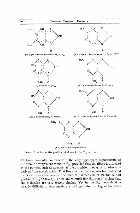
208 DOROTHY CROWFOOT HODGKIN:
(IX.) s: 6-Dimethyl-benziminazole, in B12• (X.) S-Hydroxy-benziminazoJe, in Factor "III".
(XIII.) Hypoxanthine, in Factor G.
HC N N
3 "" / "" / '\ C C , I II CH
N~ /C"" // C N I I
NH2 R (XII.) .-Methyl-adenine, in Factor A.
HC N N 3 "" / "" / '\ C C ,
I II CH N C / ~/""/ C N
I I OH R
(XIV.) 2-Methyl-hypoxanthine, in Factor H.
CH-S N N 3 ""C/ ""C/ ,
I II CH N C / ~/""/
C N I I
NH2 R (XV.) 2-Methyl-mercaptoadenine.
Note: R indicates the position of ribose in the B12 factors.
All these molecules conform with the very rigid space requirements of the atomic arrangement found in B12, provided that the ribose is attached to the purines, such as adenine, at the 7 position, not 9, as in adenosine derived from nucleic acids. That this must be the case, was first indicated by X-ray measurements of the unit cell dimension of Factor A and 'IjJ-vitamin B12 (Table 2). These are so much like B12 that it is clear that the molecules are very closely similar. Yet in the B12 molecule it is already difficult to accommodate a hydrogen atom at C(4) of the benz-

X-ray Analysis and the Structure of Vitamin Blz'
iminazole group in contact with the nucleus; it would be quite impossible to place the amino group at position 6 of adenine at this site. The recent isolation of 7-(D-ribofuranosido)-adenine from 7p-vitamin B 1z by FRIEDRICH
and BERNHAUER (2I) is welcome confirmation of the expected difference between the B12 nucleosides and adenosine.
Table 2. Unit Cell Dimensions (A) of Blz-like Factors Compared
with Blz dry.
Factor A 'P·B" B12
a 22.6 21.4 24·35
b 22.0 21.0 21.29
C I6.I I6.1 16.02
The structure of the cobalt containing nucleus is even more extraordinary. It is obvious that it is closely related biogenetically to porphyrins of Type III and to porphobilinogen-the side-chains at
HOOC.CH, .CH,' COOH +
COOH I
COOH CH I I 2 CH CH
\ 2 I' C-C
II ~ NH-CH -C- CH
2 2 'N/ H
NH .CH .COOH 2 2
HOOC.CH.CH .CO·CH ·COOH 2, I
NH,
COOH I
COOH CH I I 2 CH CH I 2 I 2
CH, .••• CO I I
..... CO. fH, NH -CH ".
2 2 NH,
(XVI.) Biosynthesis of porphyrins and of vitamin B12 •
Fortschritte d. Chern. org. Naturst. XV.
2IO DOROTHY CROWFOOT HODGKIN:
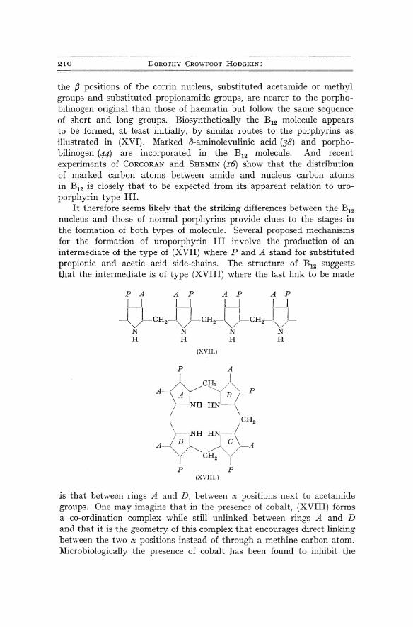
the (J positions of the corrin nucleus, substituted acetamide or methyl groups and substituted propionamide groups, are nearer to the porphobilinogen original than those of haematin but follow the same sequence of short and long groups. Biosynthetically the BI2 molecule appears to be formed, at least initially, by similar routes to the porphyrins as illustrated in (XVI). Marked 8-aminolevulinic acid (38) and porphobilinogen (44) are incorporated in the B12 molecule. And recent experiments of CORCORAN and SHEMIN (I6) show that the distribution of marked carbon atoms between amide and nucleus carbon atoms in B12 is closely that to be expected from its apparent relation to uroporphyrin type III.
I t therefore seems likely that the striking differences between the B12 nucleus and those of normal porphyrins provide clues to the stages in the formation of both types of molecule. Several proposed mechanisms for the formation of uroporphyrin III involve the production of an intermediate of the type of (XVII) where P and A stand for substituted propionic and acetic acid side-chains. The structure of B12 suggests that the intermediate is of type (XVIII) where the last link to be made
P A A PAP A P
I I I I I I I I
~)-CH2-~)-CH2-~)-CH2J~)-N N N N H H H H
(XVIL)
P P (XVIIL)
is that between rings A and D, between eX positions next to acetamide groups. One may imagine that in the presence of cobalt, (XVIII) forms a co-ordination complex while still unlinked between rings A and D and that it is the geometry of this complex that encourages direct linking between the hvo eX positions instead of through a methine carbon atom. Microbiologically the presence of cobalt has been found to inhibit the
X-ray Analysis and the Structure of Vitamin B12 • 2II
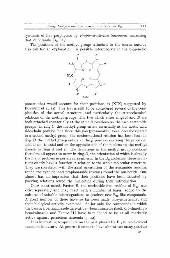
synthesis of free porphyTins by Propionibacterium Shermanii, increasing that of vitamin B12 (34).
The positions of the methyl groups attached to the corrin nucleus also call for an explanation. A possible intermediate in the biogenetic
P A
t 1* / ' * /',
A~ ',~"/ , P \ I li~ /=c-N N \
HgC /
_/-f NI=-~ A '~. (*A ~/ * '" *1
P P (XIX.)
process that would account for their positions, is (XIX) suggested by BONNETT et al. (5). This leaves still to be considered several of the complexities of the actual structure, and particularly the stereochemical relations of the methyl groups. The two which enter rings A and Bare both attached equatorially at the same f3 positions as the two acetamide groups; in ring C, the methyl group enters essentially at the acetic acid side-chain position but since this has (presumably) been decarboxylated to a second methyl group, the conformational relation has been lost; in ring D the methyl group enters at the f3 position carrying the propionic acid chain, is axial and on the opposite side of the nucleus to the methyl groups in rings A and B. The deviations in the methyl group positions therefore all appear to occur in ring D, the orientation of which is already the major problem in porphyrin synthesis. In the B12 molecule, these deviations clearly have a function in relation to the whole molecular structure. They are correlated with the ayjal orientation of the acetamide residues round the cyanide, and propionamide residues round the nucleotide. One almost has an impression that their positions have been dictated by packing relations round the nucleotide during their introduction.
Once constructed, Factor B, the nucleotide-free residue of B12, can exist separately and may react with a number of bases, added to the cultures of suitable microorganisms to produce new B12 like compounds. A great number of these have so far been made biosynthetically, and their biological activity examined. So far only the compounds in which the base is a benziminazole derivative-benziminazole itself, 5: 6-dimethylbenziminazole and Factor III have been found to be at all markedly active against pernicious anaemia (3, I4).
It is interesting to speculate on the part played by B12 in biochemical reactions in nature. At present it seems to have almost too many possible

212 DOROTHY CROWFOOT HODGKIN:
roles, e. g. in the synthesis of methyl groups, in carbohydrate metabolism, possibly through an effect on SH containing enzymes, on the synthesis of nucleic acids, perhaps also in protein synthesis.
Much more work is clearly necessary to discover the actual chemical process or processes involved in the effects observed. But one can see certain features of the B12 structure that seem significant. First, it is rather rigid. The whole of the benziminazole-phosphate-propanolamine unit is confined in a definite position, from which it can only move away bodily (as in the cyanide reaction, for example). The negatively charged oxygen atoms of the phosphate group project from the surface, easily available for reaction. Though the acetamide and propionamide chains have greater freedom of movement and may-and do-swing in and out from the centre of the molecule, the distances between them in these processes are unlikely to differ much from the average of about 7 A. It is quite easy geometrically to see how they might be involved in attaching B12 to a protein intrinsic factor or concerned in some scheme for protein synthesis. Lastly, the cyanide group could easily be attached to methyl groups* and lose them again, or be ejected from the molecule altogether, leaving the cobalt atom open to take part in further oxidationreduction reactions.
VI. The Crystal Structures of Vitamin B12 and the Hexacarboxylic Acid.
The X-ray analytical process involves placing atoms not only in the molecules studied but also in the crystals. And so we have detailed evidence of the way in which these large molecules make contact with one another in the different crystal structures examined and their relations to solvent molecules, particularly water of crystallisation, in the spaces between them.
In the case of the hexacarboxylic acid the X-ray analysis has reached a stage at which it seems likely that every atom in the unit cell has been placed reasonably closely. Fig. 24 shows two projections of the crystal structure found, which make it look complicated enough. The large disc shaped molecules are closely packed together, with the main planes of neighbouring "planar" groups making angles of roughly 45 0 to one another. There is comparatively little space between them-many of the carboxyl groups make direct hydrogen bonded contacts with one another. Two water molecules assist in bridging rather longer gaps between the active groups; in the one definite hole in the crystal structure, there appears to be an acetone molecule. It is a remarkable fact that of the different atoms capable of forming hydrogen bonds in the crystal,
* As suggested to me by Dr. L. E. ORGEL.

1\ ~
\ ...~
. .... -
.,\
;.;
·c.(~
J-
a
Fig
. 24
. P
roje
ctio
ns
of t
he a
tom
ic.
posi
tion
s fo
und
in
the
crys
tal
stru
ctur
e of
th
e he
xaca
rbox
ylic
aci
d,
(a)
proj
ecti
on
alon
g th
e a
axis
; th
e fo
ur
sym
met
ry
rela
ted
mol
ecul
es o
verl
ap,
and
are
ind
icat
ed b
y d
iffe
rent
var
ieti
es o
f li
nes
join
ing
atom
ic p
osit
ions
, (b
) pr
ojec
tion
alo
ng t
he c
axi
s.
The
hea
vy li
nes
indi
cate
the
sid
e of
th
e m
olec
ule
near
est
to
the
obse
rver
. T
he
num
bers
I,
2
, 3,
4
atta
ched
\ to
the
cob
alt
atom
s co
rrel
ate
the
mol
ecul
ar
posi
tion
s sh
own
in {
a}
and
{b)
.
~ >1
P' ~ :> ::: P' .;:r
~.
w § 0. g (fJ 8" " g (1
) 8.. ~ P'
S S· f I<J .... W

DOROTHY CROWFOOT HODGKIN;
only one oxygen of a carboxyl group is too far (3.2 A) from any neighbour actually to do so. The acetone molecule and one of the water molecules appear from the observed electron density levels to be rather less precisely fixed in the crystal than most of the atoms of the hexacarboxylic acid; one side-chain in their neighbourhood also shows signs of disorder.
Fig. 25 a.
Fig. 25 . Projected atomic positions in the crystal structure of wet B12> (a) on the a plane, (b) on the c plane. In (a) one layer of molecules is shown complete and a single molecule (thin lines) of the layer above. Only a few water molecules are shown in both projectionsJ particularly most of those in the "pool" are omitted,
leaving an apparent empty space in (b) .
In the B12 crystals the structure analysis is complicated by the relatively large amount of solvent of crystallisation present. Here there is evidence of still greater disorder in the water molecule positions and these are still being investigated. Formally from cell size and density considerations we expect to find about twenty-five water molecules per B12 molecule in the wet B12 crystal structure and perhaps eighteen in the air dried crystals (Table I, p. I75).
Fig. 25 shows the positions so far found for atoms in the crystal structure of wet B12. It is difficult in two dimenslOns to give an accurate

X-ray Analysis and the Structure of Vitamin B12.
impression of the three-dimensional relationships of these large molecules, turning about two fold screw axes of symmetry and settling side by side in the crystal with every atom so placed in relation to every other that they closely conform with all the stereochemical rules found to hold in much simpler crystal structures. The molecules lie in two layers
Fig. 25 b.
parallel with the a plane and have very approximately a close packed arrangement in these layers-the planar groups themselves of neighbouring molecules within each layer are only slightly tilted out of the same a plane. The separate layers of molecules are connected in the crystal through a very regular hydrogen bond system involving the phosphate groups projecting alternately up and down in the a direction (Fig. 25 b). This hydrogen bond system is shown diagrammatically in Fig. 26-it links the phosphate groups through two intervening water molecules. In other directions in the crystal structure the molecules make a number of direct hydrogen bonded contacts with one another, amide to amide and amide to phosphate. These contacts themselves provide very interesting evidence

216 DOROTHY CROWFOOT HODGKIN:
of the accuracy of the X-ray analysis. In e 3 for wet B12, the oxygen and nitrogen atoms of the amide groups were not distinguished in the phasing calculation but were assigned a mean scattering factor of 71/2'
In the resulting electron density maps, however, the atoms appear in all but one amide group, definitely unequal in weight, and unequal in a perfectly sensible direction. This can be traced throughout the crystal structure. \Vherever two groups make direct contact, the heavier atom,
0,- p"--O ._ ..... _ .. _. H-q/ H I \ \ O-H-·· .. ··· .. · .. ·O /0
° i. H '---..P ° i /\ ° \ \0 ! ° ° ~ I . , P : O-H .. _ .... ·· .. ·· 0- "'--0 .............. H-6
'H Fig. 26. Phosphate~water molecule chain, projected along the b axis.
oxygen of one is connected with the lighter, nitrogen atom, of the other-for example in the contact between the acetamide group on ring A and the propionamide group on ring C of the succeeding molecule in the c direction. In both cases, where an atom of an amide group is in hydrogen bonded contact with a phosphate oxygen, it is clearly the nitrogen atom that is involved.
Many of the water molecules in the crystal, e. g. in the phosphate hydrogen bonded chain, are in well defined sites, connected with active groups on the molecule. But there is also in the crystal what may almost be described as a pool of water. This extends from one side of the unit cell to the other, as Fig. 25 shows, along the c axis, parallel with the phosphate chain. It spreads out towards this chain in the direction of the b axis. In this region, it is very difficult to be sure of the arrangement of the oxygen atoms. It has been studied by the calculation of a number of difference electron density maps, the latest characterised as L1 e 5. These maps show small peaks of the order of I electronjA 3, more than enough peaks to represent the water molecules in the crystal but often conflicting with one another. At present it seems most likely that these peaks represent sites which are sometimes occupied, sometimes not. It is possible among them to select about twenty-two sites reasonably arranged, at distances of about 2.7-3.2 A from one another, usually roughly 4 co-ordinated, though the angles involved may be far from tetrahedral. To these sites, there are others that seem to be strategically related-places to which particular water molecules might move and make rather different, often quite as sensible, contacts, with another set of atoms. This seems such a reasonable situation, it is likely to be true-but it is somewhat hard to represent in terms of exact X-ray analysis.
X-ray Analysis and the Structure of Vitamin B12 • 21 7
When wet B12 crystals are picked out of their mother liquor, almost certainly some of the water molecules move out of the crystal along the channel observed. As a result, the B12 molecules move to fill the space. They tilt at a rather greater angle to the a plane and turn a little in this plane. One acetamide group, on ring B, swings round the
bond )C-CH2-, out of contact with a phosphate group on a
neighbouring molecule and into contact with the cyanide group on the same molecule. Another, on ring A, turns a little about the bond -CH2-C. The water molecule pattern in the region of the pool also changes a good deal-it is still being explored. In the drawn-out electron density contours of the atoms in this region one can almost see the turning of the amide groups, the movement of the water molecules. The atomic arrangement is only relatively stable. If the crystals are left exposed to the air, still more water passes out of the structure and this becomes much more disordered, giving very few X-ray reflections. It is tempting to see in the early conflicting analytical results on B12 the end of this process. Some of the water molecules, e. g. in the phosphate chain, are so caught in the meshes of the B12 molecules they must be very hard to remove except on most drastic drying. There are about six which appear so strongly in electron density maps at all stages of our analysis that in our early speculative period we often attempted to link them by chemical bonds to the molecule itself.
If one looks closely at the positions of the atoms found within each molecule in each crystal, they provide remarkable illustrations of the interplay of the different factors controlling them, chemical, stereochemical, conformational, intermolecular-all the various forces of attraction and repulsion. At first sight, the degree to which the atoms adopt preferred conformations is one of the most convincing features of the structure as a whole-witness, for example, the staggered relations of the substituents at the fJ position of the five-membered rings or the extended staggered form of the propionamide chains on rings Band D. But in detail, one can see small deviations, and also why they arise. The propionamide chain, for example, on ring A, is turned a little from the preferred orientation of the bond C(30) -C(31) to the bonds C(2) -C(3) and C(3)-C(4) in the five-membered ring. This enables the amide nitrogen atom to come within hydrogen bonded contact of a phosphate oxygen atom in a neighbouring molecule. The actual conformations adopted by the different side-chains differ a little in the hexacarboxylic acid and in the B12 crystals-and even from wet to air dried crystals, as described above. Clearly, within a little, they are adjustable in relation to the surrounding country, whether in the crystals or in the living organism.

218 DOROTHY CROVlFOOT HODGKIN:
VII. Conclusion.
To this necessarily imperfect account of vitamin B12, I might perhaps add. All structure analyses, once achieved, seem easy in retrospect. There is nothing about the B12 crystal structures that now suggests that they represent the limit of molecular size that can be explored by X-ray analysis. Indeed already promising stages have been reached in the study of larger molecules. There are particular dangers and difficulties in the kinds of operations we have to use in these studies but, with sufficient controls, as the B12 analysis illustrates, they do not seem insuperable. That we know so much about B12 seems marvellous until we begin to think how much more there still is to know-its precise relations with the molecules around it in living tissues, the reactions by which it prevents pernicious anaemia or promotes growth-before we can know these too, we see before us a vista of still more intimate chemical studies, and more complicated X-ray analyses.
References.
I. ALICINO, J. F.: Perchloric Acid Salt of Vitamin Bu' J. Amer. Chem. Soc. 73, 4051 (I95 I ).
2. BEAVEN, G. H., E. R. HOLIDAY, E. A. JOHNSON, B. ELLIS and V. PETROW: The Chemistry of Antipernicious Anaemia Factors. VI. The Mode of Combination of Component ex in Vitamin B l2. J. Pharm. Pharmacal. 2, 944 (1950).
3. BERNHAUER, K., K. BLUMBERGER und P. PETRIDES: Die ,Virkung des "VitaminB l2-Faktors III" bei der pernici5sen Anamie. Arzneimittel-Forsch. 5, 442 (1955)·
4. BONNETT, R., J. R. CANNON, V. M. CLARK, A. W. JOHNSON, L. F. J. PARKER, E. LESTER SMITH and A. TODD: Chemistry of the Vitamin Bl2 Group. Part V. The Structure of the Chromophoric Grouping. J. Chem. Soc. (London) I957, II58.
5. BONNETT, R., J. R. CANNON, A. ,V. JOHNSON, I. SUTHERLAND, A. R. TODD and E. LESTER SMITH: The Structure of Vitamin Bl2 and its Hexacarboxylic Acid Degradation Product. Nature (London) I76, 328 (I955).
6. BONNETT, R., J. R. CANNON, A. W. JOHNSON and A. TODD: Chemistry of the Vitamin Bl2 Group. Part. IV. The Isolation of Crystalline Nucleotide-free Degradation Products. J. Chem. Soc. (London) I957, I I48.
7. BRINK, C., D. C. HODGKIN, J. LINDSEY, J. PICKWORTH, ]. H. ROBERTSON and J. G. WHITE: X-ray Crystallographic Evidence on the Structure of Vitamin B!2' Nature (London) I74, II69 (I954).
8. BRINK, N. G., F. A. KUEHL, Jr. and K. FOLKERS: Vitamin B l2. The Identification of Vitamin B12 as a Cyano-Cobalt Coordination Complex. Science (Washington) II2, 354 (I950)'
9. BROWN, F. B. and E. LESTER SMITH: New Purines in Bl2 Vitamins. Proc. Biochem. Soc., Biochemic. J. 56, XXXIV (I954).
IO. CALLOMAN, H. J.: Studies in Molecular Spectroscopy. D. Phil. Thesis, Oxford University, 1954.
II. CANNON, J. R., A. W. JOHNSON and A. R. TODD: A Crystalline Nucleotide-free Degradation Product of Vitamin B 12. Nature (London) I74, II68 (I954).

X-ray Analysis and the Structure of Vitamin B12. 21 9
I2. CASON, J., C. GASTALDO, D. L. GLUSKER, J. ALLINGER and L. B. ASH: BranchedChain Fatty Acids. XXVII. Further Study of the Dependence of Rate of Amide Hydrolysis on Substitution Near the Amide Group. Relative Rates of Hydrolysis of Nitrile to Amide and Amide to Acid. J. Organ. Chern. (USA) 18, 1129 (1953)·
I3. CASON, J. and H. J. \VOLFHAGEN: Location of Branching Methyl Groups near Carboxyl by Rate Studies of Amide Hydrolysis. J. Organ. Chern. (USA) 14, 155 (1949)·
I4. COATES, M. E. and S. K. KON: Biological and Microbiological Activities of Purine and Benziminazole Analogues of Vitamin B 12 • Europaisches Symposion tiber Vitamin B12 und Intrinsic Factor, P.72. Stuttgart: F. Enke. 1957.
IS. COOLEY, G., B. ELLIS, V. PETROW, G. H. BEAVEN, E. R. HOLIDAY and E. A. JOHNSON: Some Transformations of Vitamin B12 b' J. Pharm. Pharmacol. 3, 271 (195 1).
I6. CORCORAN, J. W. and D. SHEMIN: On the Biosynthesis of Vitamin B 12 : the Mode of Utilisation of o-Aminolevulinic Acid. Biochim. Biophys. Acta 25, 661 (1957).
I7. ELLIS, B., V. PETROW and G. F. SNOOK: The Isolation of the Crystalline Antipernicious Anaemia Factor from Liver. J. Pharm. Pharmacol. 1, 60 (1949).
I8. FANTES, K. H., J. E. PAGE, L. F. J. PARKER and E. LESTER SMITH: Crystalline Anti-pernicious Anaemia Factor from Liver. Proc. Roy. Soc. (London) 136 B, 592 (1950).
I9. FOLKERS. K. and D. E. WOLF: Chemistry of Vitamin Bn. Vitamins and Horm. 12, I (1954),
20. FRIEDRICH, \Y. und K. BERNHAUER: Beitrage zur Chemie und Biochemie der "Cobalamine", I. Mitt.: Vber die Alkylierung von B 12-Faktor III und Vitamin B 12. Chem. Ber. 89, 2030 (1956).
2I. - - Beitrage zur Chemie und Biochemie der "Cobalamine", II. Mitt.: Vber den Abbau der "Cobalamine" mit Cer(III)-hydroxyd. 7-[D-RibofuranosidoJ-adenin, ein Abbauprodukt des Pseudovitamins B 12. Chern. Ber. 89, 2507 (1956) .
.22. - - Zur Chemie und Biochemie der "Cobalamine", VII. Mitt. z-Methylmercaptoadenin-cobalamin-analogon, ein neuer B 12-Faktor des Faulschlammes. Chern. Ber. 90, 1966 (1957).
23. HEINRICH, H. C. (Editor): Europaisches Symposion tiber Vitamin B12 nnd Intrinsic Factor. Stuttgart: F. EnIce-Verlag. 1957.
24. HODGKIN, D. C., A. \V. JOHNSON and A. R. TODD: The Structure of Vitamin B 12. Chern. Soc. (London) Special Publications 3, 109 (1955).
25· HODGKIN, D. c., J. KAMPER, J. LINDSEY, M. MACKAY, J. PICKWORTH, J. H. ROBERTSON, C. B. SHOEMAKER; J. G. WHITE, R. J. PROSEN and K. N. TRUEBLOOD: The Structure of Vitamin B l2 . I. An Outline of the Crystallographic Investigation of Vitamin B 12. Proc. Roy. Soc. (London) 242 A, zz8 (1957).
26. HODGKIN, D. C., J. KAMPER, M. MACKAY, J. PICKWORTH, K. N. TRUEBLOOD and J. G. WHITE: The Structure of Vitamin B 12. Nature (London) 178, 64 (1956).
27. HODGKIN, D. C., J. PICKWORTH, J. H. ROBERTSON, K. N. TRUEBLOOD, R. J. PROSEN and J. G. \VHITE: The Crystal Structure of the Hexacarboxylic Acid Derived from B12 and the Molecular Structure of the Vitamin. Nature (London) 176, 32 5 (1955)·
28. HODGKIN, D., M. \V. PORTER and R. C. SPILLER: Crystallographic Measurements on the Anti-pernicious Anaemia Factor. Proc. Roy. Soc. (London) 136 B, 609 (1950).

220 D. CROWFOOT HODGKIN: X-ray Analysis and the Structure of Vitamin B 12.
29. KAMPER, J. and D. C. HODGKIN: Some Observations on the Crystal Structure of a Chlorine-substituted Vitamin B 12 . Nature (London) 176, 551 (1955).
30. KUEHL, F. A., Jr., C. H. SHUNK and K. FOLKERS: Vitamin B 12. XXIV. DL-3,3-Dimethyl-2,5-dioxo-4-hydroxy-pYlTolidine-4-propionic Acid Lactone and DL-3,3-Dimethyl-2,5-dioxopyrrolidine-4-propionic Acid, new Degradation Products. J. Amer. Chern. Soc. 77, 25 1 (1955)·
3I. LUZZATI, V.: Resolution d'une structure cristalline lorsque les positions d'une partie des atomes sont connues: Traitement statistique. Acta Crystallogr. 6, 142 (1953)·
32. MATHIESON, A. McL.: The Direct Determination of Molecular Structure and Configuration of Moderately Complex Organic Compounds. Rev. Pure and Applied Chern. 5, 113 (1955).
33. MINOT, G. R. and W. P. MURPHY: Treatment of Pernicious Anaemia by a Special Diet. J. Amer. Med. Assoc. 87, 470 (1926).
34. PAWELKIEWICZ, J. and K. ZODROW: The Synthesis of Free Porphyrins by Propionibacterium. Shermanii. Acta Biochem. Polonica 3, 225 (1956).
35. PEERDEMAN, A. F., A. J. VAN BOMMEL and J. M. BI]VOET: Determination of Absolute Configuration of Optically Active Compounds by Means of X-rays. Proc. Acad. Sci. Amsterdam, B 54, 16 (1951).
36. RICKES, E. L., N. G. BRINK, F. R. KONIUSZY, T. N. WOOD and K. FOLKERS: Crystalline Vitamin B l2 . Science (\Vashington) 107, 396 (1948).
37. SCHMID, H., A. EBNOTHER und P. KARRER: Beitrag zur Kenntnis des Vitamins B12. Helv. Chim. Acta 36, 65 (1953).
38. SHEMIN, D., J. W. CORCORAN, C. ROSENBLUM and I. M. MILLER: On the Biosynthesis of the Porphyrin-like Moiety of Vitamin B 12. Science (Washington} 124, 272 (1956).
39. SMITH, E. LESTER: The Isolation and Chemistry of Vitamin B 12. Biochemical Society Symposia 13, 3 (1955).
40. SMITH, E. LESTER and L. F. J. PARKER: Purification of Anti-pernicious Anaemia. Factor. Proc. Biochem. Soc., Biochemic. J.43, VIn (1948).
4I . TODD, A. R.: Vitamin B12. Osterr. Chemiker-Zeitung 58, 113 (1957)' 42. TODD, A. R. and A. W. JOHNSON: Vitamin B 12. Rev. Pure and Applied Chern. 2,
23 (1952). 43. Vos, A.: to be published. 44. WATSON, C. J.: Porphyrin Metabolism in Anemias. GEORGE MINOT Lecture.
Arch. Internat. Medicine 99, 323 (1957). 45. WI]MENGA, H. G., W. L. C. VEER and J. LENS: Vitamin B l2. II. The Influence
of HCN on Some Factors of the Vitamin Bl2 Group. Biochim. Biophys. Acta 6, 229 (1950).
46. WOLF, D. E., \V. H. JONES, J. VALIANT and K. FOLKERS: Degradation of Vitamin B12 to D g-I-Amino-2-propanol. J. Amer. Chern. Soc. 72, 2820 (1950).
(Received, December I7, I957.)