zedoary oil (ezhu you) inhibits proliferation of ags cells
TRANSCRIPT

Shi et al. Chinese Medicine 2013, 8:13http://www.cmjournal.org/content/8/1/13
RESEARCH Open Access
Zedoary oil (Ezhu You) inhibits proliferation ofAGS cellsHailian Shi1,2,3, Bao Tan4, Guang Ji1, Lan Lu2,3, Aili Cao2,3, Songshan Shi2 and Jianqun Xie1*
Abstract
Background: Zedoary (Curcumae Rhizoma, Ezhu), a Chinese medicinal herb, has been reported to show anticanceractivity. This study aims to investigate the effect of zedoary oil (Ezhu You) on the proliferation of AGS cells which isone gastric cancer cell line.
Methods: The main ingredients of the herb were detected by GC-MS for herbal quality control. Cell viability wasmeasured by MTT assay and cell proliferation was investigated by immunocytochemical staining for proliferating cellnuclear antigen (PCNA) protein. In addition, the cell cycle distributions were detected by flow cytometry with propidiumiodine (PI) staining and the apoptosis rates were evaluated by flow cytometry with annexin V/PI double-staining. Themorphological changes associated with apoptosis were observed by Hoechst 33342/PI double-staining. Proteinexpression was determined by western blot analysis.
Results: The main ingredients of the herb, including curzerene (26.45%), eucalyptol (12.04%), curcumol (9.04%), pyridine(7.97%), germacrone (7.89%), β-elemene (7.36%), τ-elemene (4.11%) and 28 other ingredients, including curdione, wereconsistent with the chemical profiles of zedoary. Zedoary oil significantly decreased the cell viability of AGS cells (P < 0.01)and MGC 803 cells (P < 0.01), and the inhibitory effects were attenuated by elevated concentrations of FBS. At highconcentrations (≥90 μg/mL), zedoary oil killed GES-1 cells. At low concentrations (≤60 μg/mL), zedoary oil wasless inhibitory toward normal gastric epithelial cells than gastric cancer cell lines. In AGS cells, zedoary oil inhibited cellproliferation in a dose- and time-dependent manner, with decreased PCNA protein expression in the zedoary oil-treated cells, and arrested the cell cycle at S, G2/M and G0/G1 stages after treatment for 6–48 h. At concentrations of 30,60 and 90 μg/mL, which resulted in significant inhibition of proliferation and cell cycle arrest, zedoary oil induced cellapoptosis. In addition, Hoechst 33342/PI double-staining confirmed the morphological characteristics of cell apoptosisat 24 h. Zedoary oil upregulated the ratio of Bax/Bcl-2 protein expression (P < 0.01).
Conclusions: Zedoary oil inhibited AGS cell proliferation through cell cycle arrest and cell apoptosis promotion, whichwere related to Bax/Bcl-2 protein expression.
BackgroundCurcuma phaeocaulis, Curcuma kwangsiensis and Curcumawenyujin are named zedoary in the Chinese Pharmacopoeiaand used as antiviral and antimicrobial medicines [1-3].Zedoary oil is a Chinese medicine that is used for treatmentof gynecologic inflammation [4], pneumonia [5], pediatricdiseases [6], viral myocarditis [7] and malignant tumors,such as oophoroma, hepatocellular carcinoma and lung
* Correspondence: [email protected] of Digestive Disease, Longhua Hospital, Shanghai University ofTraditional Chinese Medicine, 725 South Wanping Road, XuHui District,Shanghai 200032, PR ChinaFull list of author information is available at the end of the article
© 2013 Shi et al.; licensee BioMed Central LtdCommons Attribution License (http://creativecreproduction in any medium, provided the or
cancer [8-11]. Moreover, zedoary oil is a safe drug withlow toxicity [12]. Deng et al. [13] reported that the acutetoxicity of zedoary turmeric oil gelatin microspheres mightresult from dystopic embolism rather than the zedoary tur-meric oil itself entrapped in the microspheres.Zedoary belongs to the Zingiberacea family, which is
composed of about 70 species of rhizomatous herbs athome and abroad, with approximately 20 species existingin China [1,10]. Different species of zedoary and differentpreparations have different chemical ingredients [14], whichresult in different biological actions. Many chemical analysismethods, including thin-layer chromatography scanning,high-performance liquid chromatography, gas chromatog-raphy (GC) and gas chromatography–mass spectrometry
. This is an Open Access article distributed under the terms of the Creativeommons.org/licenses/by/2.0), which permits unrestricted use, distribution, andiginal work is properly cited.

Shi et al. Chinese Medicine 2013, 8:13 Page 2 of 11http://www.cmjournal.org/content/8/1/13
(GC-MS), are used to detect the chemical compounds inessential oil of zedoary and also for quality control [15].Gastric glandular cells are replaced by intestinal-type
epithelial cells with high cell proliferation rates and fibrosisin severe inflammation of the gastric membrane, resultingin chronic atrophic gastritis with intestinal metaplasia anddysplasia, imbalance between cell proliferation and apop-tosis in the normal gastric mucosa and increasing incidenceof gastric cancer [16-19].Proliferating cell nuclear antigen (PCNA) was originally
identified as an antigen expressed in the nuclei of cellsduring the DNA synthesis phase of the cell cycle [20], andonly exists in normal proliferative cells and cancer cells.Bax and Bcl-2 are very important for cytochrome c-
dependent apoptosis. Bax inserts itself into the outermitochondrial membrane, followed by cytochrome c re-lease from mitochondria. In contrast, when Bcl-2 bindsto the outer mitochondrial membrane, the release of cyto-chrome c is blocked [21,22]. Many anticancer agents caninduce release of cytochrome c by upregulating Bax ex-pression and/or downregulating Bcl-2 expression [23-25].Chinese medicines are available for treatment of
patients with chronic atrophic gastritis [26]. Zedoary-containing Chinese herbal formulas, e.g., Weiqi decoc-tion (an empirical formula from Longhua Hospital,Shanghai University of Traditional Chinese Medicine,China), are often used for treatment of gastric diseases[27-29]. However, the effects of zedoary oil on gastricepithelial cells with high proliferation rates are unclear.The AGS cell line, a type of human gastric cancer epi-
thelial cell line, is used as a cell model for abnormal pro-liferation and apoptosis in the gastric mucosa andgastric cancer research [30,31]. The present study aimsto investigate the effect of zedoary oil (Ezhu You) onAGS cell proliferation.
MethodsMaterialsZedoary oil was purchased from Shanghai Institute forthe National Institute for the Control of Pharmaceuticaland Biological Products (Lot No. 111544-200703)(Shanghai, China). AGS (TCHu 7) and MGC 803 (TCHu84) cell lines were purchased from Cell Bank of Aca-demia Sinica (Shanghai, China). The GES-1 cell line waspurchased from Cell Bank of Chinese Academy of Med-ical Sciences (Beijing, China). Ham’s/F-12, DMEM/HighGlucose and RPMI 1640 media were purchased fromThermo Fisher Scientific Inc. (IL, USA). Fatal bovineserun (FBS) was purchased from Hangzhou Sijiqing Bio-logical Engineering Materials Co. Ltd. (Hangzhou,China). FBS (Lot No. 989268) was purchased fromGibco (NY, USA). DMSO (Lot No. 1988B176) was pur-chased from Amresco (OH, USA). MTT (Lot No.091205) was purchased from Richu BioScience Co. Ltd.
(Shanghai, China). PI (Lot No. 118 K3538) and DMSO(Lot No. 019 K2300) were purchased from Sigma Chem-ical Co. (MO, USA). A Hoechst 33342/PI Apoptosis/deathStaining Kit (Catalogue No. C1056) and Hematoxylin Stain-ing Kit (Catalogue No. C0107) were purchased fromBeyotime (Shanghai, China). Trypsin (Lot No. 632461) waspurchased from Invitrogen (CA, USA). RNase A (Lot No.3408B040) was purchased from Beijing Jingkehongda Bio-tech. Co. Ltd. (Beijing, China). Annexin V (Lot No. 40601)was purchased from BioVision Inc. (CA, USA). Anti-Bax(Lot No. 4), anti-Bcl-2 (Lot No. 2) and anti-β-actin (LotNo. 3) antibodies were purchased from Cell Signaling Tech-nology (MA, USA). An anti-PCNA (Lot No. YJ020304CS)antibody was purchased from Epitomics (CA, USA). AnSABC Kit (Lot No. 06L06AJ) and a DAB Kit (CatalogueNo. AR1022) were purchased from Wuhan Boster Bio-logical Technology Co. Ltd. (Wuhan, China). An ECL plusKit (Lot No. 84A) was purchased from GE Healthcare(NA, UK). Cell plates were purchased from Greiner Bio-One (Frickenhausen, Germany). Syringe filters were pur-chased from Pall Co. Ltd. (MI, USA).
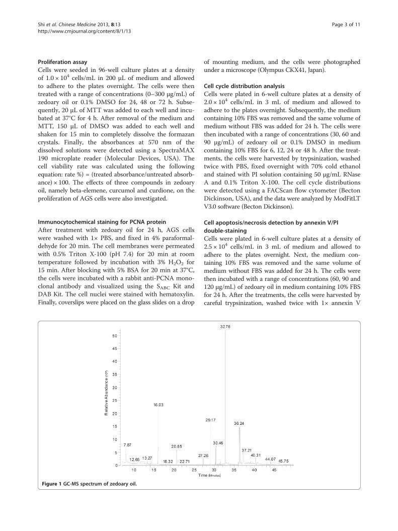
Identification of the main constituents in zedoary oil byGC-MS analysisZedoary oil was diluted with chloroform at a ratio of1:1000. The analysis was performed on a Thermo Sci-entific system composed of a DSQ mass spectrometercoupled with a Trace GC Ultra gas chromatographand an AS 3000 autosampler (Thermo Scientific,USA). The GC was equipped with a 30-m (0.25-mminternal diameter; 0.25-μm film thickness) TR-5MSfused-silica capillary column (Thermo Scientific). Thesplitless injection port temperature was set at 250°C.The column temperature program was 50°C for1 min, followed by elevation to 110°C at 5°C/min,140°C at 3°C/min and 170°C at 5°C/min. Finally, thetemperature was raised to 230°C at 3°C/min and heldat 230°C for 5 min. The constant flow rate was1 mL/min He. The MS was operated in the positiveEI mode. The ion source temperature was set at 250°C.The peaks were identified by comparisons withThe National Institute of Standards and Technology(NIST) library. Relative quantitative data wereobtained from the normalized peak areas: % area =(area/total area) × 100.
Cell cultureAGS cells, MGC 803 cells (gastric cancer cell line) andGES-1 cells (normal gastric epithelial cells) were cul-tured in Ham’s/F-12, RPMI 1640 and DMEM mediasupplemented with 10% FBS, respectively, at 37°C in a5% CO2 atmosphere.
Shi et al. Chinese Medicine 2013, 8:13 Page 3 of 11http://www.cmjournal.org/content/8/1/13
Proliferation assayCells were seeded in 96-well culture plates at a densityof 1.0 × 10⁴ cells/mL in 200 μL of medium and allowedto adhere to the plates overnight. The cells were thentreated with a range of concentrations (0–300 μg/mL) ofzedoary oil or 0.1% DMSO for 24, 48 or 72 h. Subse-quently, 20 μL of MTT was added to each well and incu-bated at 37°C for 4 h. After removal of the medium andMTT, 150 μL of DMSO was added to each well andshaken for 15 min to completely dissolve the formazancrystals. Finally, the absorbances at 570 nm of thedissolved solutions were detected using a SpectraMAX190 microplate reader (Molecular Devices, USA). Thecell viability rate was calculated using the followingequation: rate %) = (treated absorbance/untreated absorb-ance) × 100. The effects of three compounds in zedoaryoil, namely beta-elemene, curcumol and curdione, on theproliferation of AGS cells were also investigated.
Immunocytochemical staining for PCNA proteinAfter treatment with zedoary oil for 24 h, AGS cellswere washed with 1× PBS, and fixed in 4% paraformal-dehyde for 20 min. The cell membranes were permeatedwith 0.5% Triton X-100 (pH 7.4) for 20 min at roomtemperature followed by incubation with 3% H2O2 for15 min. After blocking with 5% BSA for 20 min at 37°C,the cells were incubated with a rabbit anti-PCNA mono-clonal antibody and visualized using the SABC Kit andDAB Kit. The cell nuclei were stained with hematoxylin.Finally, coverslips were placed on the glass slides on a drop
Figure 1 GC-MS spectrum of zedoary oil.
of mounting medium, and the cells were photographedunder a microscope (Olympus CKX41, Japan).
Cell cycle distribution analysisCells were plated in 6-well culture plates at a density of2.0 × 104 cells/mL in 3 mL of medium and allowed toadhere to the plates overnight. Subsequently, the mediumcontaining 10% FBS was removed and the same volume ofmedium without FBS was added for 24 h. The cells werethen incubated with a range of concentrations (30, 60 and90 μg/mL) of zedoary oil or 0.1% DMSO in mediumcontaining 10% FBS for 6, 12, 24 or 48 h. After the treat-ments, the cells were harvested by trypsinization, washedtwice with PBS, fixed overnight with 70% cold ethanoland stained with PI solution containing 50 μg/mL RNaseA and 0.1% Triton X-100. The cell cycle distributionswere detected using a FACScan flow cytometer (BectonDickinson, USA), and the data were analyzed by ModFitLTV3.0 software (Becton Dickinson).
Cell apoptosis/necrosis detection by annexin V/PIdouble-stainingCells were plated in 6-well culture plates at a density of2.5 × 104 cells/mL in 3 mL of medium and allowed toadhere to the plates overnight. Next, the medium con-taining 10% FBS was removed and the same volume ofmedium without FBS was added for 24 h. The cells werethen incubated with a range of concentrations (60, 90 and120 μg/mL) of zedoary oil in medium containing 10% FBSfor 24 h. After the treatments, the cells were harvested bycareful trypsinization, washed twice with 1× annexin V

Shi et al. Chinese Medicine 2013, 8:13 Page 4 of 11http://www.cmjournal.org/content/8/1/13
binding buffer, resuspended in binding buffer and stainedwith annexin V and PI. Cell apoptosis was detected usingthe FACScan flow cytometer.
Apoptosis and necrosis detection by Hoechst 33342/PIdouble-stainingCells were seeded in 6-well culture plates at a density of2.0 × 104 cells/mL in 3 mL of medium and allowed to
Table 1 Compounds detected in the zedoary oil
ApexRT
Area(%)
Composition AS No.
7.87 7.97 pyridine 110-86-1
11.58 0.45 1,1-ethanediol diacetate 542-10-9
12.65 0.67 α-pinene 80-56-8
13.27 1.18 camphene 79-92-5
13.91 0.21 sabinene 3387-41-5
14.19 0.95 β-pinene 127-91-3
15.82 0.73 bornylene 464-17-5
16.03 12.04 eucalyptol 470-82-6
20.65 3.18 (-)-alcanfor 464-48-2
27.26 1.05 δ-elemene 20307-84-0
29.17 7.36 β-elemene 515-13-9
30.29 0.66 β-cubebene 13744-15-5
30.46 4.11 τ-elemene 30824-67-0
30.72 0.13 β-cubebene 13744-15-5
31.3 0.27 γ-muurolene 30021-74-0
31.44 0.16 γ-cadinene 39029-41-9
31.63 0.6 α-caryophyllene 6753-98-6
32.09 0.31
32.24 0.07
32.43 0.42 germacrene-D 23986-74-5
32.51 0.26 zingiberene 495-60-3
32.76 26.45 curzerene 17910-09-7
32.96 0.39 α-selinene 473-13-2
33.45 0.35 β-cadinene 523-47-7
34.02 0.34 (+)-β-guaiene 88-84-6
36.24 7.89 germacrone 6902-91-6
36.39 7.63
36.78 0.36
37.21 9.04 curcumol 4871-97-0
39.1 0.41 butylidenephtalide 551-08-6
40.31 1.23 curdione 13657-68-6
41.24 0.11
41.53 0.24
41.74 0.23
44.07 1.13 2-[(4-methoxyphenyl)methylene]-Cyclohexanone
5765-29-7
adhere to the plates overnight. The medium containing10% FBS was removed and the same volume of mediumwithout FBS was added for 24 h. The cells were then incu-bated with a range of concentrations (30, 60 and 90 μg/mL)of zedoary oil or 0.1% DMSO in medium containing 10%FBS for 24 h. After the treatments, the cells were harvestedby careful trypsinization and resuspended in staining buffer.The cells were stained with Hoechst 33342 and PI, and ana-lyzed for apoptosis and/or necrosis under a fluorescencemicroscope (Olympus CKX41, Japan). The rates of Hoechst33342-positive cells, whose blue color was brilliant andaggregative, were analyzed by Image-Pro Plus 6.0 soft-ware (Media Cybernetics, USA).
Western blot analysisCells were plated in 6-well culture plates at a density of4.0 × 104 cells/mL in 3 mL of medium and allowed toadhere to the plates overnight. The medium containing10% FBS was removed and the same volume of mediumwithout FBS was added for 24 h. The cells were then incu-bated with a range of concentrations (60, 90 and 120 μg/mL)of zedoary oil or 0.1% DMSO in medium containing 10%FBS for 24 h. After the incubations, the cells were col-lected, lysed with cell lysis buffer and sonicated threetimes for 15 s each. The cell lysates were centrifuged for15 min at 14,000 × g and 4°C, and the supernatants werecollected. The protein samples were separated by SDS-PAGE (15% separating gel and 5% stacking gel) and
Table 2 Effects of zedoary oil and β-elemene oncell viability
Group n Cell viability
24 h 48 h 72 h
Zedoary oil
0 μg/mL 4 99.5 ± 0.9 99.8 ± 0.3 99.9 ± 0.2
1 μg/mL 4 95.8 ± 2.9 82.5 ± 2.5** 71.0 ± 7.0**
10 μg/mL 4 95.0 ± 2.3 77.1 ± 6.2** 69.2 ± 2.3**
30 μg/mL 4 81.2 ± 6.2** 74.3 ± 3.5** 58.4 ± 2.9**
60 μg/mL 4 65.3 ± 5.1** 49.5 ± 4.2** 42.5 ± 5.4**
90 μg/mL 4 47.4 ± 1.5** 19.0 ± 3.6** 16.7 ± 5.9**
100 μg/mL 4 35.4 ± 4.7** 13.7 ± 9.4** 9.8 ± 5.7**
300 μg/mL 4 14.8 ± 2.5** 5.3 ± 0.6** 2.5 ± 0.7**
β-elemene
11.5 μg/mL 3 98.2 ± 2.0 98.1 ± 10.7 91.2 ± 7.4
23 μg/mL 4 97.1 ± 5.0 89.0 ± 8.5 86.8 ± 14.4
92 μg/mL 4 94.1 ± 6.8 81.6 ± 3.1* 79.6 ± 3.5*
184 μg/mL 4 87.6 ± 2.0 60.3 ± 9.5** 70.0 ± 9.1**
368 μg/mL 4 25.1 ± 2.4** 14.0 ± 6.3** 12.0 ± 3.4**
1104 μg/mL 4 9.3 ± 1.3** 3.2 ± 0.7** 5.0 ± 4.1**
AGS cells were treated with zedoary oil in Ham’s/F-12 medium containing 10%FBS for 24, 48 and 72 h. The cell viability was detected by MTT assays. *P < 0.05,**P < 0.01 vs. control cells.

Figure 2 Effects of zedoary oil on the viability of gastric cancer cells and normal gastric epithelial cells. (A) AGS cells were treated withzedoary oil without FBS for 24, 48 and 72 h. (B) AGS cells were treated with zedoary oil containing 3% FBS for 24, 48 and 72 h. (C) MGC 803 cellswere treated with zedoary oil containing 3% FBS for 24, 48 and 72 h. (D) GES-1 cells were treated with zedoary oil containing 3% FBS for 24, 48and 72 h. Data represent means ± SD (N = 3).
Figure 3 Zedoary oil downregulates PCNA protein expression in AGS cells. (A-D) Cells were treated with zedoary oil (0, 30, 60 and 90 μg/mL)for 24 h, respectively. The brown color represents positive expression of PCNA protein. The blue color represents the cell nuclei of AGS cells (200×).The control cells were AGS cells cultured in Ham’s/F-12 medium containing 10% FBS and 0.1% DMSO.
Shi et al. Chinese Medicine 2013, 8:13 Page 5 of 11http://www.cmjournal.org/content/8/1/13
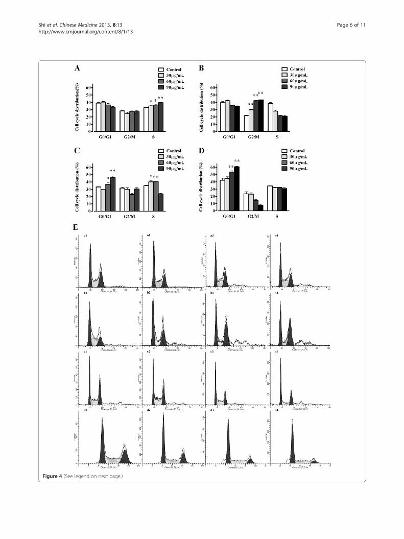
Figure 4 (See legend on next page.)
Shi et al. Chinese Medicine 2013, 8:13 Page 6 of 11http://www.cmjournal.org/content/8/1/13

(See figure on previous page.)Figure 4 Zedoary oil induces cycle arrest in AGS cells. Zedoary oil was added at the indicated concentrations and the cells were incubatedfor 6, 12, 24 and 48 h. The cell cycle proportions were determined by flow cytometry after staining with PI. (A–D) Statistical analyses of the cellcycle phase distributions after 6, 12, 24 and 48 h, respectively. (E) Typical pictures of the respective cell cycle phase distributions from flowcytometry: a1–a4, control, 30, 60 and 90 μg/mL for 6 h; b1–b4, control, 30, 60 and 90 μg/mL for 12 h; c1–c4, control, 30, 60 and 90 μg/mL for24 h; d1–d4, control, 30, 60 and 90 μg/mL for 48 h. The data represent means ± SD (*P < 0.05, **P < 0.01 vs. control cells). The control cells wereAGS cells cultured in Ham’s/F-12 medium containing 10% FBS and 0.1% DMSO.
Shi et al. Chinese Medicine 2013, 8:13 Page 7 of 11http://www.cmjournal.org/content/8/1/13
transferred onto Hybond-NC membranes by wet trans-fer. Subsequently, the NC membranes were blockedwith 5% non-fat milk solution and incubated with theprimary antibodies against Bax, Bcl-2 and β-actin over-night at 4°C. After washing with 1× TBST, the NC mem-branes were incubated with goat anti-rabbit IgG (HuaAn,China). The protein bands were visualized with the ECLplus Kit, scanned and analyzed with SmartView software(Furi, China).
Statistical analysisThe data were represented by the mean ± standard devi-ation (SD). Significant differences among three or moredata sets were analyzed by one-way ANOVA with Dunnett’stest using PrismDemo 4 software (GraphPad Software Inc.,USA). Differences between two groups were analyzed byStudent’s t-test. The PrismDemo software did not provideexact P values for ANOVA, and thus no exact P valueswere reported. Values of P < 0.05 were considered to in-dicate statistical significance.
ResultsGC-MS analysis of zedoary oil for quality controlAs shown in Figure 1 and Table 1, the GC-MS analysisdetected 35 chemical compounds in zedoary oil. Curzerene(26.45%), eucalyptol (12.04%), curcumol (9.04%), pyridine(7.97%), germacrone (7.89%), β-elemene (7.36%), τ-elemene(4.11%), curdione (1.23%), δ-elemene (1.05%), (-)-alcanfor(3.18%), camphene (1.18%), 2-β-pinene (0.95%), bornylene(0.73%), β-cubebene (0.66%), α-caryophyllene (0.6%) andα-pinene (0.67%) were indicated as the main compoundsof essential oil of zedoary, whose relative contents (%areas) were >0.5% in the total zedoary oil. Many othercompounds were also detected by the GC-MS, and severalchemical compounds could not be determined from theNIST library.
Inhibitory effects of zedoary oil on cell viabilityZedoary oil inhibited the proliferation of AGS cells in adose- and time-dependent manner after treatment for24, 48 and 72 h (P < 0.01 vs. control cells) (Table 2). TheIC50 values of zedoary oil at 24, 48 and 72 h were 72.40,64.28 and 63.83 μg/mL, respectively.Beta-elemene had inhibitory effects on AGS cell prolif-
eration (P < 0.05, P < 0.01 for different concentration vs.control cells), and its IC50 values at 24, 48 and 72 h were
280.57, 212.98 and 243.98 μg/mL, respectively. Curcumoland curdione did not show significant inhibitory effectson AGS cell proliferation.As shown in Figure 2, zedoary oil had significant in-
hibitory effects on the proliferation of MGC 803 cells.Zedoary oil increased the proliferation of GES-1 cells at1, 10 and 30 μg/mL from 24 to 48 h. After 72 h of treat-ment, zedoary oil showed low inhibitory effects on cellproliferation. At the concentration of 60 μg/mL, zedoaryoil had low inhibitory effects on the viability of GES-1cells. However, zedoary oil killed most of the AGS, MGC803 and GES-1 cells at 90 μg/mL.In the presence of different FBS concentrations (0, 3
and 10%), zedoary oil achieved different inhibitory effectson the proliferation of AGS cells. There was a negativecorrelation between the FBS concentration and the inhibi-tory effect (Figure 2).
Inhibitory effects of zedoary oil on PCNA protein expressionAs shown in Figure 3, zedoary oil significantly decreasedPCNA protein expression in AGS cells.
Zedoary oil induces cell cycle arrestAfter treatment with zedoary oil at 60 and 90 μg/mL for6 h, the population of cells in S phase increased from32.74 (in control cells) to 39.59% (in 90 μg/mL-treatedcells) (P < 0.05, P < 0.01 vs. control cells, respectively)(Figure 4A). After treatment for 12 h, the populations ofcells in G2/M phase reached 29.86 (in 30 μg/mL-treatedcells), 42.31 (in 60 μg/mL-treated cells) and 43.41% (in90 μg/mL-treated cells), respectively, compared with21.94% in the control group (P < 0.01, P < 0.01, P < 0.01vs. control cells) (Figure 4B). After treatment for 24 h,the population of cells in G0/G1 phase was elevatedfrom 33.20 (in control cells) to 45.88% (in 90 μg/mL-treated cells) (P > 0.05, P < 0.05, P < 0.01 vs. control cells)(Figure 4C), and the population of cells in S phase in-creased from 35.08 (in control cells) to 40.19 (in 30 μg/mL-treated cells) and 40.40% (in 60 μg/mL-treated cells) inzedoary oil-treated cells (P < 0.05, P < 0.01 vs. control cells),respectively. Zedoary oil (30, 60 and 90 μg/mL) elevatedthe population of cells in G0/G1 phase from 42.25 (in con-trol cells) to 60.64% (in 90 μg/mL-treated cells) after treat-ment for 48 h (P > 0.05, P < 0.01, P < 0.01 vs. control cells,respectively) (Figure 4D).

Figure 5 (See legend on next page.)
Shi et al. Chinese Medicine 2013, 8:13 Page 8 of 11http://www.cmjournal.org/content/8/1/13

(See figure on previous page.)Figure 5 Effects of essential oil of zedoary on cell apoptosis of AGS cells at 24 h. (IA) Control; (IB) 60 μg/mL; (IC) 90 μg/mL; (ID) 120 μg/mL;(IE) effects of essential oil of zedoary on cell apoptosis evaluated by annexin V/PI double-staining at 24 h. (II) Effects of zedoary oil on AGS cellapoptosis and necrosis evaluated by Hoechst 33342/PI double-staining for 24 h (200×). (III) Ratios of Hoechst 33342-positive cells whose blue colorstaining was brilliant and aggregative. The control cells were AGS cells cultured in Ham’s/F-12 medium containing 10% FBS and 0.1% DMSO. *P < 0.05,**P < 0.01 vs. control cells.
Shi et al. Chinese Medicine 2013, 8:13 Page 9 of 11http://www.cmjournal.org/content/8/1/13
Zedoary oil induces cell apoptosis/necrosisZedoary oil (60, 90 and 120 μg/mL) promoted the earlycell apoptosis rate from 5.97 (in control cells) to 18.23%(in 120 μg/mL-treated cells), as detected by flow cytome-try with annexin V/PI double-staining (P < 0.05 vs. con-trol cells) (Figure 5I). The blue staining of Hoechst33342, which crosses the cell membranes of both livingand dying cells and stains their DNA, was of low inten-sity in untreated cells. However, the blue staining wasbrilliant and aggregative in treated cells, indicating thatthe DNA had become aggregative and that cell apoptosiswas initiated after zedoary oil treatment. PI cannot pene-trate the cell membranes of living cells, but can crossthe cell membranes of dying cells and stain the DNA inthe cell nucleus. Consequently, only late apoptotic andnecrotic cells can be stained by PI. Thus, to distinguishlate apoptotic and/or necrotic cells from early apoptoticcells, the numbers of Hoechst 33342-positive, but notPI-positive, cells were measured in this study. As shownin Figure 5, both cell apoptosis characteristics of brilliant
Figure 6 Effects of zedoary oil on apoptosis-related Bcl-2 and Bax proapoptosis-related protein Bcl-2 and Bax expression in AGS cells. The controFBS and 0.1% DMSO. The cells were incubated with zedoary oil for 24 h, cowestern blot analysis. 1: control; 2: 60 μg/mL; 3: 90 μg/mL; 4: 120 μg/mL. (B
and aggregative blue color staining were observed byHoechst 33342/PI double-staining in zedoary oil-treatedcells (90 μg/mL) for 24 h (P < 0.01 vs. control cells).
Protein expressions of Bcl-2 and BaxThe western blot analyses revealed that the Bcl-2 proteinlevels in AGS cells were significantly decreased by zedo-ary oil at 60, 90 and 120 μg/mL. Zedoary oil did notupregulate the Bax protein expression level at 60 and90 μg/mL, but did increase the Bax protein expressionlevel at 120 μg/mL. Moreover, zedoary oil at 90 and120 μg/mL increased the ratio of Bax/Bcl-2 protein ex-pression (P < 0.01 vs. control cells) (Figure 6).
DiscussionSome zedoary varieties or origins do not containcurdione, curcumol and curzerenone [32-34]. Thus, it isimportant to determine the compounds in zedoary oilfor its quality control [15].
tein expression in AGS cells. (A) Effects of zedoary oil onl cells were AGS cells cultured in Ham’s/F-12 medium containing 10%llected and measured for their Bax and Bcl-2 protein expression by) Effects of zedoary oil on the ratio of Bax/Bcl-2 expression.

Shi et al. Chinese Medicine 2013, 8:13 Page 10 of 11http://www.cmjournal.org/content/8/1/13
In the present study, the GC-MS results showed thatour zedoary oil contained curcumol, β-elemene, curdioneand germacrone, which is a characteristic component ofzedoary oil [12,35]. Zedoary oil was reported to showantitumor activities toward different human cancer cellsand/or animal models with different malignant tumors,such as human oophoroma, hepatocellular carcinoma,lung cancer and leukemia, in vitro and in vivo [8-11,36].However, the inhibitory effects of zedoary oil on chronicatrophic gastritis and gastric cancer have not been exam-ined. In our study, inhibitory effects of zedoary oil on theviability of AGS cells and MGC 803 cells were observed.In previous studies [12,32], β-elemene, curcumol andcurdione showed inconsistent results for antitumor activ-ities. In the present study, β-elemene, but not curdioneand curcumol, showed inhibitory effect on AGS cell pro-liferation. The ability of β-elemene to inhibit the prolifera-tion of AGS cells was weaker than that of zedoary oil.These findings indicate that other compounds in zedoaryoil might have inhibitory effects on AGS cell viability, orthat many ingredients may have a synergistic inhibitory ef-fect on AGS cell proliferation, which should be investi-gated in further studies. Although more than 30compounds were detected in zedoary oil, we were unableto identify all the compounds and determine the activecompounds involved in the inhibition of AGS cell prolifer-ation, owing to time and cost issues.In this study, zedoary oil had stronger inhibitory effects
on AGS cell proliferation in the presence of lower FBSconcentrations (0 and 3%). These observations may indi-cate that some FBS ingredients (e.g., esterases) rapidlybreak down the active ingredients in zedoary oil, therebydecreasing the inhibitory effects of zedoary oil on thegrowth of gastric cancer cells. Furthermore, AGS cells maybe weaker and more sensitive to chemical compoundsbecause of the lack of nutrition at lower concentrationsof FBS [37,38]. We did not investigate the causes of thedifferences in the inhibitory effects of zedoary oil be-tween the presence and absence of FBS, because cancercells always exist in a nutrition-rich environment. Zedoaryoil showed weaker inhibitory effects on MGC 803 cellsthan on AGS cells in medium containing 3% FBS, becauseof differences between the two cell lines.In our investigations, zedoary oil induced cell cycle arrest
at S, G2/M and G0/G1 phases at different times during 6–48 h of treatment. Other compounds, such as tangeretinand nobiletin, have similar effects on the cell cycle [39].After treatment for 6 and 12 h, cell cycle arrest was inducedby zedoary oil at concentrations of 30 and 60 μg/mL, whichdid not have obvious inhibitory effects on AGS cell prolifer-ation. Cell cycle arrest at 6–12 h may result in DNA repairin AGS cells, with a view to escaping from cell apoptosis/necrosis. Subsequently, after treatment with zedoary oil for24 h, the cells whose DNA could not be repaired proceeded
to cell apoptosis/necrosis, which was confirmed by obser-vations of apoptotic/necrotic characteristics detected byflow cytometry and Hoechst 33342/PI double-staining inzedoary oil-treated AGS cells.In our experiments, PCNA protein expression in AGS
cells was significantly downregulated by zedoary oil treat-ment. This finding confirmed that zedoary oil inhibitedAGS cell proliferation.Zedoary oil induces cell apoptosis through a mitochondria/
caspase-dependent pathway in human hepatoma cells [10].In the present study, zedoary oil significantly decreasedBcl-2 protein expression and decreased Bax protein expres-sion in 60 μg/mL zedoary oil-treated cells, while the Baxprotein level in 120 μg/mL zedoary oil-treated cells was in-creased, indicating that the balance between Bax and Bcl-2in cytochrome c-dependent apoptosis was disturbed.
ConclusionsZedoary oil inhibited AGS cell proliferation through cellcycle arrest and cell apoptosis promotion, which wererelated to Bax/Bcl-2 protein expression.
AbbreviationsDMSO: Dimethyl sulfoxide; PI: Propidium iodine; FBS: Fetal bovine serum;MTT: 3-(4,5-dimethylthizol-2-yl)-2,5-diphenyltetrazolium bromide; TBST:Tris–HCl-buffered saline with 0.1% Tween-20; SDS-PAGE: Sodium dodecylsulfate-polyacrylamide gel electrophoresis; GC-MS: Gas chromatography–massspectrometry; GC: Gas chromatography; NIST: National Institute of Standardsand Technology; PBS: Phosphate-buffered saline; NC: Nitrocellulose;RT: Retention time; PCNA: Proliferating cell nuclear antigen.
Competing interestsThe authors declare that they have no competing interests.
Authors’ contributionsGJ and JX designed the study. HS, BT, LL, SS and AC performed theexperiments. HS, BT, GJ and JX wrote the manuscript. All authors read andapproved the final manuscript.
AcknowledgmentsThis project was supported by the High Level Project of University ofEducational Commission of Shanghai of China (No. 2008GSP19), OpeningProject of Shanghai Key Laboratory of Complex Prescription (No,11DZ2272300), Educational Commission of Shanghai of China (No. 09JW21and 2012JW19), Shanghai Leading Academic Discipline Project (No. J50305)and Shanghai Municipal Natural Science Foundation (No. 09ZR1431800).
Author details1Institute of Digestive Disease, Longhua Hospital, Shanghai University ofTraditional Chinese Medicine, 725 South Wanping Road, XuHui District,Shanghai 200032, PR China. 2Institute of Materia Medica, Shanghai Universityof Traditional Chinese Medicine, 1200 Cailun Road, Zhangjiang Hi-tech Park,Shanghai 201203, PR China. 3Shanghai Key Laboratory of ComplexPrescription, 1200 Cailun Road, Zhangjiang Hi-tech Park, Shanghai 201203, PRChina. 4Chinese Medicine Hospital of Shanxi Province, Taiyuan, ShanxiProvince 030012, PR China.
Received: 19 May 2012 Accepted: 9 June 2013Published: 28 June 2013
References1. Pharmacopeia Commission of PRC: Pharmacopoeia of the People’s Republic
of China (English edition). Beijing: Chemical Industry Press; 2000:230.

Shi et al. Chinese Medicine 2013, 8:13 Page 11 of 11http://www.cmjournal.org/content/8/1/13
2. Wilson B, Abraham G, Manju VS, Mathew M, Vimala B, Sundaresan S,Nambisan B: Antimicrobial activity of Curcuma zedoaria and Curcumamalabarica tubers. J Ethnopharmacol 2005, 99:147–151.
3. Uechi S, Ishimine Y, Hong F: Antibacterial activity of essential oil derivedfrom Curcuma zedoaria against food borne pathogenic bacteria andtheir thermal stability. Rdngbm 2000, 47:129–136.
4. Wang YF, Liu SQ, Zhao JH: Observation of therapeutic effect ofCompound Zedoary Turmeric Oil Suppositories for treating monilialvaginitis with pregnancy. Hebei Yi Yao (Chin) 2006, 28:839–840.
5. Ding YL, Xu AX: Effects of oil of Zedoary and its valid component againsttumor. Zhong Yao Cai (Chin) 2005, 28:152–156.
6. He JS: Clinic application of oil of Zedoary in paediatrics. Xiandai Zhong XiYi Jie He Za Zhi (Chin) 2006, 15:501.
7. Ding XL, Hu LC: Therapeutic effect of injection of Zedoary oil on viralmyocarditis in paediatrics. Shizhen Guo Yi Guo Yao (Chin) 2002, 13:670–671.
8. Li X, Wang G, Zhao J, Ding H, Cunningham C, Chen F, Flynn DC, Reed E, Li QQ:Antiproliferative effect of β-elemene in chemoresistant ovarian carcinomacells is mediated through arrest of the cell cycle at the G2-M phase.Cell Mol Life Sci 2005, 62:894–904.
9. Wu WY, Xu Q, Shi LC, Zhang WB: Inhibitory effects of Curcuma aromaticaoil on proliferation of hepatoma in mice. World J Gastroenterol 2000,6:216–219.
10. Xiao Y, Yang FQ, Li SP, Hu G, Li SMY, Wang YT: Essential oil of Curcumawenyujin induces apoptosis in human hepatoma cells. World JGastroenterol 2008, 14:4309–4318.
11. Zhao J, Li QQ, Zou B, Wang G, Li X, Kim JE, Cuff CF, Huang L, Reed E,Gardner K: In vitro combination characterization of the new anticancerplant drug β-elemene with taxanes against human lung carcinoma. Int JOncol 2007, 31:241–252.
12. Li GD, Xu F, Shen AJ: Research advances for zedoary turmeric oil. ZhongGuo Yao Xue Za Zhi (Chin) 2002, 37:806–809.
13. Deng S, Mo L, Ou Y, Ou R: Experimental study of the acute toxicity of thezedoary turmeric oil gelatin microsphere. Yi Yao Dao Bao (Chin) 2002,21:200–202.
14. Yang FQ, Wang YT, Li SP: Simultaneous determination of 11 characteristiccomponents in three species of Curcuma rhizomes using pressurizedliquid chromatography. J Chromatogr A (Chin) 2006, 1134:226–231.
15. Xiang X, Lü G, Chen S, Lou Z: Progress in research of quality control andpharmacological actions in essential oil of Curcuma. Zhonguo Xian DaiYing Yong Yao Xue (Chin) 2010, 27:979–982.
16. Correa P, Piazuelo MB, Camargo MC: The future of gastric cancerprevention. Gastric Cancer 2004, 7:9–16.
17. EI-Zimaity HM, Ota H, Graham DY, Akamatsu T, Katsuyama T: Patterns ofgastric atrophy in intestinal type gastric carcinoma. Cancer 2002,94:1428–1436.
18. EI-Zimaity H: Gastritis and gastric atrophy. Curr Opin Gastroenterol 2008,24:682–686.
19. Kuipers EJ: Review article: Relationship between Helicobacter pylori,atrophic gastritis and gastric cancer. Aliment Pharmacol Ther 1998, 12:25–36.
20. Leonardi E, Girlando S, Serio G, Mauri FA, Perrone G, Scampini S, DallaPalma P, Barbareschi M: PCNA and Ki 67 expression in breast carcinoma:correlations with clinical and biological variable. J Clin Pathol 1992,4:416–419.
21. Ow YP, Green DR, Hao Z, Mak TW: Cytochrome c: functions beyondrespiration. Nat Rev Mol Cell Biol 2008, 9:532–542.
22. Xu Y, Ge R, Du J, Xin H, Yi T, Sheng J, Wang Y, Ling C: Corosolic acidinduces apoptosis through mitochondrial pathway and caspasesactivation in human cervix adenocarcinoma HeLa cells. Cancer Lett 2009,284:229–237.
23. Reyes-Zurita FJ, Rufino-Palomares EE, Lupianez JA, Cascante M: Maslinicacid, a natural triterpene from Olea europaea L., induces apoptosis inHT29 human colon-cancer cells via the mitochondrial apoptoticpathway. Cancer Lett 2009, 273:44–54.
24. Das A, Banik NL, Ray SK: Mechanism of apoptosis with the involvement ofcalpain and caspase cascades in human malignant neuroblastoma SH-SY5Y cells exposed to flavonoids. Int J Cancer 2006, 119:2575–2585.
25. EI-Mahdy MA, Zhu Q, Wang QE, Wani G, Wani AA: Thymoquinone inducesapoptosis through activation of caspase-8 and mitochondrial events inp53-null myeloblastic leukemia HL-60 cells. Int J Cancer 2005,117:409–417.
26. Lin J, Huang WW: A systematic review of treating Helicobacter pyloriinfection with traditional Chinese medicine. World J Gastroenterol 2009,15:4715–4719.
27. Liu W, Hao WW, Zhu LY, Gong YP, Tang ZP, Wang ZN: Clinical efficacy of“Weiqi Beverage” in treating functional dyspepsia and its effects onmotilin. Shanghai Zhong Yi Yao Za Zhi (Chin) 2008, 42:36–38.
28. Hu HY, Zheng HB, Lu X, Gong YP, Ma GT: Mechanism of the effects of“Weiqiyin Drink” in reversing chronic atrophic gastritis. Zhongguo ZhongXi Yi Jie He Xiao Hua Za Zhi (Chin) 2001, 9:94–96.
29. Tan B, Shi HL, Ji G, Lu L, Cao AL, Shi SS, Xie JQ: Antiproliferative effects ofessential oil of a compound Chinese herbal medicine Weiqi Decoctionon AGS cells. J Chin Integr Med 2011, 9:558–564.
30. Kim JM, Kim KM, Park EH, Seo JH, Song JY, Shin SC, Kang HL, Lee WK, Cho MJ,Rhee KH, Youn HS, Baik SC: Anthocyanins from black soybean inhibitHelicobacter pylori-induced inflammation in human gastric epithelial AGScells. Microbiol Immunol 2013. doi:10.1111/j.1348-0421.12049.
31. Liu W, Chen Y, Lu G, Sun L, Si J: Down-regulation of HSP70 sensitizesgastric epithelial cells to apoptosis and growth retardation triggered byH. pylori. BMC Gastroenterol 2011, 11:146.
32. Wang Y, Wang MZ: Study on the quality of Rhizoma Curcumae. ZhongGuo Yao Li Xue Bao (Chin) 2001, 36:849–853.
33. Xie Y, Hang T, Zhang X, An D: Comparison of curcumol contents inessential oil from four species of rhizome Curcumae L. Zhong Cao Yao(Chin) 2001, 32:600–602.
34. Pisani P, Parkin DM, Bray F, Ferlay J: Estimates of the worldwide mortalityfrom 25 cancers in 1999. Int J Cancer 1999, 83:18–29.
35. Li QF, Shi HJ: Study on the methods of quality control of RhizomaCurcumae. Zhong Guo Zhong Yao Za Zhi J Chin Med Mater (Chin) 2004,27:526–527.
36. Yu Z, Wang R, Xu L, Dong J, Jing Y: N-(beta-Elemene-13-yl) tryptophanmethyl ester induces apoptosis in human leukemia cells and synergizeswith arsenic trioxide through a hydrogen peroxide dependent pathway.Cancer Lett 2008, 269:165–173.
37. Satoh H, Ishikawa H, Fujiwara M, Yamashita YT, Ohtsuka M, Ogata T,Hasegawa S, Kamma H: Production of cytokeratin 19 fragment by humansquamous lung cancer cell lines. Am J Respir Cell Mol Biol 1997, 16:597–604.
38. Ghazi NA, Hussain KIA, Malek NANN, Hamdan S: The effects of zeolite Xand Y on cancer cell lines. J Sci Technol 2012, 4:33–40.
39. Morley KL, Ferguson PJ, Koropatnick J: Tangeretin and nobiletin induce G1cell cycle arrest but not apoptosis in human breast and colon cancercells. Cancer Lett 2007, 251:168–178.
doi:10.1186/1749-8546-8-13Cite this article as: Shi et al.: Zedoary oil (Ezhu You) inhibits proliferationof AGS cells. Chinese Medicine 2013 8:13.
Submit your next manuscript to BioMed Centraland take full advantage of:
• Convenient online submission
• Thorough peer review
• No space constraints or color figure charges
• Immediate publication on acceptance
• Inclusion in PubMed, CAS, Scopus and Google Scholar
• Research which is freely available for redistribution
Submit your manuscript at www.biomedcentral.com/submit