1 a novel chromate reductase from thermus scotoductus sa-01
TRANSCRIPT

JOURNAL OF BACTERIOLOGY, Apr. 2008, p. 3076–3082 Vol. 190, No. 80021-9193/08/$08.00�0 doi:10.1128/JB.01766-07Copyright © 2008, American Society for Microbiology. All Rights Reserved.
A Novel Chromate Reductase from Thermus scotoductus SA-01Related to Old Yellow Enzyme�
Diederik Johannes Opperman, Lizelle Ann Piater, and Esta van Heerden*Department of Microbial, Biochemical and Food Biotechnology, University of the Free State, Bloemfontein 9300, South Africa
Received 7 November 2007/Accepted 2 February 2008
Bacteria can reduce toxic and carcinogenic Cr(VI) to insoluble and less toxic Cr(III). Thermus scotoductusSA-01, a South African gold mine isolate, has been shown to be able to reduce a variety of metals, includingCr(VI). Here we report the purification to homogeneity and characterization of a novel chromate reductase.The oxidoreductase is a homodimeric protein, with a monomer molecular mass of approximately 36 kDa,containing a noncovalently bound flavin mononucleotide cofactor. The chromate reductase is optimally activeat a pH of 6.3 and at 65°C and requires Ca2� or Mg2� for activity. Enzyme activity was also dependent onNADH or NADPH, with a preference for NADPH, coupling the oxidation of approximately 2 and 1.5 molNAD(P)H to the reduction of 1 mol Cr(VI) under aerobic and anaerobic conditions, respectively. The Km valuesfor Cr(VI) reduction were 3.5 and 8.4 �M for utilizing NADH and NADPH as electron donors, respectively,with corresponding Vmax values of 6.2 and 16.0 �mol min�1 mg�1. The catalytic efficiency (kcat/Km) of chromatereduction was 1.14 � 106 M�1 s�1, which was >50-fold more efficient than that of the quinone reductases and>180-fold more efficient than that of the nitroreductases able to reduce Cr(VI). The chromate reductase wasidentified to be encoded by an open reading frame of 1,050 bp, encoding a single protein of 38 kDa under theregulation of an Escherichia coli �70-like promoter. Sequence analysis shows the chromate reductase to berelated to the old yellow enzyme family, in particular the xenobiotic reductases involved in the oxidative stressresponse.
Hexavalent chromium [Cr(VI)] is acutely toxic and is aknown carcinogen (6). Cr(VI) has become a common environ-mental metal contaminant through the widespread use ofCr(VI) compounds by various industries as well as throughtheir incorrect disposal (17). Cr(VI) usually occurs as thestrongly oxidizing oxyanions chromate (CrO4
2�) and dichro-mate (Cr2O7
2�) (5), which are highly mobile within ground-water systems (17). In contrast, Cr(III) is relatively immobile insoils, as it is readily adsorbed by soil or forms insoluble oxidesand hydroxides (5) considered to be relatively innocuous.
The structural similarity of chromate to SO42� allows for the
uptake of Cr(VI) into bacteria via the sulfate transport system(5). Intracellular reduction through physiological reducingagents to transiently formed intermediates, such as Cr(V), andrelated free radical generation are considered to be majorcauses of Cr(VI) toxicity and carcinogenesis (6, 16). A va-riety of bacteria have been shown to mediate Cr(VI) reduc-tion, and several enzymes with diverse physiological func-tions (25), including glutathione reductase, lipoyl reductase,and ferredoxin-NADP� reductase, have been shown to beable to catalyze chromate reduction. Fortuitous chromatereduction via the nitroreductases (NfsA) of Pseudomonasambigua, Escherichia coli, and Vibrio harveyi has also beendemonstrated (1, 14, 27). The chromate reductases (ChrR)of Pseudomonas putida and E. coli reduce Cr(VI) to Cr(III)(2, 22), but catalytic and sequence similarities indicate these
enzymes to be probable quinone reductases. Regardless,these reductases have been shown to be able to protectagainst chromate toxicity through decreased reactive oxygenspecies (ROS) formation (2).
Old yellow enzyme (OYE) from Saccharomyces carlsbergen-sis was the first flavin oxidoreductase identified (32). OYEhomologues usually occur as homodimers with a noncovalentlybound flavin mononucleotide (FMN) cofactor. The enzymefollows a bi-bi ping-pong mechanism, with NAD(P)H as thepresumed physiological reductant. Despite over 70 years ofresearch, the physiological oxidants for OYE as well as mostmembers of the OYE family are still unknown (33). Althoughmolecular oxygen can serve as an electron acceptor, the lowreaction rate as well as the production of H2O2 makes it anunlikely physiological oxidant. Other substrates capable ofreoxidizing OYE include methylene blue, Fe3�, quinones, cy-tochrome c, and ferricyanide. Bacterial homologues include,among others, the morphinone reductase (8), xenobiotic re-ductases (9), and, more recently, the Bacillus subtilis YqjMprotein (7). The involvement of many of the OYE homologuesin the oxidative stress response as well as the range of electro-philic substrates during reactions, such as the reduction of theolefinic bond of �,�-unsaturated carbonyl compounds, the re-ductive denitration of aliphatic nitro esters, and the reductionof nitroaromatic compounds, could suggest a detoxificationenzyme in the antioxidant defense system (4, 7, 9, 12, 15).
The South African gold mine isolate Thermus scotoductusSA-01 can reduce a variety of heavy metals (11, 18), includingCr(VI) (21). This paper reports the purification, characteriza-tion, and sequence analysis of a novel cytoplasmic chromatereductase from T. scotoductus SA-01 related to OYE.
* Corresponding author. Mailing address: Department of Microbial,Biochemical and Food Biotechnology, University of the Free State,Bloemfontein 9300, South Africa. Phone: 2751 401 2472. Fax: 2751 4443219. E-mail: [email protected].
� Published ahead of print on 8 February 2008.
3076
Dow
nloa
ded
from
http
s://j
ourn
als.
asm
.org
/jour
nal/j
b on
06
Dec
embe
r 20
21 b
y 11
2.16
1.82
.93.

MATERIALS AND METHODS
Organism, growth conditions, and plasmids. Thermus scotoductus SA-01(ATCC 700910; American Type Culture Collection) was cultured in a complexorganic medium, TYG (5 g tryptone [Biolab, Wadeville, South Africa], 3 g yeastextract [Saarchem, Wadeville, South Africa], and 1 g glucose in 1 liter double-distilled H2O), at 65°C, pH 7.0, with aeration (200 rpm). pTrueBlue (GenomicsOne,Laval, Canada) vector (25) and One Shot TOP10 Escherichia coli competent cells(Invitrogen, Carlsbad, CA) were used for Thermus scotoductus SA-01 genomiclibrary construction and cultured on Luria-Bertani (LB) liquid medium with anappropriate antibiotic. For the expression of the recombinant protein, pET-22b(�) (Novagen, Darmstadt, Germany) in E. coli (BL21)DE3 (Lucigen,Middleton, MI) was used.
Enzyme assays. Chromate reductase activity was determined by measuring thedecrease of hexavalent chromium by the s-diphenylcarbazide method (30). Dur-ing purification, the enzyme was assayed in 1-ml reaction mixtures containing 20mM MOPS-NaOH buffer, 10 mM CaCl2 (pH 6.5), 0.1 mM CrO3, 0.3 mMNADPH, and 0.05 ml of the enzyme preparation at 65°C. Samples (0.3 ml) werewithdrawn and added to 2.5 ml of a 0.12 M H2SO4 stock solution. A 0.2-mlvolume of the sym-diphenylcarbazide reagent (dissolved in acetone) was addedto the reaction mixture to a final concentration of 0.4 mM. Absorbance wasmeasured using a Spectronic Genesys 5 spectrophotometer (Milton Roy Com-pany, NY) at 540 nm. One unit is defined as the amount of enzyme required toreduce 1 �mol of Cr(VI) per minute.
The pH optimum of the enzyme was determined in an equimolar morpho-lineethanesulfonic acid (MES)-morpholinepropanesulfonic acid (MOPS)-Bicine(20 mM) buffer at 65°C. The temperature optimum of the enzyme was analyzedby adjusting the pH of a 20 mM MOPS buffer to pH 6.5 at various temperatures.Kinetic parameters were determined at the optimum pH and temperature overa 5-min incubation.
Purification of Cr(VI) reductase. All purification steps were carried out atroom temperature with enzyme preparations stored at 4°C. Purification to ho-mogeneity entailed five chromatographic steps, including anion-exchange, hy-drophobic-interaction, dye-affinity, and size-exclusion chromatography, using anACTA Prime purification system (GE Healthcare AB, Uppsala, Sweden).
Thermus scotoductus SA-01 was fractionated into periplasmic, cytoplasmic,and membrane fractions as previously described (23). Crude cytoplasmic pro-teins were applied to a DEAE-Toyopearl 650 M column (6 � 2.5 cm; TosohCorporation, Tokyo, Japan) equilibrated with 20 mM MOPS-NaOH buffer (pH7; buffer A). Unbound proteins were eluted with 200 ml of the same buffer.Bound proteins were eluted with an isocratic NaCl concentration of 60 mM (100ml) in the same buffer (flow rate, 300 ml h�1). Strongly bound proteins wereeluted with buffer A containing 1 M NaCl (100 ml). Fractions with Cr(VI)reduction activity were pooled, dialyzed against 20 mM MOPS (pH 7.0), re-loaded onto the DEAE column, and again eluted with a linear NaCl gradient of0 to 0.1 M NaCl in buffer A (400 ml). Active fractions were pooled, and(NH4)2SO4 was added to a final concentration of 0.4 M.
For hydrophobic-interaction chromatography, a phenyl-Toyopearl 650 M (6 �2.5 cm; Tosoh Corporation, Tokyo, Japan) column was equilibrated with 0.4 M(NH4)2SO4 in buffer A. Unbound proteins were eluted with the equilibrationbuffer (200 ml; flow rate, 300 ml h�1), after which bound proteins were elutedwith a 200-ml linear gradient of 0.4 to 0 M (NH4)2SO4.
The dye-affinity chromatography step employed a Blue Sepharose CL-6B(Sigma-Aldrich, Steinheim, Germany) column (17 � 1.3 cm) equilibrated withbuffer A (flow rate, 120 ml h�1). After binding of the dialyzed sample, thecolumn was washed sequentially with 100 ml buffer A containing 0.1 M NaCl andbuffer A containing 10 mM NAD�. The chromate reductase was then eluted with100 ml of buffer A containing both 0.1 M NaCl and 10 mM NAD�. Activefractions were pooled and concentrated to 3 ml using ultrafiltration on a 10-kDa-cutoff cellulose membrane Vivaspin column (VivaScience, Hanover, Ger-many).
The final purification step was size-exclusion chromatography, whereby thenative molecular mass of the chromate reductase was also determined. Theconcentrate was loaded onto a Sephacryl S-100 HR column (2.6 � 62 cm;Sigma-Aldrich, St. Louis, MO) equilibrated with 20 mM MOPS-NaOH (pH 7.0)containing 50 mM NaCl. Proteins were eluted with the same buffer at a flow rateof 0.5 ml min�1. Cytochrome c (12.4 kDa), chymotrypsin (25 kDa), and bovineserum albumin (66 kDa) were used as molecular mass standards.
Gel electrophoresis. Electrophoresis under denaturing conditions or sodiumdodecyl sulfate-polyacrylamide gel electrophoresis (SDS-PAGE) was performedaccording to the protocol described by Laemmli (15), using a 10% resolving geland a 4% stacking gel. Precision Plus protein standards (Bio-Rad, Hercules, CA)
were used as molecular mass markers, and proteins were visualized by staining ofthe polyacrylamide gels with Coomassie brilliant blue R-250.
Determination of flavin content. UV-visible spectra of the purified enzymewere recorded on a Beckman Coulter DU-800 spectrophotometer (Fullerton,CA). Flavin cofactor content was determined through thin-layer chromatogra-phy. The cofactor was released from the enzyme by autoclaving the sample,followed by centrifugation (14,000 � g for 10 min). The supernatant was appliedto Silicagel 60 thin-layer chromatography aluminum plates (Merck, Darmstadt,Germany), using butanol-acetic acid-water (12:3:5) as the solvent. FMN andflavin adenine dinucleotide (FAD) were used as standards, and fluorescencespots were visualized using UV light.
Determination of N-terminal amino acid sequences. N-terminal amino acidsequencing was performed using an Applied Biosystems 4774A gas-phase se-quencer (Foster City, CA) at the Protein Chemistry Facility of the Centro deInvestigaciones Biologicas (CSIC, Madrid, Spain).
DNA library construction. Total genomic DNA (gDNA) of SA-01 was isolatedusing a modified SDS-proteinase K treatment method (29) and restriction di-gested using the endonuclease Sau3AI (New England BioLabs, Beverly, MA),using serial dilutions of enzyme ranging from 250 U ml�1 to 4.6 U ml�1 (15 minat 37°C). Digested DNA fragments ranging from 3 to 6 kbp were excised fromthe agarose gel and purified using a GFX PCR DNA and gel band purificationkit (GE Healthcare, Buckinghamshire, United Kingdom).
pTrueBlue was linearized using BamHI (New England Biolabs) for 3 h at 37°C(1 U �l�1), followed by dephosphorylation using Antarctic phosphatase (NewEngland BioLabs) (0.93 U �l�1; 4 h at 37°C). Size-fractionated DNA fragmentswere ligated into pTrueBlue at 1:2 and 1:3 vector/insert ratios, using an average4.5-kb insert size for conversion of molar ratios to mass ratios, with T4 DNAligase (New England BioLabs) (6 Weiss units) at 16°C for 12 h. Again, theplasmids were purified using a GFX PCR DNA and gel band purification kit.
Ligation mixtures were transformed into One Shot TOP10 E. coli competentcells (Invitrogen, Carlsbad, CA) according to the manufacturer’s recommenda-tions. Transformed cells were plated onto LB medium plates (100 �l plate�1)containing ampicillin (0.06 mg ml�1), isopropyl-�-D-thiogalactopyranoside(IPTG; 0.01 mg ml�1), and 5-bromo-4-chloro-3-indolyl-�-D-galactopyranoside(0.04 mg ml�1) and incubated overnight at 37°C to verify DNA ligation usingblue-white selection.
Screening of genome library. An oligonucleotide probe was designed from theN-terminal amino acid sequence of the chromate reductase. A codon usage tablewas constructed from four complete protein coding sequences available forThermus scotoductus from the GenBank DNA sequence database. Codon pref-erence was analyzed using Kazusa CountCodon (20). The degenerate oligonu-cleotide probe (5� GCC CTS CTS TTY ACS CCC CTS GAD GGS 3�, where S �G/C, Y � C/T, and D � A/G/T) was labeled with digoxigenin (DIG) at both the5� and 3� ends and purified by high-performance liquid chromatography (TIBMolbiol, Berlin, Germany).
Colony hybridization using the DIG-labeled oligonucleotide probe was doneas described in the DIG application manual for filter hybridization (Roche,Mannheim, Germany). Membranes were hybridized for 4 h with 10 ml DIG EasyHyb solution containing 25 ng ml�1 DIG-labeled DNA probe at 33°C. Anti-DIGantibodies conjugated with alkaline phosphatase were used for the detection ofDIG-labeled oligonucleotide probes. The membranes were incubated overnightwith 4-nitroblue tetrazolium chloride (0.34 mg ml�1) and 5-bromo-4-chloro-3-indolylphosphate (0.175 mg ml�1) for colorimetric detection, and correspondingpositive clones were identified on the master plates. Positive clones were inoc-ulated into 5 ml LB medium supplemented with 0.1 mg ml�1 ampicillin andgrown overnight at 37°C. Plasmid extractions were performed using a GeneJETplasmid miniprep kit (Fermentas, Glen Burnie, MD) per the manufacturer’sinstructions.
DNA sequencing and analysis. Plasmid inserts were sequenced by InqabaBiotechnical Industries (South Africa), using a Spectrumedix SCE2410 geneticanalysis system (SpectruMedix, State College, PA). Sequencing reactions wereperformed using a BigDye (v3.1) Terminator cycle sequencing kit (AppliedBiosystems, Foster City, CA), using the universal T7 promoter primer and thepTrueBlueRev primer (5�-GGGATGCGCAGCTAATC-3�), which were lo-cated on the plasmid vector. Further sequencing was performed using aprimer walking strategy. Sequences were analyzed using Vector NTI 9.0.0(InforMax, Frederick, MD).
Homology searches against the GenBank database were performed using thebasic local alignment search tool (BLAST) (3). Multiple alignments were doneusing CLUSTAL W multiple sequence alignment software (28). Secondary struc-ture prediction was done using 3D-PSSM (10).
Cloning and heterologous expression of the chromate reductase. The com-plete open reading frame (ORF) for the chromate reductase was amplified by
VOL. 190, 2008 NOVEL CHROMATE REDUCTASE FROM THERMUS SCOTODUCTUS 3077
Dow
nloa
ded
from
http
s://j
ourn
als.
asm
.org
/jour
nal/j
b on
06
Dec
embe
r 20
21 b
y 11
2.16
1.82
.93.

PCR from genomic DNA, using the Expand high-fidelity PCR system (Roche,Mannheim, Germany). PCRs were performed using the primers CrS_F_Nde (5�CAT ATG GCC TTG CTC TTC ACC CCC CTG GAA CTC 3�) andCrS_R_Eco (5� GAA TTC CTA AAA CCC CCT TTG GTA CTG GGG GGGTAC 3�), which contained an NdeI and an EcoRI restriction site, respectively(underlined). Reaction mixtures (50 �l) consisted of 10� Expand high-fidelitybuffer with 15 mM MgCl2 (5 �l), deoxynucleoside triphosphates (0.8 mM),Expand high-fidelity enzyme mix (1 �l), 50 ng of gDNA, and 0.2 �M of both theforward and reverse primers. Reaction conditions consisted of an initial dena-turing step at 95°C for 5 min, followed by 25 cycles of denaturing at 95°C (30 s),annealing at 65°C (40 s), and elongation at 72°C (1.5 min), with a final extensionat 72°C for 10 min.
The PCR product was ligated into pGEM-T Easy (Promega, Madison, WI)vector at a 1:1 molar ratio overnight at 4°C according to the manufacturer’sinstructions and proliferated in One Shot TOP10 E. coli competent cells (In-vitrogen, Carlsbad, CA). Plasmids were isolated using a GeneJet miniprep kit(Fermentas, Glen Burnie, MD). Plasmids containing inserts were double di-gested with the restriction enzymes NdeI (0.5 U �l�1; Fermentas) and EcoRI(0.5 U �l�1; Fermentas) at 37°C (3 h) for ligation into pET22b(�), which wassimilarly digested. Inserts and digested pET22b(�) vectors were cleaned from alow-melting-point agarose gel by using a GFX PCR DNA and gel band purifi-cation kit. Cohesive end ligations were performed at a 1:1 molar ratio, using 40ng of vector. Ligations were performed in 20-�l reaction volumes overnight at4°C with 1.5 Weiss U �l�1 T4 DNA ligase (New England Biolabs, Beverly, MA).Ligation mixtures were again transformed into TOP10 E. coli cells, and positiveclones were identified through plasmid isolation and restriction digestion asdescribed above.
The pET22-chromate reductase construct was transformed into E. coliBL21(DE3) competent cells (Lucigen, Middleton, WI) for expression. Positiveclones were identified through selection on LB plates containing 100 �g ml�1
ampicillin and inoculated into LB medium also containing the same appropriateantibiotic. Cells were incubated with shaking (200 rpm) until an optical densityat 600 nm of approximately 0.8 to 1 was reached. IPTG was added as an inducerto a final concentration of 1 mM, and cells were grown for an additional 4 h. Cellswere harvested through centrifugation (8,000 � g, 10 min) and washed using 20mM MOPS-NaOH (pH 7).
Harvested cells were resuspended in 20 mM MOPS-NaOH (pH 7) and werebroken by ultrasonic treatment for 5 min (100 W), after which unbroken cells anddebris were removed through centrifugation (8,000 � g for 10 min). The solublefraction (cytoplasm) was separated from the insoluble fraction (membranes)through ultracentrifugation (100,000 � g, 90 min). The recombinant chromatereductase was purified from the E. coli proteins through a single heat denatur-ation step of 1.5 h at 70°C.
Analytical techniques. Protein concentrations were determined using thebicinchoninic acid (BCA) method (26). A BCA protein assay kit and a MicroBCA protein assay kit from Pierce (Rockford, IL) were used according to themanufacturer’s instructions, with bovine serum albumin as the standard.
DNA concentrations were determined using a NanoDrop spectrophotometer(NanoDrop Technologies, Wilmington, DE).
NAD(P)H consumption was related to Cr(VI) reduction by measuring theoxidation of NADH through the decrease in absorbance at 340 nm, using aBeckman Coulter DU-800 spectrophotometer or a Beckman Coulter DU-650spectrophotometer (Fullerton, CA) within an anoxic Coy anaerobic chamber(Coy Laboratory Products, Grass Lake, MI). The NAD(P)H concentration wasdetermined from the A340 by using the extinction coefficient 6.3 mM cm�1 (34).
Nucleotide sequence accession number. The DNA sequence reported here hasbeen submitted to the EMBL nucleotide sequence database under accessionnumber AM902709.
RESULTS
Purification of chromate reductase. Previous studies haveshown that Thermus scotoductus SA-01 has more than oneprotein capable of chromate reduction (21). In addition to thedihydrolipoamide dehydrogenase fortuitously catalyzing Cr(VI)reduction (22), SA-01 also has a constitutively produced cyto-plasmic protein capable of chromate reduction.
The cytoplasmic chromate reductase was purified from thecrude soluble fraction of T. scotoductus to homogeneitythrough DEAE-Toyopearl, phenyl-Toyopearl, Blue Sepha-rose, and Sephacryl S100HR chromatography (Table 1), withan overall purification of 450-fold relative to the crude cyto-plasmic fraction and with a final yield of 9.1%.
Physical characterization. The molecular mass of the proteinwas determined through SDS-PAGE analysis to be approximately36 kDa. The native molecular mass of the chromate reductasewas determined to be 72.4 kDa through size-exclusion chro-matography, suggesting a homodimeric quaternary structurefor the native protein.
Activity corresponded with a light yellow color resistant toultrafiltration, with UV-visible absorbance maxima at 375 nmand 460 nm, indicative of flavoproteins (Fig. 1). The nonco-valently bound flavin was identified as FMN through thin-layerchromatography (data not shown).
Enzymatic properties. Chromate reductase activity was op-timal at a pH of 6.3. The enzyme was optimally active at 65°Cbut was also active over a wide temperature range, retaining70% of its activity at 80°C. Activity was dependent on thepresence of the divalent metal Ca2� or Mg2�, which increasedthe activity 4-fold, whereas Zn2�, Mn2�, and EDTA inhib-ited the reaction (Table 2). The chromate reductase couldaccept electrons from both NADH and NADPH, with a pref-erence toward NADPH.
The apparent Km values obtained for Cr(VI) were 3.5 0.3�M and 8.4 1.1 �M with NADH and NADPH as electrondonors, respectively, with corresponding Vmax values of 6.2 0.2 �mol min�1 mg�1 and 16.0 0.6 �mol min�1 mg�1 (Fig.2, top panel). At concentrations above 0.1 mM, NADPHshowed substrate inhibition, but it was still more efficient thanNADH at these concentrations (Fig. 2, bottom panel).
TABLE 1. Purification of cytoplasmic chromate reductase from T. scotoductus
Purification step Volume(ml)
Total protein(mg)
Total activity(U)a
Sp act(U mg�1) Yield (%) Purification
(fold)
Cytoplasm 150 480.0 9.42 0.02 100.0 1.0DEAE-Toyopearl chromatography 49 79.4 4.81 0.06 51.0 3.1
84 15.4 2.80 0.18 29.7 9.3Phenyl-Toyopearl chromatography 88 2.0 2.55 1.25 27.1 63.7Blue Sepharose chromatography 39.5 NDb 0.63 NDb 6.7 NDb
Sephacryl S100HR chromatography 17.5 0.1 0.85 8.84 9.1 450
a One unit (U) of activity is defined as the amount of enzyme required to reduce 1 �mol of Cr(VI) per minute, using NADH as the electron donor.b NAD� interference.
3078 OPPERMAN ET AL. J. BACTERIOL.
Dow
nloa
ded
from
http
s://j
ourn
als.
asm
.org
/jour
nal/j
b on
06
Dec
embe
r 20
21 b
y 11
2.16
1.82
.93.

NAD(P)H consumption and stoichiometry. Stoichiometricanalysis relating NAD(P)H oxidation to Cr(VI) reductionshowed that 2.1 0.1 mol NADH or 1.9 0.2 mol NADPHis consumed per mol Cr(VI) reduced under aerobic conditions,whereas only 1.3 0.2 mol NADH or 1.5 0.2 mol NADPHis consumed under anaerobic conditions. Since the reductionof Cr(VI) to Cr(III) requires only three electrons, the fourthelectron is most probably involved in ROS formation throughthe reduction of molecular oxygen. NAD(P)H oxidation withFMN and FAD as electron acceptors was determined due tosequence similarities of the chromate reductase (see below)with predicted NADH-dependent flavin oxidoreductases. Al-though an increase in NAD(P)H oxidation was observed withFAD and FMN as substrates, the reaction rates were signifi-cantly lower than that observed for Cr(VI) (Table 3) and weremore comparable to the NAD(P)H oxidase rates, where elec-trons are donated to oxygen, suggesting that the reactions arefortuitous.
Sequencing of the gene. The N-terminal sequence of thepurified chromate reductase, determined through Edman deg-radation, was ALLFTPLELGG. A degenerate oligonucleotideprobe labeled with DIG was used to screen a genomic DNAlibrary of Thermus scotoductus for the reductase gene. Colonyhybridization of the gDNA library gave two positive clones,both containing inserts of approximately 5 kbp. Sequence anal-ysis of the clones confirmed the presence of the oligonucleo-tide probe sequence and revealed an ORF of 1,050 bp. AGenBank similarity search (BLASTn) showed the ORF to
have the highest sequence identity to a probable NADH-de-pendent flavin oxidoreductase from Thermus thermophilusHB8 (86%), from whole-genome sequencing. Computationalanalysis of the chromate reductase gene showed it to encode asingle protein of 349 amino acids with a predicted molecularmass of 37.98 kDa, consistent with the experimental molecularmass, and a theoretical pI of 7.83.
A putative ribosome binding site (GGAGG) was identifieddirectly upstream of the ORF but uncharacteristically close tothe start codon, with only a 1-nucleotide (nt) separation. The5�-flanking region of the ORF also included putative E. coli
FIG. 1. UV-visible absorbance spectrum of the purified cytoplasmicchromate reductase from Thermus scotoductus SA-01 (0.02 mg ml�1).
TABLE 2. Effects of metals and EDTA on specific activity ofchromate reductase
Metal orEDTA (10 mM)
Sp act (mean SD��mol min�1 mg�1�)
None ............................................................................... 3.1 0.5Ca....................................................................................14.5 2.2Mg...................................................................................13.6 0.7Zn ................................................................................... NAa
Mn................................................................................... 1.5 0.1EDTA............................................................................. 1.0 0.6
a NA, no activity.
FIG. 2. Steady-state kinetics of purified chromate reductase. Reactionmixtures contained 20 mM MOPS-NaOH (pH 6.5), 10 mM CaCl2, and 0.3mM NADPH (F) or 0.3 mM NADH (f) (top) or 0.1 mM Cr(VI) withvarious NADPH (F) or NADH (f) concentrations (bottom) and were in-cubated under optimal conditions. Error bars indicate standard errors.
TABLE 3. Rates of NAD(P)H oxidation by different substrates
SubstrateSp act (mean SD ��mol min�1 mg�1�)
NADH NADPH
Cr(VI) 12.0 1.1 122.5 13.0FAD 1.0 0.4 2.0 0.02FMN 1.8 1.0 4.2 1.4Molecular oxygen 1.1 0.3 1.0
VOL. 190, 2008 NOVEL CHROMATE REDUCTASE FROM THERMUS SCOTODUCTUS 3079
Dow
nloa
ded
from
http
s://j
ourn
als.
asm
.org
/jour
nal/j
b on
06
Dec
embe
r 20
21 b
y 11
2.16
1.82
.93.
70-like �10 and �35 hexamer promoter elements approxi-mately 100 nt upstream of the translation initiation site. A 9-bpinverted repeat was detected immediately downstream of theORF, indicating a putative transcription termination sequence.
Amino acid sequence comparison. BLAST analysis of thepredicted amino acid sequence against the RCSB Protein DataBank showed the highest sequence identity (50%) to be withan OYE homologue (YqjM) from Bacillus subtilis involved inthe oxidative stress response through the reductive degrada-tion of various xenobiotics (7, 12). High identity (45%) with thexenobiotic reductase (XenA) from Pseudomonas putida 86,catalyzing the NAD(P)H-dependent reduction of electrophilicxenobiotics (9), was also found. Multiple alignments of thegene product with the predicted flavin oxidoreductase from T.thermophilus HB8 (NCBI:YP 143423), XenA from P. putida 86(PDB:2H8X), YqjM from B. subtilis (PDB:1Z41A), and a pre-dicted NADH:flavin oxidoreductase from Geobacter metallire-ducens (NCBI:YP 385521) are shown in Fig. 3.
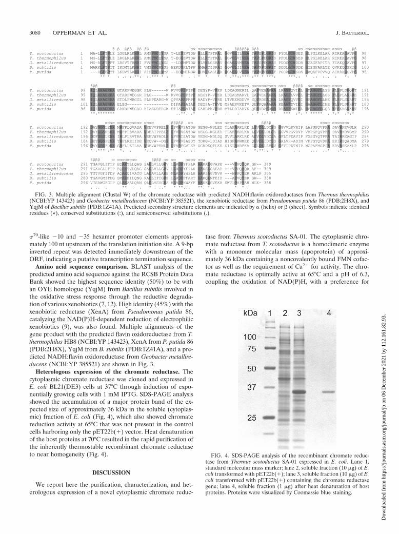
Heterologous expression of the chromate reductase. Thecytoplasmic chromate reductase was cloned and expressed inE. coli BL21(DE3) cells at 37°C through induction of expo-nentially growing cells with 1 mM IPTG. SDS-PAGE analysisshowed the accumulation of a major protein band of the ex-pected size of approximately 36 kDa in the soluble (cytoplas-mic) fraction of E. coli (Fig. 4), which also showed chromatereduction activity at 65°C that was not present in the controlcells harboring only the pET22b(�) vector. Heat denaturationof the host proteins at 70°C resulted in the rapid purification ofthe inherently thermostable recombinant chromate reductaseto near homogeneity (Fig. 4).
DISCUSSION
We report here the purification, characterization, and het-erologous expression of a novel cytoplasmic chromate reduc-
tase from Thermus scotoductus SA-01. The cytoplasmic chro-mate reductase from T. scotoductus is a homodimeric enzymewith a monomer molecular mass (apoprotein) of approxi-mately 36 kDa containing a noncovalently bound FMN cofac-tor as well as the requirement of Ca2� for activity. The chro-mate reductase is optimally active at 65°C and a pH of 6.3,coupling the oxidation of NAD(P)H, with a preference for
FIG. 3. Multiple alignment (Clustal W) of the chromate reductase with predicted NADH:flavin oxidoreductases from Thermus thermophilus(NCBI:YP 143423) and Geobacter metallireducens (NCBI:YP 385521), the xenobiotic reductase from Pseudomonas putida 86 (PDB:2H8X), andYqjM of Bacillus subtilis (PDB:1Z41A). Predicted secondary structure elements are indicated by � (helix) or � (sheet). Symbols indicate identicalresidues (*), conserved substitutions (:), and semiconserved substitutions (.).
FIG. 4. SDS-PAGE analysis of the recombinant chromate reduc-tase from Thermus scotoductus SA-01 expressed in E. coli. Lane 1,standard molecular mass marker; lane 2, soluble fraction (10 �g) of E.coli transformed with pET22b(�); lane 3, soluble fraction (10 �g) of E.coli transformed with pET22b(�) containing the chromate reductasegene; lane 4, soluble fraction (1 �g) after heat denaturation of hostproteins. Proteins were visualized by Coomassie blue staining.
3080 OPPERMAN ET AL. J. BACTERIOL.
Dow
nloa
ded
from
http
s://j
ourn
als.
asm
.org
/jour
nal/j
b on
06
Dec
embe
r 20
21 b
y 11
2.16
1.82
.93.

NADPH, to the reduction of Cr(VI). The catalytic efficiency ofthe chromate reductase with NADPH as an electron donor(kcat/Km) was 1.14 � 106 M�1 s�1, which is at least 180-foldmore efficient than the NfsA proteins (1, 14) and 50-foldmore efficient than the ChrR proteins (2). Low levels ofNAD(P)H oxidase as well as flavin reductase activity wereobserved.
N-terminal sequencing of the chromate reductase revealedthe absence of an N-terminal methionine residue. Whetherthis is due to the uncharacteristically closely spaced ribosomebinding site or to posttranslational modification is unknown.The chromate reductase is encoded by an ORF of 1,050 bpwith putative promoter elements and a ribosome binding sitelocated in the 5� regulatory flanking region. The putative �10and �35 hexamers showed high similarity to E. coli 70 pro-moter consensuses (TATAAT and TTGACA, respectively).Comparison of the primary structure of the chromate reduc-tase with the PDB showed the highest homology to the OYEhomologues YqjM from Bacillus subtilis (7, 12) and XenA fromPseudomonas putida (9). Both of these reductases have beenshown to reduce a variety of xenobiotic substrates. Due to thebroad substrate specificity of YqjM and other members of thefamily, as well as their involvement in the oxidative stressresponse, Fitzpatrick and coworkers (12) speculated that it isunlikely that these enzymes have a single specific physiologicalsubstrate in vivo and that these enzymes could function in themaintenance of the cellular redox state. Reduction of Cr(VI)by cellular components to Cr(V) as well as a transiently formedCr(V) intermediate before complete reduction to Cr(III) bysome flavoenzymes is thought to be a major cause of chromateoxidative stress and carcinogenesis through the formation ofROS (16, 24). The NfsA (nitroreductase) from E. coli has beenshown to be able to preempt ROS generation and minimizeoxidative stress during chromate reduction, protecting againstchromate toxicity as well as increasing Cr(VI) tolerance (1).
The high degree of similarity between the chromate reduc-tase and the OYE homologues suggests the characteristic(��)8-barrel (TIM barrel) fold. Secondary structure predictionalso indicated eight alternating �-sheets and �-helixes (10).(��)8-Barrel-folded proteins catalyze a variety of biochemicalreactions and are among the most frequent folds observed(19, 31).
Multiple alignments of the chromate reductase with theseenzymes showed that the sequence identities corresponded tothe functionally important amino acids identified in YqjM andXenA involved in lining the active site, especially those in-volved in the interaction between the apoprotein and FMN. InYqjM, a sulfate ion from the crystallization solution occupiedthe catalytic site on the isoalloxazine ring of the FMN, and inXenA a sulfate ion could be modeled into the same position.The residues capable of interaction with the sulfate ion are alsoconserved in the chromate reductase (specifically His173,His176, and Tyr178) (Fig. 4). The structural similarity betweensulfate and chromate could be an indication of how chromatebinds as a substrate in the catalytic site. In addition, Kitzingand coworkers (12) showed that the active site of YqjM ishydrophobic, wide open, and easily accessible and that thehuge substrate binding pocket allows the binding of a variety ofdifferent substrates (including the possibility of other proteins)to control the cellular redox state.
ACKNOWLEDGMENTS
This research was supported by the National Research Foundation,South Africa.
We thank Victor Parro (Centro de Astrobiologica, Spain) for helpwith the N-terminal sequencing and for helpful advice as well as JoseBerenguer (Centro de Biologıa Molecular, Spain) for help with het-erologous expression studies.
REFERENCES
1. Ackerley, D. F., C. F. Gonzalez, M. Keyhan, R. Blake II, and A. Matin. 2004.Mechanisms of chromate reduction by the Escherichia coli protein, NfsA,and the role of different chromate reductases in minimizing oxidative stressduring chromate reduction. Environ. Microbiol. 6:851–860.
2. Ackerley, D. F., C. F. Gonzalez, C. H. Park, R. Blake II, M. Keyhan, and A.Matin. 2004. Chromate-reducing properties of soluble flavoproteins fromPseudomonas putida and Escherichia coli. Appl. Environ. Microbiol. 70:873–882.
3. Altschul, S. F., W. Gish, W. Miller, E. W. Myers, and D. J. Lipman. 1990.Basic local alignment search tool. J. Mol. Biol. 215:403–410.
4. Blehert, D. S., B. G. Fox, and G. H. Chambliss. 1999. Cloning and sequenceanalysis of two Pseudomonas flavoprotein xenobiotic reductases. J. Bacteriol.181:6254–6263.
5. Cervantes, C., J. Campos-Garcıa, S. Devars, F. Gutierrez-Corona, H. Loza-Tavera, J. C. Torres-Guzman, and R. Moreno-Sanchez. 2001. Interactions ofchromium with microorganisms and plants. FEMS Microbiol. Rev. 25:335–347.
6. Codd, R., C. T. Dillon, T. Levina, and P. A. Lay. 2001. Studies on thegenotoxicity of chromium: from the test tube to the cell. Coord. Chem. Rev.216:537–582.
7. Fitzpatrick, T. B., N. Amrhein, and P. Macheroux. 2003. Characterization ofYqjM, and old yellow enzyme homolog from Bacillus subtilis involved in theoxidative stress response. J. Mol. Biol. 278:19891–19897.
8. French, C. E., and N. C. Bruce. 1995. Bacterial morphinone reductase isrelated to old yellow enzyme. Biochem. J. 312:671–678.
9. Griese, J. J., R. P. Jakob, S. Schwarzinger, and H. Dobbek. 2006. Xenobioticreductase A in the degradation of quinoline by Pseudomonas putida 86:physiological function, structure and mechanism of 8-hydroxycoumarin re-duction. J. Mol. Biol. 361:140–152.
10. Kelley, L. A., R. M. MacCallum, and M. J. E. Sternberg. 2000. Enhancedgenome annotation using structural profiles in the program 3D-PSSM. J.Mol. Biol. 299:499–520.
11. Kieft, T. L., J. K. Fredrickson, T. C. Onstott, Y. A. Gorby, H. M. Kostan-darithes, T. J. Bailey, D. W. Kennedy, S. W. Li, A. E. Plymale, C. M. Spadoni,and M. S. Gray. 1999. Dissimilatory reduction of Fe(III) and other electronacceptors by a Thermus isolate. Appl. Environ. Microbiol. 65:1214–1221.
12. Kitzing, K., T. B. Fitzpatrick, C. Wilken, J. Sawa, G. P. Bourenkov, P.Macheroux, and T. Clausen. 2005. The 1.3 Å crystal structure of the flavo-protein YqjM reveals a novel class of old yellow enzymes. J. Biol. Chem.280:27904–27913.
13. Kohli, R. M., and V. Massey. 1998. The oxidation half-reaction of old yellowenzyme. J. Biol. Chem. 273:32763–32770.
14. Kwak, Y. H., D. S. Lee, and H. B. Kim. 2003. Vibrio harveyi nitroreductase isalso a chromate reductase. Appl. Environ. Microbiol. 69:4390–4395.
15. Laemmli, U. K. 1970. Cleavage of structural proteins during the assembly ofthe head of bacteriophage T4. Nature 227:680–685.
16. Liu, K. J., and X. Shi. 2001. In vivo reduction of chromium (VI) and itsrelated free radical generation. Mol. Cell. Biochem. 222:41–47.
17. Losi, M. E., C. Amrhein, and W. T. S. Frankenberger. 1994. Environmentalbiochemistry of chromium. Rev. Environ. Contam. Toxicol. 136:91–121.
18. Moller, C., and E. van Heerden. 2006. Isolation of a soluble and membrane-associated Fe(III) reductase from the thermophile, Thermus scotoductus(SA-01). FEMS Microbiol. Lett. 265:237–243.
19. Nagano, N., C. A. Orengo, and J. M. Thornton. 2002. One fold with manyfunctions: the evolutionary relationship between TIM barrel families basedon their sequences, structures and functions. J. Mol. Biol. 321:741–765.
20. Nakamura, Y., T. Gojobori, and T. Ikemura. 2000. Codon usage tabulatedfrom international DNA sequence database: status for the year 2000. NucleicAcids Res. 28:292.
21. Opperman, D. J., and E. van Heerden. 2007. Aerobic Cr(VI) reduction byThermus scotoductus strain SA-01. J. Appl. Microbiol. 103:1907–1913.
22. Opperman, D. J., and E. van Heerden. 22 January 2008, posting date. Amembrane-associated protein with Cr(VI)-reducing activity from Thermusscotoductus SA-01. FEMS Microbiol. Lett. doi:10.1111/j.1574-6968.2007.01063.x.(Subsequently published, FEMS Microbiol. Lett. 208:210–218, 2008.)
23. Park, C. H., M. Keyhan, B. Wielinga, S. Fendorf, and A. Matin. 2000.Purification to homogeneity and characterization of a novel Pseudomonasputida chromate reductase. Appl. Environ. Microbiol. 66:1788–1795.
24. Shi, X., and N. S. Dalal. 1990. NADPH-dependent flavoenzymes catalyzeone electron reduction of metal ions and molecular oxygen and generatehydroxyl radicals. FEBS Lett. 276:189–191.
VOL. 190, 2008 NOVEL CHROMATE REDUCTASE FROM THERMUS SCOTODUCTUS 3081
Dow
nloa
ded
from
http
s://j
ourn
als.
asm
.org
/jour
nal/j
b on
06
Dec
embe
r 20
21 b
y 11
2.16
1.82
.93.

25. Slilaty, S. N., and S. Lebel. 1998. Accurate insertional inactivation of lacZ�:construction of pTrueBlue and M13TrueBlue cloning vectors. Gene 213:83–91.
26. Smith, P. K., R. I. Krohn, G. T. Hermanson, A. K. Mallia, F. H. Gartner,M. D. Provenzano, E. K. Fujimoto, N. M. Goeke, B. J. Olson, and D. C.Klenk. 1985. Measurement of protein using bicinchoninic acid. Anal. Bio-chem. 150:76–85.
27. Suzuki, T., N. Miyata, H. Horitsu, K. Kawai, K. Takamizawa, Y. Tai, and M.Okazaki. 1992. NAD(P)H-dependent chromium(VI) reductase of Pseudo-monas ambigua G-1: a Cr(V) intermediate is formed during the reduction ofCr(VI) to Cr(III). J. Bacteriol. 174:5340–5345.
28. Thompson, J. D., D. G. Higgins, and T. J. Gibson. 1994. CLUSTAL W:improving the sensitivity of progressive multiple sequence alignment throughsequence weighting, position-specific gap penalties and weight matrix choice.Nucleic Acids Res. 22:4673–4680.
29. Towner, P. 1991. Isolation of DNA by SDS-proteinase K treatment, p. 52–53.In T. A. Brown (ed.) Essential molecular biology, vol. 1. A practical ap-proach. Oxford University Press, Oxford, United Kingdom.
30. Urone, P. F. 1955. Stability of colorimetric reagent for chromium, s-diphe-nylcarbazide, in various solvents. Anal. Chem. 27:1354–1355.
31. Vega, M. C., E. Lorentzen, A. Linden, and M. Wilmanns. 2003. Evolutionarymarkers in the (�/�)8-barrel fold. Curr. Opin. Chem. Biol. 7:694–701.
32. Warburg, O., and W. Christian. 1933. Uber das gelbe Ferment und seineWirkungen. Biochem. Z. 266:377–411.
33. Williams, R. E., and N. C. Bruce. 2002. “New uses for an old enzyme”—theold yellow enzyme family of flavoenzymes. Microbiology 148:1607–1614.
34. Ziegenhorn, J., M. Senn, and T. Bucher. 1976. Molar absorptivities of�-NADH and �-NADPH. Clin. Chem. 22:151–160.
3082 OPPERMAN ET AL. J. BACTERIOL.
Dow
nloa
ded
from
http
s://j
ourn
als.
asm
.org
/jour
nal/j
b on
06
Dec
embe
r 20
21 b
y 11
2.16
1.82
.93.