1 nr240 nursing ii care of clients with coma & increased intracranial pressure review self study...
Post on 21-Dec-2015
216 views
TRANSCRIPT

1
NR240Nursing II
Care of clients with coma & increased intracranial pressure
Review self study slides 1-6

2
Review Chapt 43 neuro A & P key terms Structure of Neurons Mechanism of nerve impulse
conduction Neurotransmitters
Acetylcholine Serotonin Dopamine Norepinephrine
Structures of the brain Supratentorial/infratentorial
Cerebral circulation Circle of Willis Blood-brain barrier Cerebrospinal fluid
circulation Spinal cord structures
Ascending tracts Spinothalamic tracts Spinocerebellar tracts
descending tracts Extrapyramidal tracts Basal ganglia
Peripheral nervous system Sensory receptors Plexuses Lower motor neuron Reflexes Cranial nerves

3
Review Chapt 43 neuro diagnostic assessment Emphasize understanding of prep, indications and
outcomes Radiographic exam Cerebral angiography CT scanning MRI MRA EEG EMG Lumbar puncture

4
Review Terms related to Coma Obtundation
Reduced alertness Lethargy
Abnormal drowsiness Persistent vegetative state
state results when the cerebrum, which controls thought and behavior, is destroyed, but the thalamus and brain stem, which control sleep cycles, body temperature, breathing, and heart rate, are spared
Locked- in state people are conscious and able to think but are so severely
paralyzed that they can communicate only by opening and closing the eyes in response to questions
5
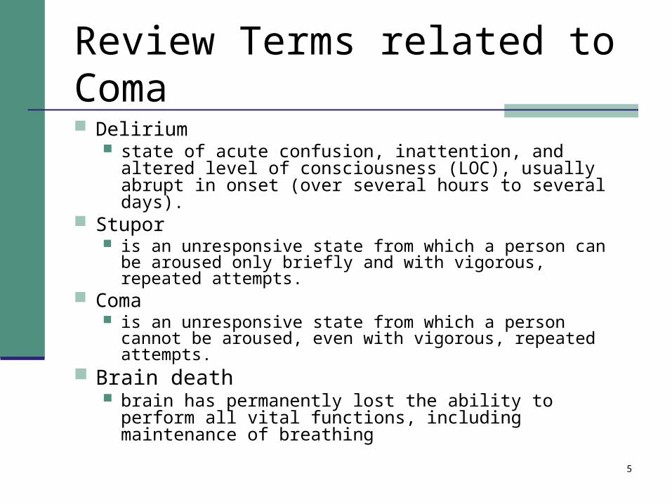
Review Terms related to Coma
Delirium state of acute confusion, inattention, and altered level
of consciousness (LOC), usually abrupt in onset (over several hours to several days).
Stupor is an unresponsive state from which a person can be
aroused only briefly and with vigorous, repeated attempts. Coma
is an unresponsive state from which a person cannot be aroused, even with vigorous, repeated attempts.
Brain death brain has permanently lost the ability to perform all vital
functions, including maintenance of breathing

6
Defining Altered Mental State
Change in neurological function on a continuum affecting: Arousability Cognition, verbal response ability to follow commands Motor function Sensory function Presence of reflexes

7
Neurological Assessment
Level of consciousness (LOC),Mental status Cognition, emotional status
cranial nerves reflexes motor function
Cerebellar strength
sensory function
8
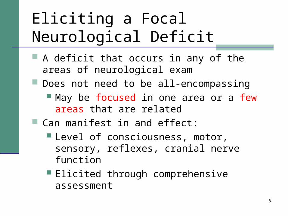
Eliciting a Focal Neurological Deficit
A deficit that occurs in any of the areas of neurological exam
Does not need to be all-encompassing May be focused in one area or a few areas that
are related Can manifest in and effect:
Level of consciousness, motor, sensory, reflexes, cranial nerve function
Elicited through comprehensive assessment

9
Performing a neurocheck
Rapid neurocheck: Glasgow coma scale (eye opening, motor
response, verbal response) Pupilary response Motor strength Vital signs Sensation Seizure activity

10
Documenting Neuro status
Neurological Flowsheet Key points
Must be compared to baseline Must evaluate right and left separately when
possible Should be performed with vital signs Physician notification must be timely

11
Reporting criteria based and neurocheck results Drop in GCS of 2 points or more Deterioration in neuro status Abnormal vitals signs:
rising systolic with unchanged diastolic (widened pulse pressure), bradycardia and change in respiratory pattern (Cushings triad)
Rising body temperature (can increase brain oxygen demand)
New onset seizure activity CSF leakage

12
Acute changes requiring emergency intervention
Notify MD within 5 minutes of discovering: Unilateral pupil dilation, Loss of pupil response Abnormal flexion or extension Loss of brain stem reflexes (gag reflex, corneal
reflex) Initiate emergency response
Ensure airway, provide oxygen, increase frequency of assessment establish IV access
13
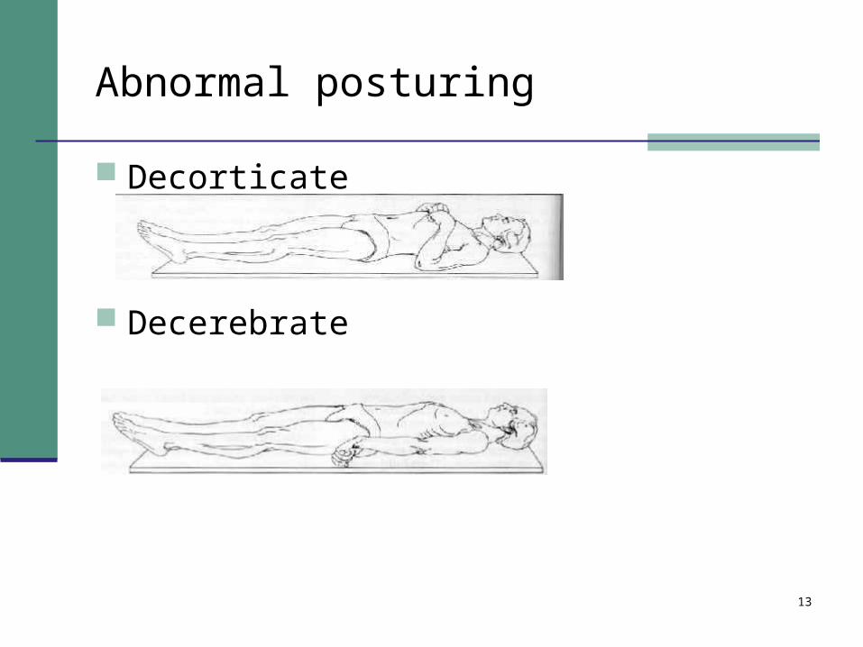
Abnormal posturing
Decorticate
Decerebrate

14
Brain stem reflexes (3 types)
Caloric stimulation Cold calorics video (performed by MD) Injection of 20-30 cc syringe with an 18 gauge angiocath filled
with ice water and squirted into the ear while evaluating eye movement.
In a Normal response, eyes conjugately deviate away from the cold ear, then snap back to midline
Corneal ReflexTouch the lateral lower corner of the cornea.
In a Normal response, ipsalateral eye blinks
Cough, gag reflexJiggle the endotracheal tube or NG tube to stimulate the larynx or
pharynx
In a Normal response, patient coughs or gags
15
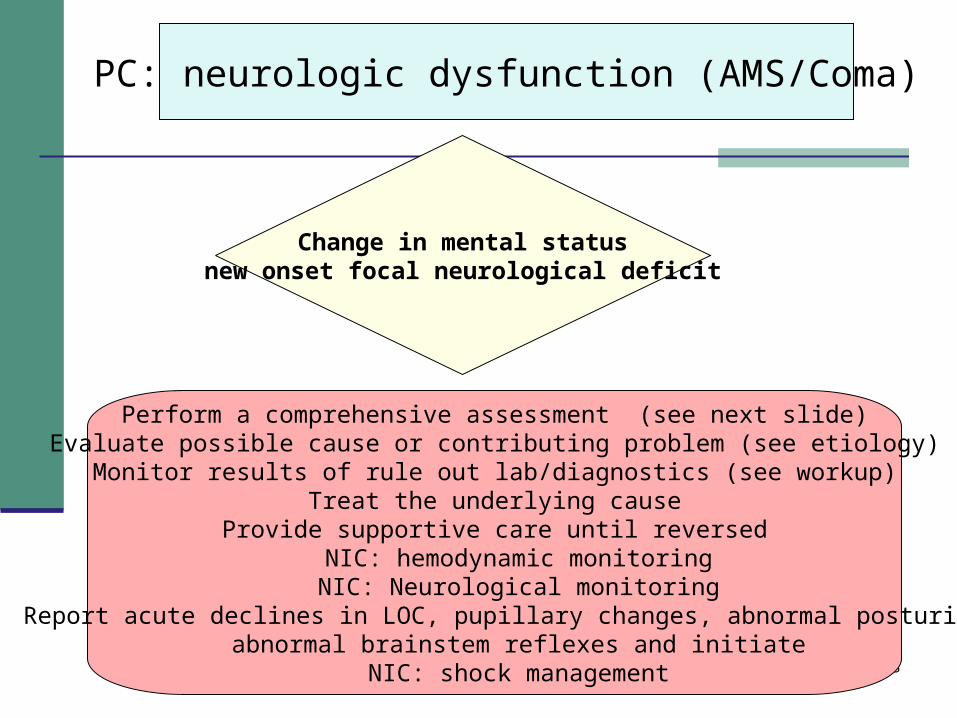
PC: neurologic dysfunction (AMS/Coma)
Change in mental statusnew onset focal neurological deficit
Perform a comprehensive assessment (see next slide)Evaluate possible cause or contributing problem (see etiology)
Monitor results of rule out lab/diagnostics (see workup)Treat the underlying cause
Provide supportive care until reversedNIC: hemodynamic monitoringNIC: Neurological monitoring
Report acute declines in LOC, pupillary changes, abnormal posturing, abnormal brainstem reflexes and initiate
NIC: shock management
16
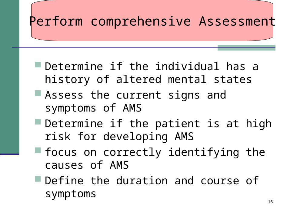
Determine if the individual has a history of altered mental states
Assess the current signs and symptoms of AMS
Determine if the patient is at high risk for developing AMS
focus on correctly identifying the causes of AMS
Define the duration and course of symptoms
Perform comprehensive Assessment
17
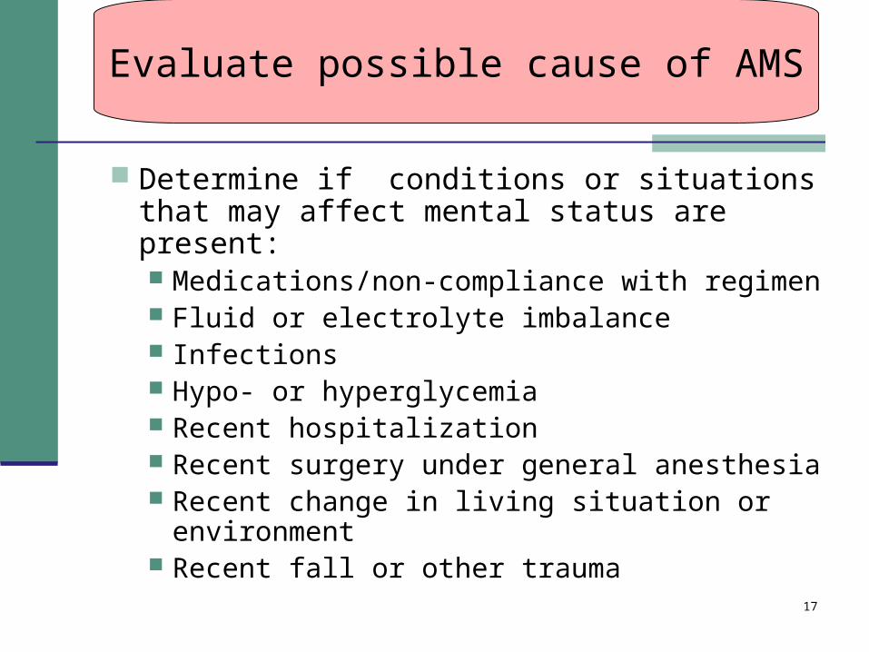
Determine if conditions or situations that may affect mental status are present: Medications/non-compliance with regimen Fluid or electrolyte imbalance Infections Hypo- or hyperglycemia Recent hospitalization Recent surgery under general anesthesia Recent change in living situation or
environment Recent fall or other trauma
Evaluate possible cause of AMS
18
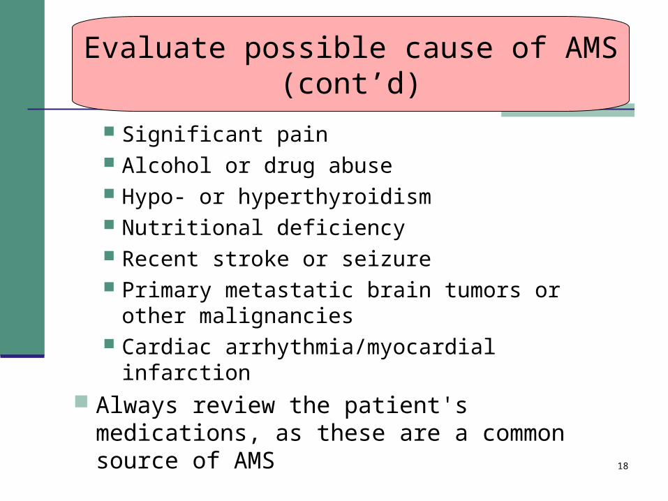
Significant pain Alcohol or drug abuse Hypo- or hyperthyroidism Nutritional deficiency Recent stroke or seizure Primary metastatic brain tumors or other
malignancies Cardiac arrhythmia/myocardial infarction
Always review the patient's medications, as these are a common source of AMS
Evaluate possible cause of AMS(cont’d)
19
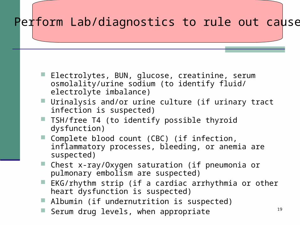
Electrolytes, BUN, glucose, creatinine, serum osmolality/urine sodium (to identify fluid/ electrolyte imbalance)
Urinalysis and/or urine culture (if urinary tract infection is suspected)
TSH/free T4 (to identify possible thyroid dysfunction) Complete blood count (CBC) (if infection, inflammatory
processes, bleeding, or anemia are suspected) Chest x-ray/Oxygen saturation (if pneumonia or pulmonary
embolism are suspected) EKG/rhythm strip (if a cardiac arrhythmia or other heart
dysfunction is suspected) Albumin (if undernutrition is suspected) Serum drug levels, when appropriate
Perform Lab/diagnostics to rule out cause
20
Radiological examination CT MRI
Perform Lab/diagnostics to rule out cause

21
Nursing Priorities for the unconscious client (source: Carpenito) PC: Respiratory insufficiency PC: Pneumonia/Atelectasis PC: Increased intracranial pressure PC: Seizures PC: Sepsis PC: Thrombophlebitis PC: Fluid/electrolyte imbalance PC: Negative nitrogen balance PC: Bladder distention PC: Stress ulcers PC: Renal calculi PC: Urinary tract infection

22
Nursing Priorities for the unconscious client (source: Carpenito) cont’d Nursing Diagnoses Infection, Risk for related to immobility and invasive devices
(tracheostomy, Foley catheter, venous lines)• Risk for Tissue Integrity, Impaired: Corneal related to corneal
drying secondary to open eyes and lower tear production Family Anxiety/Fear related to present state of individual and
uncertain prognosis• Risk for Oral Mucous Membrane, Impaired related to inability to
perform own mouth care and pooling of secretions• Total Incontinence related to unconscious state Disuse Syndrome Powerlessness (family) related to feelings of loss of control and
restrictions on lifestyle Risk for Ineffective Airway Clearance related to stasis of
secretions secondary to inadequate cough and decreased mobility

23
Understanding ICP
24
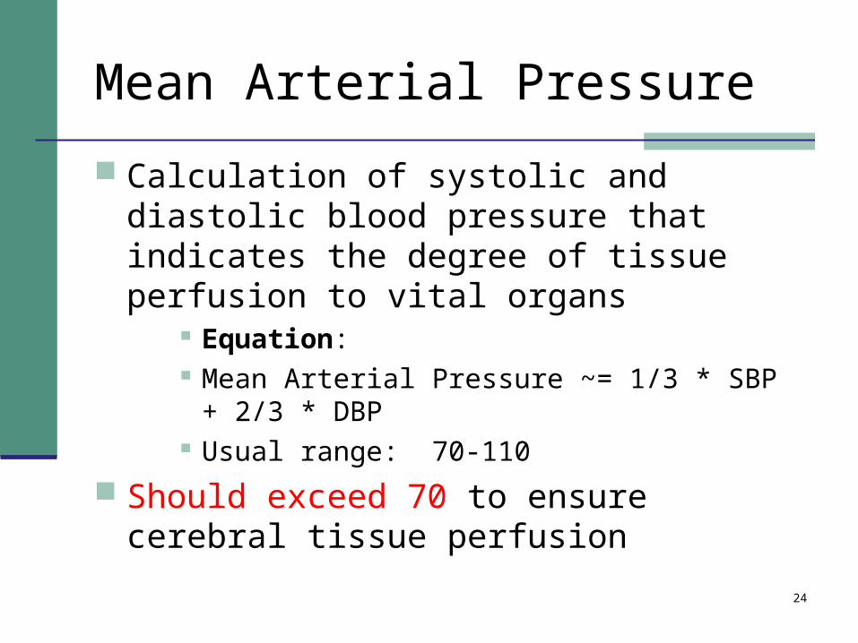
Mean Arterial Pressure
Calculation of systolic and diastolic blood pressure that indicates the degree of tissue perfusion to vital organs
Equation: Mean Arterial Pressure ~= 1/3 * SBP + 2/3 * DBP Usual range: 70-110
Should exceed 70 to ensure cerebral tissue perfusion
25
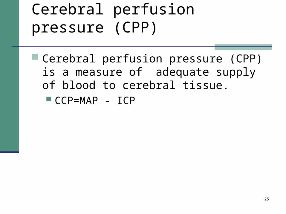
Cerebral perfusion pressure (CPP)
Cerebral perfusion pressure (CPP) is a measure of adequate supply of blood to cerebral tissue. CCP=MAP - ICP

26
cerebral blood flow (CBF)
cerebral blood flow (CBF) is ensured through regulation of arterial blood supply and cerebrovascular resistance (CVR) CBF=CPP ÷ CVR.
Determinants of supply occur as a result of: Vasomotor control of cerebral arteries
Influenced by circulating levels of carbon dioxide, oxygen, products of metabolism, and pH.
Autoregulatory response to changes in MAP

27
Factors contributing to Cerebral arterial vasodilation to preserve Cerebral blood flow
Contributing Factors
Increased PaCo2
Decreased PaO2 < 50
pH<7.35
Decreased blood pressure
28
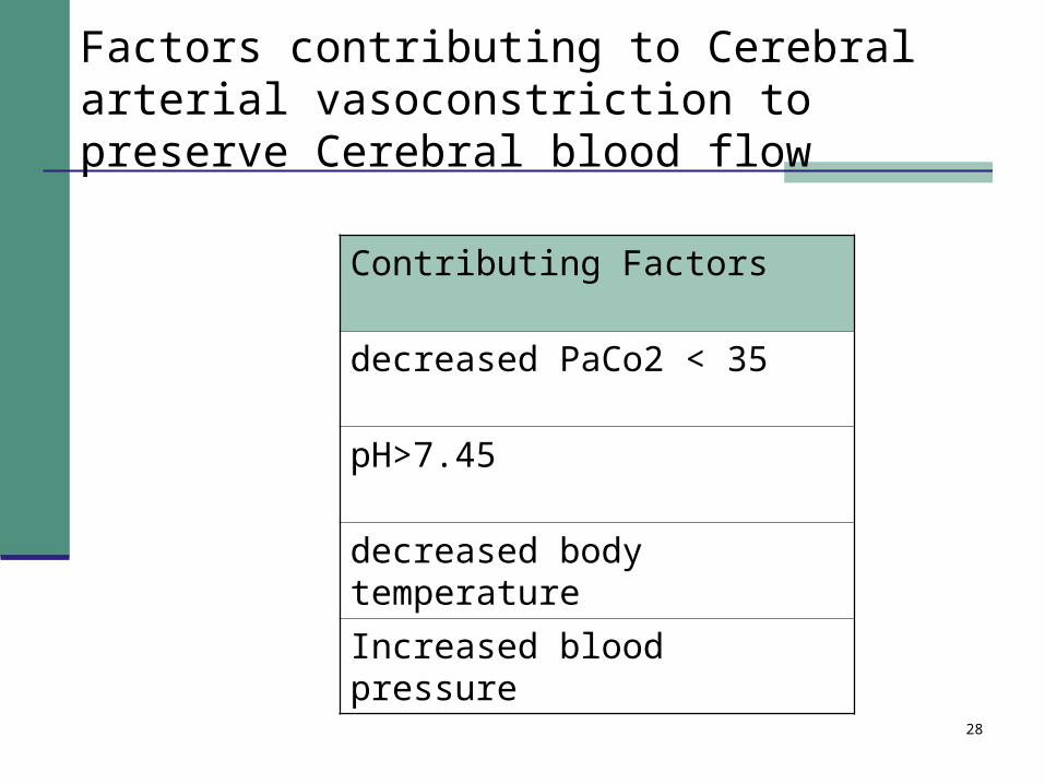
Factors contributing to Cerebral arterial vasoconstriction to preserve Cerebral blood flow
Contributing Factors
decreased PaCo2 < 35
pH>7.45
decreased body temperature
Increased blood pressure

29
Maladaptation in Autoregulation
Decreased systolic BP results in decreased CPP
Decreased CPP leads to increased vasodilation
Increased vasodilation increased cerebral blood volume
Increased cerebral blood volume increases ICP which in turn decreases cerebral perfusion pressure and the cycle repeats itself

30
Defining Intracranial Pressure
measure of pressure inside the cranium has an arbitrary numeric amount
Can be monitored using pressure devices Intracranial pressure monitoring

31
Causes of an increased ICP
Conditions Increasing Brain Volume intracranial mass (tumor, hematoma, aneurysm, AVM) cerebral edema CNS infection (abscess, inflammatory process)
Conditions Increasing Blood Volume obstruction of venous outflow hyperemia hypercapnea
Conditions Increasing CSF Volume increased production decreased reabsorption of CSF (meningitis, SAH) obstruction to flow of CSF

32
High Risk Populations for Increased ICP Intracerebral masses blood clots blockage of venous outflow head injuries inflammatory diseases cranial surgery
33
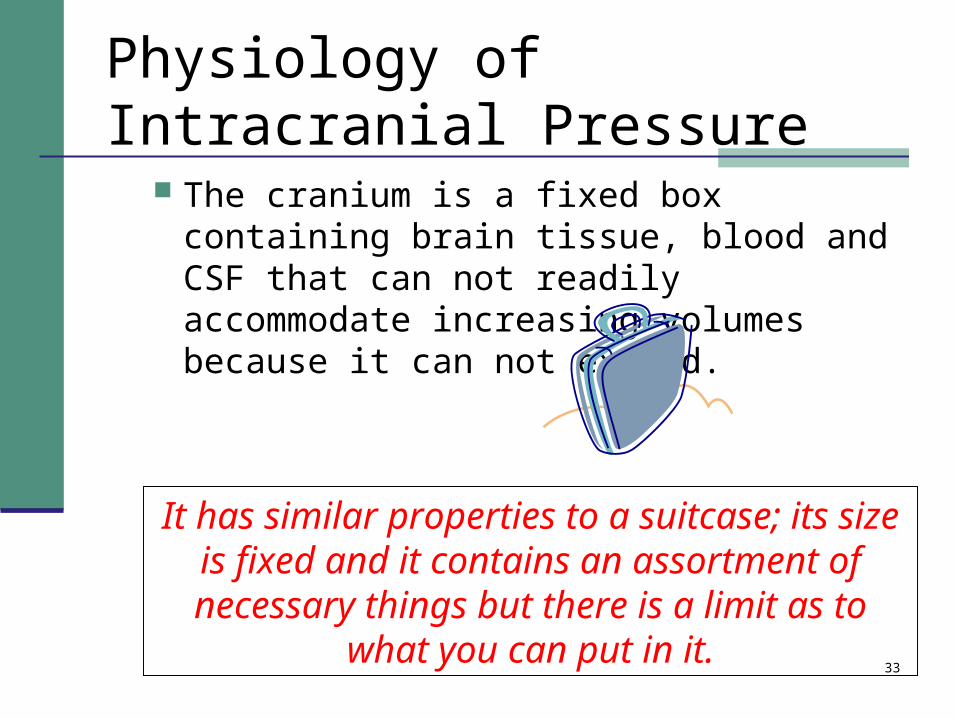
Physiology of Intracranial Pressure
The cranium is a fixed box containing brain tissue, blood and CSF that can not readily accommodate increasing volumes because it can not expand.
It has similar properties to a suitcase; its size is fixed and it contains an assortment of necessary things but there is a limit as to
what you can put in it.

34
Physiology of Intracranial Pressure
When the volume inside the cranium is subject to stressors that can increase it precipitously, it results in an increase in intracranial pressure.
Such events include; Cerebral vasodilation and edema, decreased
venous return, masses and lesions
It is like an overstuffed suitcase
35
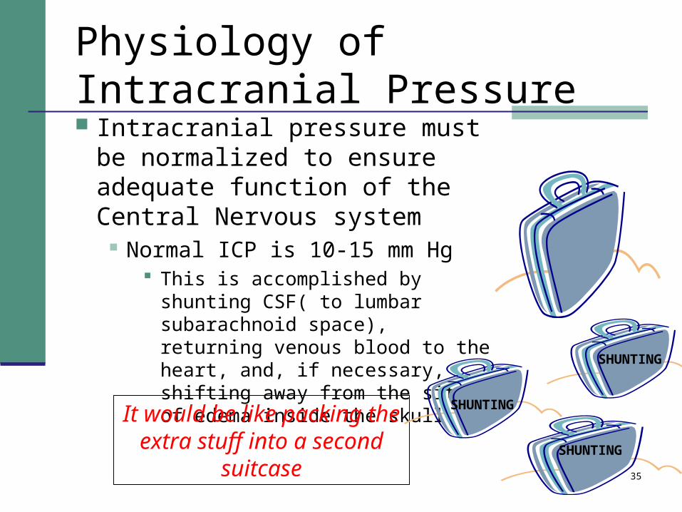
Physiology of Intracranial Pressure Intracranial pressure must be
normalized to ensure adequate function of the Central Nervous system
Normal ICP is 10-15 mm Hg This is accomplished by shunting CSF(
to lumbar subarachnoid space), returning venous blood to the heart, and, if necessary, shifting away from the site of edema inside the skull.
It would be like packing the extra stuff into a second
suitcase
SHUNTING
SHUNTING
SHUNTING

36
Relationship of volume to pressure
Monroe-Kellie Hypothesisto maintain a normal ICP,
a change in the volume of one compartment must be offset by a reciprocal change in the volume of another compartment

37
When you have too much in your suitcase, you have to unpack some of it
Your brain needs to do the same thing when the ICP is too high.

38
Physiology of Intracranial Pressure
If the stressors that increase volume are too great inside the cranium it becomes difficult to get anything else in such as;
Oxygenated blood and nutrients, exacerbating cerebral edema and intracranial pressure
The only way you could get anything else in is
by force

39
Physiology of Intracranial Pressure
Mean arterial pressure will reflexively rise to overcome a rising intracranial pressure to restore perfusion
There is only just much force that can
be applied
40
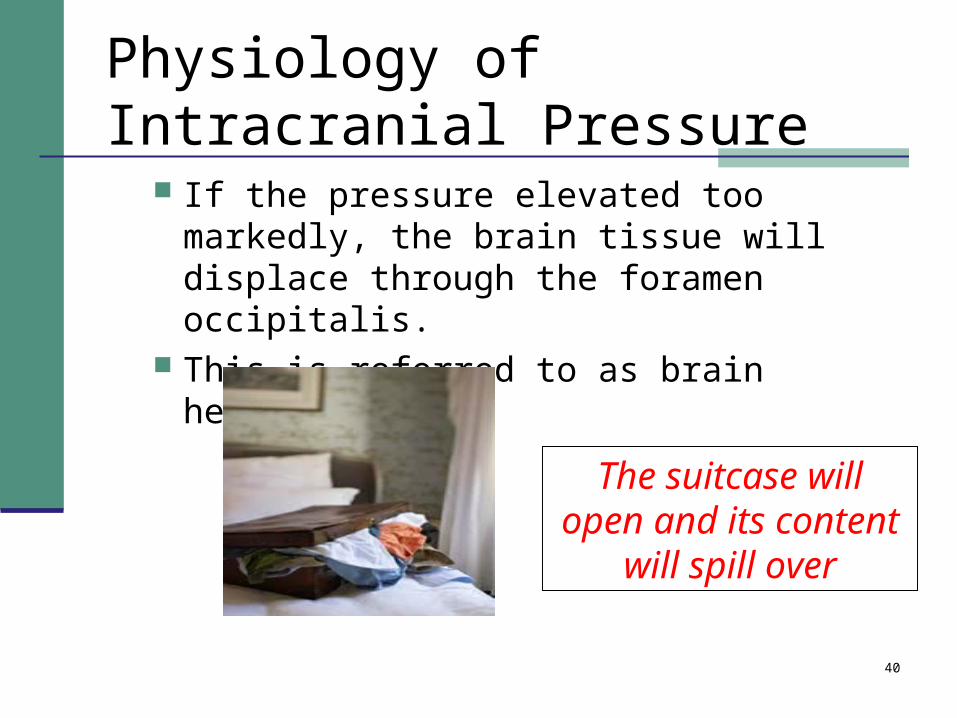
Physiology of Intracranial Pressure
If the pressure elevated too markedly, the brain tissue will displace through the foramen occipitalis.
This is referred to as brain herniation
The suitcase will open and its content
will spill over
41
Brain Herniation
Profound Neurological dysfunction Progressive loss of consciousness Coma Irregular breathing Respiratory arrest (no breathing) Irregular pulse Cardiac arrest (no pulse) Loss of all brainstem reflexes (blink, gag, pupillary
reaction to light) Source Medline plus Determining brain death

42
Management of increased ICP
Identification of clients at risk Initiation of ICP monitoring if indicated Airway maintenance and ventilation Oxygenation and low normal PaCO2 Fluid balance to maintain cerebral perfusion Avoiding positions that increase ICP Sedation and decreased external stimulation Osmotic and loop diuretics Temperature maintenance Blood glucose control Pain management and stool softeners
See ICP sheet

43
Definition of ICP monitoring
type of device that is calibrated to detect the internal pressure readings
Interpretation of the readings assist in guiding actions to restore cerebral tissue perfusion. Types
Ventriculostomy Subarachnoid Epidural Subdural Parenchymal
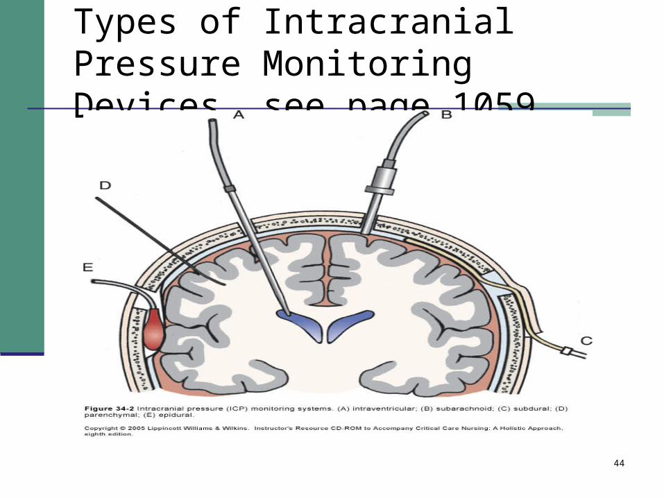
44
Types of Intracranial Pressure Monitoring Devices see page 1059
45
Indications for ICP monitoring
Head injury Craniotomy Intracranial hemorrhage Cerebral edema
46
Goal if ICP monitoring
CFS clear ICP< 20 CPP between 60-75

47
Strategies to maintain normal ICP
Source: UNC Policy and Procedure
48
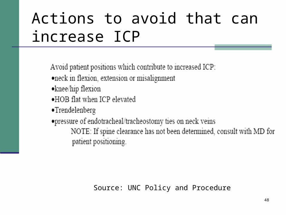
Actions to avoid that can increase ICP
Source: UNC Policy and Procedure
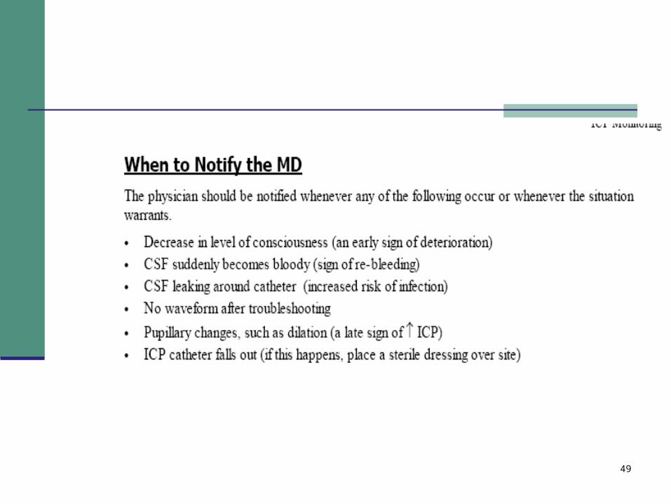
49
50
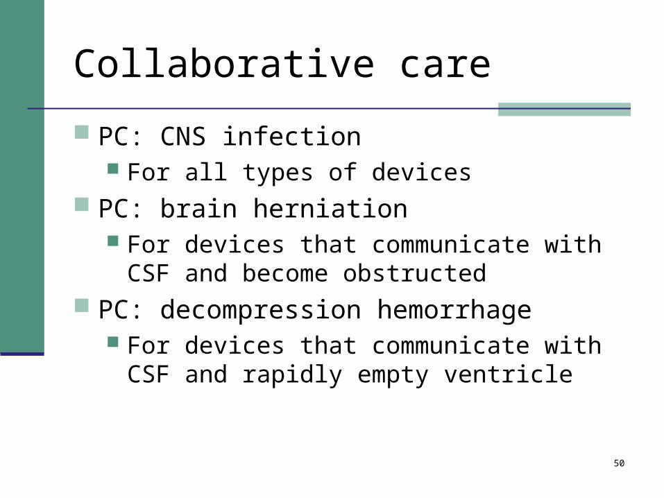
Collaborative care
PC: CNS infection For all types of devices
PC: brain herniation For devices that communicate with CSF and
become obstructed PC: decompression hemorrhage
For devices that communicate with CSF and rapidly empty ventricle
51
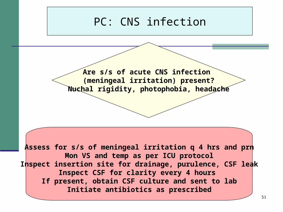
PC: CNS infection
Are s/s of acute CNS infection (meningeal irritation) present?
Nuchal rigidity, photophobia, headache
Assess for s/s of meningeal irritation q 4 hrs and prnMon VS and temp as per ICU protocol
Inspect insertion site for drainage, purulence, CSF leakInspect CSF for clarity every 4 hours
If present, obtain CSF culture and sent to labInitiate antibiotics as prescribed

52
PC: brain herniation
Perform neurological assessment as per protocolKeep system free from kinks to avoid disruption in CSF drainage.
Assess for the presence of obstruction and call MD
If present , initiate emergency interventions to minimize herniationAdminister O2, Intubate, Initiate shock management
Call MD
Are s/s brain herniation present?Pupillary changes, loss of brainstem reflexes,
Change in LOC
53
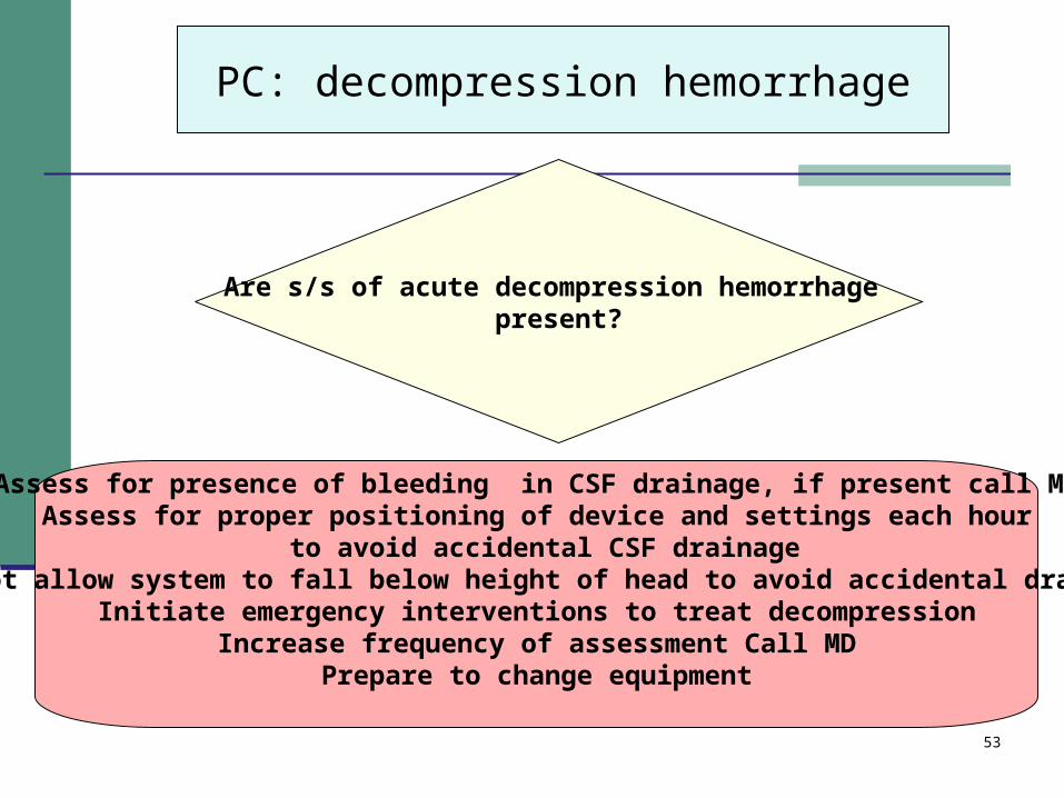
PC: decompression hemorrhage
Assess for presence of bleeding in CSF drainage, if present call MDAssess for proper positioning of device and settings each hour
to avoid accidental CSF drainageDo not allow system to fall below height of head to avoid accidental drainage
Initiate emergency interventions to treat decompressionIncrease frequency of assessment Call MD
Prepare to change equipment
Are s/s of acute decompression hemorrhage present?
54
Summary of Plan for PC: increased ICP Assess for s/s of increased ICP Monitor labs/vitals and diagnostics and
collaborate if indicators require treatment Perform ICP monitoring if indicated Avoid positions, maneuvers, situations that
increase ICP Administer agents that restore cerebral
perfusion