2013 antioxidant properties of xanthones
TRANSCRIPT

Blanco-Ayala et al. BMC Complementary and Alternative Medicine 2013, 13:262http://www.biomedcentral.com/1472-6882/13/262
RESEARCH ARTICLE Open Access
Antioxidant properties of xanthones fromCalophyllum brasiliense: prevention of oxidativedamage induced by FeSO4Tonali Blanco-Ayala1, Rafael Lugo-Huitrón1, Elizabeth M Serrano-López1, Ricardo Reyes-Chilpa2,Edgar Rangel-López3, Benjamín Pineda4, Omar Noel Medina-Campos5, Laura Sánchez-Chapul6, Enrique Pinzón7,Trejo-Solis Cristina4, Daniela Silva-Adaya1, José Pedraza-Chaverrí5, Camilo Ríos1, Verónica Pérez de la Cruz1*
and Mónica Torres-Ramos8*
Abstract
Background: Reactive oxygen species (ROS) are important mediators in a number of degenerative diseases.Oxidative stress refers to the imbalance between the production of ROS and the ability to scavenge these speciesthrough endogenous antioxidant systems. Since antioxidants can inhibit oxidative processes, it becomes relevant todescribe natural compounds with antioxidant properties which may be designed as therapies to decrease oxidativedamage and stimulate endogenous cytoprotective systems. The present study tested the protective effect of twoxanthones isolated from the heartwood of Calophyllum brasilienses against FeSO4-induced toxicity.
Methods: Through combinatory chemistry assays, we evaluated the superoxide (O2●—), hydroxyl radical (OH●),
hydrogen peroxide (H2O2) and peroxynitrite (ONOO—) scavenging capacity of jacareubin (xanthone III) and 2-(3,3-dimethylallyl)-1,3,5,6-tetrahydroxyxanthone (xanthone V). The effect of these xanthones on murine DNA and bovineserum albumin degradation induced by an OH• generator system was also evaluated. Additionally, we investigatedthe effect of these xanthones on ROS production, lipid peroxidation and glutathione reductase (GR) activity inFeSO4-exposed brain, liver and lung rat homogenates.
Results: Xanthone V exhibited a better scavenging capacity for O2●—, ONOO- and OH● than xanthone III, although
both xanthones were unable to trap H2O2. Additionally, xanthones III and V prevented the albumin and DNAdegradation induced by the OH● generator system. Lipid peroxidation and ROS production evoked by FeSO4 weredecreased by both xanthones in all tissues tested. Xanthones III and V also prevented the GR activity depletioninduced by pro-oxidant activity only in the brain.
Conclusions: Altogether, the collected evidence suggests that xanthones can play a role as potential agents toattenuate the oxidative damage produced by different pro-oxidants.
Keywords: Xanthones, Antioxidant capacity, ROS production
* Correspondence: [email protected]; [email protected] de Neuroquímica, Instituto Nacional de Neurología yNeurocirugía Manuel Velasco Suárez, Insurgentes Sur 3877, S.S.A., México, DF14269, MéxicoFull list of author information is available at the end of the article
© 2013 Blanco-Ayala et al.; licensee BioMed Central Ltd. This is an open access article distributed under the terms of theCreative Commons Attribution License (http://creativecommons.org/licenses/by/2.0), which permits unrestricted use,distribution, and reproduction in any medium, provided the original work is properly cited.

b
c
a
Figure 1 Chemical structures of xanthones: a) jacareubin (III), b)2-(3,3-dimethylallyl)-1,3,5,6-tetrahydroxyxanthone (V), andc) α-mangostin.
Blanco-Ayala et al. BMC Complementary and Alternative Medicine 2013, 13:262 Page 2 of 15http://www.biomedcentral.com/1472-6882/13/262
BackgroundRedox homeostasis is maintained in organisms as anintracellular equilibrium between oxidant and antioxi-dant levels. An imbalance in favor of pro-oxidants re-sults in oxidative stress which leads cells to damagethrough the alteration of endogenous macromoleculessuch as proteins, lipids and DNA [1,2]. Cells possess sev-eral antioxidant defense mechanisms designed to main-tain homeostasis in response to oxidative stressors;however, under pathological conditions, such antioxi-dant defenses are depleted, thus eliciting oxidativedamage. The most promising strategy to prevent theoxidative damage caused by reactive oxygen and nitro-gen species (ROS/RNS) is the use of antioxidant mole-cules. These compounds can act either as direct antioxidantthrough free radical scavenging mechanisms or as indirectantioxidants by enhancing the antioxidant status towardboth enzymatic and non-enzymatic systems. Different nat-ural compounds have been described as antioxidants, asthey are capable to decrease ROS levels in cells and areconsequently useful to attenuate aging-related complica-tions [3,4] and for treatment of some human diseases,including atherosclerosis, cardiovascular diseases [5],inflammatory injury [6], cancer [7] and neurodegenera-tive diseases [8]. Natural antioxidants can therefore serveas innovative tools for preventive medicine. Calophyllumbrasiliense Cambess is a big tree found in the tropicalrain forests from Brazil to Mexico. It has been widelyused in Latin American folk medicine to treat a varietyof maladies including pain [9], inflammation, diabetes,hypertension [10,11], diarrhea, herpes and rheumatism[12]. Despite its extensive use, only a few biological ac-tivities have been reported in the scientific literature.Plants of this species are a rich source of xanthonesand coumarins. Particularly, xanthones (9H-xanthen-9-ones) are heterocyclic compounds with the dibenzo-γ-pyrone framework (Figure 1) [13]. Several xanthoneshave been isolated from Calophyllum brasiliense heartwood,including jacareubin (III) and 2-(3,3-dimethylallyl)-1,3,5,6-tetrahydroxyxanthone (V). Xanthones from Callophyllumbrasiliense Cambess have shown to be inhibitorsof sulfotransferases, SULT1A1 and SULT2A1 [14], an-tibiotics [15], and inhibitors of gastric H+, K+−ATPaseactivity [16]. Furthermore, it has been reported thatxanthones from other sources, like α-mangostin, are ef-fective antioxidants and could be important free radicalscavengers [17-21]. This study was conducted to explore thescavenging effects and antioxidant properties of xanthonesIII and V (Figure 1b and 1c, respectively) isolated fromCallophylum brasiliensis, a species collected in the Mexicanrain forest. Here, we evaluated the activities of these xan-thones as scavengers of different ROS in synthetic systems.Also, we tested their effects against oxidative damage in-duced by FeSO4 in brain, liver and lung rat homogenates.
MethodsReagentsCalophyllum brasiliense Cambess (Clusiaceae) was col-lected by J.I. Calzada from the Selva Lancandona, Chiapas,México [22]. Identification was done by the collector, avoucher (JIC-3116), and wood simple (00011-XALw) aredeposited in the Xal Herbarium, and Dr. Faustino Mi-randa Xylarium, both of the Instituto de Ecología, A.C., atXalapa, Ver. México. Five xanthones (I-V) were isolatedfrom Calophyllum brasiliense heartwood; xanthones IIIand V were obtained from a methanolic extract and werethe most abundant constituents, as previously described,and identified by proton nuclear magnetic resonance andmass spectrometry [23]. α-Mangostin was isolated fromthe dried pericarp of Garcinia mangostana, as previouslydescribed by Marquez-Valadez et al. [24].2′,7′-dichlorofluorescein (DCF), 2′,7′-dichlorodihydro-
fluorescein diacetate (DCFH-DA), FeSO4, ascorbic acid,histidine, xylenol orange, ammonium iron (II) sulfate hexa-hydrate, 2,2′-azinobis-(3-ethylbenzothiazoline-6-sulfonic acid)(ABTS), 2,2-diphenyl-1-picrylhydrazyl (DPPH), bovine serumalbumin (BSA), dimethylsulfoxide (DMSO), DL-penicillamine,
Blanco-Ayala et al. BMC Complementary and Alternative Medicine 2013, 13:262 Page 3 of 15http://www.biomedcentral.com/1472-6882/13/262
glutathione (GSH), nitroblue tetrazolium (NBT), phenazinemethosulfate (PMS), ethylenediaminetetraacetic acid (EDTA),nicotinamide adenine dinucleotide (NADH), N-acetyl cysteine(NAC), N-2-hydroxyethyl-piperazine-N'-2-ethane sulphonicacid (HEPES), diethylenetriaminepentaacetic acid (DTPA),nicotinamide adenine dinucleotide reduced form (NADPH),Coomassie brilliant blue R, mercaptoethanol, bromophenolblue, sodium dodecyl sulfate (SDS), sodium hypochlorite(NaOCl), hydrogen peroxide (H2O2) and N,N-dimethyl-4-nitrosoaniline (DMNA) were all obtained from Sigma Al-drich Chemical Company (St. Louis, MO, USA). All otherreagents were analytical grade and obtained from knowncommercial suppliers. Solutions were prepared using de-ionized water obtained from a Milli-RQ (Millipore) purifiersystem.
AnimalsTissue samples were obtained from the whole brain ofmale Wistar rats (280–320 g) from the vivarium ofFacultad de Medicina (UNAM, Mexico City). A totalof 40 rats were employed throughout the study. Beforethey were subjected to experimental treatments, animalswere housed five per cage in acrylic cages and providedwith standard commercial rat diet (Laboratory rodent diet5001, PMI Feeds Inc., Richmond, IN, USA) and water adlibitum. Housing room was maintained under constantconditions of temperature (25 ± 3°C), humidity (50 ± 10%)and lighting (12 h light/dark cycles). Tissues were col-lected by decapitation and immediately dissected out onice and preserved for limited times at −70°C. All proce-dures with animals were carried out according to theNational Institutes of Health Guide for the Care andUse of Laboratory Animals and the local guidelines onthe ethical use of animals from the Health Ministry ofMexico. During the dissections, all efforts were madeto minimize animal suffering. The study was approvedby Instituto Nacional de Neurología y Neurocirugía’sethics committee (30/12).
Superoxide anion scavenging assessmentSuperoxide (O2
●—) scavenging was assessed according topreviously reported methods [25] based on the reductionof NBT. The non-enzymatic PMS/NADH system gener-ates superoxide radicals that reduce NBT into a purple-colored formazan. The reaction mixture contained HEPESbuffer (20 mM, pH 7.4), 196 μM NADH, 39.2 μM NBT,3.92 μM PMS and increasing concentrations of XanthoneIII or V (0 – 1,000 μM). Final mixture volume was1.3 mL. After incubation for 5 min at room temperature,the absorbance was taken at 560 nm against an appropri-ate blank solution. All tests were performed six times inan independent manner. Results are shown as percent ofO2
●— scavenging capacity.
Hydroxyl radical (OH●) scavenging assayThe ability of xanthones III and V to scavenge OH● wasestimated through the Fe3+-EDTA-H2O2-deoxyribosesystem [26,27]. The system contained different concen-trations of xanthones III or V (0 – 50 μM or an equiva-lent volume of vehicle (ethanol, 0.025% final concentrationused in the assay)) or distilled water for the control),0.2 mM ascorbic acid, 0.2 mM FeCl3, 0.208 mM EDTA,1 mM H2O2, 0.56 mM deoxyribose, and 20 mM phosphatebuffer (pH 7.4). OH● radicals were generated by incubatingthe mixture (final volume 1 mL) at 37°C for 60 min. Theiron salt (FeCl3) was mixed with EDTA before its additionto the reaction mixture. The extent of deoxyribose degrad-ation by the formed OH● was measured directly in theaqueous phase by the thiobarbituric acid (TBA) test.Briefly, 100 μL of the 0.05 M TBA solution was added tothe samples and final solutions were re-incubated in a boil-ing water bath (94°C) for 10 min. The optical density wasestimated at a wavelength of 532 nm in a Genesys 8spectrophotometer.
Peroxynitrite (ONOO—) scavenging activity assessmentONOO— was synthesized as previously described [28].Briefly, 5 mL of an acidic solution (0.6 M HCl) of H2O2
(0.7 M) was mixed with 5 mL of 0.6 M KNO2 on an ice bathfor 1 second, and the reaction mixture was quenched with5 mL of ice-cold 1.2 M NaOH. Residual H2O2 was removedusing granular MnO2 prewashed with 1.2 M NaOH, andthis mixture was then left overnight at −20°C. The resultingyellow liquid layer on the top of the frozen mixture was col-lected for the experiment. Concentrations of ONOO— weredetermined before each experiment at 302 nm using a molarextinction coefficient of 1,670 M-1 cm-1. ONOO— scaven-ging activity was measured by monitoring the oxidation ofDCFH-DA to DCF by the modified method of Beckmanand Crow [28,29]. The reaction mixture (in a final volumeof 1.45 mL in 0.1 M phosphate buffer pH 7.4) consisted of14 μM DTPA plus 36.2 μMDCFH-DA plus the samples ex-posed to different xanthones III and V at increasing concen-trations (0–1,000 μM) and 35 μM ONOO—. The opticaldensity was determined at 500 nm in a Genesys 8 spectro-photometer. A probe containing the reaction mixture, butnot a sample, was considered as 0% scavenging capacity or100% of DCFH-DA oxidation by the ONOO— added to theassay. To calculate the ONOO— scavenging ability, the read-ings of the tubes containing the xanthones or referencecompounds were expressed as percentages of DCFH-DAoxidation and converted to percentages of scavengingability, using as reference the tube with 100% DCFH-DA oxidation [26].
H2O2 assayThe ability of xanthones III or V to scavenge H2O2 wasmeasured by the method described by Long et al. [30].
Blanco-Ayala et al. BMC Complementary and Alternative Medicine 2013, 13:262 Page 4 of 15http://www.biomedcentral.com/1472-6882/13/262
Briefly, 9 volumes of 4.4 mM butylated hydroxytoluenein HPLC-grade methanol were mixed with 1 volume of1 mM xylenol orange and 2.56 mM ammonium ferroussulfate in 0.25 M H2SO4 to give the "working" FOX re-agent. A solution of 75 μM H2O2 was mixed with differ-ent concentrations of xanthones III and V (0–1000 μM)and added with 0.01 mL of HPLC-grade methanol, im-mediately followed by the addition of 0.9 mL of FOX re-agent, vortexed for 5 seconds, and then incubated atroom temperature for 30 min (final volume 1.3 mL).Tubes were centrifuged at 15,000 × g for 10 min and ab-sorbance was read at 560 nm against a methanol blank.
OH●-mediated protein degradationExperiments for detection of OH●-mediated BSA oxida-tion were carried out using a metal-catalyzed reactionbased on the method described by Kocha et al. [31] withmodifications. A solution of ascorbic acid (1.6 mM)/EDTA (0.8 mM)/(NH4)2Fe(SO4)2 (0.8 mM) was preparedin 50 mM phosphate buffer (pH 7.4). Briefly, 1% BSA and250 μL of the ascorbic acid/EDTA/(NH4)2Fe(SO4)2•6H2Osolution were mixed in the presence or absence of xan-thones III and V or α-mangostin (0.5, 1 and 2.5 μM). Thegeneration of OH● was initiated through the addition of15 μL of 2% H2O2. In control tubes (without the OH●
generator system), H2O2 was replaced by water. The finalvolume for all probes was 250 μL. The maximum concen-tration of EtOH (vehicle) in reaction tube was 0.01%. After1 h of incubation at room temperature, 250 μL of 20%trichloroacetic acid was added, and the mixture was thencentrifuged at 2,236 x g for 30 min at 4°C. The superna-tants were discarded and the pellets were resuspended in500 μL of 0.1 M NaOH. To evaluate the oxidative damageto proteins induced by OH●, the samples were subjectedto SDS-polyacrilamide gel electrophoresis. BSA (50 μg;from resuspended pellets) was mixed (1:1) with loadingbuffer (10% glycerol, 2% SDS, 25 mM Tris–HCl (pH 6.8),5% mercaptoethanol, 0.1% bromophenol blue) and heatedat 100°C for 1 min. The protein sample (40 μg per well)was loaded in a 12% polyacrylamide gel and the electro-phoresis was run at 150 V for 1 h. After running out, gelswere stained with 0.2% Coomassie brilliant blue R for 1 h.Images were visualized and captured in a BioRad GelDocumentation System (Gel Doc 1000 BioRad). Proteinlevels were densitometrically evaluated using the QuantityOne Program 4.2 [32].
OH●-mediated DNA degradationDNA samples were obtained from mice tails and incu-bated overnight at 55°C in lysis buffer (Tris 100 mM,pH 8.5, 5 mM EDTA, 0.2% SDS, 200 mM NaCl and100 μg/ml proteinase K). The mixtures were deproteinizedwith phenol:chlorophorm:isoamilic ethanol (25:24:1). TheDNA in the recovered supernatant was precipitated by
adding cold isopropanol and washed with 70% ethanol so-lution, then dissolved in endonucleases-free water for theassay. Experiments for detection of OH●-mediated DNAoxidation were carried out by using a metal-catalyzed re-action based on Kocha et al. [31] with modifications. A so-lution of ascorbic acid (1.6 mM)/EDTA (0.8 mM)/(NH4)2Fe(SO4)2 (0.8 mM) was prepared in 20 mM phosphatebuffer (pH 7.4) plus DNA, xanthones III or xanthone V,and α-mangostin (0.5, 1 and 2.5 μM). Briefly, 20 μg ofpurified DNA was mixed with 25 μL of the ascorbic acid/EDTA/(NH4)2Fe(SO4)2•6H2O solution, in presence or ab-sence of xanthones and α-mangostin. The generation ofOH● was initiated through the addition of 15 μl of 2%H2O2. In control tubes (without the OH● generator sys-tem), H2O2 was replaced by water. The final volume forall probes was 250 μL. The maximum concentration ofEtOH (vehicle) in reaction tube was 0.01%. After 15 minof incubation at room temperature, 50 μL of loadingbuffer was added to stop the reaction. To evaluate theoxidative damage to DNA induced by OH●, 10 μL ofthe samples were subjected to agarose (2%) gel electro-phoresis for 20 min at 90 V. After running out, gelswere stained with ethidium bromide (0.1 mg/ml). Im-ages were visualized and captured in a BioRad GelDocumentation System (Gel Doc 1000 BioRad). Inorder to estimate DNA degradation; the main DNAband per well was densitometrically evaluated usingthe Quantity One Program 4.2 [32].
Assay of lipid peroxidationLipid peroxidation (LP) was assessed in homogenates ofdifferent tissues (brain, liver and kidney) by estimation ofTBA-reactive substances (TBA-RS), according to a previ-ous report [33]. Each tissue was homogenized (brain 1:10,liver 1:300, kidney 1:100 w/v) in Krebs buffer (pH 7.4)containing 19 mM NaCl, 5 mM KCl, 2 mM CaCl2,1.2 mM MgSO4, 5 mM glucose, 13 mM NaH2PO4 and3 mM Na2HPO4. Aliquots of 250 μL of the homogenatewere incubated in the presence of FeSO4 (5 μM) and/orxanthone III and V (0.5, 1 and 2.5 μM). The final vol-ume for all probes was 500 μL. All incubations weredone at 37°C for 2 h in a shaking water bath. At theend of the incubation time, 500 μL of the TBA reagent(containing 0.75 g of TBA + 15 g of trichloroaceticacid + 2.54 mL of HCl) were added and final solutionswere re-incubated in a boiling water bath (94°C) foradditional 20 min. Samples were then kept on ice for5 min and centrifuged at 3,000 x g for 15 min. The op-tical density of supernatants was estimated at a wave-length of 532 nm in a Genesys 8 spectrophotometer.Concentrations of malondialdehyde (MDA) were cal-culated by interpolation in a standard curve of MDA,constructed in parallel. Results were expressed as per-cent of LP vs. control.
Blanco-Ayala et al. BMC Complementary and Alternative Medicine 2013, 13:262 Page 5 of 15http://www.biomedcentral.com/1472-6882/13/262
Determination of ROS in homogenatesROS were detected by DCF fluorescence [34,35]. Ali-quots (500 μL) of the homogenates (brain, liver and kid-ney) were incubated in the absence (control) or presenceof 5 μM FeSO4 and/or xanthones III or V (0.5, 1 and2.5 μM) at 37°C in a shaking-water bath for 2 h. Thefinal volume for all probes was 1 mL. Then 100 μL of75 μM DCF were added to samples and incubated for30 min in the abscence of light. Finally, samples werecentrifuged at 6,000 x g for 15 min. ROS were convention-ally detected in supernatants by fluorescent spectrometryin a Perkin-Elmer LS50 spectrometer at 488 nm (excitationwavelength) and 532 nm (emission wavelength). Resultswere expressed as percent of ROS formation vs. control.
Glutathione reductase (GR) activityGR activity in forebrain, liver and kidney homogenates wasassayed using oxidized glutathione (GSSG) as substrate andmeasuring the consumption of NADPH at 340 nm [36].Briefly, aliquots of 250 μL of the homogenate were incu-bated in the presence of 5 μM FeSO4 and/or xanthone IIIand V (0.5, 1 and 2.5 μM). The final volume for all probeswas 500 μL. All incubations were done at 37°C for 2 h in ashaking water bath. After incubation 50 μL of homogenatewere mixed with 950 μL of the reaction mixture (1.25 mMGSSG, 0.1 mM NADPH, 0.5 mM Na2EDTA in 100 mMphosphate buffer, pH 7.6). One unit of GR was defined asthe amount of enzyme that oxidizes 1 μmol NADPH/min.Data were obtained as units per milligram of protein andexpressed as percent of GR activity vs. control.
Statistical analysisResults were expressed as mean values ± S.E.M. ROS scaven-ging capacity was expressed as 50% of the inhibitory concen-tration (IC50) value, which denotes the concentration of thexanthone and standard (μM) required to reach a 50% reduc-tion in the respective molecule oxidation relative to the probewithout xanthones or reference compound. IC50 was calcu-lated by the least square method. The lower the IC50 valuethe higher the scavenging capacity of the compound. In re-gard to the experiments carried out in tissue homogenates,n = 6 experiments (each experiment corresponding to one rat)were considered. All data were analyzed by one-way ANOVAfollowed by Tukey’s test for multiple comparisons, using thePrism 3.02 software (GraphPad, San Diego, CA, USA). Valuesof P < 0.05 were considered as statistically significant.
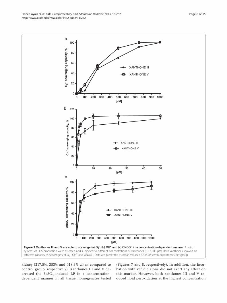
ResultsScavenging activities of xanthones III and VXanthones III and V were able to scavenge O2
•−
(Figure 2A), OH● (Figure 2B) and ONOO- (Figure 2C)in a concentration-dependent manner, in contrast,they were unable to scavenge H2O2 (data not shown).The IC50 values, calculated from the linear portion of
the dose–response curve, are shown in Table 1. Allthese values are in the μM range. Xanthone V wasmore effective to scavenge ROS than xanthone III.
Xanthones III and V prevent the oxidative proteindegradation in a concentration-dependent mannerXanthones III and V (0.5, 1 and 2.5 μM; Figure 3) were ableto prevent, in a concentration-dependent manner, the BSAdegradation induced by OH●. A densitometric assessmentwas employed for protein detection. Protection was foundsince the lowest concentration of each xanthone used(0.5 μM), although this protection was more evident athigher concentrations (1 and 2.5 μM; Figures 3a and 3b).The EtOH did not have effect in the DNA degradation in-duced by OH● (Figure 4c; line 3). These data confirm previ-ous observations on the OH●-scavenging capacity ofxanthones. α-Mangostin prevented BSA degradationinduced by OH● at all concentrations used.
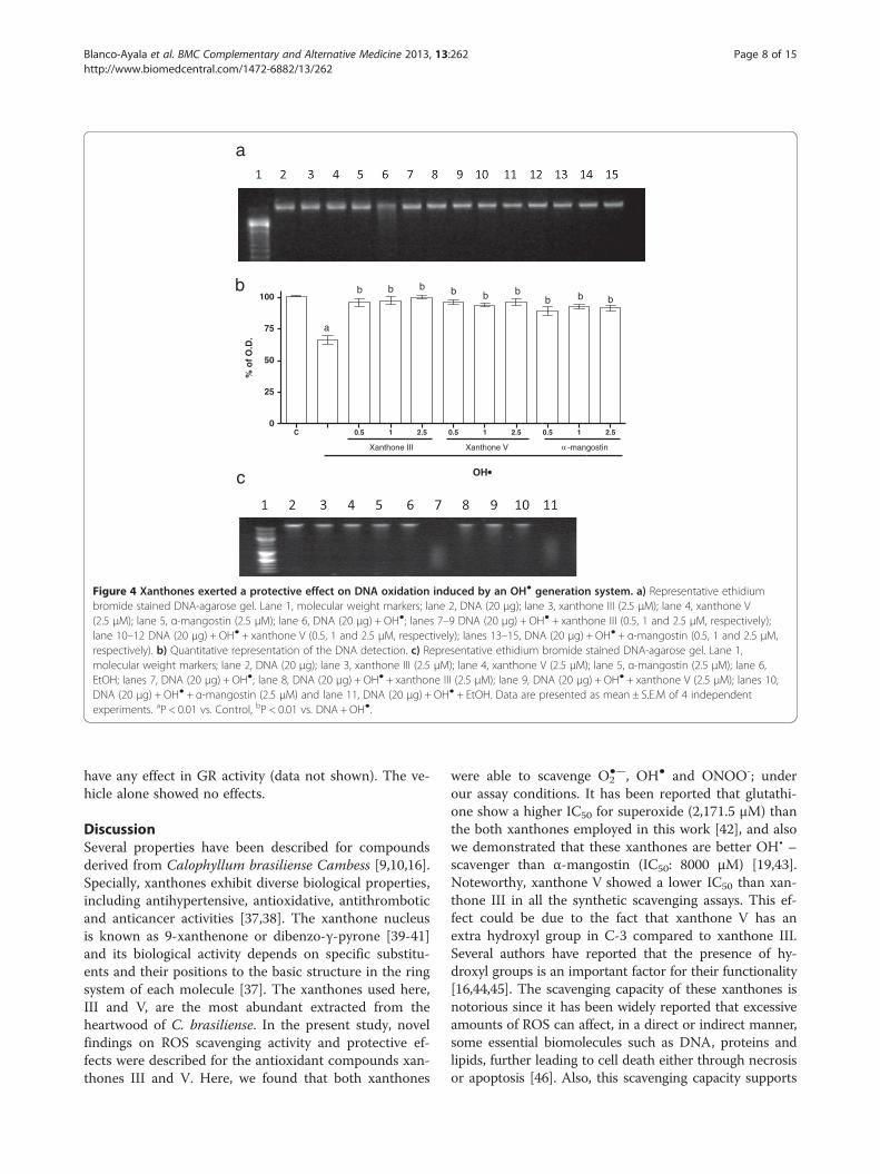
Xanthones III and V block DNA degradation induced by OH•Figure 4a shows the protective effect of xanthones III(lanes 7–9) and V (lanes 10–12) on DNA degradationinduced by OH● (lane 6). The effect of α-mangostin wasalso evaluated in this paradigm (lanes 13–15), as a refer-ence. The OH● generator system induced around 35% ofDNA degradation (Figure 4b), which was completelyabolished by xanthones III and V and α-mangostin, aneffect not dependent of the concentration used. Figure 4cshows that the three compounds tested did not exertany effect when compared to control group at thehighest concentration used (2.5 μM; lanes 3, 4 and 5)and also shows that the EtOH at the maximum concen-tration used (0.01%) does not have any effect on theDNA degradation induced by OH• (line 11).
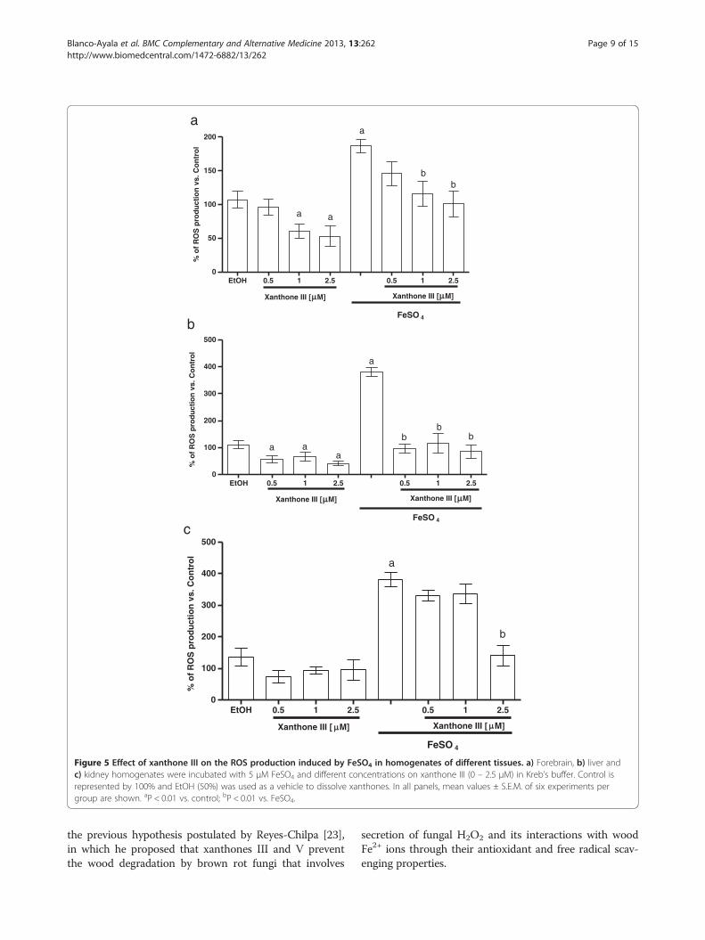
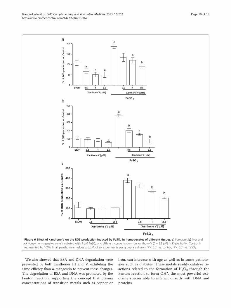
Xanthones III and V reduce the ROS production inducedby FeSO4
FeSO4 induced an increase in ROS formation in fore-brain, liver and kidney (186.7% 380%, and 380.3% whencompared to control group, respectively (Figures 5 and6a, 5 and 6b and 5 and 6c). In all preparations, xan-thones III and V (0.5, 1 and 2.5 μM; Figures 5 and 6, re-spectively) reduced ROS formation to basal levels andalso decreased ROS formation in a concentration-dependent manner in the homogenates exposed tothe ferrous iron. Upon these conditions, ethanol (usedas vehicle) did not have any effect when compared tocontrol group. Both xanthones showed the same ef-fectiveness to decrease this marker.
Xanthones III and V reduced the lipid peroxidationproduced by FeSO4
Figures 7 and 8 show the FeSO4-induced increase inLP in homogenates of (a) forebrain, (b) liver and (c)
0 100 200 300 400 500 600 700 800 900 10000
20
40
60
80
100
XANTHONE III
XANTHONE V
[ M]
O2.-
sca
ven
gin
g c
apac
ity,
%
0 10 20 30 40 500
20
40
60
80
100
120
XANTHONE III
XANTHONE V
[ M]
OH
sca
ven
gin
g c
apac
ity,
%
0 100 200 300 400 500 600 700 800 900 10000
20
40
60
80
100
XANTHONE III
XANTHONE V
[ M]
ON
OO-
sca
ven
gin
g c
apac
ity,
%
b
c
a
Figure 2 Xanthones III and V are able to scavenge (a) O2•−, (b) OH● and (c) ONOO− in a concentration-dependent manner. In vitro
systems of ROS production were assessed and subjected to different concentrations of xanthones (0.5-1,000 μM). Both xanthones showed aneffective capacity as scavengers of O2
•−, OH● and ONOO−. Data are presented as mean values ± S.E.M. of seven experiments per group.
Blanco-Ayala et al. BMC Complementary and Alternative Medicine 2013, 13:262 Page 6 of 15http://www.biomedcentral.com/1472-6882/13/262
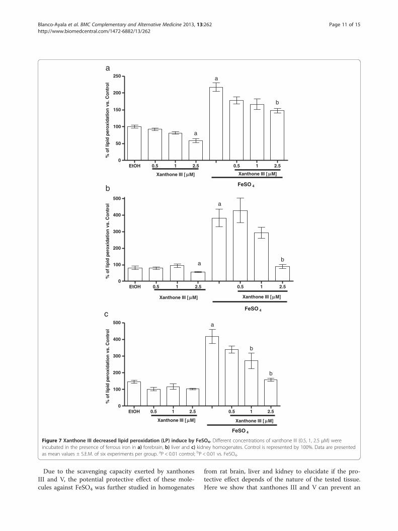
kidney (217.5%, 383% and 418.3% when compared tocontrol group, respectively). Xanthones III and V de-creased the FeSO4-induced LP in a concentration-dependent manner in all tissue homogenates tested
(Figures 7 and 8, respectively). In addition, the incu-bation with vehicle alone did not exert any effect onthis marker. However, both xanthones III and V re-duced lipid peroxidation at the highest concentration
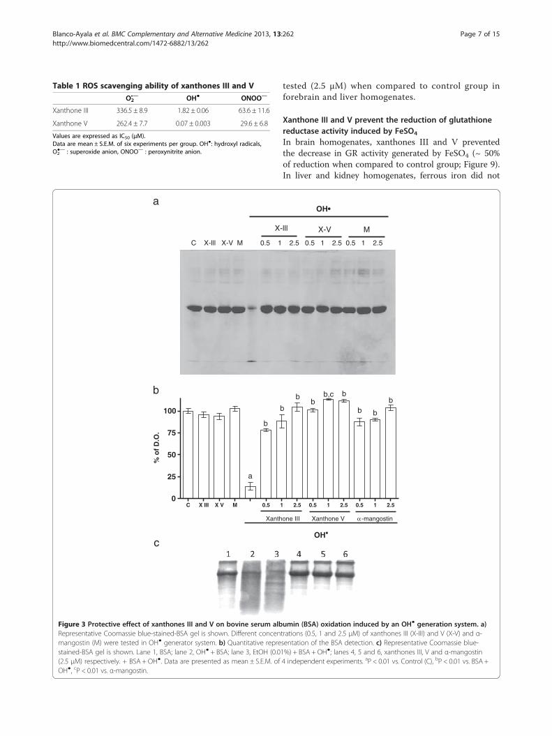
Table 1 ROS scavenging ability of xanthones III and V
O2•— OH● ONOO—
Xanthone III 336.5 ± 8.9 1.82 ± 0.06 63.6 ± 11.6
Xanthone V 262.4 ± 7.7 0.07 ± 0.003 29.6 ± 6.8
Values are expressed as IC50 (μM).Data are mean ± S.E.M. of six experiments per group. OH●: hydroxyl radicals,O2●— : superoxide anion, ONOO— : peroxynitrite anion.
C X III X V M 0.5 10
25
50
75
100
Xanth
a
b
b
% o
f D
.O.
a
b
C X-lll X-V M 0.5 1
X-
c
Figure 3 Protective effect of xanthones III and V on bovine serum albRepresentative Coomassie blue-stained-BSA gel is shown. Different concenmangostin (M) were tested in OH● generator system. b) Quantitative represtained-BSA gel is shown. Lane 1, BSA; lane 2, OH● + BSA; lane 3, EtOH (0.0(2.5 μM) respectively. + BSA + OH●. Data are presented as mean ± S.E.M. ofOH●, cP < 0.01 vs. α-mangostin.
Blanco-Ayala et al. BMC Complementary and Alternative Medicine 2013, 13:262 Page 7 of 15http://www.biomedcentral.com/1472-6882/13/262
tested (2.5 μM) when compared to control group inforebrain and liver homogenates.
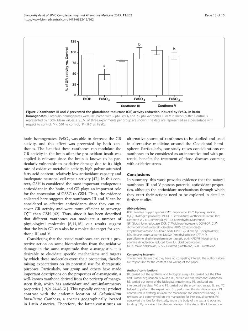
Xanthone III and V prevent the reduction of glutathionereductase activity induced by FeSO4
In brain homogenates, xanthones III and V preventedthe decrease in GR activity generated by FeSO4 (~ 50%of reduction when compared to control group; Figure 9).In liver and kidney homogenates, ferrous iron did not
2.5 0.5 1 2.5 0.5 1 2.5
Xanthone Vone III -mangostin
OH
bb
b,c b
b b
b
2.5 0.5 1 2.5 0.5 1 2.5
lll X-V M
OH•
umin (BSA) oxidation induced by an OH● generation system. a)trations (0.5, 1 and 2.5 μM) of xanthones III (X-III) and V (X-V) and α-sentation of the BSA detection. c) Representative Coomassie blue-1%) + BSA + OH●; lanes 4, 5 and 6, xanthones III, V and α-mangostin4 independent experiments. aP < 0.01 vs. Control (C), bP < 0.01 vs. BSA +
C 0.5 1 2.5 0.5 1 2.5 0.5 1 2.50
25
50
75
100
Xanthone III Xanthone V -mangostin
OH
a
b b b b b bb b b
% o
f O
.D.
a
b
c
Figure 4 Xanthones exerted a protective effect on DNA oxidation induced by an OH● generation system. a) Representative ethidiumbromide stained DNA-agarose gel. Lane 1, molecular weight markers; lane 2, DNA (20 μg); lane 3, xanthone III (2.5 μM); lane 4, xanthone V(2.5 μM); lane 5, α-mangostin (2.5 μM); lane 6, DNA (20 μg) + OH●; lanes 7–9 DNA (20 μg) + OH● + xanthone III (0.5, 1 and 2.5 μM, respectively);lane 10–12 DNA (20 μg) + OH● + xanthone V (0.5, 1 and 2.5 μM, respectively); lanes 13–15, DNA (20 μg) + OH● + α-mangostin (0.5, 1 and 2.5 μM,respectively). b) Quantitative representation of the DNA detection. c) Representative ethidium bromide stained DNA-agarose gel. Lane 1,molecular weight markers; lane 2, DNA (20 μg); lane 3, xanthone III (2.5 μM); lane 4, xanthone V (2.5 μM); lane 5, α-mangostin (2.5 μM); lane 6,EtOH; lanes 7, DNA (20 μg) + OH●; lane 8, DNA (20 μg) + OH● + xanthone III (2.5 μM); lane 9, DNA (20 μg) + OH● + xanthone V (2.5 μM); lanes 10,DNA (20 μg) + OH● + α-mangostin (2.5 μM) and lane 11, DNA (20 μg) + OH● + EtOH. Data are presented as mean ± S.E.M of 4 independentexperiments. aP < 0.01 vs. Control, bP < 0.01 vs. DNA + OH●.
Blanco-Ayala et al. BMC Complementary and Alternative Medicine 2013, 13:262 Page 8 of 15http://www.biomedcentral.com/1472-6882/13/262
have any effect in GR activity (data not shown). The ve-hicle alone showed no effects.
DiscussionSeveral properties have been described for compoundsderived from Calophyllum brasiliense Cambess [9,10,16].Specially, xanthones exhibit diverse biological properties,including antihypertensive, antioxidative, antithromboticand anticancer activities [37,38]. The xanthone nucleusis known as 9-xanthenone or dibenzo-γ-pyrone [39-41]and its biological activity depends on specific substitu-ents and their positions to the basic structure in the ringsystem of each molecule [37]. The xanthones used here,III and V, are the most abundant extracted from theheartwood of C. brasiliense. In the present study, novelfindings on ROS scavenging activity and protective ef-fects were described for the antioxidant compounds xan-thones III and V. Here, we found that both xanthones
were able to scavenge O2●—, OH● and ONOO-; under
our assay conditions. It has been reported that glutathi-one show a higher IC50 for superoxide (2,171.5 μM) thanthe both xanthones employed in this work [42], and alsowe demonstrated that these xanthones are better OH• –scavenger than α-mangostin (IC50: 8000 μM) [19,43].Noteworthy, xanthone V showed a lower IC50 than xan-thone III in all the synthetic scavenging assays. This ef-fect could be due to the fact that xanthone V has anextra hydroxyl group in C-3 compared to xanthone III.Several authors have reported that the presence of hy-droxyl groups is an important factor for their functionality[16,44,45]. The scavenging capacity of these xanthones isnotorious since it has been widely reported that excessiveamounts of ROS can affect, in a direct or indirect manner,some essential biomolecules such as DNA, proteins andlipids, further leading to cell death either through necrosisor apoptosis [46]. Also, this scavenging capacity supports
EtOH 0.5 1 2.5 0.5 1 2.50
50
100
150
200
Xanthone III [ M] Xanthone III [ M]
a
bb
a a
FeSO 4
% o
f R
OS
pro
du
ctio
n v
s. C
on
tro
l
EtOH 0.5 1 2.5 0.5 1 2.50
100
200
300
400
500
Xanthone III [ M] Xanthone III [ M]
a
bb
b
FeSO 4
a aa
% o
f R
OS
pro
du
ctio
n v
s. C
on
tro
l
a
b
EtOH 0.5 1 2.5 0.5 1 2.50
100
200
300
400
500
Xanthone III [ M] Xanthone III [ M]
a
b
FeSO 4
% o
f R
OS
pro
du
ctio
n v
s. C
on
tro
l
c
Figure 5 Effect of xanthone III on the ROS production induced by FeSO4 in homogenates of different tissues. a) Forebrain, b) liver andc) kidney homogenates were incubated with 5 μM FeSO4 and different concentrations on xanthone III (0 – 2.5 μM) in Kreb’s buffer. Control isrepresented by 100% and EtOH (50%) was used as a vehicle to dissolve xanthones. In all panels, mean values ± S.E.M. of six experiments pergroup are shown. aP < 0.01 vs. control; bP < 0.01 vs. FeSO4.
Blanco-Ayala et al. BMC Complementary and Alternative Medicine 2013, 13:262 Page 9 of 15http://www.biomedcentral.com/1472-6882/13/262
the previous hypothesis postulated by Reyes-Chilpa [23],in which he proposed that xanthones III and V preventthe wood degradation by brown rot fungi that involves
secretion of fungal H2O2 and its interactions with woodFe2+ ions through their antioxidant and free radical scav-enging properties.
EtOH 0.5 1 2.5 0.5 1 2.50
50
100
150
200
Xanthone V [ M] Xanthone V [ M]
a
b
b
a aa
FeSO 4
% o
f R
OS
pro
du
ctio
n v
s. C
on
tro
l
EtOH 0.5 1 2.5 0.5 1 2.50
100
200
300
400
500
Xanthone V [ M] Xanthone V [ M]
a
b
b
b
FeSO 4
a
% o
f R
OS
pro
du
ctio
n v
s. C
on
tro
l
c
a
b
EtOH 0.5 1 2.5 0.5 1 2.50
100
200
300
400
500
Xanthone V [ M] Xanthone V [ M]
a
b
b
FeSO 4
% o
f R
OS
pro
du
ctio
n v
s. C
on
tro
l
Figure 6 Effect of xanthone V on the ROS production induced by FeSO4 in homogenates of different tissues. a) Forebrain, b) liver andc) kidney homogenates were incubated with 5 μM FeSO4 and different concentrations on xanthone V (0 – 2.5 μM) in Kreb’s buffer. Control isrepresented by 100%. In all panels, mean values ± S.E.M. of six experiments per group are shown. aP < 0.01 vs. control; bP < 0.01 vs. FeSO4.
Blanco-Ayala et al. BMC Complementary and Alternative Medicine 2013, 13:262 Page 10 of 15http://www.biomedcentral.com/1472-6882/13/262
We also showed that BSA and DNA degradation wereprevented by both xanthones III and V, exhibiting thesame efficacy than α-mangostin to prevent these changes.The degradation of BSA and DNA was promoted by theFenton reaction, supporting the concept that plasmaconcentrations of transition metals such as copper or
iron, can increase with age as well as in some patholo-gies such as diabetes. These metals readily catalyze re-actions related to the formation of H2O2 through theFenton reaction to form OH●, the most powerful oxi-dizing species able to interact directly with DNA andproteins.
EtOH 0.5 1 2.5 0.5 1 2.50
50
100
150
200
250
Xanthone III [ M] Xanthone III [ M]
a
b
FeSO 4
a
% o
f lip
id p
ero
xid
atio
n v
s. C
on
tro
l
EtOH 0.5 1 2.5 0.5 1 2.50
100
200
300
400
500
Xanthone III [ M] Xanthone III [ M]
a
ba
FeSO 4
% o
f lip
id p
ero
xid
atio
n v
s. C
on
tro
l
b
a
EtOH 0.5 1 2.5 0.5 1 2.50
100
200
300
400
500
Xanthone III [ M]Xanthone III [ M]
a
b
b
FeSO 4
% o
f lip
id p
ero
xid
atio
n v
s. C
on
tro
l
c
Figure 7 Xanthone III decreased lipid peroxidation (LP) induce by FeSO4. Different concentrations of xanthone III (0.5, 1, 2.5 μM) wereincubated in the presence of ferrous iron in a) forebrain, b) liver and c) kidney homogenates. Control is represented by 100%. Data are presentedas mean values ± S.E.M. of six experiments per group. aP < 0.01 control; bP < 0.01 vs. FeSO4.
Blanco-Ayala et al. BMC Complementary and Alternative Medicine 2013, 13:262 Page 11 of 15http://www.biomedcentral.com/1472-6882/13/262
Due to the scavenging capacity exerted by xanthonesIII and V, the potential protective effect of these mole-cules against FeSO4 was further studied in homogenates
from rat brain, liver and kidney to elucidate if the pro-tective effect depends of the nature of the tested tissue.Here we show that xanthones III and V can prevent an

EtOH 0.5 1 2.5 0.5 1 2.50
50
100
150
200
250
Xanthone V [ M] Xanthone V [ M]
a
b
b
a
FeSO 4
% o
f lip
id p
ero
xid
atio
n v
s. C
on
tro
l
EtOH 0.5 1 2.5 0.5 1 2.50
100
200
300
400
500a
b
b
Xanthone V [ M] Xanthone V [ M]
aaa
FeSO 4
% o
f lip
id p
ero
xid
atio
n v
s co
ntr
ol
b
a
c
EtOH 0.5 1 2.5 0.5 1 2.50
100
200
300
400
500
Xanthone V [ M] Xanthone V [ M]
b
b
a
FeSO 4
% o
f lip
id p
ero
xid
atio
n v
s. C
on
tro
l
Figure 8 Concentration-response effect of xanthone V on the lipid peroxidation (LP) induced by FeSO4 in homogenates. a) Forebrain,b) liver and c) kidney homogenates were incubated with 5 μM FeSO4 and different concentrations of xanthone V (0, 0.5, 1 and 2.5 μM) in Kreb’sbuffer. Control is represented by 100%. In all panels, mean values ± S.E.M. of six experiments per group are shown. aP < 0.01 vs. control; bP < 0.01vs. FeSO4.
Blanco-Ayala et al. BMC Complementary and Alternative Medicine 2013, 13:262 Page 12 of 15http://www.biomedcentral.com/1472-6882/13/262
increase in the levels of the oxidative markers tested,which in turn are related with the scavenging capacityexhibited by these xanthones in synthetic assays.
Interestingly, the antioxidant effect showed by xan-thones on ROS production and lipid peroxidation wasnot dependent on the tissue studied. Moreover, only in
EtOH FeSO 4 FeSO 4 FeSO 4
0
25
50
75
100
125
a
bb
Xanthone III Xanthone V
% o
f G
R a
ctiv
ity
vs. C
on
tro
l
Figure 9 Xanthones III and V prevented the glutathione reductase (GR) activity reduction induced by FeSO4 in brainhomogenates. Forebrain homogenates were incubated with 5 μM FeSO4 and 2.5 μM xanthones III or V in Kreb’s buffer. Control isrepresented by 100%. Mean values ± S.E.M. of three experiments per group are shown. The data are represented as a percentage withrespect to control. aP < 0.01 vs control; bP < 0.01vs. FeSO4.
Blanco-Ayala et al. BMC Complementary and Alternative Medicine 2013, 13:262 Page 13 of 15http://www.biomedcentral.com/1472-6882/13/262
brain homogenates, FeSO4 was able to decrease the GRactivity, and this effect was prevented by both xan-thones. The fact that these xanthones can modulate theGR activity in the brain after the pro-oxidant insult wasapplied is relevant since the brain is known to be par-ticularly vulnerable to oxidative damage due to its highrate of oxidative metabolic activity, high polyunsaturatedfatty acid content, relatively low antioxidant capacity andinadequate neuronal cell repair activity [47]. In this con-text, GSH is considered the most important endogenousantioxidant in the brain, and GR plays an important rolefor the conversion of GSSG to GSH. Thus, the evidencecollected here suggests that xanthones III and V can beconsidered as effective antioxidants since they can re-cover GR activity and were more efficient to scavengeO2
●— than GSH [42]. Thus, since it has been describedthat different xanthones can modulate a number ofphysiological molecules [6,14,16], our results suggestthat the brain GR can also be a molecular target for xan-thone III and V.Considering that the tested xanthones can exert a pro-
tective action on some biomolecules from the oxidativedamage in the same magnitude than α-mangostin, it isdesirable to elucidate specific mechanisms and targetsby which these molecules exert their protection, therebyraising expectations on its potential use for therapeuticpurposes. Particularly, our group and others have madeimportant descriptions on the properties of α-mangostin, awell-known xanthone derived from the pericarp of mango-steen fruit, which has antioxidant and anti-inflammatoryproperties [19,21,24,48-51]. This typically oriental productcontrast with the endemic location of Calophyllumbrasiliense Cambess, a species geographically locatedin Latin America. Therefore, the latter constitutes an
alternative source of xanthones to be studied and usedin alternative medicine around the Occidental hemi-sphere. Particularly, our study raises considerations onxanthones to be considered as an innovative tool with po-tential benefits for treatment of those diseases coursingwith oxidative stress.
ConclusionsIn summary, this work provides evidence that the naturalxanthones III and V possess potential antioxidant proper-ties, although the antioxidant mechanisms through whichthey exert their actions need to be explored in detail infurther studies.
AbbreviationsROS: Reactive oxygen species; O2
●—: Superoxide; OH●: Hydroxyl radical;H2O2: Hydrogen peroxide; ONOO—: Peroxynitrite; xanthone III: Jacareubin;xanthone V: 2-(3,3-dimethylallyl)-1,3,5,6-tetrahydroxyxanthone;GR: Glutathione reductase; DCF: 2′,7′-dichlorofluorescein; DCFH-DA: 2′,7′-dichlorodihydrofluorescein diacetate; ABTS: 2,2′-azinobis-(3-ethylbenzothiazoline-6-sulfonic acid); DPPH: 2,2-diphenyl-1-picrylhydrazyl;BSA: Bovine serum albumin; DMSO: Dimethylsulfoxide; DTPA: DL-penicillamine, diethylenetriaminepentaacetic acid; NADPH: Nicotinamideadenine dinucleotide reduced form; LP: Lipid peroxidation;MDA: Malondialdehyde; GSSG: Oxidized glutathione; GSH: Glutathione.
Competing interestsThe authors declare that they have no competing interest. The authors aloneare responsible for the content and writing of the paper.
Authors’ contributionsBT, carried out the synthetic and biological assays. LR, carried out the DNAand Protein degradation. SEM and RR, carried out the xanthones extraction.RE, carried out some of the biological experiments. PB, analyzed andinterpreted the data. MO and PE, carried out the enzymatic assays. SL and TChelped to perform the experiment. SD, performed the statistical analysis. PJ,contributed in drafting, revision the manuscript and obtained funding. RC,reviewed and commented on the manuscript for intellectual context. PV,conceived the idea for the study, wrote the body of the text and obtainedfunding. TM, conceived the idea and design of the study. All of the authors

Blanco-Ayala et al. BMC Complementary and Alternative Medicine 2013, 13:262 Page 14 of 15http://www.biomedcentral.com/1472-6882/13/262
read the manuscript, contributed in correcting it and approving its finalversion.
AcknowledgementsThis work was supported in part by PAPIIT IN210713, and CONACYT Grants129838, 204474 and 183867.
Author details1Departamento de Neuroquímica, Instituto Nacional de Neurología yNeurocirugía Manuel Velasco Suárez, Insurgentes Sur 3877, S.S.A., México, DF14269, México. 2Instituto de Química, Universidad Nacional Autónoma deMéxico, México, DF 04510, México. 3Laboratorio de Aminoácidos Excitadores,Instituto Nacional de Neurología y Neurocirugía Manuel Velasco Suárez, S.S.A., México, DF 14269, México. 4Laboratorio de Neuroinmunología, InstitutoNacional de Neurología y Neurocirugía Manuel Velasco Suárez, S.S.A., México,DF 14269, México. 5Departamento de Biología, Facultad de Química,Universidad Nacional Autónoma de México, México, DF 04510, México.6Laboratorio de Bioquímica Muscular, Instituto Nacional de Rehabilitación, S.S.A., México, DF 14389, México. 7Unidad del Bioterio, Facultad de Medicina,Universidad Nacional Autónoma de México, México, DF 04510, México.8Unidad Periférica de Neurociencias Facultad de Medicina UNAM-INNN,México, DF 14269, México.
Received: 12 April 2013 Accepted: 7 October 2013Published: 11 October 2013
References1. Nordberg J, Arner ES: Reactive oxygen species, antioxidants, and the
mammalian thioredoxin system. Free Radic Biol Med 2001, 31(11):1287–1312.2. Valko M, Leibfritz D, Moncol J, Cronin MT, Mazur M, Telser J: Free radicals
and antioxidants in normal physiological functions and human disease.Int J Biochem Cell Biol 2007, 39(1):44–84.
3. Charles AL, Meyer A, Dal-Ros S, Auger C, Keller N, Gali Ramamoorthy T, Zoll J,Metzger D, Schini-Kerth V, Geny B: Polyphenols prevents aging-relatedimpairment in skeletal muscle mitochondrial function through decreasedreactive oxygen species production. Exp Physiol 2012.
4. Corcoran MP, McKay DL, Blumberg JB: Flavonoid basics: chemistry,sources, mechanisms of action, and safety. J Nutr Gerontol Geriatr 2012, 31(3):176–189.
5. Widmer RJ, Freund MA, Flammer AJ, Sexton J, Lennon R, Romani A,Mulinacci N, Vinceri FF, Lerman LO, Lerman A: Beneficial effects of polyphenol-rich olive oil in patients with early atherosclerosis. Eur J Nutr 2012.
6. Gonzalez R, Ballester I, Lopez-Posadas R, Suarez MD, Zarzuelo A, Martinez-Augustin O, Sanchez De Medina F: Effects of flavonoids and otherpolyphenols on inflammation. Crit Rev Food Sci Nutr 2011, 51(4):331–362.
7. Shan T, Ma Q, Guo K, Liu J, Li W, Wang F, Wu E: Xanthones frommangosteen extracts as natural chemopreventive agents: potentialanticancer drugs. Curr Mol Med 2011, 11(8):666–677.
8. Campos-Esparza Mdel R, Torres-Ramos MA: Neuroprotection by naturalpolyphenols: molecular mechanisms. Cent Nerv Syst Agents Med Chem2010, 10(4):269–277.
9. Lewis WH, McNaughton DR, Goh SH, LeJohn HB, Wright JA: Inhibition ofmammalian ribonucleotide reductase by a dinucleotide produced ineucaryotic cells. J Cell Physiol 1977, 93(3):345–352.
10. Duke JA, Vásquez R: Amazonian ethnobotanical dictionary. Boca Raton, Fla:CRC Press; 1994.
11. Grenand P: Pharmacopées traditionnelles en Guyane: Créoles, Wayãpi, Palikur,Ed. entièrement rev. et complétée edn. Paris: IRD Éditions, Institut derecherche pour le développement; 2004.
12. Rutter R: Catalogo de las Plantas Utiles de la Amazonia Peruana. Perú:Instituto Lingüistico de verano; 1990.
13. Katritzky AR, Rees CW: Comprehensive heterocyclic chemistry: the structure,reactions, synthesis, and uses of heterocyclic compounds. 1st edition. OxfordOxfordshire, New York: Pergamon Press; 1984.
14. Mesia-Vela S, Sanchez RI, Estrada-Muniz E, Alavez-Solano D, Torres-Sosa C,Jimenez M, Estrada, Reyes-Chilpa R, Kauffman FC: Natural products isolatedfrom Mexican medicinal plants: novel inhibitors of sulfotransferases,SULT1A1 and SULT2A1. Phytomedicine 2001, 8(6):481–488.
15. Yasunaka K, Abe F, Nagayama A, Okabe H, Lozada-Perez L, Lopez-Villafranco E,Muniz EE, Aguilar A, Reyes-Chilpa R: Antibacterial activity of crude extracts
from Mexican medicinal plants and purified coumarins and xanthones.J Ethnopharmacol 2005, 97(2):293–299.
16. Reyes-Chilpa R, Baggio CH, Alavez-Solano D, Estrada-Muniz E, Kauffman FC,Sanchez RI, Mesia-Vela S: Inhibition of gastric H+, K + −ATPase activity byflavonoids, coumarins and xanthones isolated from Mexican medicinalplants. J Ethnopharmacol 2006, 105(1–2):167–172.
17. Campos-Esparza MR, Sanchez-Gomez MV, Matute C: Molecularmechanisms of neuroprotection by two natural antioxidant polyphenols.Cell Calcium 2009, 45(4):358–368.
18. Martinez A, Galano A, Vargas R: Free radical scavenger properties ofalpha-mangostin: thermodynamics and kinetics of HAT and RAFmechanisms. J Phys Chem B 2011, 115(43):12591–12598.
19. Pedraza-Chaverri J, Reyes-Fermin LM, Nolasco-Amaya EG, Orozco-Ibarra M,Medina-Campos ON, Gonzalez-Cuahutencos O, Rivero-Cruz I, Mata R: ROSscavenging capacity and neuroprotective effect of alpha-mangostinagainst 3-nitropropionic acid in cerebellar granule neurons. Exp ToxicolPathol 2009, 61(5):491–501.
20. Buelna-Chontal M, Correa F, Hernandez-Resendiz S, Zazueta C, Pedraza-Chaverri J:Protective effect of alpha-mangostin on cardiac reperfusion damage byattenuation of oxidative stress. J Med Food 2011, 14(11):1370–1374.
21. Reyes-Fermin LM, Gonzalez-Reyes S, Tarco-Alvarez NG, Hernandez-Nava M,Orozco-Ibarra M, Pedraza-Chaverri J: Neuroprotective effect of alpha-mangostin and curcumin against iodoacetate-induced cell death. NutrNeurosci 2012, 15(5):34–41.
22. Barcenas-Pazos: Caracterización tecnológica de veinte especiesmaderables de la Selva Lacandona. Madera y Bosques 1995, 1(1):29.
23. Reyes-Chilpa R, Jimenez-Estrada M, Estrada-Muniz E: Antifungal xanthonesfrom Calophyllum brasiliensis heartwood. J Chem Ecol 1997, 23(7):1901–1911.
24. Marquez-Valadez B, Lugo-Huitron R, Valdivia-Cerda V, Miranda-Ramirez LR, Perez-De La Cruz V, Gonzalez-Cuahutencos O, Rivero-Cruz I, Mata R, Santamaria A,Pedraza-Chaverri J: The natural xanthone alpha-mangostin reduces oxidativedamage in rat brain tissue. Nutr Neurosci 2009, 12(1):35–42.
25. Fontana M, Mosca L, Rosei MA: Interaction of enkephalins withoxyradicals. Biochem Pharmacol 2001, 61(10):1253–1257.
26. Floriano-Sanchez E, Villanueva C, Medina-Campos ON, Rocha D, Sanchez-Gonzalez DJ, Cardenas-Rodriguez N, Pedraza-Chaverri J:Nordihydroguaiaretic acid is a potent in vitro scavenger of peroxynitrite,singlet oxygen, hydroxyl radical, superoxide anion and hypochlorousacid and prevents in vivo ozone-induced tyrosine nitration in lungs. FreeRadic Res 2006, 40(5):523–533.
27. Halliwell B, Gutteridge JM, Aruoma OI: The deoxyribose method: a simple"test-tube" assay for determination of rate constants for reactions ofhydroxyl radicals. Anal Biochem 1987, 165(1):215–219.
28. Beckman JS, Chen J, Ischiropoulos H, Crow JP: Oxidative chemistry ofperoxynitrite. Methods Enzymol 1994, 233:229–240.
29. Crow JP, Beckman JS: The importance of superoxide in nitric oxide-dependent toxicity: evidence for peroxynitrite-mediated injury. Adv ExpMed Biol 1996, 387:147–161.
30. Long LH, Evans PJ, Halliwell B: Hydrogen peroxide in human urine:implications for antioxidant defense and redox regulation. BiochemBiophys Res Commun 1999, 262(3):605–609.
31. Kocha T, Yamaguchi M, Ohtaki H, Fukuda T, Aoyagi T: Hydrogen peroxide-mediated degradation of protein: different oxidation modes of copper-and iron-dependent hydroxyl radicals on the degradation of albumin.Biochim Biophys Acta 1997, 1337(2):319–326.
32. Galano A, Macias-Ruvalcaba NA, Medina Campos ON, Pedraza-Chaverri J:Mechanism of the OH radical scavenging activity ofnordihydroguaiaretic acid: a combined theoretical and experimentalstudy. J Phys Chem B 2010, 114(19):6625–6635.
33. Perez-De La Cruz V, Elinos-Calderon D, Carrillo-Mora P, Silva-Adaya D,Konigsberg M, Moran J, Ali SF, Chanez-Cardenas ME, Perez-De La Cruz G,Santamaria A: Time-course correlation of early toxic events in threemodels of striatal damage: modulation by proteases inhibition.Neurochem Int 2010, 56(6–7):834–842.
34. Ali SF, LeBel CP, Bondy SC: Reactive oxygen species formation as abiomarker of methylmercury and trimethyltin neurotoxicity. Neurotoxicol1992, 13(3):637–648.
35. Herrera-Mundo N, Sitges M: Mechanisms underlying striatal vulnerabilityto 3-nitropropionic acid. J Neurochem 2010, 114(2):597–605.
36. Tapia E, Sanchez-Gonzalez DJ, Medina-Campos ON, Soto V, Avila-Casado C,Martinez-Martinez CM, Johnson RJ, Rodriguez-Iturbe B, Pedraza-Chaverri J,
Blanco-Ayala et al. BMC Complementary and Alternative Medicine 2013, 13:262 Page 15 of 15http://www.biomedcentral.com/1472-6882/13/262
Franco M, et al: Treatment with pyrrolidine dithiocarbamate improvesproteinuria, oxidative stress, and glomerular hypertension in overloadproteinuria. Am J Physiol Renal Physiol 2008, 295(5):F1431–1439.
37. Na Y: Recent cancer drug development with xanthone structures.J Pharm Pharmacol 2009, 61(6):707–712.
38. Minami H, Takahashi E, Fukuyama Y, Kodama M, Yoshizawa T, Nakagawa K:Novel xanthones with superoxide scavenging activity from Garciniasubelliptica. Chem Pharm Bull 1995, 43(2):347–349.
39. Pedraza-Chaverri J, Cardenas-Rodriguez N, Orozco-Ibarra M, Perez-Rojas JM:Medicinal properties of mangosteen (Garcinia mangostana). Food ChemToxicol 2008, 46(10):3227–3239.
40. Vieira LM, Kijjoa A: Naturally-occurring xanthones: recent developments.Curr Med Chem 2005, 12(21):2413–2446.
41. Gales L, Damas AM: Xanthones–a structural perspective. Curr Med Chem2005, 12(21):2499–2515.
42. Lugo-Huitron R, Blanco-Ayala T, Ugalde-Muniz P, Carrillo-Mora P, Pedraza-Chaverri J, Silva-Adaya D, Maldonado PD, Torres I, Pinzon E, Ortiz-Islas E, et al:On the antioxidant properties of kynurenic acid: free radical scavengingactivity and inhibition of oxidative stress. Neurotoxicol Teratol 2011, 33(5):538–547.
43. Sun D, Zhang S, Wei Y, Yin L: Antioxidant activity of mangostin in cell-freesystem and its effect on K562 leukemia cell line in photodynamictherapy. Acta Biochim Biophys Sin 2009, 41(12):1033–1043.
44. Ito C, Itoigawa M, Mishina Y, Filho VC, Mukainaka T, Tokuda H, Nishino H,Furukawa H: Chemical constituents of Calophyllum brasiliensis: structureelucidation of seven new xanthones and their cancer chemopreventiveactivity. J Nat Prod 2002, 65(3):267–272.
45. Sang S, Lapsley K, Jeong WS, Lachance PA, Ho CT, Rosen RT: Antioxidativephenolic compounds isolated from almond skins (Prunus amygdalusBatsch). J Agric Food Chem 2002, 50(8):2459–2463.
46. Almeida RD, Manadas BJ, Carvalho AP, Duarte CB: Intracellular signalingmechanisms in photodynamic therapy. Biochim Biophys Acta 2004, 1704(2):59–86.
47. Traystman RJ, Kirsch JR, Koehler RC: Oxygen radical mechanisms of brain injuryfollowing ischemia and reperfusion. J Appl Physiol 1991, 71(4):1185–1195.
48. Sanchez-Perez Y, Morales-Barcenas R, Garcia-Cuellar CM, Lopez-Marure R,Calderon-Oliver M, Pedraza-Chaverri J, Chirino YI: The alpha-mangostinprevention on cisplatin-induced apoptotic death in LLC-PK1 cells isassociated to an inhibition of ROS production and p53 induction.Chem Biol Interact 2010, 188(1):144–150.
49. Chen LG, Yang LL, Wang CC: Anti-inflammatory activity of mangostinsfrom Garcinia mangostana. Food Chem Toxicol 2008, 46(2):688–693.
50. Tewtrakul S, Wattanapiromsakul C, Mahabusarakam W: Effects ofcompounds from Garcinia mangostana on inflammatory mediators inRAW264.7 macrophage cells. J Ethnopharmacol 2009, 121(3):379–382.
51. Marquez-Valadez B, Maldonado PD, Galvan-Arzate S, Mendez-Cuesta LA,Perez-De La Cruz V, Pedraza-Chaverri J, Chanez-Cardenas ME, Santamaria A:Alpha-mangostin induces changes in glutathione levels associated withglutathione peroxidase activity in rat brain synaptosomes. Nutr Neurosci2012, 15(5):13–19.
doi:10.1186/1472-6882-13-262Cite this article as: Blanco-Ayala et al.: Antioxidant properties ofxanthones from Calophyllum brasiliense: prevention of oxidativedamage induced by FeSO4. BMC Complementary and Alternative Medicine2013 13:262.
Submit your next manuscript to BioMed Centraland take full advantage of:
• Convenient online submission
• Thorough peer review
• No space constraints or color figure charges
• Immediate publication on acceptance
• Inclusion in PubMed, CAS, Scopus and Google Scholar
• Research which is freely available for redistribution
Submit your manuscript at www.biomedcentral.com/submit