evaluation of antinociceptive and antioxidant properties
TRANSCRIPT
ORIGINAL ARTICLE
Evaluation of antinociceptive and antioxidant propertiesof 3-[4-(3-trifluoromethyl-phenyl)-piperazin-1-yl]-dihydrofuran-2-one in mice
Kinga Sałat & Katarzyna Gawlik & Jadwiga Witalis &
Dorota Pawlica-Gosiewska & Barbara Filipek &
Bogdan Solnica & Krzysztof Więckowski &Barbara Malawska
Received: 22 October 2012 /Accepted: 28 February 2013 /Published online: 14 March 2013# The Author(s) 2013. This article is published with open access at Springerlink.com
Abstract The aim of this study was to evaluate the influ-ence of 3-[4-(3-trifluoromethyl-phenyl)-piperazin-1-yl]-dihydrofuran-2-one (LPP1) on nociceptive thresholds inmouse models of persistent pain. Influence of LPP1 onmotor coordination and its antioxidant capacity in mousebrain tissue homogenates were also assessed. Pain sensitiv-ity thresholds in animals treated with LPP1 were establishedusing 5 % formalin solution in normoglycemic mice and instreptozotocin (STZ)-treated diabetic mice in the von Frey,hot plate, innocuous, and noxious cold water tests (water at10 °C and 4 °C, respectively). Motor deficits were assessedin the rotarod test, whereas antioxidant capacities wereevaluated using ferric reducing ability of plasma (FRAP)assay, catalase (CAT), and superoxide dismutase (SOD)activities. LPP1was antinociceptive in both phases of theformalin test, in particular, in the late phase (at doses 0.9–30 mg/kg for 66–99 % vs. control normoglycemic mice) and
in a statistically significant manner increased nociceptivethresholds in response to mechanical, heat, and noxious coldstimulation in neuropathic mice (at 30 mg/kg for 274, 192,and 316 %, respectively vs. diabetic control). LPP1 did notimpair motor coordination of mice in the rotarod revolvingat 6 or 18 rpm. In brain tissue homogenates, it demonstratedantioxidant capacity in FRAP assay and increased SODactivity for 63 % (acute administration) and 28 % (chronicadministration) vs. control. No influence on CAT activitywas observed. LPP1 has significant antinociceptive proper-ties in the formalin model and elevates pain thresholds inneuropathic mice. It has antioxidant capacity and is devoidof negative influence on animals' motor coordination.
Keywords Dihydrofuran-2-one . Enzymatic antioxidantdefense . Formalin-induced tonic pain . Streptozotocin .
Total antioxidant status
AbbreviationsAEDs Antiepileptic drugsCAT CatalaseCNS Central nervous systemFRAP Ferric reducing ability of plasmaH2O2 Hydrogen peroxidei.p. IntraperitonealMC MethylcellulosePGB PregabalinROS Reactive oxygen speciesSOD Superoxide dismutaseSTZ StreptozotocinTPTZ 2,4,6-Tripyridyl-S-triazine
K. Sałat (*) : J. Witalis :B. FilipekDepartment of Pharmacodynamics, Jagiellonian University,Medical College, Medyczna 9, 30-688 Cracow, Polande-mail: [email protected]
K. Gawlik :D. Pawlica-Gosiewska : B. SolnicaDepartment of Diagnostics, Jagiellonian University, MedicalCollege, Kopernika 15a, 31-501 Cracow, Poland
K. Więckowski :B. MalawskaDepartment of Physicochemical Drug Analysis, JagiellonianUniversity, Medical College, Medyczna 9, 30-688 Cracow, Poland
Naunyn-Schmiedeberg's Arch Pharmacol (2013) 386:493–505DOI 10.1007/s00210-013-0847-2
Introduction
Neuropathic pain is a debilitating form of chronic pain thatresults from dysfunction or damage to the peripheral orcentral nervous system (CNS). This type of pain is consid-ered as a drug-resistant complication that still remains aserious medical problem worldwide. The term “neuropathicpain” comprises a variety of painful conditions, includingpostamputation pain, painful neuropathies (e.g., painful di-abetic neuropathy and postherpetic neuralgia), posttraumaticneuralgia, and others. So far, multiple factors responsible fordevelopment of neuropathic pain have been identified: met-abolic diseases (e.g., diabetes), neuronal tissue injuriescaused by ischemia, toxicological factors or mechanicaldamage to the spinal cord, and others (Nickel et al. 2012;Woolf and Mannion 1999). Hence, pharmacotherapy used torelieve neuropathic pain comprises several pharmacologicalclasses, of which antiepileptic drugs (AEDs), antidepressantdrugs, opioid analgesics, and local anesthetic agents play apivotal role (Christoph et al. 2011; Davis 2007; Davis 2010;Gilron et al. 2009; Miranda et al. 2012; Takeuchi et al. 2007;Yamama et al. 2010). Despite this, approximately 10–30 %of patients suffering from neuropathic pain syndromes aredrug resistant (Blackburn-Munro and Erichsen 2005), sostill, there is a great need for seeking new analgesic com-pounds able to attenuate neuropathic pain episodes.
Many lines of evidence indicate that oxidative stress isimplicated in a variety of disorders, including degenerativediseases (Kasznicki et al. 2012; Reynolds et al. 2007;Trushina and McMurray 2007; Uttara et al. 2009), athero-sclerosis, inflammation (Barton et al. 2007; Reuter et al.2010; Salvemini et al. 2011), and chronic pain (Janes et al.2012; Salvemini et al. 2011). Painful diabetic neuropathy isone of the most serious complications of diabetes in whichthe role of oxidative stress has been postulated. Imbalancebetween enhanced generation of reactive oxygen and nitro-gen species and diminished activity of enzymatic andnonenzymatic antioxidant defenses as a key factor underly-ing diabetic neuropathy in mammals has been demonstrated(Di Naso et al. 2011; Pacher et al. 2005).
In our previous studies, we demonstrated significantantinociceptive, antiinflammatory, and local anesthetic activi-ties (Salat et al. 2009; Salat et al. 2012a; Salat et al. 2012b;Więckowski et al. 2012) as well as antioxidant properties (Salatet al. 2012a; Salat et al. 2012b) of several dihydrofuran-2-onederivatives, including the 3-[4-(3-trifluoromethyl-phenyl)-piperazin-1-yl]-dihydrofuran-2-one (LPP1). Observed cellmembrane-stabilizing properties of these derivatives, togetherwith their antioxidant capacity, suggest that they might beeffective as antiallodynic and antihyperalgesic agents indiabetes-induced neuropathic pain models.
In the present study, we focus on antinociceptiveactivity of the compound LPP1. We evaluate its efficacy
in tonic (formalin) and neuropathic pain models innormoglycemic and diabetic mice, respectively. In view ofthe observed significant antioxidant capacity in 2,2'-azino-bis-3-ethylbenzothiazoline-6-sulfonic acid (ABTS) radical cationscavenging assay, we assess influence of LPP1 on selectedmarkers of oxidative stress in mouse brain tissues (total anti-oxidant status in ferric reducing ability of plasma (FRAP)assay, activity of superoxide dismutase (SOD), and catalase(CAT)). The influence of LPP1 on motor coordination ofdiabetic animals in the rotarod test is also presented below.
Materials and methods
Animals and housing conditions
Adult male Albino Swiss (CD-1) mice weighing 18–24 g wereused in behavioral experiments. The animals were kept ingroups of 15 mice in cages at room temperature of 22±2 °Cunder light/dark (12:12) cycle and had free access to food andwater before experiments. Ambient temperature of the roomand humidity were kept consistent throughout all tests. For theexperiments, the animals were randomly selected. Each groupconsisted of eight to 18 animals per dose and each mouse wasused only once. The mice were allowed to acclimate to holdingcages prior to the test for a minimum of 30 min. The experi-ments were performed between 8 a.m. and 3 p.m. Behavioralmeasures were scored by trained observers blind to experimen-tal conditions. The animals were killed by cervical dislocationimmediately after the assay. All the procedures were approvedby the local ethics committee of the Jagiellonian University inCracow (ZI/595/2011).
Chemicals used in pharmacological tests
Synthesis of the investigated compound, LPP1, wasdescribed previously (Salat et al. 2009). For behavioralexperiments, LPP1, pregabalin (a reference drug in theneuropathic pain model), and morphine (a reference com-pound in the formalin test) were suspended in a 0.5 %methylcellulose solution (Loba Chemie, Germany) and ad-ministered by the intraperitoneal (i.p.) route 30 min beforethe test. Control animals were given an appropriate amountof vehicle (0.5 % methylcellulose suspension; i.p.) 30 minbefore the test. To evaluate antioxidant capacity, LPP1 andpregabalin were administered in two protocols: acutely (asingle i.p. injection of each compound) and chronically (a10-day i.p. administration of LPP1 or pregabalin).
Formalin (37 % formaldehyde solution), acetic acid,hydrochloric acid, toluidine blue, glutaraldehyde (solution3 % pure), and potassium phosphate were purchased fromPolskie Odczynniki Chemiczne (Poland). Streptozotocin(STZ), 2,4,6-tripyridyl-S-triazine (TPTZ), and iron (III)
494 Naunyn-Schmiedeberg's Arch Pharmacol (2013) 386:493–505
chloride water solution (FeCl3) were purchased from Sigma-Aldrich (Poland). Morphine hydrochloride and pregabalinwere provided by Polfa Kutno (Poland) and TocrisBioscience (Germany), respectively. Hydrogen peroxide(H2O2) was provided by Stanlab (Poland) and 0.1 % adren-aline solution was purchased from Warszawskie ZakładyFarmaceutyczne Polfa (Poland).
Behavioral testing paradigm
Evaluation of antinociceptive activity and influence on pawedema formation in the formalin model in normoglycemic mice
In mice, i.p. injection of diluted formalin produces a biphasicnocifensive behavioral response (i.e., licking or biting theinjected paw). The acute nociceptive (neurogenic) phase lastsfor the first 5 min, whereas the second (inflammatory) phaseoccurs between 15 and 30 min after formalin injection. Theformalin test in mice was performed according to Laughlin etal. (2002). The mice were pretreated with the test compoundor the vehicle and were allowed to acclimate in Plexiglasobservation chambers (20×30×15 cm) for 30 min before thetest. Then, 20 μl of a 5 % formalin solution was injectedintraplantarly into the right hind paw using a 26-gauge needle.Immediately after formalin injection, the animals were placedindividually into glass beakers and were observed during thenext 30 min. Time (in seconds) spent on licking or biting theinjected paw in selected intervals, 0–5, 15–20, 20–25, and 25–30 min, was measured in each experimental group and was anindicator of nociceptive behavior.
To investigate whether or not nociception caused byformalin is associated with development of edema formationand to assess influence of LPP1 on edema formation informalin-treated mice, paw edema was measured afterintraplantar formalin injection in control and LPP1-treatedanimals. For this purpose, immediately after the formalintest, the mice were sacrificed by cervical dislocation andboth paws were cut at the ankle joint and weighted on ananalytical balance. The difference in weight of the formalin-treated paw and the weight of the nontreated (control) pawwas compared (Beirith et al. 2002).
Influence on pain sensitivity thresholds in STZ-inducedneuropathic pain model in diabetic mice Inductionand assessment of diabetes
STZ kills insulin-secreting islet cells. To induce type Idiabetes, mice were intraperitoneally injected with STZ (asingle injection of STZ—200 mg/kg) dissolved in 0.1 Ncitrate buffer. Age-matched control mice received an equalvolume of citrate buffer. Body weight and blood glucoselevel were measured 1 day before (referred to as “day 0”)and repeatedly 1, 2, and 3 weeks after STZ injection using a
blood glucose monitoring system (Accu-Chek Active,Roche, France). Blood samples for measurement of glucoseconcentration were obtained from the tail vein of the mice.The animals were considered as diabetic when their bloodglucose concentration exceeded 300 mg/dl (Tanabe et al.2008) and only these mice (diabetic mice) were used insubsequent pain tests. In order to follow development ofdiabetic neuropathy in STZ-treated mice, time courses ofmechanical and thermal nociceptive thresholds were evalu-ated for naïve and diabetic animals in the von Frey and hotplate tests along with blood glucose and body weight mon-itoring on day 0 as well as 1, 2, and 3 weeks later.
Evaluation of mechanical nociceptive thresholds in diabeticmice Mechanical hypersensitivity (tactile allodynia) in micewas assessed using an electronic von Frey unit (Panlab, Spain)supplied with a single flexible filament applying increasingforce (from 0 to 10 g) against the plantar surface of the hindpaw of the mouse. The nocifensive paw withdrawal responseautomatically turned off the stimulus and the mechanicalpressure that evoked the response was recorded.
On the day of the experiment, the mice were placedindividually in test compartments with a wire mesh bottomand were allowed to habituate for 1 h. After the habituationperiod, in order to obtain baseline values, each mouse wastested three times alternately in each hind paw, allowing atleast 30 s between each measurement. Then, the mice werepretreated with the test compound or vehicle. Thirty minuteslater, the animals were tested again and mean values foreach mouse were obtained (Tanabe et al. 2008).
Evaluation of pain sensitivity thresholds for cold stimuli indiabetic mice Cold allodynia and cold hyperalgesia wereassessed as paw withdrawal latencies in response to tempera-ture, either non-noxious or noxious cold stimulation of hindpaws when dipped in water bath maintained at 10 °C or 4 °C,respectively (Barriere et al. 2012; Pabreja et al. 2011). Afterestablishment of baseline values of latency time for eachmouse, the animals were pretreated with the test compoundor vehicle. Thirty minutes later, they were observed until pawwithdrawal or struggle signs with a cutoff time of 30 s wereestablished to avoid paw tissue damage. Mice not respondingwithin 30 s were removed from the apparatus and assigned ascore of 30 s. The reaction time was measured two to threetimes, with an interval of at least 15 min between the twomeasurements to obtain two consecutive values that differedby nomore than 10%. The hind paws were immediately driedwith cellulose paper to avoid paw cooling between the twomeasurements. Final results were expressed as a percentageaccording to the following formula:
Cold threshold (%)=100 %− [(mean value in drug-treated group×100 %)/mean value of the vehicle-treatedgroup)].
Naunyn-Schmiedeberg's Arch Pharmacol (2013) 386:493–505 495
Evaluation of pain sensitivity thresholds for heat stimuli indiabetic mice Thermal hyperalgesia was assessed in the hotplate test as described by Eddy and Leimbach (1953) withsome minor modification. Briefly, mice were treated i.p.either with the test compound or vehicle 30 min beforeplacing the animal on the hot plate apparatus (Hot Plate2AType Omega, Poland). This apparatus has an electricallyheated surface and is supplied with a temperature controllerthat maintains the temperature at 55–56 °C. The time untilthe animal licked its hind paws or jumped was recorded bymeans of a stopwatch. In this assay, the cutoff time wasestablished (30 s) to avoid tissue damage, and mice notresponding within 30 s were removed from the apparatusand assigned a score of 30 s.
Formation of degenerative changes (e.g., demyelin-ation and axonal degeneration), i.e., toxic effects ofSTZ within nerves, could explain the differences in painsensitivity in experimental mice, including hypoalgesiadue to STZ-induced fiber degeneration. In order toconfirm correctness of results obtained in pain tests,we investigated whether STZ induced abnormalitieswithin the nerve structure. For this purpose, we usedthe sciatic nerve that is relatively easy to isolate. Weinvestigated whether these changes (if any) could beseen in a light microscope. Immediately after the paintests, the sciatic nerves were isolated from control(nondiabetic) mice and STZ-treated mice. The nerveswere fixed in 3 % glutaraldehyde in 0.1 % phosphatebuffer (pH7.4). After dehydration in a graded series ofethanol followed by acetone, the nerves were embeddedin Epon. Blocks were cut on an ultramicrotome.Semithin sections were stained with toluidine blue andexamined with a light microscope.
Evaluation of motor impairing properties in diabetic mice(the rotarod test)
The test was performed according to the method describedby Talarek et al. (2010) with some minor modifications.Mice were trained daily for 3 days on the rotarod apparatus(rotarod apparatus, May Commat RR0711, Turkey; roddiameter: 2 cm) rotating at a constant speed of 18 rpm.During each training session, the animals were placed on arotating rod for 3 min with an unlimited number of trials.Proper experimentation was conducted at least 24 h after thefinal training trial. On the test day, 30 min before therotarod test, the mice were intraperitoneally pretreatedwith the test compound or vehicle. Then, the animalswere tested on the rotarod, revolving at 6 or 18 rpm.Motor impairments, defined as the inability to remainon the rotating rod for 1 min, were measured at eachspeed and were expressed as the mean time to fall offthe rotating rod.
Evaluation of antioxidant capacity
For evaluation of antioxidant capacity of LPP1 andpregabalin, whole-brain tissues of normoglycemic mice re-ceiving these compounds at a dose of 30 mg/kg (i.p.) acutelyor chronically (for details, see “Chemicals used in pharma-cological tests”) were homogenized in 0.05 M potassiumphosphate buffer (pH7.0) to obtain 20 % homogenates. Thehomogenates were centrifuged (800×g, 20 min, 4 °C), andthe supernatants were used in all further assays. Proteinconcentrations were measured by means of a biochemicalanalyzer MaxMat PL (Maxmat, France) using ready-madereagents and applications from AllMed (Poland).
Determination of total antioxidant status in the FRAP assay
The FRAP assay was used to measure the total antioxidanteffect of LPP1 and pregabalin in mouse brain tissues. TheFRAP method i s based on reduc t ion of fe r r ictripyridyltriazine (Fe3+–TPTZ) complex to the ferrous(Fe2+) form at low pH by the low molecular weight plasmaantioxidants. Reduced Fe2+–TPTZ forms an intense bluecolor, with an absorption maximum at 593 nm. Absorptionchanges proportionally to antioxidant concentration (Benzieand Strain 1996). FRAP measurement was performed usingthe following reagents: 0.2 mol/l acetate buffer (pH=3.6),0.01 mol/l TPTZ solution in 0.04 mol/l hydrochloric acid,and 0.02 mol/l iron (III) chloride water solution (FeCl3).Briefly, 5 μl of plasma along with 15 μl deionized water wasadded to 150 μl freshly prepared FRAP reagent containingacetate buffer, TPTZ, and FeCl3 at 10:1:1 ratio. Blanksamples consisted of 20 μl deionized water against plasma.Absorbance reading was taken after 6-min incubation periodusing a biochemical analyzer MaxMat PL. Change in ab-sorption (ΔA) between the sample reading (A) and the re-agent blank reading (A1) was calculated for each sample andrelated to ΔA of a Fe2+ standard solutions tested in parallel.The calibration curve was prepared with the use of five Fe2+
standard solutions: 0.2, 0.4, 0.8, 1.2, and 1.6 mmol/l foreach set of sample measurements.
Determination of SOD activity
SOD activity was determined by the method described byMisra and Fridovich (1972) that is based on inhibition ofautooxidation of adrenaline to adrenochrome at alkaline pH.Briefly, the assay mixture consisted of 240 μl 50 mM car-bonate buffer (pH10.2), 40 μl of the brain homogenatesupernatant, 10 μl of 0.1 % adrenaline, and 10 μl of20 mM FeCl3 solution (fivefold diluted in 0.05 M potassiumphosphate buffer pH7.0). The initial absorbance wasrecorded after FeCl3 addition and the final absorbance after2 min. The reaction was followed at 480 nm on the
496 Naunyn-Schmiedeberg's Arch Pharmacol (2013) 386:493–505
biochemical analyzer, MaxMat PL. The control samplecontained 40 μl 0.05 M potassium phosphate buffer (pH7.0) against the sample homogenate. One unit of SODactivity was defined as the amount of the enzyme thatcaused 50 % reduction in the autooxidation of adrenaline.The final results were expressed as units per gram of protein.
Determination of CAT activity
CAT activity was evaluated by Aebi method (Aebi 1983)that is based on reduction of H2O2 to generate H2O and O2.Activity of the enzyme was determined by measuring thedecrease in absorbance at 240 nm. The reagent mixture wascomposed of 30 mM H2O2 in 0.05 M potassium phosphatebuffer (pH7.0). The sample (50 μl) was added to 1 ml of thereagent mixture. Initial absorbance was recorded after addi-tion of the sample and final absorbance after 1 min. Thereaction was followed at 240 nm with the aid of the U-2800A UV/Vis spectrophotometer (Hitachi, Japan). Resultswere expressed as units per gram of protein. One unit ofCAT activity is defined as the amount of enzymedecomposing 1 μmol of H2O2 per minute.
Data analysis
Data analysis of the results was provided by GraphPadPrism Software (v.5). Numerical results from behavioraltests are expressed as mean±standard error of the mean(SEM). Results were statistically evaluated using Student'st test or one-way analysis of variance (ANOVA), followedby Tukey's or Dunnett's post hoc comparisons to comparethe results obtained in drug-treated and control groups. Two-way repeated measures ANOVA, followed by Bonferroni'scomparison were applied for statistical evaluation of timecourses of effects obtained in pharmacological tests. Inevery case, p<0.05 was considered significant.
The log-probit method (Litchfield and Wilcoxon 1949)was applied to establish median effective doses (ED50) forLPP1 and morphine in the first phase of the formalin test.Here, the ED50 value is defined as the dose of the investi-gated compound that reduces the duration of formalin-induced licking or biting response (i.e., the dose that di-minishes pain reaction) for 50 % as compared to vehicle-treated animals.
Results
Antinociceptive activity in the formalin testin normoglycemic mice
In both phases of the formalin test, LPP1 demonstrated ahighly significant antinociceptive activity (Table 1), reducing
duration of nocifensive responses as compared to vehicle-treated mice. The ED50 value obtained for this compound inthe first phase of the assay (2.1 mg/kg) was comparable to thatof morphine (3.0 mg/kg). A very pronounced antinociceptiveactivity of LPP1 was also observed in the second(inflammatory) phase of the test, in which this compoundreduced duration of paw licking response from 66 % at0.9 mg/kg to 99.9 % at 30 mg/kg (significant at p<0.001).
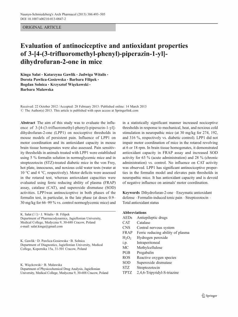
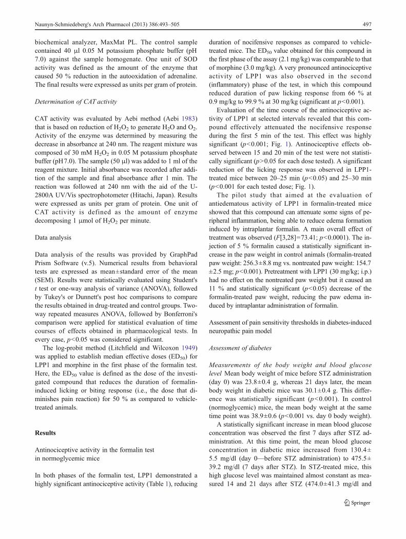
Evaluation of the time course of the antinociceptive ac-tivity of LPP1 at selected intervals revealed that this com-pound effectively attenuated the nocifensive responseduring the first 5 min of the test. This effect was highlysignificant (p<0.001; Fig. 1). Antinociceptive effects ob-served between 15 and 20 min of the test were not statisti-cally significant (p>0.05 for each dose tested). A significantreduction of the licking response was observed in LPP1-treated mice between 20–25 min (p<0.05) and 25–30 min(p<0.001 for each tested dose; Fig. 1).
The pilot study that aimed at the evaluation ofantiedematous activity of LPP1 in formalin-treated miceshowed that this compound can attenuate some signs of pe-ripheral inflammation, being able to reduce edema formationinduced by intraplantar formalin. A main overall effect oftreatment was observed (F[3,28]=73.41; p<0.0001). The in-jection of 5 % formalin caused a statistically significant in-crease in the paw weight in control animals (formalin-treatedpaw weight: 256.3±8.8 mg vs. nontreated paw weight: 154.7±2.5 mg; p<0.001). Pretreatment with LPP1 (30 mg/kg; i.p.)had no effect on the nontreated paw weight but it caused an11 % and statistically significant (p<0.05) decrease of theformalin-treated paw weight, reducing the paw edema in-duced by intraplantar administration of formalin.
Assessment of pain sensitivity thresholds in diabetes-inducedneuropathic pain model
Assessment of diabetes
Measurements of the body weight and blood glucoselevel Mean body weight of mice before STZ administration(day 0) was 23.8±0.4 g, whereas 21 days later, the meanbody weight in diabetic mice was 30.1±0.4 g. This differ-ence was statistically significant (p<0.001). In control(normoglycemic) mice, the mean body weight at the sametime point was 38.9±0.6 (p<0.001 vs. day 0 body weight).
A statistically significant increase in mean blood glucoseconcentration was observed the first 7 days after STZ ad-ministration. At this time point, the mean blood glucoseconcentration in diabetic mice increased from 130.4±5.5 mg/dl (day 0—before STZ administration) to 475.5±39.2 mg/dl (7 days after STZ). In STZ-treated mice, thishigh glucose level was maintained almost constant as mea-sured 14 and 21 days after STZ (474.0±41.3 mg/dl and
Naunyn-Schmiedeberg's Arch Pharmacol (2013) 386:493–505 497
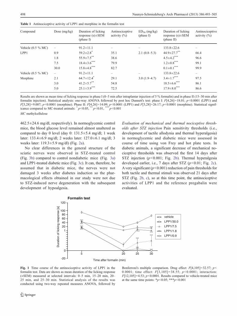
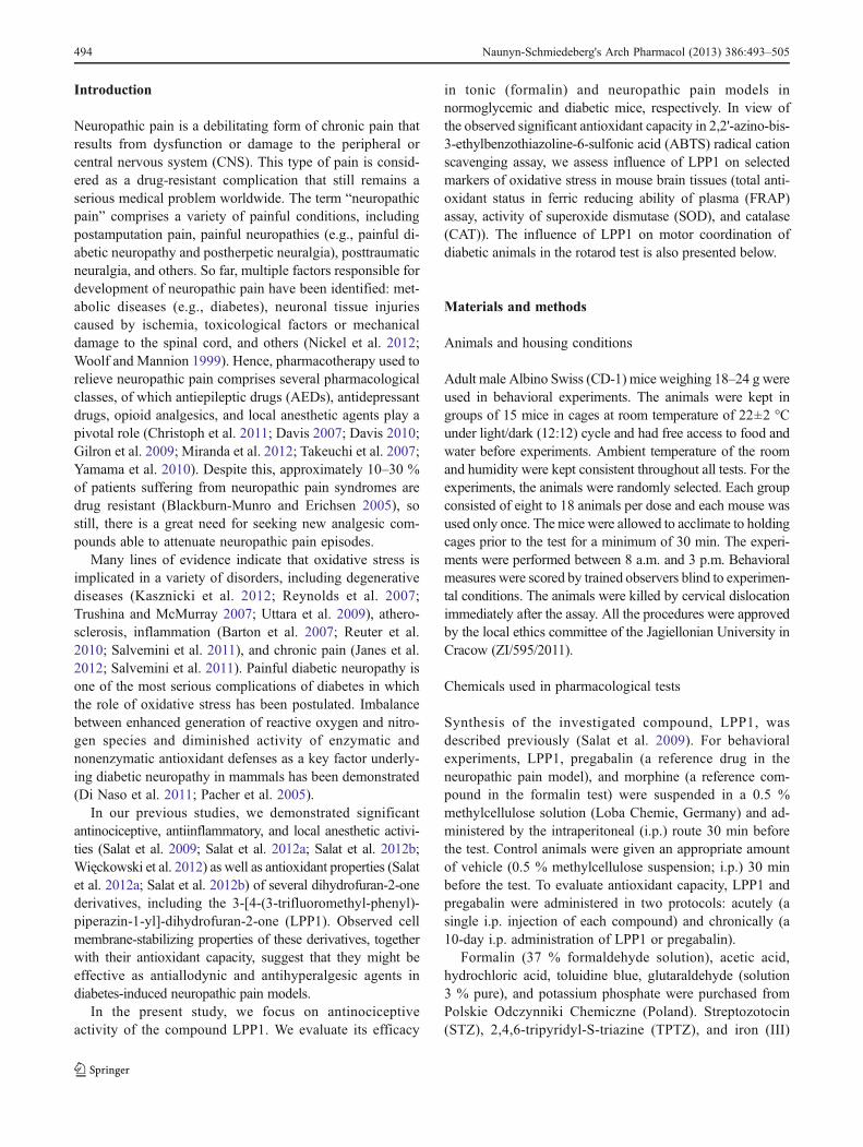
462.5±24.6 mg/dl, respectively). In normoglycemic controlmice, the blood glucose level remained almost unaltered ascompared to day 0 level (day 0: 131.5±5.4 mg/dl; 1 weeklater: 133.4±6.9 mg/dl; 2 weeks later: 127.0±6.1 mg/dl; 3weeks later: 119.3±5.9 mg/dl) (Fig. 2a).
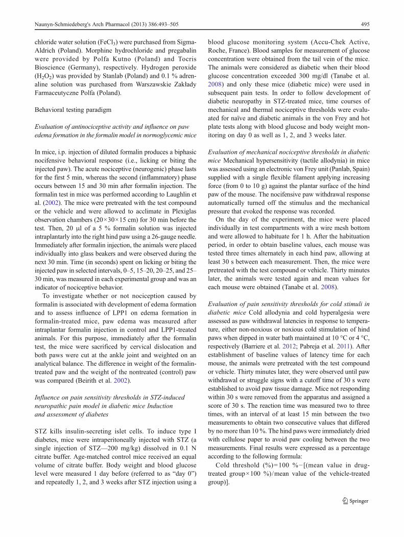
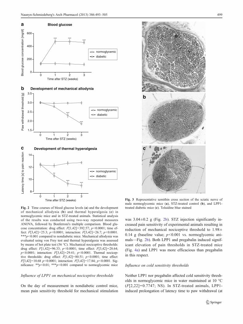
No clear differences in the general structure of thesciatic nerves were observed in STZ-treated control(Fig. 3b) compared to control nondiabetic mice (Fig. 3a)and LPP1-treated diabetic mice (Fig. 3c). It can, therefore, beassumed that in diabetic mice, the nerves were notdamaged 3 weeks after diabetes induction so the phar-macological effects obtained in our study were not dueto STZ-induced nerve degeneration with the subsequentdevelopment of hypoalgesia.
Evaluation of mechanical and thermal nociceptive thresh-olds after STZ injection Pain sensitivity thresholds (i.e.,development of tactile allodynia and thermal hyperalgesia)in normoglycemic and diabetic mice were assessed incourse of time using von Frey and hot plate tests. Indiabetic animals, a significant decrease of mechanical no-ciceptive thresholds was observed the first 14 days afterSTZ injection (p<0.001; Fig. 2b). Thermal hyperalgesiadeveloped earlier, i.e., 7 days after STZ (p<0.01; Fig. 2c).Avery significant (p<0.001) reduction of pain thresholds forboth tactile and thermal stimuli was observed 21 days afterSTZ (Fig. 2b, c), so at this time point, the antinociceptiveactivities of LPP1 and the reference pregabalin wereevaluated.
Table 1 Antinociceptive activity of LPP1 and morphine in the formalin test
Compound Dose (mg/kg) Duration of lickingresponse (s)±SEM(phase I)
Antinociceptiveactivity (%)
ED50 (mg/kg)(phase I)
Duration of lickingresponse (s)±SEM(phase II)
Antinociceptiveactivity (%)
Vehicle (0.5 % MC) – 91.2±11.1 – – 133.8±22.6 –
LPP1 0.9 59.2±2.8* 35.1 2.1 (0.8–5.3) 44.9±27.7** 66.4
1.8 55.9±7.5** 38.6 4.5±4.2*** 96.6
7.5 18.4±3.6*** 79.9 1.2±0.8*** 99.1
30.0 15.8±4.8*** 82.7 0.1±0.1*** 99.9
Vehicle (0.5 % MC) – 91.2±11.1 – – 133.8±22.6 –
Morphine 2.1 64.7±12.4* 29.1 3.0 (1.9–4.7) 3.4±1.7*** 97.5
3.0 41.2±5.7** 54.8 18.5±6.6*** 86.1
5.0 25.1±3.9*** 72.5 17.9±8.0*** 86.6
Results are shown as mean time of licking response in phase I (0–5 min after intraplantar injection of 5 % formalin) and in phase II (15–30 min afterformalin injection). Statistical analysis: one-way ANOVA followed by post hoc Dunnett's test: phase I: F[4,26]=18.01; p<0.0001 (LPP1) andF[3,28]=9.007; p<0.0001 (morphine). Phase II: F[4,26]=14.09; p<0.0001 (LPP1) and F[3,28]=26.17; p<0.0001 (morphine). Statistical signif-icance compared to MC-treated animals: * p<0.05, ** p<0.01, *** p<0.001
MC methylcellulose
Dur
atio
n of
lick
ing
resp
onse
[s]
5 20 25 30
-20
0
120406080
100
120Formalin test
Time after formalin (min)
vehicle
LPP1/0.9
LPP1/30.0
LPP1/7.5
LPP1/1.8
Fig. 1 Time course of the antinociceptive activity of LPP1 in theformalin test. Data are shown as mean duration of the licking response(±SEM) measured at selected intervals: 0–5 min, 15–20 min, 20–25 min, and 25–30 min. Statistical analysis of the results wasconducted using two-way repeated measures ANOVA, followed by
Bonferroni's multiple comparison. Drug effect: F[4,105]=32.57; p<0.0001; time effect: F[3,105]=38.55; p<0.0001; interaction:F[12,105]=4.53; p<0.0001. Results compared to vehicle-treated miceat the same time points: *p<0.05; ***p<0.001
498 Naunyn-Schmiedeberg's Arch Pharmacol (2013) 386:493–505
Influence of LPP1 on mechanical nociceptive thresholds
On the day of measurement in nondiabetic control mice,mean pain sensitivity threshold for mechanical stimulation
was 3.04±0.2 g (Fig. 2b). STZ injection significantly in-creased pain sensitivity of experimental animals resulting inreduction of mechanical nociceptive threshold to 1.98±0.14 g (baseline value; p<0.001 vs. normoglycemic ani-mals—Fig. 2b). Both LPP1 and pregabalin induced signif-icant elevation of pain thresholds in STZ-treated mice(Fig. 4a) and LPP1 was more efficacious than pregabalinin this respect.
Influence on cold sensitivity thresholds
Neither LPP1 nor pregabalin affected cold sensitivity thresh-olds in normoglycemic mice in water maintained at 10 °C(F[2,22]=0.7747; NS). In STZ-treated animals, LPP1-induced prolongation of latency time to paw withdrawal (at
Blood glucose
0 1 2 30
200
400
600
Time after STZ (weeks)
Blo
od g
luco
se c
once
ntra
tion
[mg/
dl]
Development of mechanical allodynia
0 1 2 3
1.5
2.0
2.5
3.0
3.5
Time after STZ (weeks)
Paw
with
draw
al th
resh
old
[g]
Development of thermal hyperalgesia
0 1 2 3
8
10
12
14
Time after STZ (weeks)
Late
ncy
time
[s] t
o pa
in r
eact
ion
normoglycemic
diabetic
normoglycemic
diabetic
normoglycemic
diabetic
a
b
c
Fig. 2 Time courses of blood glucose levels (a) and the developmentof mechanical allodynia (b) and thermal hyperalgesia (c) innormoglycemic mice and in STZ-treated animals. Statistical analysisof the results was conducted using two-way repeated measuresANOVA, followed by Bonferroni's multiple comparison. Blood glu-cose concentration: drug effect: F[1,42]=392.57; p<0.0001; time ef-fect: F[3,42]=25.3; p<0.0001; interaction: F[3,42]=26.7; p<0.0001.***p<0.001 compared to nondiabetic mice. Mechanical allodynia wasevaluated using von Frey test and thermal hyperalgesia was assessedby means of hot plate test (56 °C). Mechanical nociceptive thresholds:drug effect: F[1,42]=96.33; p<0.0001; time effect: F[3,42]=28.64;p<0.0001; interaction: F[3,42]=29.41; p<0.0001. Thermal nocicep-tive thresholds: drug effect: F[1,42]=80.51; p<0.0001; time effect:F[3,42]=10.68 p<0.0001; interaction: F[3,42]=17.84; p<0.0001. Sig-nificance: **p<0.01; ***p<0.001 compared to normoglycemic mice
Fig. 3 Representative semithin cross section of the sciatic nerve ofmale normoglycemic mice (a), STZ-treated control (b), and LPP1-treated diabetic mice (c). Toluidine blue stained
Naunyn-Schmiedeberg's Arch Pharmacol (2013) 386:493–505 499
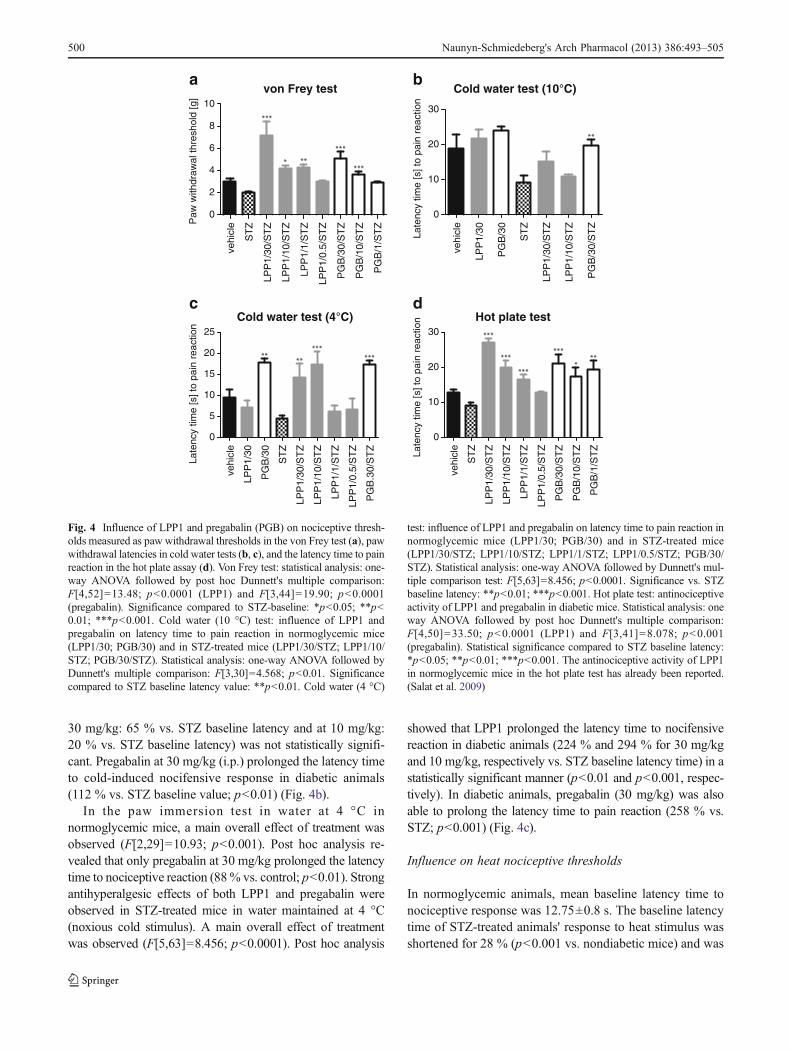
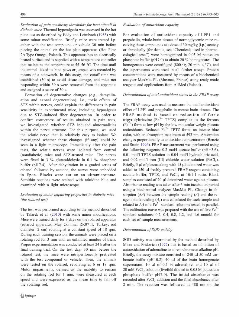
30 mg/kg: 65 % vs. STZ baseline latency and at 10 mg/kg:20 % vs. STZ baseline latency) was not statistically signifi-cant. Pregabalin at 30 mg/kg (i.p.) prolonged the latency timeto cold-induced nocifensive response in diabetic animals(112 % vs. STZ baseline value; p<0.01) (Fig. 4b).
In the paw immersion test in water at 4 °C innormoglycemic mice, a main overall effect of treatment wasobserved (F[2,29]=10.93; p<0.001). Post hoc analysis re-vealed that only pregabalin at 30 mg/kg prolonged the latencytime to nociceptive reaction (88% vs. control; p<0.01). Strongantihyperalgesic effects of both LPP1 and pregabalin wereobserved in STZ-treated mice in water maintained at 4 °C(noxious cold stimulus). A main overall effect of treatmentwas observed (F[5,63]=8.456; p<0.0001). Post hoc analysis
showed that LPP1 prolonged the latency time to nocifensivereaction in diabetic animals (224 % and 294 % for 30 mg/kgand 10 mg/kg, respectively vs. STZ baseline latency time) in astatistically significant manner (p<0.01 and p<0.001, respec-tively). In diabetic animals, pregabalin (30 mg/kg) was alsoable to prolong the latency time to pain reaction (258 % vs.STZ; p<0.001) (Fig. 4c).
Influence on heat nociceptive thresholds
In normoglycemic animals, mean baseline latency time tonociceptive response was 12.75±0.8 s. The baseline latencytime of STZ-treated animals' response to heat stimulus wasshortened for 28 % (p<0.001 vs. nondiabetic mice) and was
von Frey test
vehi
cle
ST
Z
LPP
1/30
/ST
Z
LPP
1/10
/ST
Z
LPP
1/1/
ST
Z
LPP
1/0.
5/S
TZ
PG
B/3
0/S
TZ
PG
B/1
0/S
TZ
PG
B/1
/ST
Z
0
2
4
6
8
10
Paw
with
draw
al th
resh
old
[g]
Cold water test (10°C)
vehi
cle
LPP
1/30
PG
B/3
0
ST
Z
LPP
1/30
/ST
Z
LPP
1/10
/ST
Z
PG
B/3
0/S
TZ
0
10
20
30
Late
ncy
time
[s] t
o pa
in r
eact
ion
Cold water test (4°C)
vehi
cle
LPP
1/30
PG
B/3
0
ST
Z
LPP
1/30
/ST
Z
LPP
1/10
/ST
Z
LPP
1/1/
ST
Z
LPP
1/0.
5/S
TZ
PG
B.3
0/S
TZ
0
5
10
15
20
25
Late
ncy
time
[s] t
o pa
in r
eact
ion
Hot plate test
vehi
cle
ST
Z
LPP
1/30
/ST
Z
LPP
1/10
/ST
Z
LPP
1/1/
ST
Z
LPP
1/0.
5/S
TZ
PG
B/3
0/S
TZ
PG
B/1
0/S
TZ
PG
B/1
/ST
Z
0
10
20
30
Late
ncy
time
[s] t
o pa
in r
eact
ion
a b
c d
Fig. 4 Influence of LPP1 and pregabalin (PGB) on nociceptive thresh-olds measured as paw withdrawal thresholds in the von Frey test (a), pawwithdrawal latencies in cold water tests (b, c), and the latency time to painreaction in the hot plate assay (d). Von Frey test: statistical analysis: one-way ANOVA followed by post hoc Dunnett's multiple comparison:F[4,52]=13.48; p<0.0001 (LPP1) and F[3,44]=19.90; p<0.0001(pregabalin). Significance compared to STZ-baseline: *p<0.05; **p<0.01; ***p<0.001. Cold water (10 °C) test: influence of LPP1 andpregabalin on latency time to pain reaction in normoglycemic mice(LPP1/30; PGB/30) and in STZ-treated mice (LPP1/30/STZ; LPP1/10/STZ; PGB/30/STZ). Statistical analysis: one-way ANOVA followed byDunnett's multiple comparison: F[3,30]=4.568; p<0.01. Significancecompared to STZ baseline latency value: **p<0.01. Cold water (4 °C)
test: influence of LPP1 and pregabalin on latency time to pain reaction innormoglycemic mice (LPP1/30; PGB/30) and in STZ-treated mice(LPP1/30/STZ; LPP1/10/STZ; LPP1/1/STZ; LPP1/0.5/STZ; PGB/30/STZ). Statistical analysis: one-way ANOVA followed by Dunnett's mul-tiple comparison test: F[5,63]=8.456; p<0.0001. Significance vs. STZbaseline latency: **p<0.01; ***p<0.001. Hot plate test: antinociceptiveactivity of LPP1 and pregabalin in diabetic mice. Statistical analysis: oneway ANOVA followed by post hoc Dunnett's multiple comparison:F[4,50]=33.50; p<0.0001 (LPP1) and F[3,41]=8.078; p<0.001(pregabalin). Statistical significance compared to STZ baseline latency:*p<0.05; **p<0.01; ***p<0.001. The antinociceptive activity of LPP1in normoglycemic mice in the hot plate test has already been reported.(Salat et al. 2009)
500 Naunyn-Schmiedeberg's Arch Pharmacol (2013) 386:493–505
an indicative of hyperalgesia. In diabetic mice, both LPP1and pregabalin at doses 1–30 mg/kg prolonged the latencytime to jump or lick the hind paw (Fig. 4d).
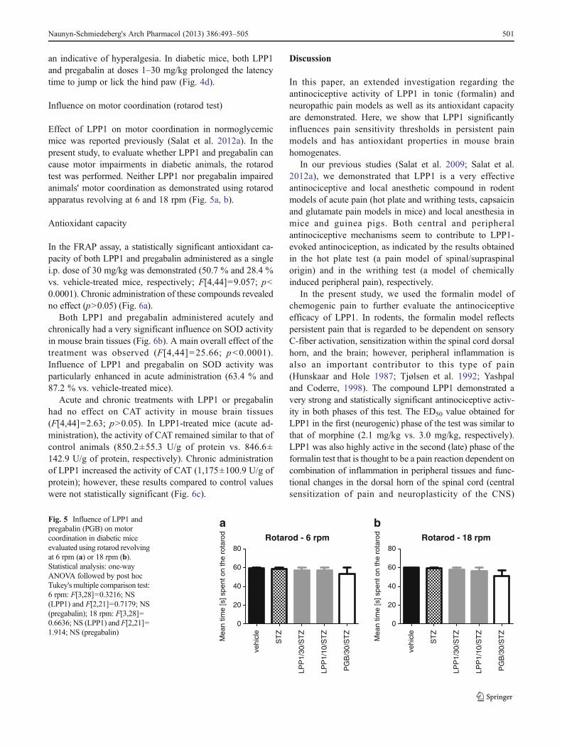
Influence on motor coordination (rotarod test)
Effect of LPP1 on motor coordination in normoglycemicmice was reported previously (Salat et al. 2012a). In thepresent study, to evaluate whether LPP1 and pregabalin cancause motor impairments in diabetic animals, the rotarodtest was performed. Neither LPP1 nor pregabalin impairedanimals' motor coordination as demonstrated using rotarodapparatus revolving at 6 and 18 rpm (Fig. 5a, b).
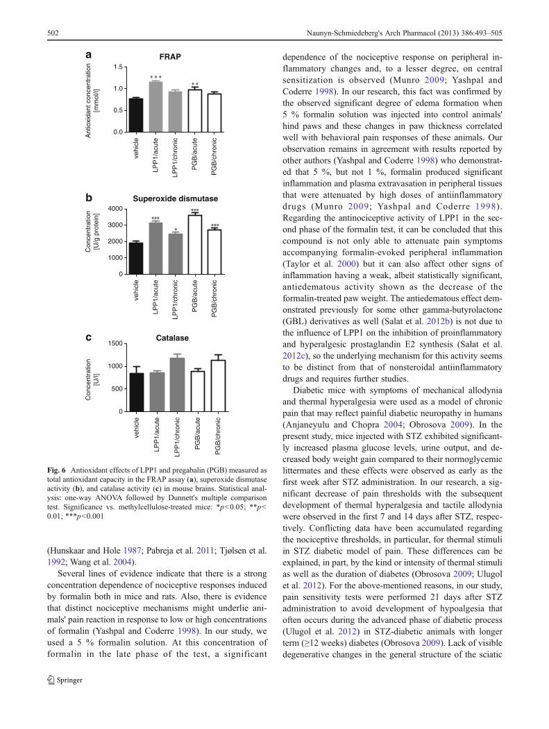
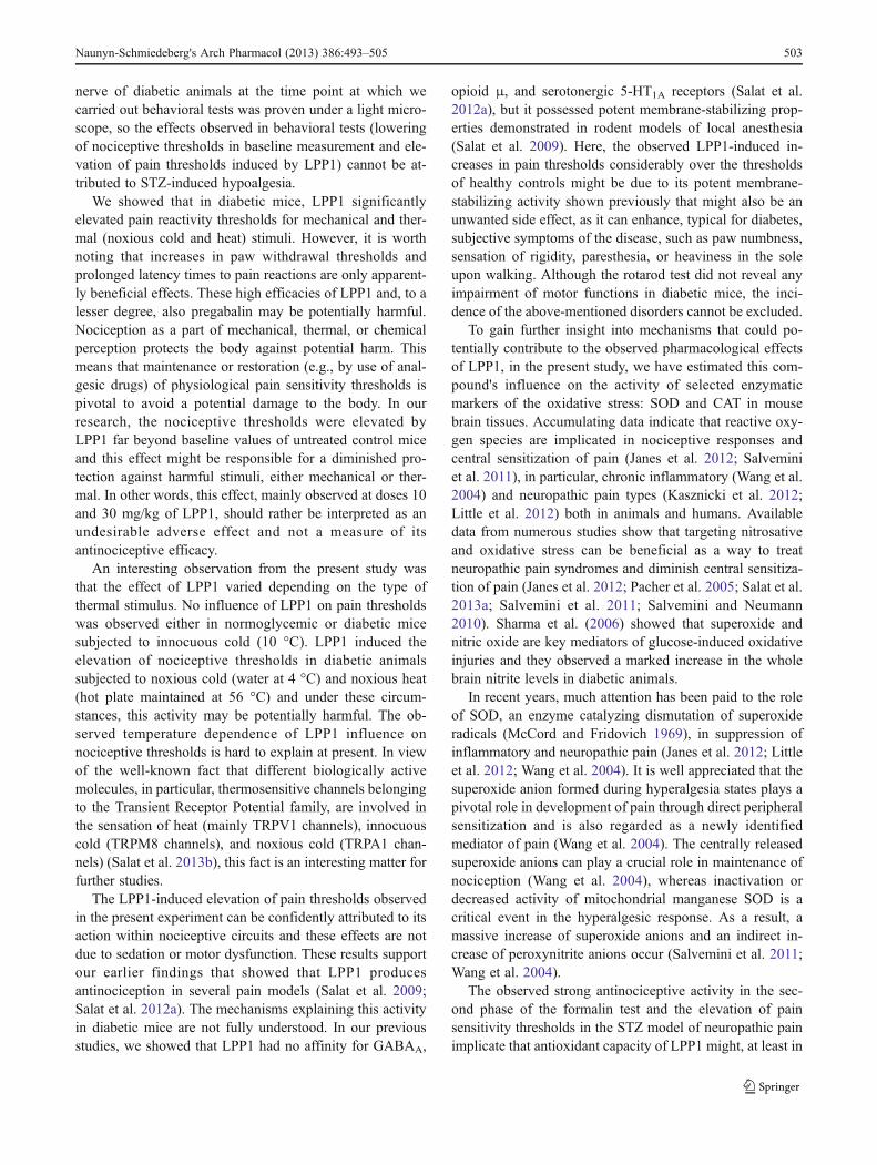
Antioxidant capacity
In the FRAP assay, a statistically significant antioxidant ca-pacity of both LPP1 and pregabalin administered as a singlei.p. dose of 30 mg/kg was demonstrated (50.7 % and 28.4 %vs. vehicle-treated mice, respectively; F[4,44]=9.057; p<0.0001). Chronic administration of these compounds revealedno effect (p>0.05) (Fig. 6a).
Both LPP1 and pregabalin administered acutely andchronically had a very significant influence on SOD activityin mouse brain tissues (Fig. 6b). A main overall effect of thetreatment was observed (F[4,44]=25.66; p<0.0001).Influence of LPP1 and pregabalin on SOD activity wasparticularly enhanced in acute administration (63.4 % and87.2 % vs. vehicle-treated mice).
Acute and chronic treatments with LPP1 or pregabalinhad no effect on CAT activity in mouse brain tissues(F[4,44]=2.63; p>0.05). In LPP1-treated mice (acute ad-ministration), the activity of CAT remained similar to that ofcontrol animals (850.2±55.3 U/g of protein vs. 846.6±142.9 U/g of protein, respectively). Chronic administrationof LPP1 increased the activity of CAT (1,175±100.9 U/g ofprotein); however, these results compared to control valueswere not statistically significant (Fig. 6c).
Discussion
In this paper, an extended investigation regarding theantinociceptive activity of LPP1 in tonic (formalin) andneuropathic pain models as well as its antioxidant capacityare demonstrated. Here, we show that LPP1 significantlyinfluences pain sensitivity thresholds in persistent painmodels and has antioxidant properties in mouse brainhomogenates.
In our previous studies (Salat et al. 2009; Salat et al.2012a), we demonstrated that LPP1 is a very effectiveantinociceptive and local anesthetic compound in rodentmodels of acute pain (hot plate and writhing tests, capsaicinand glutamate pain models in mice) and local anesthesia inmice and guinea pigs. Both central and peripheralantinociceptive mechanisms seem to contribute to LPP1-evoked antinociception, as indicated by the results obtainedin the hot plate test (a pain model of spinal/supraspinalorigin) and in the writhing test (a model of chemicallyinduced peripheral pain), respectively.
In the present study, we used the formalin model ofchemogenic pain to further evaluate the antinociceptiveefficacy of LPP1. In rodents, the formalin model reflectspersistent pain that is regarded to be dependent on sensoryC-fiber activation, sensitization within the spinal cord dorsalhorn, and the brain; however, peripheral inflammation isalso an important contributor to this type of pain(Hunskaar and Hole 1987; Tjølsen et al. 1992; Yashpaland Coderre, 1998). The compound LPP1 demonstrated avery strong and statistically significant antinociceptive activ-ity in both phases of this test. The ED50 value obtained forLPP1 in the first (neurogenic) phase of the test was similar tothat of morphine (2.1 mg/kg vs. 3.0 mg/kg, respectively).LPP1 was also highly active in the second (late) phase of theformalin test that is thought to be a pain reaction dependent oncombination of inflammation in peripheral tissues and func-tional changes in the dorsal horn of the spinal cord (centralsensitization of pain and neuroplasticity of the CNS)
Rotarod - 6 rpm
vehi
cle
ST
Z
LPP
1/30
/ST
Z
LPP
1/10
/ST
Z
PG
B/3
0/S
TZ
0
20
40
60
80
Mea
n tim
e [s
] spe
nt o
n th
e ro
taro
d
Rotarod - 18 rpm
vehi
cle
ST
Z
LPP
1/30
/ST
Z
LPP
1/10
/ST
Z
PG
B/3
0/S
TZ
0
20
40
60
80
Mea
n tim
e [s
] spe
nt o
n th
e ro
taro
d
a bFig. 5 Influence of LPP1 andpregabalin (PGB) on motorcoordination in diabetic miceevaluated using rotarod revolvingat 6 rpm (a) or 18 rpm (b).Statistical analysis: one-wayANOVA followed by post hocTukey's multiple comparison test:6 rpm: F[3,28]=0.3216; NS(LPP1) and F[2,21]=0.7179; NS(pregabalin); 18 rpm: F[3,28]=0.6636; NS (LPP1) and F[2,21]=1.914; NS (pregabalin)
Naunyn-Schmiedeberg's Arch Pharmacol (2013) 386:493–505 501
(Hunskaar and Hole 1987; Pabreja et al. 2011; Tjølsen et al.1992; Wang et al. 2004).
Several lines of evidence indicate that there is a strongconcentration dependence of nociceptive responses inducedby formalin both in mice and rats. Also, there is evidencethat distinct nociceptive mechanisms might underlie ani-mals' pain reaction in response to low or high concentrationsof formalin (Yashpal and Coderre 1998). In our study, weused a 5 % formalin solution. At this concentration offormalin in the late phase of the test, a significant
dependence of the nociceptive response on peripheral in-flammatory changes and, to a lesser degree, on centralsensitization is observed (Munro 2009; Yashpal andCoderre 1998). In our research, this fact was confirmed bythe observed significant degree of edema formation when5 % formalin solution was injected into control animals'hind paws and these changes in paw thickness correlatedwell with behavioral pain responses of these animals. Ourobservation remains in agreement with results reported byother authors (Yashpal and Coderre 1998) who demonstrat-ed that 5 %, but not 1 %, formalin produced significantinflammation and plasma extravasation in peripheral tissuesthat were attenuated by high doses of antiinflammatorydrugs (Munro 2009; Yashpal and Coderre 1998).Regarding the antinociceptive activity of LPP1 in the sec-ond phase of the formalin test, it can be concluded that thiscompound is not only able to attenuate pain symptomsaccompanying formalin-evoked peripheral inflammation(Taylor et al. 2000) but it can also affect other signs ofinflammation having a weak, albeit statistically significant,antiedematous activity shown as the decrease of theformalin-treated paw weight. The antiedematous effect dem-onstrated previously for some other gamma-butyrolactone(GBL) derivatives as well (Salat et al. 2012b) is not due tothe influence of LPP1 on the inhibition of proinflammatoryand hyperalgesic prostaglandin E2 synthesis (Sałat et al.2012c), so the underlying mechanism for this activity seemsto be distinct from that of nonsteroidal antiinflammatorydrugs and requires further studies.
Diabetic mice with symptoms of mechanical allodyniaand thermal hyperalgesia were used as a model of chronicpain that may reflect painful diabetic neuropathy in humans(Anjaneyulu and Chopra 2004; Obrosova 2009). In thepresent study, mice injected with STZ exhibited significant-ly increased plasma glucose levels, urine output, and de-creased body weight gain compared to their normoglycemiclittermates and these effects were observed as early as thefirst week after STZ administration. In our research, a sig-nificant decrease of pain thresholds with the subsequentdevelopment of thermal hyperalgesia and tactile allodyniawere observed in the first 7 and 14 days after STZ, respec-tively. Conflicting data have been accumulated regardingthe nociceptive thresholds, in particular, for thermal stimuliin STZ diabetic model of pain. These differences can beexplained, in part, by the kind or intensity of thermal stimulias well as the duration of diabetes (Obrosova 2009; Ulugolet al. 2012). For the above-mentioned reasons, in our study,pain sensitivity tests were performed 21 days after STZadministration to avoid development of hypoalgesia thatoften occurs during the advanced phase of diabetic process(Ulugol et al. 2012) in STZ-diabetic animals with longerterm (≥12 weeks) diabetes (Obrosova 2009). Lack of visibledegenerative changes in the general structure of the sciatic
FRAP
vehi
cle
LPP
1/ac
ute
LPP
1/ch
roni
c
PG
B/a
cute
PG
B/c
hron
ic
0.0
0.5
1.0
1.5
Ant
ioxi
dant
con
cent
ratio
n[m
mol
/l]
Superoxide dismutase
vehi
cle
LPP
1/ac
ute
LPP
1/ch
roni
c
PG
B/a
cute
PG
B/c
hron
ic
0
1000
2000
3000
4000
Con
cent
ratio
n[U
/g p
rote
in]
Catalase
vehi
cle
LPP
1/ac
ute
LPP
1/ch
roni
c
PG
B/a
cute
PG
B/c
hron
ic
0
500
1000
1500
Con
cent
ratio
n[U
/l]
a
b
c
Fig. 6 Antioxidant effects of LPP1 and pregabalin (PGB) measured astotal antioxidant capacity in the FRAP assay (a), superoxide dismutaseactivity (b), and catalase activity (c) in mouse brains. Statistical anal-ysis: one-way ANOVA followed by Dunnett's multiple comparisontest. Significance vs. methylcellulose-treated mice: *p<0.05; **p<0.01; ***p<0.001
502 Naunyn-Schmiedeberg's Arch Pharmacol (2013) 386:493–505
nerve of diabetic animals at the time point at which wecarried out behavioral tests was proven under a light micro-scope, so the effects observed in behavioral tests (loweringof nociceptive thresholds in baseline measurement and ele-vation of pain thresholds induced by LPP1) cannot be at-tributed to STZ-induced hypoalgesia.
We showed that in diabetic mice, LPP1 significantlyelevated pain reactivity thresholds for mechanical and ther-mal (noxious cold and heat) stimuli. However, it is worthnoting that increases in paw withdrawal thresholds andprolonged latency times to pain reactions are only apparent-ly beneficial effects. These high efficacies of LPP1 and, to alesser degree, also pregabalin may be potentially harmful.Nociception as a part of mechanical, thermal, or chemicalperception protects the body against potential harm. Thismeans that maintenance or restoration (e.g., by use of anal-gesic drugs) of physiological pain sensitivity thresholds ispivotal to avoid a potential damage to the body. In ourresearch, the nociceptive thresholds were elevated byLPP1 far beyond baseline values of untreated control miceand this effect might be responsible for a diminished pro-tection against harmful stimuli, either mechanical or ther-mal. In other words, this effect, mainly observed at doses 10and 30 mg/kg of LPP1, should rather be interpreted as anundesirable adverse effect and not a measure of itsantinociceptive efficacy.
An interesting observation from the present study wasthat the effect of LPP1 varied depending on the type ofthermal stimulus. No influence of LPP1 on pain thresholdswas observed either in normoglycemic or diabetic micesubjected to innocuous cold (10 °C). LPP1 induced theelevation of nociceptive thresholds in diabetic animalssubjected to noxious cold (water at 4 °C) and noxious heat(hot plate maintained at 56 °C) and under these circum-stances, this activity may be potentially harmful. The ob-served temperature dependence of LPP1 influence onnociceptive thresholds is hard to explain at present. In viewof the well-known fact that different biologically activemolecules, in particular, thermosensitive channels belongingto the Transient Receptor Potential family, are involved inthe sensation of heat (mainly TRPV1 channels), innocuouscold (TRPM8 channels), and noxious cold (TRPA1 chan-nels) (Salat et al. 2013b), this fact is an interesting matter forfurther studies.
The LPP1-induced elevation of pain thresholds observedin the present experiment can be confidently attributed to itsaction within nociceptive circuits and these effects are notdue to sedation or motor dysfunction. These results supportour earlier findings that showed that LPP1 producesantinociception in several pain models (Salat et al. 2009;Salat et al. 2012a). The mechanisms explaining this activityin diabetic mice are not fully understood. In our previousstudies, we showed that LPP1 had no affinity for GABAA,
opioid μ, and serotonergic 5-HT1A receptors (Salat et al.2012a), but it possessed potent membrane-stabilizing prop-erties demonstrated in rodent models of local anesthesia(Salat et al. 2009). Here, the observed LPP1-induced in-creases in pain thresholds considerably over the thresholdsof healthy controls might be due to its potent membrane-stabilizing activity shown previously that might also be anunwanted side effect, as it can enhance, typical for diabetes,subjective symptoms of the disease, such as paw numbness,sensation of rigidity, paresthesia, or heaviness in the soleupon walking. Although the rotarod test did not reveal anyimpairment of motor functions in diabetic mice, the inci-dence of the above-mentioned disorders cannot be excluded.
To gain further insight into mechanisms that could po-tentially contribute to the observed pharmacological effectsof LPP1, in the present study, we have estimated this com-pound's influence on the activity of selected enzymaticmarkers of the oxidative stress: SOD and CAT in mousebrain tissues. Accumulating data indicate that reactive oxy-gen species are implicated in nociceptive responses andcentral sensitization of pain (Janes et al. 2012; Salveminiet al. 2011), in particular, chronic inflammatory (Wang et al.2004) and neuropathic pain types (Kasznicki et al. 2012;Little et al. 2012) both in animals and humans. Availabledata from numerous studies show that targeting nitrosativeand oxidative stress can be beneficial as a way to treatneuropathic pain syndromes and diminish central sensitiza-tion of pain (Janes et al. 2012; Pacher et al. 2005; Salat et al.2013a; Salvemini et al. 2011; Salvemini and Neumann2010). Sharma et al. (2006) showed that superoxide andnitric oxide are key mediators of glucose-induced oxidativeinjuries and they observed a marked increase in the wholebrain nitrite levels in diabetic animals.
In recent years, much attention has been paid to the roleof SOD, an enzyme catalyzing dismutation of superoxideradicals (McCord and Fridovich 1969), in suppression ofinflammatory and neuropathic pain (Janes et al. 2012; Littleet al. 2012; Wang et al. 2004). It is well appreciated that thesuperoxide anion formed during hyperalgesia states plays apivotal role in development of pain through direct peripheralsensitization and is also regarded as a newly identifiedmediator of pain (Wang et al. 2004). The centrally releasedsuperoxide anions can play a crucial role in maintenance ofnociception (Wang et al. 2004), whereas inactivation ordecreased activity of mitochondrial manganese SOD is acritical event in the hyperalgesic response. As a result, amassive increase of superoxide anions and an indirect in-crease of peroxynitrite anions occur (Salvemini et al. 2011;Wang et al. 2004).
The observed strong antinociceptive activity in the sec-ond phase of the formalin test and the elevation of painsensitivity thresholds in the STZ model of neuropathic painimplicate that antioxidant capacity of LPP1 might, at least in
Naunyn-Schmiedeberg's Arch Pharmacol (2013) 386:493–505 503
part, contribute to its pharmacological properties, includingefficacy in some rodent pain models. Our earlier studiesregarding the antioxidant capacity of LPP1 proved its highABTS radical cation-scavenging properties (Salat et al.2012a). The results from the present research confirm thatLPP1 has antioxidant activity in tissues.
In conclusion, the significant antinociceptive efficacy ofLPP1 in acute and tonic pain models in rodents as well as itsability to elevate nociceptive thresholds in the diabetic neu-ropathic pain model indicate an interesting lead structure inthe search for new analgesic agents active in a wide range ofpainful conditions, provided carefully selecting dose rangesthat maintain or restore physiological nociceptive thresholds.The pharmacological activity of LPP1 in mice, together withthe antioxidant effect of this compound, in particular, itsinfluence on SOD activity, is interesting for further studies.
Acknowledgments This study was financially supported by theJagiellonian University grant K/ZDS/003329.
Conflict of interest None.
Open Access This article is distributed under the terms of the CreativeCommons Attribution License which permits any use, distribution, andreproduction in any medium, provided the original author(s) and thesource are credited.
References
Aebi HE (1983) Catalase. In: “Method of enzymatic analysis”, VCH,Weinheim.(3):273-286
Anjaneyulu M, Chopra K (2004) Fluoxetine attenuates thermalhyperalgesia through 5-HT1/2 receptors in streptozotocin-induced diabetic mice. Eur J Pharmacol 497:285–292
Barrière DA, Rieusset J, Chanteranne D, Busserolles J, Chauvin MA,Chapuis L, Salles J, Dubray C, Morio B (2012) Paclitaxel therapypotentiates cold hyperalgesia in streptozotocin-induced diabeticrats through enhanced mitochondrial reactive oxygen speciesproduction and TRPA1 sensitization. Pain 153(3):553–561
Barton M, Minotti R, Haas E (2007) Inflammation and atherosclerosis.Circ Res 101:750–751
Beirith A, Santos AR, Calixto JB (2002) Mechanisms underlying thenociception and paw oedema caused by injection of glutamateinto the mouse paw. Brain Res 924(2):219–228
Benzie IF, Strain JJ (1996) The ferric reducing ability of plasma(FRAP) as measure of antioxidant power: the FRAP assay. AnalBiochem 239:70–76
Blackburn-Munro G, Erichsen HK (2005) Antiepileptics and the treat-ment of neuropathic pain: evidence from animal models. CurrPharm Des 11(23):2961–2976
Christoph T, De Vry J, Schiene K, Tallarida RJ, Tzschentke TM (2011)Synergistic antihypersensitive effects of pregabalin and tapentadolin a rat model of neuropathic pain. Eur J Pharmacol 666:72–79
Davis MP (2007) What is new in neuropathic pain? Suppor CareCancer 15(4):363–372
Davis MP (2010) Recent advances in the treatment of pain, F1000 MedRep 2 63. doi: 10.3410/M2-63
Di Naso FC, Simões Dias A, Porawski M, Marroni NA (2011)Exogenous superoxide dismutase: action on liver oxidative stress
in animals with streptozotocin-induced diabetes. Exp DiabetesRes 2011:754132. doi:10.1155/2011/754132
Eddy N, Leimbach D (1953) Synthetic analgesics. II. Dithienylbutenyl-and dithienylbutylamines. J Pharmacol Exp Ther 107:385–393
Gilron I, Bailey JM, Tu D, Holden RR, Jackson AC, Houlden RL(2009) Nortriptyline and gabapentin, alone and in combination forneuropathic pain: a double-blind, randomised controlled cross-over trial. Lancet 374(9697):1252–1261
Hunskaar S, Hole K (1987) The formalin test in mice: dissociationbetween inflammatory and non-inflammatory pain. Pain 30:103–114
Janes K, Neumann WL, Salvemini D (2012) Anti-superoxide and anti-peroxynitrite strategies in pain suppression. Biochim BiophysActa 1822(5):815–821
Kasznicki J, Kosmalski M, Sliwinska A, Mrowicka M, Stanczyk M,Majsterek I, Drzewoski J (2012) Evaluation of oxidative stressmarkers in pathogenesis of diabetic neuropathy. Mol Biol Rep39(9):8669–8678
Laughlin TM, Tram KV, Wilcox GL, Birnbaum AK (2002)Comparison of antiepileptic drugs tiagabine, lamotrigine, andgabapentin in mouse models of acute, prolonged, and chronicnociception. J Pharmacol Exp Ther 302(3):1168–1175
Litchfield JT, Wilcoxon E (1949) A simplified method of evaluatingdose–effect experiments. J Pharmacol Exp Ther 96:99–113
Little JW, Doyle T, Salvemini D (2012) Reactive nitroxidative speciesand nociceptive processing: determining the roles for nitric oxide,superoxide, and peroxynitrite in pain. Amino Acids 42(1):75–94
McCord JM, Fridovich I (1969) Superoxide dismutase. An enzymicfunction for erythrocuprein (hemocuprein). J Biol Chem244(22):6049–6055
Miranda HF, Noriega V, Prieto JC (2012) Previous administration ofnaltrexone did not change synergism between paracetamol andtramadol in mice. Pharmacol Biochem Behav 102(1):72–76
Misra HP, Fridovich I (1972) The role of superoxide anion in theautoxidation of epinephrine and a simple assay for superoxidedismutase. J Biol Chem 247(10):3170–3175
Munro G (2009) Pharmacological assessment of the rat formalin testutilizing the clinically used analgesic drugs gabapentin,lamotrigine, morphine, duloxetine, tramadol and ibuprofen: influ-ence of low and high formalin concentrations. Eur J Pharmacol605:95–102
Nickel FT, Seifert F, Lanz S, Maihöfner C (2012) Mechanisms ofneuropathic pain. Eur Neuropsychopharmacol 22(2):81–91.doi:10.1016/j.euroneuro.2011.05.005
Obrosova IG (2009) Diabetic painful and insensate neuropathy: path-ogenesis and potential treatments. Neurotherapeutics 6(4):638–647. doi:10.1016/j.nurt.2009.07.004
Pabreja K, Dua K, Sharma S, Padi SS, Kulkarni SK (2011)Minocycline attenuates the development of diabetic neuropathicpain: possible anti-inflammatory and anti-oxidant mechanisms.Eur J Pharmacol 661(1–3):15–21
Pacher P, Obrosova IG, Mabley JG, Szabó C (2005) Role of nitrosativestress and peroxynitrite in the pathogenesis of diabetic complica-tions. Emerging new therapeutical strategies. Curr Med Chem12(3):267–275
Reynolds A, Laurie C, Mosley RL, Gendelman HE (2007) Oxidativestress and the pathogenesis of neurodegenerative disorders. IntRev Neurobiol 82:297–325
Reuter S, Gupta SC, Chaturvedi MM, Aggarwal BB (2010) Oxidativestress, inflammation, and cancer: how are they linked? Free RadicBiol Med 49:1603–1616
Sałat K, Filipek B,Wieckowski K,Malawska B (2009) Analgesic activityof 3-mono-substituted derivatives of dihydrofuran-2-one in experi-mental rodent models of pain. Pharmacol Rep 61(5):807–818
Salat K, Moniczewski A, Salat R, Janaszek M, Filipek B, Malawska B,Wieckowski K (2012a) Analgesic, anticonvulsant and antioxidantactivities of 3-[4-(3-trifluoromethyl-phenyl)-piperazin-1-yl]
504 Naunyn-Schmiedeberg's Arch Pharmacol (2013) 386:493–505
dihydrofuran-2-one dihydrochloride in mice. Pharmacol BiochemBehav 101(1):138–147
Salat K, Librowski T, Moniczewski A, Stanisz-Wallis K, WieckowskiK, Malawska B (2012b) Analgesic, antioedematous and antioxi-dant activity of γ-butyrolactone derivatives in rodents. BehavPharmacol 23(4):407–416
Sałat K, Librowski T, Gdula-Argasińska J, Tyszka-Czochara M,Jastrzębska-Więsek M, Moniczewski A, Rapacz A, Pytka K,Filipek B, Więckowski K, Malawska B (2012) Influence ofgamma-butyrolactone derivatives with analgesic properties onthe prostaglandin E2 level and gastric mucosa in rodents. ActaBiologica Cracoviensia Series Zoologia 53:45–52
Sałat K, Moniczewski A, Librowski T (2013a) Nitrogen, oxygen orsulfur containing heterocyclic compounds as analgesic drugs usedas modulators of the nitroxidative stress. Mini Rev Med Chem13(3):335–352
Sałat K, Moniczewski A, Librowski T (2013b) Transient receptorpotential channels—emerging novel drug targets for the treatmentof pain. Curr Med Chem (in press)
Salvemini D, Little JW, Doyle T, Neumann WL (2011) Roles ofreactive oxygen and nitrogen species in pain. Free Radic BiolMed 51(5):951–966
Salvemini D, Neumann W (2010) Targeting peroxynitrite drivennitroxidative stress with synzymes: a novel therapeutic approachin chronic pain management. Life Sci 86(15–16):604–614
Sharma S, Kulkarni SK, Agrewala JN, Chopra K (2006) Curcuminattenuates thermal hyperalgesia in a diabetic mouse model ofneuropathic pain. Eur J Pharmacol 536:256–261
Takeuchi Y, Takasu K, Ono H, Tanabe M (2007) Pregabalin, S-(+)-3-isobutylgaba, activates the descending noradrenergic system toalleviate neuropathic pain in the mouse partial sciatic nerve liga-tion model. Neuropharmacology 53(7):842–853
Talarek S, Orzelska J, Listos J, Fidecka (2010) Effect of sildenafiltreatment on the development of tolerance to diazepam-inducedmotor impairment and sedation in mice. Pharmacol Rep 62:627–634
Tanabe M, Murakami T, Ono H (2008) Zonisamide suppresses painsymptoms of formalin-induced inflammatory and streptozotocin-induced diabetic neuropathy. J Pharmacol Sci 107(2):213–220
Taylor BK, Peterson MA, Roderick RE, Tate J, Green PG, Levine JO,Basbaum AI (2000) Opioid inhibition of formalin-inducedchanges in plasma extravasation and local blood flow in rats.Pain 84(2–3):263–270
Tjølsen A, Berge OG, Hunskaar S, Rosland JH, Hole K (1992) Theformalin test: an evaluation of the method. Pain 51:5–17
Trushina E, McMurray CT (2007) Oxidative stress and mitochondrialdysfunction in neurodegenerative diseases. Neuroscience145:1233–1248
Ulugol A, Oltulu C, Gunduz O, Citak C, Carrara R, Shaqaqi MR,Sanchez AM, Dogrul A (2012) 5-HT7 receptor activation attenu-ates thermal hyperalgesia in streptozocin-induced diabetic mice.Pharmacol Biochem Behav 102:344–347
Uttara B, Singh AV, Zamboni P, Mahajan RT (2009) Oxidative stress andneurodegenerative diseases: a review of upstream and downstreamantioxidant therapeutic options. Curr Neuropharmacol 7:65–74
Wang ZQ, Porreca F, Cuzzocrea S, Galen K, Lightfoot R, Masini E,Muscoli C, Mollace V, Ndengele M, Ischiropoulos H, SalveminiD (2004) A newly identified role for superoxide in inflammatorypain. J Pharmacol Exp Ther 309(3):869–878
Więckowski K, Sałat K, Bytnar J, Bajda M, Filipek B, Stables JP,Malawska B (2012) Search for anticonvulsant and analgesic ac-tive derivatives of dihydrofuran-2(3H)-one. Bioorg Med Chem20(21):6533–6544
Woolf CJ, Mannion RJ (1999) Neuropathic pain: aetiology, symptoms,mechanisms, and management. Lancet 353(9168):1959–1964
Yamama Y, Nishikawa K, Funao T, Mori T, Asada A (2010) Intrathecalgabapentin and clonidine synergistically inhibit allodynia in spi-nal nerve-ligated rats. Life Sci 87(17–18):565–571
Yashpal K, Coderre TJ (1998) Influence of formalin concentration on themantinociceptive effects of antiinflammatory drugs in the formalintest in rats: separate mechanisms underlying the nociceptive effectsof low- and high-concentration formalin. Eur J Pain 2:63–68
Naunyn-Schmiedeberg's Arch Pharmacol (2013) 386:493–505 505