2nd trimester ultrasound markers and down’s · pdf file2nd trimester ultrasound markers...
TRANSCRIPT
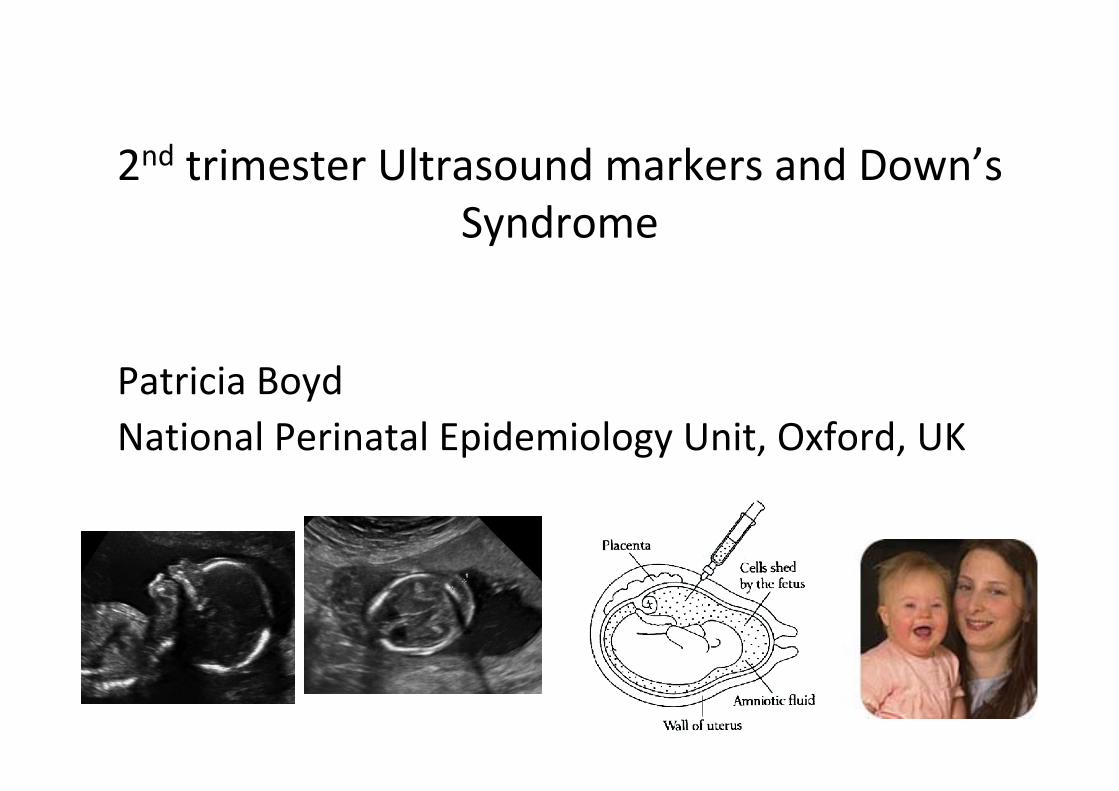
2nd trimester Ultrasound markers and Down’s Syndrome
Patricia BoydNational Perinatal Epidemiology Unit, Oxford, UK
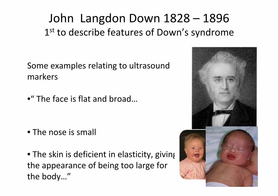
John Langdon Down 1828 – 18961st to describe features of Down’s syndrome
Some examples relating to ultrasound markers
•“ The face is flat and broad…
• The nose is small
• The skin is deficient in elasticity, giving the appearance of being too large for the body…”
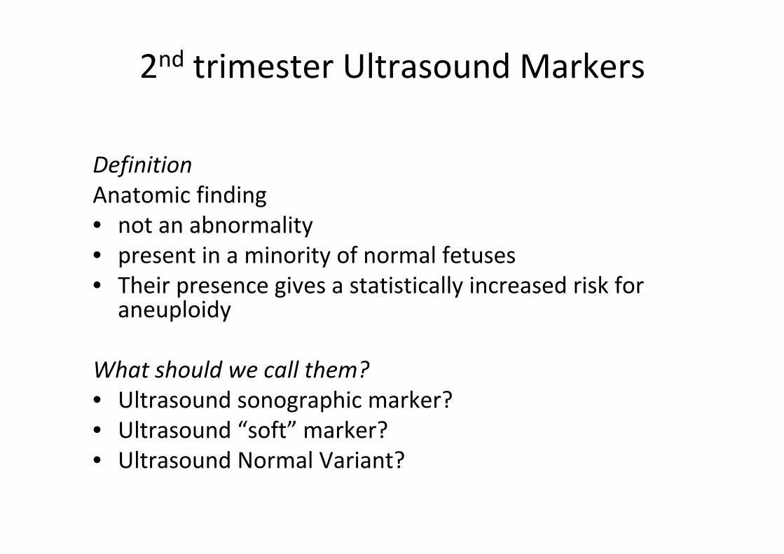
2nd trimester Ultrasound Markers
DefinitionAnatomic finding• not an abnormality• present in a minority of normal fetuses• Their presence gives a statistically increased risk for aneuploidy
What should we call them?• Ultrasound sonographic marker?• Ultrasound “soft” marker?• Ultrasound Normal Variant?
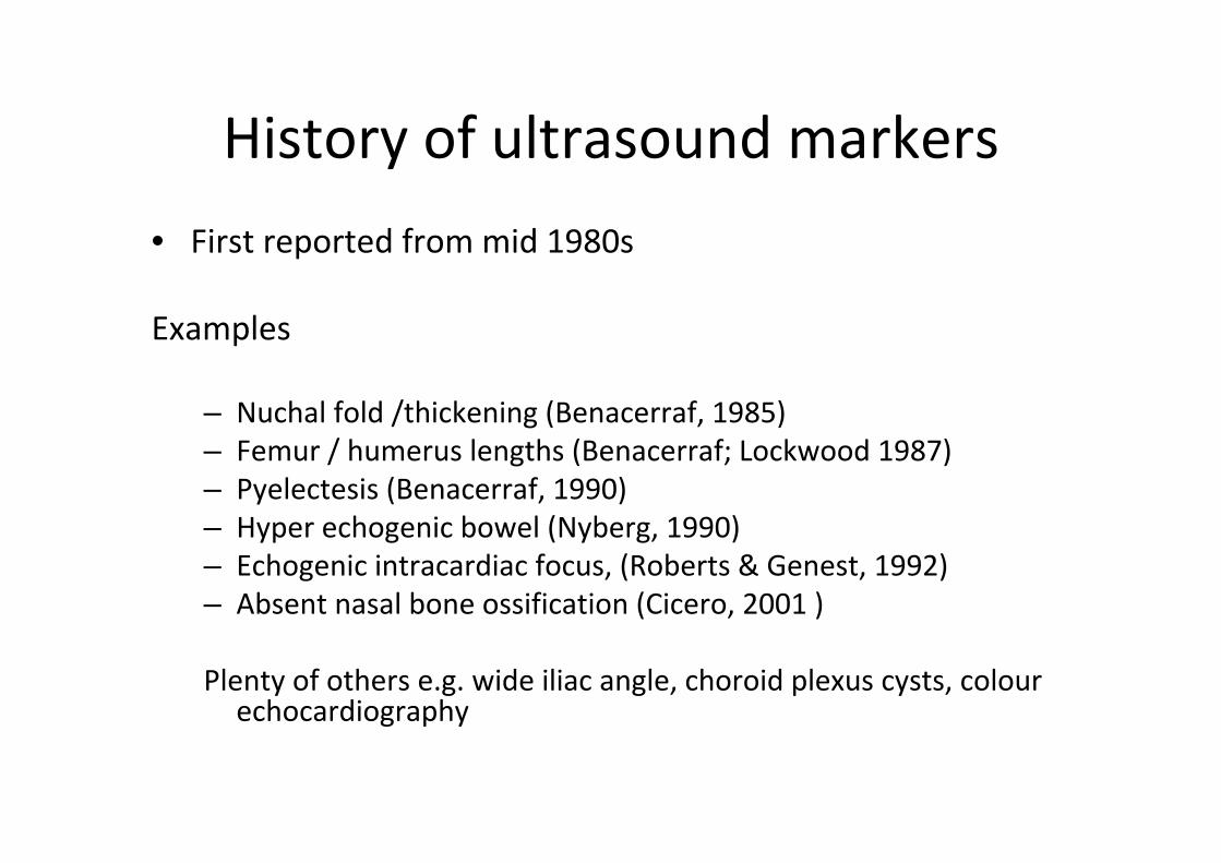
History of ultrasound markers
• First reported from mid 1980s
Examples
– Nuchal fold /thickening (Benacerraf, 1985)– Femur / humerus lengths (Benacerraf; Lockwood 1987)– Pyelectesis (Benacerraf, 1990)– Hyper echogenic bowel (Nyberg, 1990)– Echogenic intracardiac focus, (Roberts & Genest, 1992)– Absent nasal bone ossification (Cicero, 2001 )
Plenty of others e.g. wide iliac angle, choroid plexus cysts, colour echocardiography
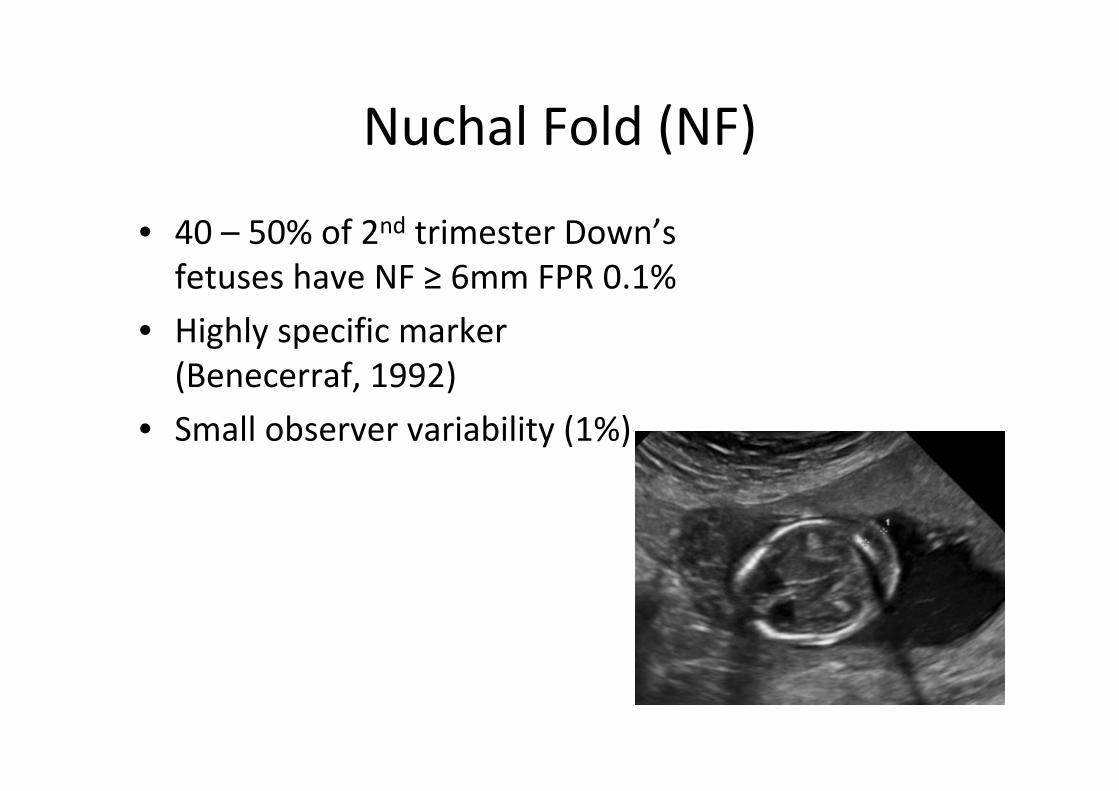
Nuchal Fold (NF)
• 40 – 50% of 2nd trimester Down’s fetuses have NF ≥ 6mm FPR 0.1%
• Highly specific marker (Benecerraf, 1992)
• Small observer variability (1%)

Mild pyelectesis (renal pelvic dilatation) and Down’s syndrome
Definition• Anterior‐posterior diameter of renal pelvis ≥4mm (or 5, 6, 7?) in Transverse section
• Present in 17‐25% of Down’s fetuses, and 2‐3% of normal controls (Benacerraf, 1990)
• Minor marker

Fetal hyperechogenic bowel
Definition• Fetal bowel at least as echogenic
as bone
• Present in 3 – 27% of Down’s fetuses and <1% normal
controls
• Subjective finding
• Note ‐ risk for cystic fibrosis, cytomegalovirus, growth restriction
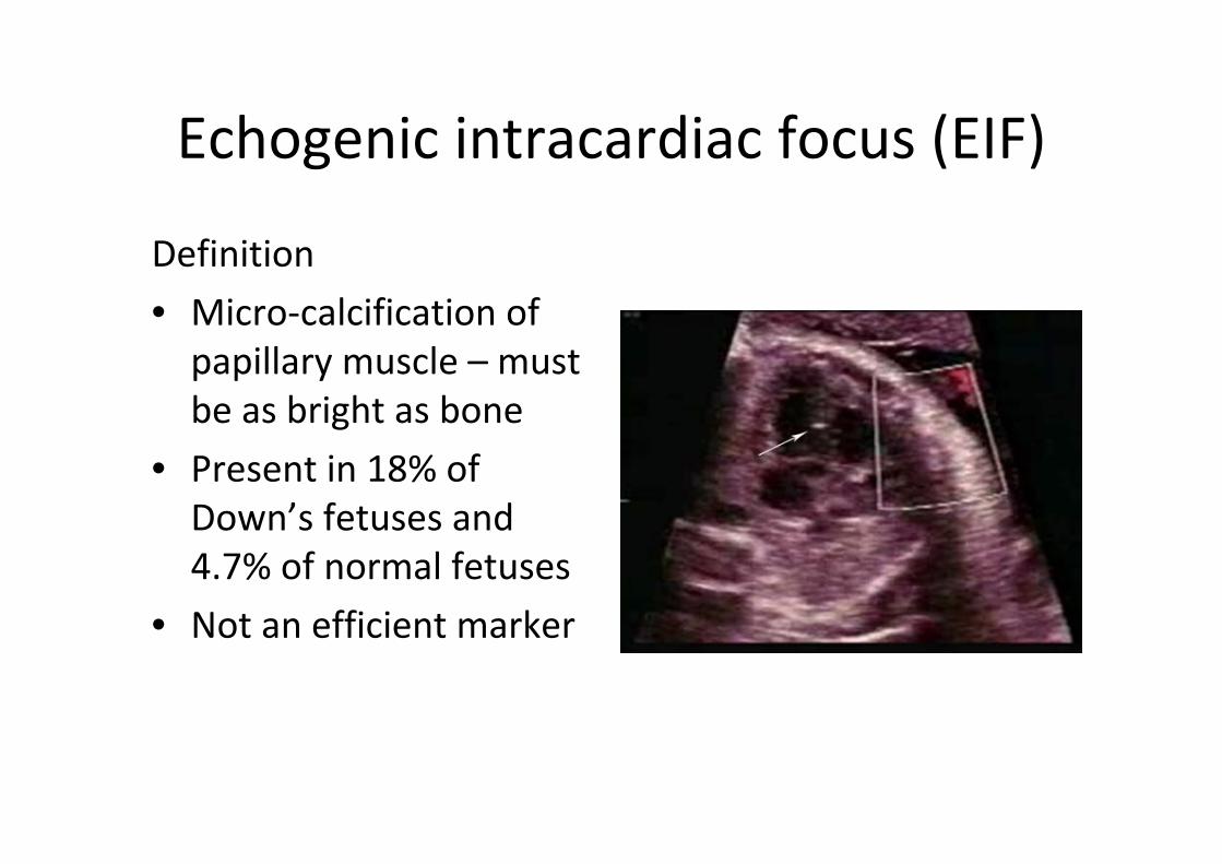
Echogenic intracardiac focus (EIF)
Definition
• Micro‐calcification of papillary muscle – must be as bright as bone
• Present in 18% of Down’s fetuses and 4.7% of normal fetuses
• Not an efficient marker
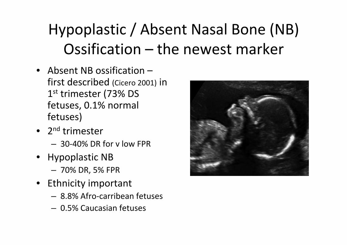
Hypoplastic / Absent Nasal Bone (NB) Ossification – the newest marker
• Absent NB ossification –first described (Cicero 2001) in 1st trimester (73% DS fetuses, 0.1% normal fetuses)
• 2nd trimester– 30‐40% DR for v low FPR
• Hypoplastic NB – 70% DR, 5% FPR
• Ethnicity important– 8.8% Afro‐carribean fetuses– 0.5% Caucasian fetuses

Should all soft markers continue to be reported?
• In 1998 Boyd et al reported 6 years experience in an unselected population
“ultrasound soft markers were responsible for a 4% increase in detection of malformations (from 51% to 55%) and a 12 fold increase in false positive rate (1 in 2332 to 1 in 188)”
Implementing the use of markers –scoring systems / genetic sonogram
• Challenge of how to interpret presence / absence of markers, in particular whether to refine the risk given after a 1st or 2nd trimester screening test
• i.e. Should the prior risk for Down’s syndrome be modified?
• Different countries – different solutions
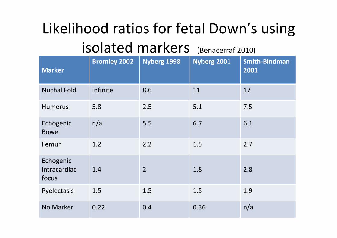
Likelihood ratios for fetal Down’s using isolated markers (Benacerraf 2010)
MarkerBromley 2002 Nyberg 1998 Nyberg 2001 Smith‐Bindman
2001
Nuchal Fold Infinite 8.6 11 17
Humerus 5.8 2.5 5.1 7.5
Echogenic Bowel
n/a 5.5 6.7 6.1
Femur 1.2 2.2 1.5 2.7
Echogenic intracardiac focus
1.4 2 1.8 2.8
Pyelectasis 1.5 1.5 1.5 1.9
No Marker 0.22 0.4 0.36 n/a
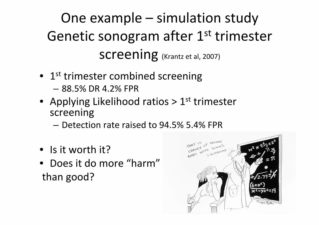
One example – simulation study Genetic sonogram after 1st trimester
screening (Krantz et al, 2007)• 1st trimester combined screening
– 88.5% DR 4.2% FPR• Applying Likelihood ratios > 1st trimester screening– Detection rate raised to 94.5% 5.4% FPR
• Is it worth it?• Does it do more “harm”than good?
UK policy• UK NSC reviewed evidence on role of minor markers to modify prior DS screening risks.
• Concluded that, with exception of nuchal thickening, minor markers should not be used to modify a prior DS screening risk, either by increasing the risk in the presence of a marker or decreasing it if the scan is normal.
FaSTER Trial (First and Second Trimester Estimation of risk)
(Aagard‐Tillery et al, 2009)
• Showed that the genetic sonogram can provide benefit to patients who have had 1st trimester screening– e.g DR increased from 81% to 90% with the combined test
• BUT scan must be performed in centres with experience
• More work needed to redefine Likelihood ratios of 2ndtrimester markers on populations already screened in the 1st trimester
The Future
• More markers, especially 1st trimester ?
• Contingency / sequential testing?
• Non‐invasive prenatal detection?
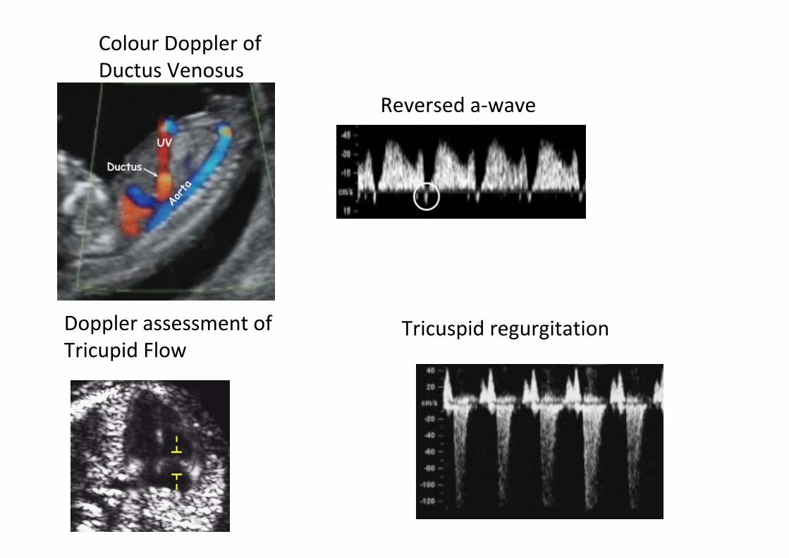
Colour Doppler of Ductus Venosus
Reversed a‐wave
Doppler assessment of Tricupid Flow
Tricuspid regurgitation
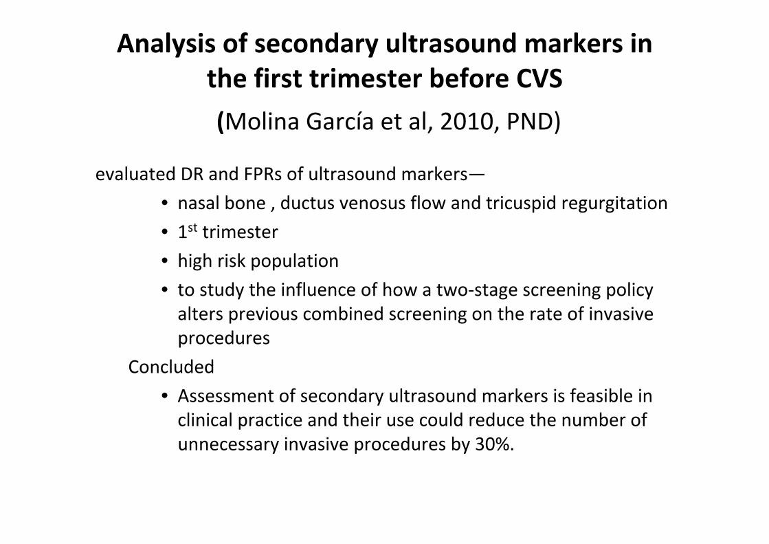
Analysis of secondary ultrasound markers in the first trimester before CVS
(Molina García et al, 2010, PND)
evaluated DR and FPRs of ultrasound markers—
• nasal bone , ductus venosus flow and tricuspid regurgitation
• 1st trimester
• high risk population
• to study the influence of how a two‐stage screening policy alters previous combined screening on the rate of invasive procedures
Concluded
• Assessment of secondary ultrasound markers is feasible in clinical practice and their use could reduce the number of unnecessary invasive procedures by 30%.
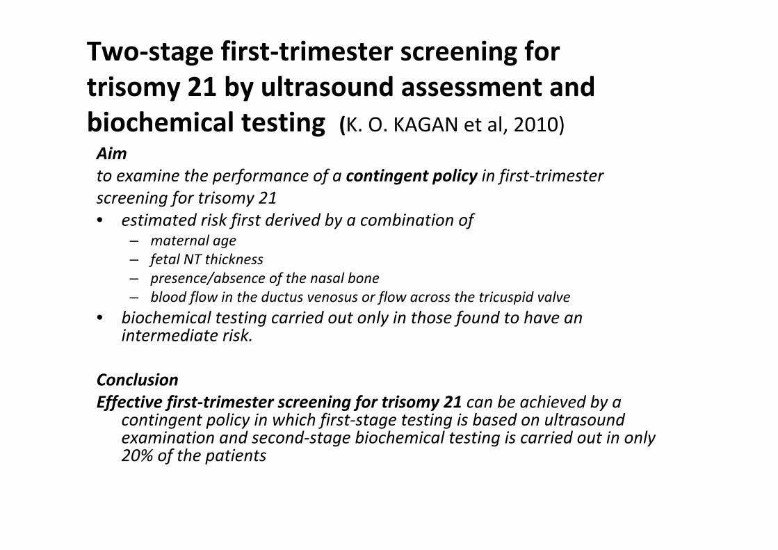
Two‐stage first‐trimester screening for trisomy 21 by ultrasound assessment and biochemical testing (K. O. KAGAN et al, 2010)Aimto examine the performance of a contingent policy in first‐trimesterscreening for trisomy 21• estimated risk first derived by a combination of
– maternal age– fetal NT thickness– presence/absence of the nasal bone– blood flow in the ductus venosus or flow across the tricuspid valve
• biochemical testing carried out only in those found to have an intermediate risk.
Conclusion Effective first‐trimester screening for trisomy 21 can be achieved by a
contingent policy in which first‐stage testing is based on ultrasound examination and second‐stage biochemical testing is carried out in only 20% of the patients
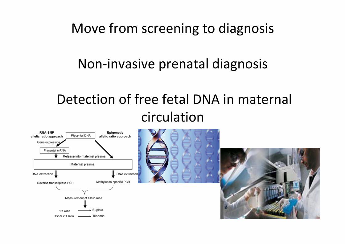
Move from screening to diagnosis
Non‐invasive prenatal diagnosis
Detection of free fetal DNA in maternal circulation
Non‐invasive Prenatal Diagnosis (NIPD) in 2020 (Lo, 2010 Prenatal Diagnosis)
“Thus, I would predict that by 2020
• Many groups would routinely be using massively parallel sequencing technology for the NIPD of multiple monogenic diseases from maternal plasma.
• Furthermore, the same tests would also provide diagnostic information on fetal chromosomal aneuploidies...
• These developments would likely lead to a drastic reduction in the use of invasive techniques, such as amniocentesis and chorionic villus sampling.
• I would foresee that in 2020, there would be multiple articles in Prenatal Diagnosis debating about the ethical and social implications of the information explosion from NIPD.”

Muchísimas gracias por invitarme a Bilbao!