a conserved domain of the arabidopsis gnom protein mediates
TRANSCRIPT

The Plant Cell, Vol. 12, 343–356, March 2000, www.plantcell.org © 2000 American Society of Plant Physiologists
A Conserved Domain of the Arabidopsis GNOM Protein Mediates Subunit Interaction and Cyclophilin 5 Binding
Markus Grebe,
a
José Gadea,
a
Thomas Steinmann,
a
Marika Kientz,
a
Jens-Ulrich Rahfeld,
b
Klaus Salchert,
c
Csaba Koncz,
c
and Gerd Jürgens
a,1
a
Entwicklungsgenetik, Zentrum für Molekularbiologie der Pflanzen, Universität Tübingen, Auf der Morgenstelle 1, D-72076 Tübingen, Germany
b
Forschungsstelle der Max-Planck-Gesellschaft, Enzymologie der Proteinfaltung, D-06120 Halle, Germany
c
Max-Planck-Institut für Züchtungsforschung, D-50829 Cologne, Germany
The Arabidopsis GNOM protein, a guanine nucleotide exchange factor (GEF) that acts on ADP ribosylation factor (ARF)–type G proteins, is required for coordination of cell polarity along the apical–basal embryo axis. Interallelic complemen-tation of
gnom
mutants suggested that dimerization is involved in GNOM function. Here, direct interaction betweenGNOM molecules is demonstrated in vitro and by using a yeast two-hybrid system. Interaction was confined to anN-terminal domain conserved within a subgroup of large ARF GEFs. The same domain mediated in vitro binding to cy-clophilin 5 (Cyp5), which was identified as a GNOM interactor in two-hybrid screening. Cyp5 displayed peptidylprolyl
cis
/
trans
–isomerase and protein refolding activities that were sensitive to cyclosporin A. Cyp5 protein accumulated inseveral plant organs and, like GNOM, was partitioned between cytosolic and membrane fractions. Cyp5 protein wasalso expressed in the developing embryo. Our results suggest that Cyp5 may regulate the ARF GEF function of theGNOM protein during embryogenesis.
INTRODUCTION
The
GNOM
gene was identified by multiple mutant alleles ina search for mutations affecting body organization of the Ar-abidopsis embryo (Mayer et al., 1991). All
gnom
alleles aredefective in establishing the apical–basal axis of embryo po-larity. The earliest defect observed in the
gnom
embryo,which is a perturbed division of the zygote, is followed by ir-regular cell division and elongation patterns at subsequentstages (Mayer et al., 1993).
gnom
seedlings lack a root, aredefective in coordinated alignment of vascular cells, anddisplay variably reduced apical structures (Mayer et al.,1993). These alterations have been attributed to defects inthe establishment of coordinated cell polarity along the api-cal–basal axis in early embryos (Steinmann et al., 1999).Cloning of the
GNOM
gene (also called
EMB30
) revealed se-quence similarity to the yeast vesicle trafficking proteinSec7p, including a central region called the Sec7 domain(Shevell et al., 1994; Busch et al., 1996). Proteins with Sec7domains catalyze guanine nucleotide exchange on smallGTP binding proteins of the ADP ribosylation factor (ARF)family required for vesicle coating in membrane trafficking(Chardin et al., 1996; Sata et al., 1998; Springer et al., 1999).
Guanine nucleotide exchange factors (GEFs) that act on
ARFs (ARF GEFs) group into small and large family mem-bers, and large ARF GEFs include the yeast proteins Gea1p,Gea2p, and Sec7p (Chardin et al., 1996; Peyroche et al.,1996; Sata et al., 1998). GNOM is a membrane-associated,functional ARF GEF of the Gea type, suggesting its involve-ment in vesicular trafficking (Steinmann et al., 1999). Apartfrom the Sec7 domain, large ARF GEFs contain as yet func-tionally uncharacterized N- and C-terminal regions (Mossand Vaughan, 1998). In the case of GNOM, however, there isgenetic evidence for subunit interaction, because
gnom
al-leles with different mutations in the Sec7 domain exhibit fullcomplementation (Busch et al., 1996). In this study, wedemonstrate a physical interaction of GNOM molecules byusing a yeast two-hybrid system and in vitro, thereby defininga novel N-terminal interaction domain that is conservedwithin a distinct subgroup of large ARF GEFs.
Large ARF GEFs, such as Gea1p, exist in high molecularweight complexes (Peyroche et al., 1996), but their interactingpartners have not been characterized. We have screened forGNOM interactors by using the yeast two-hybrid system,and we have identified cyclophilin 5 (Cyp5) as an interactingprotein. Cyp proteins catalyze
cis
/
trans
–isomerization ofpeptidylprolyl bonds, a rate-limiting step in protein folding,and their activity is inhibited by the immunosuppressivedrug cyclosporin A (Fischer et al., 1989; Schönbrunner et al.,1991). Cyp proteins are involved in different signal transduc-tion pathways, such as T cell activation and heat shock
1
To whom correspondence should be addressed. E-mail [email protected]; fax 49-7071-29-5797.

344 The Plant Cell
protein Hsp90–dependent signal transduction in glucocorti-coid receptor regulation (Mattila et al., 1990; Bram andCrabtree, 1994; Duina et al., 1996).
Only a few studies have addressed the role of Cyp pro-teins during dimerization or oligomerization of target mole-cules. In the mouse glucocorticoid receptor complex, forexample, Cyp40 interacts with the dimerization domain ofHsp90 (Carrello et al., 1999), and a yeast Cyp40 homolog,Cpr6, can reactivate the ATPase activity of Hsp90 in vitro(Prodromou et al., 1999). As an example of Cyp interactionwith an oligomeric target in vivo, human CypA was shown tobind to the human immunodeficiency virus HIV-1 capsidprotein Gag (Luban et al., 1993; Franke et al., 1994). Muta-tions in Gag abolish both CypA incorporation into virionsand virion infectivity, mimicking the effects of cyclosporin Atreatment of infected cells (Franke et al., 1994).
Our study identifies a conserved protein domain of a largeARF GEF that mediates both subunit interaction and Cypbinding. GNOM-interacting Cyp5 is a cyclosporin A–sensitivepeptidylprolyl
cis
/
trans
–isomerase (PPIase) with protein re-folding activity that is also expressed during embryogenesis.We propose that Cyp5 is a potential regulator of GNOMfunction in Arabidopsis embryogenesis.
RESULTS
Interaction of GNOM Subunits Mediated by an N-Terminal Domain
Genetic complementation between different
gnom
mutantalleles suggested that GNOM function involves physical in-teraction of GNOM subunits (Mayer et al., 1993; Busch etal., 1996). To examine this model, we generated a series ofGNOM deletion constructs in interaction trap vectors of theyeast two-hybrid system (Gyuris et al., 1993). As shown inFigures 1A and 1B, interaction was observed betweennearly full-length GNOM proteins lacking the first 17 aminoacids. The region required for self-interaction was mappedto GNOM amino acids 1 to 246 (GNOM
1–246
) encoded by thefirst exon of the
GNOM
gene (Figure 1C; see also Figure 3A).To analyze interaction in an independent test system, we
performed in vitro protein binding assays, as displayed in Fig-ure 2. A purified glutathione
S
-transferase (GST)–GNOM
1–246
fusion protein pulled down the full-length GNOM proteinfrom Arabidopsis protein extracts (Figure 2A). The sameGST–GNOM fusion also bound to GNOM
1–246
synthesizedby in vitro translation (Figure 2B). As already observed dur-ing two-hybrid analysis (Figure 1), smaller subfragments ofGNOM did not interact (J. Gadea and G. Jürgens, unpub-lished results), suggesting a structural requirement of thewhole domain for binding. These results demonstrate a di-rect interaction between GNOM molecules mediated by adistinct N-terminal domain. For simplicity, we refer to thisminimal domain as the dimerization domain.
Sequence Conservation of the GNOM Dimerization Domain among Large ARF GEFs of the GNOM/Gea Type
In contrast to the well-characterized Sec7 domain of largeARF GEF proteins, little is known about the role of the N- andC-terminal regions of their proteins (Moss and Vaughan,1998). By searching the databases, we identified five large
Figure 1. Interaction between GNOM Subunits and Mapping of theInteraction Domain in Yeast Two-Hybrid Assays.
(A) GNOM fragments fused to an activation domain (AD–GNOM)tested for interaction with two LexA–GNOM fusions are representedby bars with amino acid positions indicated. Amino acids 1 to 246encoded by the first exon and the Sec7 domain (Sec7D) are shaded.Vector, negative control.(B) Interaction of AD–GNOM fragments with nearly full-lengthGNOM protein fused to a DNA binding domain (LexA–GNOM18–1451).(C) Interaction of AD–GNOM fragments with N-terminal 246 aminoacids of GNOM protein fused to a DNA binding domain (LexA–GNOM1–246).Activation of leucine growth reporter (-Leu growth) is indicated by11 or 2; LacZ reporter activity is displayed as relative b-galactosi-dase units determined by liquid culture assay (Ausubel et al., 1995).Error bars represent standard deviations from three to five indepen-dent transformants.

GNOM Interaction Domain 345
ARF GEF sequences with significant homology to theGNOM dimerization domain, which are compared in Figure 3.In each case, the homologous region represents the N-ter-minal part of the protein, indicating a conserved positionwithin the overall protein structure (Figure 3A). The GNOMdimerization domain is most similar in a GNOM-like putativeprotein identified by the Arabidopsis genome sequencingproject, followed by human Golgi-specific
brefeldin A–resis-tance factor 1 (GBF1), a
Caenorhabditis elegans
open read-ing frame, and yeast Gea1p and Gea2p proteins. Bycontrast, yeast Sec7p, protein sequences with higher overallhomology to Sec7p, and mammalian small ARF GEFs didnot display homology to the GNOM dimerization domain(Figure 3A). Coiled-coil domains, leucine zippers, or otherknown protein interaction motifs were not found in theGNOM dimerization domain. However, a predicted hydro-phobic helix spanning amino acids 152 to 169, which is con-served among GNOM, the Arabidopsis GNOM-like putativeprotein, and human GBF1, contains leucine and valine inregularly spaced seven–amino acid intervals (Figure 3B).This hydrophobic region may act as a protein–protein inter-action surface. In addition, all five GNOM/Gea–type se-quences share a PFL motif flanked by conserved aminoacids, giving the consensus sequence (L/I)XPFLX(V/I)(I/V). Insummary, both the sequence and the N-terminal position ofthe GNOM dimerization domain are conserved within a dis-tinct subgroup of large ARF GEFs, suggesting functionalconstraints.
Isolation of GNOM Interacting Proteins
Genetic screening for Arabidopsis embryo pattern mutantsidentified
GNOM
as the earliest-acting zygotic gene cur-rently known to be involved in apical–basal axis formation(Mayer et al., 1991, 1993). However, no genes encoding po-tential direct interactors have been identified by mutation,which prompted us to perform an interaction trap screeningfor presumptive regulators of GNOM function. The nearlyfull-length GNOM protein was used to screen two Arabidop-sis cDNA libraries generated from suspension cells andyoung siliques (see Methods). In each case, 3.6
3
10
6
pri-mary transformants were screened for galactose-dependentactivation of leucine growth and
lacZ
reporters. Clones forstrong interactors from the suspension cell library weregrouped into 17 cDNA classes and subjected to furtherspecificity tests. One of the GNOM interactors was a puta-tive Cyp, Cyp5, which was represented by one clone fromthe cell suspension and 23 clones from the silique library.Among 17 different GNOM-interacting proteins testedagainst LexA–GNOM baits, only Cyp5 interacted with LexA–GNOM
18–360
, as shown in Figure 4A (also shown in M. Grebeand G. Jürgens, unpublished results). Interaction with Cyp5was mediated by the GNOM dimerization domain, whichwas therefore designated the dimerization and Cyp binding(DCB) domain (Figure 4A; cf. Figure 3). Cyp5 was subjected
to further domain mapping and chosen for more detailedcharacterization.
Specific Interaction between GNOM and Cyp5 Is Mediated by the DCB Domain
To assess whether the GNOM–Cyp5 interaction was inde-pendent of the fusion context, we generated a LexA–Cyp5fusion and analyzed its interaction with GNOM fragmentsfused to the transactivation domain. As shown in Figure 4B,the Cyp5–GNOM interaction was confirmed and mapped toGNOM
1–246
, whereas smaller subfragments of GNOM
1–246
did not mediate interaction in the two-hybrid system. Speci-ficity of the GNOM–Cyp5 interaction was addressed by ana-lyzing possible interactions of GNOM with other members ofthe Cyp family, such as the related rotamase Cyp proteinROC1 and the more divergent ROC4 protein. In contrast toCyp5, these two Cyp proteins did not interact with GNOM inthe two-hybrid system (Figure 4C).
In the in vitro assay shown in Figure 5A, a purified GST–Cyp5 fusion protein pulled down full-length GNOM proteinfrom Arabidopsis protein extracts. We also analyzed thebinding of in vitro–translated ROC1 and Cyp5 to the GNOMDCB domain. The GST–GNOM
1–246
fusion protein boundCyp5 but not ROC1 (Figure 5C). Conversely, the in vitro–translated GNOM DCB domain interacted with a purifiedGST–Cyp5 fusion but not with the GST–ROC1 fusion protein
Figure 2. Interaction between GNOM Subunits in Vitro.
In vitro binding assays of the GST–GNOM1–246 fusion are shown.(A) Arabidopsis protein extracts (protein gel blot; 165-kD band de-tected by anti-GNOM Sec7 antiserum).(B) In vitro–translated 35S-methionine–labeled GNOM1–246 (autora-diograph of 15% SDS–polyacrylamide gel).The controls were glutathione–Sepharose beads (beads) and GST-coupled beads (GST).

346 The Plant Cell
Figure 3.
Sequence Conservation of the GNOM DCB Domain.
(A)
GNOM homologs identified by BLAST P search (BLAST plus BEAUTY, ungapped alignments, expect value 0.0001; Altschul et al., 1997).Horizontal lines aligned with GNOM amino acid positions (bottom) represent regions of sequence conservation
.
38%. Asterisks indicate se-quences with higher overall homology to Sec7p. GenBank accession numbers are indicated, and other references are as follows:
A.th.
GNOM,Arabidopsis GNOM/EMB30 (Shevell et al., 1994; Busch et al., 1996);
A.th.
GNOM-like, Arabidopsis GNOM-like putative protein (Sato et al.,1998);
H.s.
GBF-1, human GBF1 (Mansour et al., 1998);
B.t.
GEP, bovine Sec7-like GEP (Morinaga et al., 1997);
S.c.
Gea1p and Gea2p, budding

GNOM Interaction Domain 347
(Figure 5B). Consistent with the two-hybrid results, in vitro–translated subfragments of the GNOM DCB domain didnot interact with GST–Cyp5 (Figure 4B; J. Gadea and G.Jürgens, unpublished results). In summary, our results dem-onstrate that the GNOM–Cyp5 interaction is specific com-pared with other Arabidopsis Cyp proteins analyzed andsuggest that Cyp5 binding and GNOM subunit interactiondepend on similar structural features within the GNOM DCBdomain.
Cyp5 Shares Features of Both Cytosolic andSecreted Cyp Proteins
Cyp proteins are cyclosporin A binding proteins(Handschumacher et al., 1984). Cyclosporin A inhibits bothcatalytic activities of Cyp proteins: peptidylprolyl
cis
/
trans
–isomerization (PPI) of oligopeptides and the refolding of pro-tein substrates (Fischer et al., 1989). The GNOM-interactingCyp5 is the product of the Arabidopsis
AtCYP5
gene, whichrecently has been shown to encode a Cyp-like protein withan N-terminal endoplasmic reticulum–transport signal se-quence (Saito et al., 1999). Searching for Cyp5-related pro-teins, we observed closest similarity with putative cytosolicCyp proteins from nematodes, the plant Digitalis, and Arabi-dopsis rotamase Cyp protein ROC1, as presented in Figure6. Specifically, Cyp5 and those homologs, as well as otherplant cytosolic Cyp proteins, contain a seven–amino acid in-sertion, which seems to have originated early in eukaryoticevolution (Chou and Gasser, 1997).
The central region required for PPIase activity in func-tional Cyp proteins is more strongly conserved across spe-cies than between Cyp5 and Arabidopsis ROC1 (Figure 6).By overall sequence similarity, Cyp5 is more closely relatedto putative cytosolic Cyp proteins than to those involved inthe secretory pathway (data not shown). Thus, Cyp5 ap-pears to share some functional aspects with cytosolic Cypproteins from other species. On the other hand, Cyp5 differsfrom its most similar Cyp proteins in two of nine conservedamino acids that are involved in cyclosporin A binding of hu-man CypA (Theriault et al., 1993; Figure 6). It was thus not
obvious whether Cyp5 has cyclosporin A–sensitive PPIaseor protein refolding activities.
Cyp5 Catalyzes PPI and Protein Refolding in a Cyclosporin A–Sensitive Manner
Cyp5 PPIase and protein refolding activities could be func-tionally relevant for Cyp5 interaction with GNOM or otherproteins. Therefore, we analyzed both activities and their in-hibition by cyclosporin A in vitro by using a purified GST–Cyp5
19–201
fusion protein lacking most of the hydrophobicendoplasmic reticulum transport signal, as shown in Figure7A. We measured PPIase activity of GST–Cyp5
19–201
at aconcentration of 3 nM, which was within the range at whichfirst-order rate constants of the reaction increased linearlywith enzyme concentration. The fusion protein displayed ac-tivity with a catalytic efficiency
K
cat
/
K
m
of 5.7
3
10
6
M
2
1
sec
2
1
for the Suc-Ala-Ala-Pro-Phe-4-nitroanilide standardtetrapeptide substrate. Table 1 shows that the PPIase activ-ity of Cyp5 was within the range of activities measured forother eukaryotic Cyp proteins. Catalytic activity was stronglyinhibited by cyclosporin A with a calculated K
I
of 8 nM (Fig-ure 7B). We analyzed protein refolding activity of the GST–Cyp5
19–201
fusion protein toward the artificial substrateRNase T1 (reduced carboxymethylated RNase T1, RCM-T1,variant S54G/P55N; Mücke and Schmid, 1994). The refold-ing of 0.7
m
M RCM-T1 was accelerated approximately six-fold when 77 nM GST–Cyp5
19–201
fusion protein was added(Figure 7C). The GST–Cyp5
19–201
isomerase concentrationwas selected in a range at which first-order rate constantsfor catalysis showed linear dependence on enzyme concen-tration. The catalytic efficiency
K
cat
/
K
m
was estimated to be4.8
3
10
4
M
2
1
sec
2
1
. The catalysis of protein refolding wasinhibited to approximately half-maximal activity by 50 nMcyclosporin A (Figure 7C). In control experiments, the GSTprotein had no effect on PPIase or on protein refoldingactivity (Table 1; M. Grebe, J.-U. Rahfeld, and G. Jürgens,unpublished results). These results thus demonstrate thatCyp5 is a functional Cyp with cyclosporin A–sensitive PPI-ase and protein refolding activities, suggesting Cyp5 as atarget for cyclosporin A action in Arabidopsis.
yeast Gea1p and Gea2p (Peyroche et al., 1996);
H.s.
ARNO and ARNO 3, human ARNO (Chardin et al., 1996) and ARNO3 (Franco et al., 1998);
M.m.
cytohesin-1, -2, and –3, mouse cytohesin-1, -2, and -3 (Klarlund et al., 1997; Kim et al., 1998); cytohesin-1/B2-1 (Liu and Pohajdak, 1992);
R.n.
U83895, U83896, and U83897, rat sec7 domain proteins (Telemenakis et al., 1997);
C.e.
Z8145.1 and AL032650,
C. elegans
protein; and
S.p.
Z98602, fission yeast protein.
(B)
Clustal W (Thompson et al., 1994) alignment of GNOM DCB domain homology regions. Sequence identities (conservation) to the DCB do-main are 70% (81%) for Arabidopsis GNOM-like putative protein amino acids 20 to 225, 38% (58%) for human GBF1 amino acids 34 to 227,30% (53%) for Gea2p amino acids 88 to 230, 24% (53%) for Gea1p amino acids 87 to 226, and 26% (48%) for
C.e.
Z81451.1 amino acids 88 to226 (Z81451.1 has a 33–amino acid insertion at position 138, which is not shown for simplicity of alignment). Black boxes indicate identity, andgrey boxes indicate homology between at least three sequences. Consensus derived from identical and conserved amino acids of all six se-quences is indicated as stars and dots. Black bars overline a conserved proline-phenylalanine-leucine (PFL) motif and a hydrophobic helix.
Figure 3.
(continued).

348 The Plant Cell
Developmental and Subcellular Distribution ofCyp5 Protein
We raised an antiserum against a nonconserved Cyp5 C-ter-minal oligopeptide (see Figure 6) to determine the develop-mental expression and subcellular distribution of Cyp5. Asshown in Figure 8A, the antiserum recognized a singleband of 19 kD in siliques at high serum dilutions that wasnot detected by preimmune serum. The signal was dimin-ished in a concentration-dependent manner by antiserumpreincubation with the peptide antigen or the GST–Cyp5
19–201
fusion protein, indicating specific recognition of the Cyp5protein (Figure 8A). The Cyp5-specific 19-kD band is con-sistent with the predicted molecular mass of 19.2 kD forthe endoplasmic reticulum–processed Cyp5 protein, incontrast to the predicted 21.5 kD for the nonprocessedform. In vitro translation products of the nonprocessed andpredicted processed form of Cyp5 showed the expectedsize difference (J. Gadea and G. Jürgens, unpublished re-sults). Thus, Cyp5 appeared to be most abundant in itsprocessed form.
The Cyp5 protein was detected in several tissues, includ-ing flowers and siliques (Figure 8B). The subcellular distri-bution of Cyp5 was analyzed by differential centrifugationof extracts from suspension cells expressing a Golgi appa-ratus marker, Myc-tagged sialyl transferase (ST2-11; Weeet al., 1998). Cyp5 localized mainly to the cytosolic fractionbut was also present in the membrane fraction (Figure 8C).By contrast, the sialyl transferase marker was confined tothe microsomal pellet, indicating that microsomal mem-brane integrity was preserved during extraction. To deter-mine whether the processed Cyp5 protein was secretedfrom cells, we analyzed the medium from a suspension cellculture (Figure 8D). Upon 280-fold concentration of the cul-ture supernatant, secreted Cyp5 protein could not be de-tected, whereas a strong signal was found in the cellextract when equal amounts of total protein were loaded. Insummary, Cyp5 cofractionated with GNOM in cytosolic andmembrane fractions, as would be required for their interac-tion in vivo.
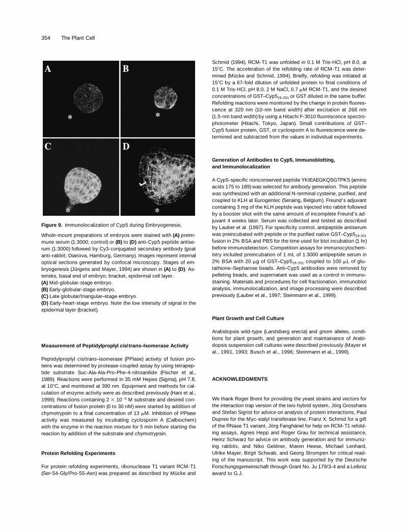
Cyp5 Protein Localization in Embryogenesis
The Cyp5 protein was detected by indirect immunofluores-cence in the developing embryo from the early globularstage, as shown in Figure 9. As a control for the specificityof the signal, embryos were stained with the preimmune se-rum and with the antiserum preincubated with native GST–Cyp5
19–201
protein. In both cases, no specific signal was ob-served (Figure 9A; data not shown). Early-globular-stageembryos gave a weak Cyp5 signal in all cells, displaying apunctate distribution in the cytoplasm (Figure 9B). At the lateglobular/transition stage, the signal became more intense inall cells (Figure 9C). From the early heart stage, epidermalcells displayed less Cyp5-specific staining than did the innercells (Figure 9D), and this lessening of staining became
Figure 4. Specificity and Domain Mapping of Cyp5–GNOM Interac-tion in the Yeast Two-Hybrid System.
(A) Interaction of the activation domain (AD)–Cyp5 fusion with differ-ent LexA–GNOM fragments (amino acid positions indicated). Activa-tion of -Leu growth is indicated by 1 or 2; b-galactosidase activityis given as arbitrary units. Error bars represent standard deviationsfrom five independent transformants. LexA-bicoid (Bicoid) is thenegative control.(B) LexA–Cyp5 interaction with AD–GNOM subfragments (aminoacid positions indicated). -Leu growth and b-galactosidase assayare as given in (A).(C) Specificity of GNOM–Cyp5 interaction compared with other Ara-bidopsis cyclophilins. LexA–GNOM fusion tested against AD fusionsof full-length Cyp5, ROC1, ROC4, and AD vector pJG4.5 (Vector).b-Galactosidase activity and growth on -Leu medium of two trans-formants streaked on plates and grown for 3 days are shown.

GNOM Interaction Domain 349
more pronounced at the torpedo stage (data not shown). Insummary, these results show that Cyp5 protein is expressedduring Arabidopsis embryogenesis.
DISCUSSION
In this study, we demonstrated molecular interaction be-tween identical subunits of GNOM, a large ARF GEF in-
volved in apical–basal axis formation in the Arabidopsisembryo. The N-terminal domain required for interaction isconserved within a distinct subgroup of eukaryotic largeARF GEFs and is also essential for binding to the Cyp5 pro-tein. Cyp5, a PPIase with protein refolding activity, is stronglyexpressed during Arabidopsis embryogenesis. These resultssuggest that Cyp5 may be a potential regulator of GNOMfunction.
Previous genetic studies revealed full complementationbetween
gnom
alleles with different mutations in the Sec7domain (Busch et al., 1996), strongly suggesting that themolecular interaction of GNOM subunits reported here isfunctionally relevant during Arabidopsis embryogenesis.The GNOM DCB domain is conserved by sequence andN-terminal position among large ARF GEF proteins fromseveral eukaryotes. These include the yeast proteinsGea1p and Gea2p, which can be functionally comple-mented by GNOM, and also a human GBF1 protein and aputative protein encoded by a
C. elegans
open readingframe (Mansour et al., 1998; Steinmann et al., 1999). More-over, the intron following the coding sequence for theGNOM DCB domain is conserved in a homologous se-quence identified in the Arabidopsis genome sequencingproject. These findings suggest functional conservation ofthe DCB domain in the Gea/GNOM subgroup of eukaryoticlarge ARF GEFs. By contrast, the Sec7 subgroup repre-sented by yeast Sec7p and Sec7p-related large ARF GEFsfrom Arabidopsis and other eukaryotes lack the DCB do-main. Mammalian small ARF GEFs contain an N-terminalcoiled-coil domain, which, in the case of human ARNO,mediates dimerization in vitro (Chardin et al., 1996). Thesefindings add to the notion that dimerization may be a con-served feature of ARF GEF function and suggest that thecoiled-coil domain of mammalian small ARF GEFs mayplay a role similar to that of the DCB domain of the GNOM/Gea–type large ARF GEFs.
After a search for GNOM interactors in the yeast two-hybrid system, we identified Cyp5 as a new protein thatspecifically bound to the GNOM DCB domain in yeast and invitro. Cofractionation of GNOM and Cyp5 proteins in cytosoland membrane fractions from Arabidopsis extracts sug-gested multiple possible locations for in vivo interaction. Todetermine precisely the subcellular distribution of Cyp5 pro-tein, one would have to perform immunolocalization at theelectron microscopic level. Our attempts to do this were un-successful, because the anti-Cyp5 antiserum did not detectthe epitope on ultrathin sections (T. Steinmann, H. Schwarz,and G. Jürgens, unpublished observations). However, cellfractionation experiments suggested that Cyp5 protein ac-cumulates mainly in the cytosol but is also associated withmembranes. We could not detect Cyp5 in the supernatantfrom cultured cell suspensions, which does not exclude thepossibility that a small fraction of Cyp5 may be secretedfrom cells. Membrane localization of Cyp5 may be due to lo-calization to endoplasmic reticulum subcompartments, assuggested by its functional N-terminal endoplasmic reticu-lum transport signal (Saito et al., 1999). The abundance of
Figure 5. Specific GNOM and GNOM DCB Domain Binding to Cyp5in Vitro.
(A) GST–Cyp5 binding of GNOM protein from Arabidopsis extracts.Protein gel blot analysis detected a 165-kD full-length GNOM bandwith anti-GNOM Sec7 antiserum (Anti-GNOM). Controls are beadsor GST-coupled beads (GST) incubated with plant extract.(B) and (C) Binding of 35S-methionine–labeled in vitro translationproducts to GST fusion proteins. Autoradiographs are of 15% SDS–polyacrylamide gels. In (B), specific binding of GNOM DCB domain(amino acids 1 to 246) to GST–Cyp5 is shown. Equal amounts oftranslation product were incubated with equal concentrations ofGST, GST–ROC1, and GST–Cyp5. In (C), specific binding of Cyp5translation product to GST–GNOM DCB domain (amino acids 1 to246) is shown. Equal amounts of translation product (10% input) ofROC1 and Cyp5 (arrows) were incubated with equal amounts ofGST and GST–GNOM amino acids 1 to 246.

350 The Plant Cell
Cyp5 in the cytosol is in agreement with the cytoplasmic lo-calization of mouse and rat secretory Cyp, CypB, which hasbeen attributed to abortion of protein translocation aftersignal peptide cleavage (Arber et al., 1992; Schumacher etal., 1994).
The mechanism underlying the abortion of translocationhas been described for the hepatitis B virus precore proteinwhose translocation is aborted after signal peptide cleavage,releasing the major part of the mature form into the cytosol(Garcia et al., 1988). Cyp5 may be localized to the cytosol in asimilar way. Cyp5 interaction with cytosolic targets is alsosuggested by sequence similarity to cytoplasmic Cyp pro-teins from other eukaryotes. Together, our data suggest thatthe cytosol and possibly membrane fractions are cellularcompartments relevant for the GNOM and Cyp5 interaction.
Saito et al. (1999) proposed that Cyp5 plays a role inpostembryonic development rather than acting as a stress-responsive chaperone. Our study shows that Cyp5 protein isan enzyme with protein folding activities that is also ex-pressed during embryogenesis, accumulating preferentiallyin inner cells at later stages. Early ubiquitous localization ofCyp5 coincides with irregular divisions affecting wholegnom embryos during early stages (Mayer et al., 1993).Moreover, epidermal cells are less strongly affected in gnomembryos (Mayer et al., 1993), which is consistent with ceas-ing Cyp5 localization in epidermal cells at the heart stage.Thus, expression of Cyp5 in cells affected in gnom mutantembryos is consistent with a presumed interaction of the
GNOM and Cyp5 proteins, as suggested by their specific in-teractions in yeast and in vitro.
Proteins involved in signal transduction, such as receptorkinases, are known to be activated by dimerization (Weissand Schlessinger, 1998). By analogy, GNOM dimerizationmay be required for ARF GEF activity because GNOM func-tion is restored by genetic interaction of certain gnom mu-tant alleles producing full-length protein that is inactive onits own (Busch et al., 1996; Steinmann et al., 1999). The DCBdomain required for GNOM subunit interaction also binds toCyp5. So what role might Cyp5 play in the presumed regula-tion of the ARF GEF activity of GNOM? The Cyp5 proteindisplayed PPIase and protein refolding in vitro activities, whichwere effectively inhibited by cyclosporin A, thus identifyingCyp5 as a new potential cyclosporin A target in Arabidopsis.These observations suggest a role for Cyp5-mediated pro-tein folding in regulating GNOM activity, although the detailsof this interaction remain to be determined.
Interactions between cyclophilins and respective dimeriz-ing or oligomerizing target molecules have been analyzed ina few cases. A competitive activation mechanism has beenimplicated in the interaction of yeast Hsp90 with the cyclo-philin-40 homolog Cpr6 (Prodromou et al., 1999). Cpr6 dis-places the inhibitory cochaperone STI1 in a competitivemanner, restoring Hsp90 ATPase activity in vitro (Prodromouet al., 1999). The mouse STI1 homolog, Hop, and Cyp40have been shown to bind to the Hsp90 dimerization domain(Carrello et al., 1999). However, this Cyp40 interaction in-
Figure 6. Alignment of Arabidopsis Cyp5 with Related Cyp Sequences.
Alignment of seven putative cytosolic eukaryotic Cyp proteins as identified and generated by BLAST P and BEAUTY search with multiple se-quence alignment (Altschul et al., 1997). Dots indicate amino acid identity; dashes represent gaps. The Cyp5 sequence contains the predictedN-terminal endoplasmic reticulum (ER) transport signal (Saito et al., 1999) and PPIase consensus (PROSITE, underlined). Peptide antigen forrabbit immunization and two nonconserved amino acids (asterisks) involved in cyclosporin A binding of human CypA are underlined (Theriault etal., 1993). An ecotype polymorphism I30V between the Landsberg erecta and Columbia sequences is indicated (top line). The more divergent Ar-abidopsis ROC4 lacks a seven–amino acid insertion present in the other Cyp proteins.

GNOM Interaction Domain 351
volves tetratricopeptide repeat domains that are not foundin Cyp5. Another example is the cyclosporin A–sensitive in-teraction of oligomeric HIV-1 capsid protein Gag with hu-man CypA, which is required for virion infectivity (Luban etal., 1993; Franke et al., 1994). Although the PPIase domainof CypA is essential for this interaction, the exact underlyingmechanism is not known. However, CypA has been sug-gested to be involved in Gag protein complex disassembly(Luban, 1996). These examples may illustrate how Cyp5might regulate GNOM activity. Further in vivo analysis ofGNOM DCB domain–Cyp5 interaction will give insight intothe developmental function of Cyp-mediated protein foldingand regulation of large ARF GEF activity in Arabidopsis em-bryogenesis. Mechanisms underlying GNOM dimerizationmay also reveal how large ARF GEF activity is regulated inother eukaryotic systems.
METHODS
Cloning Constructs for Interaction Assays
The analysis and mapping of GNOM–GNOM and GNOM–Cyp5 inter-actions required modifications of interaction trap vectors pJG4-5and pEG202 (Gyuris et al., 1993). The NotI sites were eliminated fromboth vectors by NotI restriction digest, fill in, and ligation. The derivedvectors were subjected to EcoRI restriction digest, followed by fill inand insertion of the NotI linker d(TTGCGGCCGCAA) (New EnglandBiolabs, Beverly, MA). The vectors were designated pMG5 (pJG4-5derivative) and pMG8 (pEG202 derivative). A NotI linker was insertedinto the Bsu36I site preceding codon 18 of GNOM cDNA clone c96(Busch et al., 1996). The cDNA fragments NotI-ScaI and NotI-BstXI,from which protruding 39 ends had been removed, were inserted intopMG5 and pMG8, both of which had been digested with XhoI, filledin, and digested with NotI. The resulting constructs expressed the DNAbinding fusion proteins LexA–GNOM18–1451 and LexA–GNOM18–360
and the activation domain (AD) fusion proteins AD–GNOM18–1451 andAD–GNOM18–360.
Plasmids expressing LexA–GNOM551–1451 and AD–GNOM551–1451
fusion proteins were generated by inserting a BstXI-ScaI cDNA frag-ment, from which protruding 39 ends had been removed, into thefilled-in EcoRI sites of pEG202 and pJG4-5. The vector pJG4-5 wasmodified by inserting the ClaI linker d(CATCGATG) (New England Bi-olabs) into the filled-in XhoI site and was designated pMG2. After re-moval of protruding 39 termini, a BstXI-ClaI cDNA fragment wasinserted into the filled-in EcoRI and the ClaI site of pMG2 generatingthe AD–GNOM551–818 fusion expression construct. pJG4-5 wasmodified by digestion with EcoRI, filling in, and insertion of a ClaIlinker, resulting in vector pMG1. A ClaI-ScaI cDNA fragment was in-serted into the ClaI and filled-in XhoI site of pMG1, generating a con-struct expressing the AD–GNOM818–1451 fusion. Coding regions forGNOM1–246 and subfragments were amplified with Pwo-DNA poly-merase (peQlab, Erlangen, Germany) by using polymerase chain re-action primers with EcoRI and XhoI restriction sites; they werecloned into pJG4-5 and pEG202 for two-hybrid experiments, pBlue-script KS1 (Stratagene, La Jolla, CA) for in vitro translation, orpGEX4T-1 (Pharmacia, Braunschweig, Germany) for expression ofglutathione S-transferase (GST) fusions. Expression from pJG4-5
Figure 7. Cyclosporin A Inhibition of Cyp5 PPIase and Protein Re-folding Activities.
(A) A Coomassie blue–stained SDS–polyacrylamide gel with purifiedGST and GST–Cyp519–201 fusion used in PPIase and protein refoldingassays. Numbers indicate molecular weight markers in kilodaltons.(B) Inhibition of Cyp5 PPIase activity by different cyclosporin A con-centrations. GST–Cyp519–201 (3 nM) was preincubated with varyingconcentrations of cyclosporin A, and PPIase activity was measuredin 35 mM Hepes buffer, pH 7.8, at 108C.(C) Cyp5 catalysis of slow protein refolding of RNAse T1 (0.7 mM)and inhibition by cyclosporin A. The increase of fluorescence at 320nm is shown as a function of the time of protein refolding. Curve 1shows 77 nM Cyp5 without cyclosporin A; curve 2, 77 nM Cyp5 with50 nM cyclosporin A; curve 3, 77 nM Cyp5 with 100 nM cyclosporinA; and curve 4, without Cyp5 and cyclosporin A. GST did not havean effect on refolding (not shown). Measurements were performed in35 mM Hepes buffer, pH 7.8, at 108C.

352 The Plant Cell
derivatives was tested by immunoblotting using 12CA5 anti-HA(Boehringer Mannheim, Mannheim, Germany) or anti-GNOM Sec7 sera(Steinmann et al., 1999). DNA binding of fusion proteins from pEG202derivatives was tested by repression assay (Ausubel et al., 1995) andexpression tested with anti-GNOM Sec7 antibody (Steinmann et al.,1999). All steps involved standard molecular cloning procedures(Sambrook et al., 1989).
Construction of Interaction Trap cDNA Libraries
cDNA libraries from an Arabidopsis thaliana (ecotype Columbia) cellsuspension culture and young siliques (ecotype Landsberg erecta)were generated in pJG4-5. RNA isolation and poly(A)1 mRNA gener-ation involved standard procedures (Sambrook et al., 1989).Oligo(dT)-primed cDNA synthesis and directional cloning were per-formed using a Stratagene cDNA synthesis kit according to the man-ufacturer’s instructions. Reverse transcriptase was replaced bySuperscript reverse transcriptase (GIBCO BRL Life Technologies,Eggenstein, Germany). Two million primary transformants were re-covered for the silique library, and 3 3 107 were recovered for the cellsuspension library. Plasmid DNA was prepared from both libraries bystandard procedures (Sambrook et al., 1989). Insert sizes averaged1.2 kb, and inserts .3 kb were present in both libraries.
Search for GNOM Interacting Proteins
pMG8 expressing a LexA–GNOM18–1451 fusion protein served as baitto screen the cDNA libraries. Materials and procedures for testing thebaits, GNOM–GNOM interactions, quantative b-galactosidase as-says, interaction trap screening, isolation of plasmids, and specificitytests were as described previously (Gyuris et al., 1993; Ausubel et al.,1995). Specifically, using the GNOM protein as a bait, yeast strainEGY48, and the lacZ reporter pSH18-34, 3.5 3 106 primary clonesfrom each library transformation were replated to 2 3 107 colonies onLeu2 medium. One thousand five hundred eighty-five clones growing
on Leu2 medium up to day 4 were restreaked for further testing. Twohundred and fourteen clones of the silique library and 170 clones ofthe suspension library screen displayed activation of both reporters.Grouping of galactose-dependent colonies from the suspension li-brary screen into 17 classes of cDNAs was achieved by cross-hybridization of amplified plasmid inserts. The longest clone from eachgroup was sequenced using a Sequenase version 2.0 kit (Amersham,Braunschweig, Germany) and rehybridized to amplified plasmid in-serts from clones of the silique screen. The plasmid with Cyp5 cDNA,isolated as a full-length clone, was retransformed and tested againstGNOM constructs and different arbitrary proteins including Lexa-bicoid (pRFHM1; Ausubel et al., 1995; data not shown). Cyp5 cDNAsequence was cloned into pEG202. Coding regions of ROC1 andROC4 (Lippuner et al., 1994) were amplified from cDNA by usingPwo-DNA polymerase (peQlab) and polymerase chain reaction prim-ers with restriction sites and subcloned into pJG4-5.
Expression and Purification of GST Fusion Proteins
The cDNA sequences encoding the Cyp5 and rotamase Cyp1(ROC1) proteins were cloned into pGEX4T-1 (Pharmacia) for expres-sion of GST fusions and into pBluescript KS1 for in vitro translation.The amplified sequence encoding Cyp519–201 was cloned intopGEX4T-1. Expression of GST fusions in Escherichia coli DH5a wasinduced with 0.1 mM isopropyl thiogalactoside for 3 hr at 378C. Bac-terial lysis, coupling of fusion proteins to glutathione–Sepharose(Pharmacia), washes, and elution were performed according to themanufacturer’s instructions and standard protocols (Ausubel et al.,1995). Briefly, for large-scale purification of GST fusion proteins, glu-tathione–Sepharose columns (2-mL bed volume) were prepared, andextracts were cleared by ultracentrifugation, passed through col-umns, and washed three times with PBS and 1% Triton X-100 andtwice with PBS. GST fusions were eluted with 10 mM reduced glu-tathione. Eppendorf (Hamburg, Germany) centrifugal filter tubeswere used for removing glutathione by PBS washes and for proteinconcentration.
Table 1. Comparison of Cyp5 and Other Cyp PPIase Activitiesa
Source Cyp ProteinKcat/Km
(31026 M21 sec21) References
Arabidopsis GST–Cyp5 5.7 This studyGST 0 This study; Price et al. (1994)
C. elegans MBP–Cyp3 0.4 Page et al. (1996)C. elegans MBP–Cyp6 8.4 Page et al. (1996)Human Cyp A 5.1 Schönbrunner et al. (1991)S. cerevisiae Cytosolic 3.3 Schönbrunner et al. (1991)Porcine kidney (17 kD) 5.9 Schönbrunner et al. (1991)Human CypB 6.3 Price et al. (1994)Human GST–CypB 4.9 Price et al. (1994)Maize Microsomal 25.0 Sheldon and Venis (1996)Maize Cytosolic 11.0 Sheldon and Venis (1996)
a Shown are PPIase activities determined as catalysis of cis-trans interconversion of the Suc-Ala-Ala-Pro-Phe-4-nitroanilide substrate in pro-tease-coupled assay. Except for budding yeast (S. cerevisiae) and maize Cyp proteins, which represent activities of native purified protein, allother activities were determined for recombinant protein or fusion protein. Kcat/Km (specific catalytic constant/Michaelis-Menten constant) withthe unit (M21 sec21) gives the catalytic efficiency for the reaction.

GNOM Interaction Domain 353
In Vitro Binding Assays
In vitro binding assays (Ausubel et al., 1995) were modified for re-quirements of precipitation from Arabidopsis extracts. Glutathione–Sepharose beads (25-mL bed volume) with 1 to 3 mg of coupled GSTfusion protein were blocked with 1% milk powder in PBS and 1%Triton X-100 for 30 min, washed with PBS and 1% Triton X-100,blocked with 2% BSA in PBS and 1% Triton X-100 for 30 min, andwashed once with PBS. Native protein extracts were prepared fromArabidopsis suspension culture cells (Steinmann et al., 1999). Pro-tein extraction and binding assays were performed in the cold (68C).Suspensions filtered through Miracloth (Calbiochem, Bad Soden,Germany) were frozen in liquid nitrogen and homogenized. Threevolumes of bead binding (BB) buffer (50 mM potassium phosphate,pH 7.5, 150 mM KCl, and 1 mM MgCl2) containing proteinase inhib-itor mix (Sigma, Deisenhofen, Germany), 1 mM phenylmethylsulfonylfluoride (Sigma), and 1 mg/mL pepstatin A (Sigma) were added, fol-lowed by further homogenization and sonication, as applied for bac-terial protein extracts (Ausubel et al., 1995). After centrifugation at12,000g for 20 min, protein concentration of supernatant was deter-mined (Bradford, 1976). Total Arabidopsis protein (150 to 200 mg) in150 mL of BB buffer was supplemented to give a reaction mixture of300 mL containing 2 mM DTT, 5% glycerol, 1% Triton X-100, andproteinase inhibitors (see above). This was added to glutathione–Sepharose beads (25-mL bed volume) carrying 1 to 3 mg of fusionprotein. Reactions were incubated for 1 hr at 68C under gentle rota-tion. After three washes with BB buffer, 5% glycerol, and 1% TritonX-100, beads were resuspended in 25 mL of Laemmli buffer(Laemmli, 1970), and proteins were eluted at 958C for 5 min. Beadswere pelleted, and supernatants (24 mL) were split for duplicateSDS-PAGE, allowing Coomassie Brilliant Blue R 250 staining andimmunodetection with anti-GNOM Sec7 antiserum at 1:6000 dilu-tion (Steinmann et al., 1999). For binding assays with in vitro trans-lation products, transcription and translation (TNT) reactions,including 35S-methionine (ICN, Eschwege, Germany), were per-formed using a TNT T7-coupled wheat germ extract system(Promega, Mannheim, Germany) according to the manufacturer’sinstructions. A 20-mL in vitro translation reaction was added to 1 to3 mg of a coupled GST fusion protein supplemented with 300 mL BBbuffer, 1% Triton X-100, and 5% glycerol. Binding reactions wereperformed on ice under agitation for 1 hr. Samples were washed,eluted, and subjected to SDS-PAGE. Gels were analyzed by usingCoomassie Brilliant Blue R 250 for staining; then they were driedand autoradiographed.
Figure 8. Expression and Subcellular Localization of Cyp5 Protein.
(A) Specificity of polyclonal anti-Cyp5 peptide antiserum. Shown isan immunoblot of total silique protein. Preimmune and immune se-rum dilutions are as follows: lane 1, 1:6000; lane 2, 1:9000; lane 3,1:12,000; lane 4, 1:15,000; and lane 5, 1:18,000. Antiserum specific-ity determined by preincubation with peptide or purified GST–Cyp519–201 fusion protein is as follows: lane 6, 5 mg; lane 7, 500 ng;lane 8, 50 ng; and lane 9, 5 ng. Numbers at left indicate molecularweight markers in kilodaltons.(B) Immunoblot of 30 mg of total protein from different Arabidopsisorgans detected with 1:6000 dilutions of anti-Cyp5 peptide antise-rum and anti-GNOM Sec7 antiserum (Steinmann et al., 1999). The19-kD Cyp5 and 165-kD GNOM bands are shown.(C) Localization of Cyp5 protein by subcellular fractionation. Immu-noblot of protein extracts from cell suspensions expressing theGolgi apparatus marker Myc-sialyl transferase (ST2-11) (Wee et al.,1998) subjected to differential centrifugation. S10, supernatant of10,000g centrifugation; S100, cytosolic supernatant; P100, microso-mal membrane pellet of 100,000g centrifugation. Protein gel blotswere probed with an anti-Myc antibody (A14; Santa Cruz Biotech-
nologies, Santa Cruz, CA; 1:1000) for control of membrane integrity,as well as anti-GNOM Sec7 and anti-Cyp5 peptide antisera. The19-kD Cyp5, 165-kD GNOM (arrow), and 46- and 48-kD ST2-11(Myc) bands are shown with short and long exposure of Cyp5 detec-tion.(D) Localization of Cyp5 in cell suspension cultures. Cyp5 protein inextracts from suspension cells (Culture cells) and cell culture super-natant (Culture medium) on day 5 after passage was detected by im-munoblotting with anti-Cyp5 peptide antiserum. The 19-kD Cyp5band is shown. Culture supernatant was concentrated by ammo-nium sulphate precipitation (Saito et al., 1999).
354 The Plant Cell
Measurement of Peptidylproplyl cis/trans–Isomerase Activity
Peptidylproplyl cis/trans–isomerase (PPIase) activity of fusion pro-teins was determined by protease-coupled assay by using tetrapep-tide substrate Suc-Ala-Ala-Pro-Phe-4-nitroanilide (Fischer et al.,1989). Reactions were performed in 35 mM Hepes (Sigma), pH 7.8,at 108C, and monitored at 390 nm. Equipment and methods for cal-culation of enzyme activity were as described previously (Hani et al.,1999). Reactions containing 2 3 1025 M substrate and desired con-centrations of fusion protein (0 to 30 nM) were started by addition ofchymotrypsin to a final concentration of 13 mM. Inhibition of PPIaseactivity was measured by incubating cyclosporin A (Calbiochem)with the enzyme in the reaction mixture for 5 min before starting thereaction by addition of the substrate and chymotrypsin.
Protein Refolding Experiments
For protein refolding experiments, ribonuclease T1 variant RCM-T1(Ser-54-Gly/Pro-55-Asn) was prepared as described by Mücke and
Schmid (1994). RCM-T1 was unfolded in 0.1 M Tris-HCl, pH 8.0, at158C. The acceleration of the refolding rate of RCM-T1 was deter-mined (Mücke and Schmid, 1994). Briefly, refolding was initiated at158C by a 67-fold dilution of unfolded protein to final conditions of0.1 M Tris-HCl, pH 8.0, 2 M NaCl, 0.7 mM RCM-T1, and the desiredconcentrations of GST–Cyp519–201 or GST diluted in the same buffer.Refolding reactions were monitored by the change in protein fluores-cence at 320 nm (10-nm band width) after excitation at 268 nm(1.5-nm band width) by using a Hitachi F-3010 fluorescence spectro-photometer (Hitachi, Tokyo, Japan). Small contributions of GST–Cyp5 fusion protein, GST, or cyclosporin A to fluorescence were de-termined and subtracted from the values in individual experiments.
Generation of Antibodies to Cyp5, Immunoblotting,and Immunolocalization
A Cyp5-specific nonconserved peptide YKIEAEGKQSGTPKS (aminoacids 175 to 189) was selected for antibody generation. This peptidewas synthesized with an additional N-terminal cysteine, purified, andcoupled to KLH at Eurogentec (Seraing, Belgium). Freund’s adjuvantcontaining 3 mg of the KLH peptide was injected into rabbit followedby a booster shot with the same amount of incomplete Freund’s ad-juvant 4 weeks later. Serum was collected and tested as describedby Lauber et al. (1997). For specificity control, antipeptide antiserumwas preincubated with peptide or the purified native GST–Cyp519–201
fusion in 2% BSA and PBS for the time used for blot incubation (1 hr)before immunodetection. Competition assays for immunocytochem-istry included preincubation of 1 mL of 1:3000 antipeptide serum in2% BSA with 20 mg of GST–Cyp519–201 coupled to 100 mL of glu-tathione–Sepharose beads. Anti-Cyp5 antibodies were removed bypelleting beads, and supernatant was used as a control in immuno-staining. Materials and procedures for cell fractionation, immunoblotanalysis, immunolocalization, and image processing were describedpreviously (Lauber et al., 1997; Steinmann et al., 1999).
Plant Growth and Cell Culture
Arabidopsis wild-type (Landsberg erecta) and gnom alleles, condi-tions for plant growth, and generation and maintainance of Arabi-dopsis suspension cell cultures were described previously (Mayer etal., 1991, 1993; Busch et al., 1996; Steinmann et al., 1999).
ACKNOWLEDGMENTS
We thank Roger Brent for providing the yeast strains and vectors forthe interaction trap version of the two-hybrid system, Jörg Grosshansand Stefan Sigrist for advice on analysis of protein interactions, PaulDupree for the Myc-sialyl transferase line, Franz X. Schmid for a giftof the RNase T1 variant, Jörg Fanghänel for help on RCM-T1 refold-ing assays, Agnes Hepp and Roger Grau for technical assistance,Heinz Schwarz for advice on antibody generation and for immuniz-ing rabbits, and Niko Geldner, Maren Heese, Michael Lenhard,Ulrike Mayer, Birgit Schwab, and Georg Strompen for critical read-ing of the manuscript. This work was supported by the DeutscheForschungsgemeinschaft through Grant No. Ju 179/3-4 and a Leibnizaward to G.J.
Figure 9. Immunolocalization of Cyp5 during Embryogenesis.
Whole-mount preparations of embryos were stained with (A) preim-mune serum (1:3000; control) or (B) to (D) anti-Cyp5 peptide antise-rum (1:3000) followed by Cy3-conjugated secondary antibody (goatanti–rabbit; Dianova, Hamburg, Germany). Images represent internaloptical sections generated by confocal microscopy. Stages of em-bryogenesis (Jürgens and Mayer, 1994) are shown in (A) to (D). As-terisks, basal end of embryo; bracket, epidermal cell layer.(A) Mid-globular-stage embryo.(B) Early-globular-stage embryo.(C) Late globular/triangular–stage embryo.(D) Early-heart-stage embryo. Note the low intensity of signal in theepidermal layer (bracket).

GNOM Interaction Domain 355
Received September 13, 1999; accepted December 30, 1999.
REFERENCES
Altschul, S.F., Madden, T.L., Schäffer, A.A., Zhang, J., Zhang, Z.,Miller, W., and Lipman, D.J. (1997). Gapped BLAST and PSI-BLAST: A new generation of protein database search programs.Nucleic Acids Res. 25, 3389–3402.
Arber, S., Krause, K.H., and Caroni, P. (1992). S-cyclophilin isretained intracellularly via a unique COOH-terminal sequence andcolocalizes with the calcium storage protein calreticulin. J. CellBiol. 116, 113–125.
Ausubel, F., Brent, R., Kingston, R.E., Moore, D.D., Seidman,J.G., Smith, J.A., and Struhl, K., eds (1995). Current Protocols inMolecular Biology. (New York: John Wiley).
Bradford, M.M. (1976). A rapid and sensitive method for the quanti-tation of microgram quantities of protein utilizing the principle ofprotein–dye binding. Anal. Biochem. 72, 248–254.
Bram, R.J., and Crabtree, G.R. (1994). Calcium signaling in T cellsstimulated by a cyclophilin B-binding protein. Nature 371, 355–358.
Busch, M., Mayer, U., and Jürgens, G. (1996). Molecular analysisof the Arabidopsis pattern formation gene GNOM: Gene structureand intragenic complementation. Mol. Gen. Genet. 250, 681–691.
Carrello, A., Ingley, E., Minchin, R.F., Tsai, S., and Ratajczak, T.(1999). The common tetratricopeptide repeat acceptor site forsteroid receptor-associated immunophilins and hop is located inthe dimerization domain of Hsp90. J. Biol. Chem. 274, 2682–2689.
Chardin, P., Paris, S., Antonny, B., Robineau, S., Beraud-Dufour,S., Jackson, C.L., and Chabre, M. (1996). A human exchangefactor for ARF contains Sec7- and pleckstrin-homology domains.Nature 384, 481–484.
Chou, I.T., and Gasser, C.S. (1997). Characterization of the cyclo-philin gene family of Arabidopsis thaliana and phylogenetic analy-sis of known cyclophilin proteins. Plant Mol. Biol. 35, 873–892.
Duina, A.A., Chang, H.C., Marsh, J.A., Lindquist, S., and Gaber,R.F. (1996). A cyclophilin function in Hsp90-dependent signaltransduction. Science 274, 713–715.
Fischer, G., Wittmann-Liebold, B., Lang, K., Kiefhaber, T., andSchmid, F.X. (1989). Cyclophilin and peptidyl-prolyl cis-transisomerase are probably identical proteins. Nature 337, 476–478.
Franco, M., Boretto, J., Robineau, S., Monier, S., Goud, B.,Chardin, P., and Chavrier, P. (1998). ARNO 3, a Sec7-domainguanine nucleotide exchange factor for ADP ribosylation factor 1,is involved in the control of Golgi structure and function. Proc.Natl. Acad. Sci. USA 95, 9926–9931.
Franke, E.K., Yuan, H.E., and Luban, J. (1994). Specific incorpora-tion of cyclophilin A into HIV-1 virions. Nature 372, 359–362.
Garcia, P.D., Ou, J.H., Rutter, W.J., and Walter, P. (1988). Target-ing of the hepatitis B virus precore protein to the endoplasmicreticulum membrane: After signal peptide cleavage translocationcan be aborted and the product released into the cytoplasm. J.Cell Biol. 106, 1093–1104.
Gyuris, J., Golemis, E., Chertkov, H., and Brent, R. (1993). Cdi1, a
human G1 and S phase protein phosphatase that associates withCdk2. Cell 75, 791–803.
Handschumacher, R.E., Harding, M.W., Rice, J., Drugge, R.J.,and Speicher, D.W. (1984). Cyclophilin: A specific cytosolic bind-ing protein for cyclosporin A. Science 226, 544–547.
Hani, J., Schelbert, B., Bernhardt, A., Domdey, H., Fischer, G.,Wiebauer, K., and Rahfeld, J.-U. (1999). Mutations in a peptidyl-prolyl-cis/trans-isomerase gene lead to a defect in 39-end forma-tion of a pre-mRNA in Saccharomyces cerevisiae. J. Biol. Chem.274, 108–116.
Jürgens, G., and Mayer, U. (1994). Arabidopsis. In Embryos: ColorAtlas of Development, J.B.L. Bard, ed (London: Wolfe Publishing),pp. 7–21.
Kim, H.S., Chen, Y., and Lonai, P. (1998). Complex regulation ofmultiple cytohesin-like genes in murine tissues and cells. FEBSLett. 433, 312–316.
Klarlund, J.K., Guilherme, A., Holik, J.J., Virbasius, J.V., Chawla,A., and Czech, M.P. (1997). Signaling by phosphoinositide-3,4,5-trisphosphate through proteins containing pleckstrin and Sec7homology domains. Science 275, 1927–1930.
Laemmli, U.K. (1970). Cleavage of structural proteins during theassembly of the head of bacteriophage T4. Nature 227, 680–685.
Lauber, M.H., Waizenegger, I., Steinmann, T., Schwarz, H.,Mayer, U., Hwang, I., Lukowitz, W., and Jürgens, G. (1997). TheArabidopsis KNOLLE protein is a cytokinesis-specific syntaxin. J.Cell Biol. 139, 1485–1493.
Lippuner, V., Chou, I.T., Scott, S.V., Ettinger, W.F., Theg, S.M.,and Gasser, C.S. (1994). Cloning and characterization of chloro-plast and cytosolic forms of cyclophilin from Arabidopsis thaliana.J. Biol. Chem. 269, 7863–7868.
Liu, L., and Pohajdak, B. (1992). Cloning and sequencing of ahuman cDNA from cytolytic NK/T cells with homology to yeastSEC7. Biochim. Biophys. Acta 1132, 75–78.
Luban, J. (1996). Absconding with the chaperone: Essential cyclo-philin–Gag interaction in HIV-1 virions. Cell 87, 1157–1159.
Luban, J., Bossolt, K.L., Franke, E.K., Kalpana, G.V., and Goff,S.P. (1993). Human immunodeficiency virus type 1 Gag proteinbinds to cyclophilins A and B. Cell 73, 1067–1078.
Mansour, S.J., Herbrick, J.A., Scherer, S.W., and Melancon, P.(1998). Human GBF1 is a ubiquitously expressed gene of theSec7 domain family mapping to 10q24. Genomics 54, 323–327.
Mattila, P.S., Ullman, K.S., Fiering, S., Emmel, E.A., McCutcheon,M., Crabtree, G.R., and Herzenberg, L.A. (1990). The actions ofcyclosporin A and FK506 suggest a novel step in the activation ofT lymphocytes. EMBO J. 9, 4425–4433.
Mayer, U., Torrez-Ruiz, R.A., Berleth, T., Misera, S., and Jürgens,G. (1991). Mutations affecting body organization in the Arabidop-sis embryo. Nature 353, 402–407.
Mayer, U., Büttner, G., and Jürgens, G. (1993). Apical–basal pat-tern formation in the Arabidopsis embryo: Studies on the role ofthe gnom gene. Development 117, 149–162.
Morinaga, N., Moss, J., and Vaughan, M. (1997). Cloning andexpression of a cDNA encoding a bovine brain brefeldin A–sensi-tive guanine nucleotide-exchange protein for ADP-ribosylationfactor. Proc. Natl. Acad. Sci. USA 94, 12926–12931.

356 The Plant Cell
Moss, J., and Vaughan, M. (1998). Molecules in the ARF orbit. J.Biol. Chem. 273, 21431–21434.
Mücke, M., and Schmid, F.X. (1994). Folding mechanism of ribonu-clease T1 in the absence of the disulfide bonds. Biochemistry 33,14608–14619.
Page, A.P., MacNiven, K., and Hengartner, M.O. (1996). Cloningand biochemical characterization of the cyclophilin homologuesfrom the free-living nematode Caenorhabditis elegans. Biochem.J. 317, 179–185.
Peyroche, A., Paris, S., and Jackson, C.L. (1996). Nucleotideexchange on ARF mediated by yeast Gea1 protein. Nature 384,479–481.
Price, E.R., Jin, M., Lim, D., Pati, S., Walsh, C.T., and McKeon,F.D. (1994). Cyclophilin B trafficking through the secretory path-way is altered by binding of cyclosporin A. Proc. Natl. Acad. Sci.USA 91, 3931–3935.
Prodromou, C., Siligardi, G., O’Brien, R., Woolfson, D.N., Regan,L., Panaretou, B., Ladbury, J.E., Piper, P.W., and Pearl, L.H.(1999). Regulation of Hsp90 ATPase activity by tetratricopeptiderepeat (TPR)-domain co-chaperones. EMBO J. 18, 754–762.
Saito, T., Niwa, Y., Ashida, H., Tanaka, K., Kawamukai, M., Matsuda,H., and Nakagawa, T. (1999). Expression of a gene for cyclophilinwhich contains an amino-terminal endoplasmic reticulum–target-ing signal. Plant Cell Physiol. 40, 77–87.
Sambrook, J., Fritsch, E.F., and Maniatis, T. (1989). MolecularCloning: A Laboratory Manual, 2nd ed. (Cold Spring Harbor, NY:Cold Spring Harbor Laboratory Press).
Sata, M., Donaldson, J.G., Moss, J., and Vaughan, M. (1998).Brefeldin A–inhibited guanine nucleotide-exchange activity ofSec7 domain from yeast Sec7 with yeast and mammalian ADPribosylation factors. Proc. Natl. Acad. Sci. USA 95, 4204–4208.
Sato, S., Kaneko, T., Kotani, H., Nakamura, Y., Asamizu, E.,Miyajima, N., and Tabata, S. (1998). Structural analysis of Arabi-dopsis thaliana chromosome 5. IV. Sequence features of theregions of 1,456,315 bp covered by nineteen physically assignedP1 and TAC clones. DNA Res. 5, 41–54.
Schönbrunner, E.R., Mayer, S., Tropschug, M., Fischer, G.,Takahashi, N., and Schmid, F.X. (1991). Catalysis of protein fold-ing by cyclophilins from different species. J. Biol. Chem. 266,3630–3635.
Schumacher, A., Westermann, B., Osborn, M., and Nordheim, A.(1994). The N-terminal signal peptide of the murine cyclophilin
mCyP-S1 is required in vivo for ER localization. Eur. J. Cell Biol.63, 182–191.
Sheldon, P.S., and Venis, M.A. (1996). Purification and character-ization of cytosolic and microsomal cyclophilins from maize (Zeamays). Biochem. J. 315, 965–970.
Shevell, D.E., Leu, W.M., Gillmor, C.S., Xia, G., Feldmann, K.A.,and Chua, N.H. (1994). EMB30 is essential for normal cell divi-sion, cell expansion, and cell adhesion in Arabidopsis andencodes a protein that has similarity to Sec7. Cell 77, 1051–1062.
Springer, S., Spang, A., and Schekman, R. (1999). A primer onvesicle budding. Cell 97, 145–148.
Steinmann, T., Geldner, N., Grebe, M., Mangold, S., Jackson,C.L., Paris, S., Gälweiler, L., Palme, K., and Jürgens, G. (1999).Coordinated polar localization of auxin efflux carrier PIN1 byGNOM ARF GEF. Science 286, 316–318.
Telemenakis, I., Benseler, F., Stenius, K., Südhof, T.C., andBrose, N. (1997). Rat homologues of yeast Sec7p. Eur. J. CellBiol. 74, 143–149.
Theriault, Y., Logan, T.M., Meadows, R., Yu, L., Olejniczak, E.T.,Holzman, T.F., Simmer, R.L., and Fesik, S.W. (1993). Solutionstructure of the cyclosporin A/cyclophilin complex by NMR.Nature 361, 88–91.
Thompson, J.D., Higgins, D.G., and Gibson, T.J. (1994). CLUSTALW: Improving the sensitivity of progressive multiple sequencealignment through sequence weighting, positions-specific gappenalties and weight matrix choice. Nucleic Acids Res. 22, 4673–4680.
Wee, E.G.-T., Sharrier, D.J., Prime, T.A., and Dupree, P. (1998).Targeting of active sialyltransferase to the plant Golgi apparatus.Plant Cell 10, 1759–1768.
Weiss, A., and Schlessinger, J. (1998). Switching signals on or offby receptor dimerization. Cell 94, 277–280.
NOTE ADDED IN PROOF
Since this manuscript was accepted, K. Jackson and D. Soell([1999]. Mutations in a new Arabidopsis cyclophilin disrupt interac-tion with protein phosphatase 2A. Mol. Gen. Genet. 262, 830–838)have reported the cloning of a new Arabidopsis cyclophilin, ROC7,most closely related to Cyp5, as an interaction partner for the proteinphosphatase 2A, RCN1.

DOI 10.1105/tpc.12.3.343 2000;12;343-356Plant Cell
Csaba Koncz and Gerd JürgensaMarkus Grebe, José Gadea, Thomas Steinmann, Marika Kientz, Jens-Ulrich Rahfeld, Klaus Salchert,
Cyclophilin 5 BindingA Conserved Domain of the Arabidopsis GNOM Protein Mediates Subunit Interaction and
This information is current as of January 28, 2019
References /content/12/3/343.full.html#ref-list-1
This article cites 47 articles, 19 of which can be accessed free at:
Permissions https://www.copyright.com/ccc/openurl.do?sid=pd_hw1532298X&issn=1532298X&WT.mc_id=pd_hw1532298X
eTOCs http://www.plantcell.org/cgi/alerts/ctmain
Sign up for eTOCs at:
CiteTrack Alerts http://www.plantcell.org/cgi/alerts/ctmain
Sign up for CiteTrack Alerts at:
Subscription Information http://www.aspb.org/publications/subscriptions.cfm
is available at:Plant Physiology and The Plant CellSubscription Information for
ADVANCING THE SCIENCE OF PLANT BIOLOGY © American Society of Plant Biologists