a monoclonal antibody that blocks adhesion - the journal of cell
TRANSCRIPT
A Monoclonal Antibody that Blocks Adhesion of Chlamydomonas mt ÷ Gametes W i l l i a m J. Snell , M a r i a G. Kosfiszer, Adr i an ClauseU, N a n c y Perillo, Syed I m a m , a n d G a r y H u n n i c u t t
Department of Cell Biology, University of Texas Health Science Center at Dallas, Dallas, Texas 75235
Abstract . During the mating reaction (fertilization) in the biflagellated alga, Chlamydomonas reinhardtii, mt* and mt- gametes adhere to each other via their flagella and subsequently fuse to form quadriflagellated zygotes. In the studies reported here, we describe a monoclonal antibody directed against an mt + flagellar surface molecule. The antibody blocks the adhesive- ness of mt + gametes, isolated mt ÷ flagella, and deter- gent extracts thereof. It has no effect on mt- gametes. Cyanogen bromide-activated Sepharose beads deriva- tized with the antibody bind only mt + gametes; mt- ga- metes and mt ÷ and mt- vegetative cells are unreac-
tive with the derivatized beads. The interaction of mt + gametes with the beads is dynamic and cells continuously bind, detach, and rebind to the beads. Surprisingly, antibody-derivatized beads that have been incubated with mt + gametes acquire the ability to bind mr- gametes. Moreover, extraction of the preincubated beads with detergents releases active mt + adhesion molecules. The evidence suggests that binding of the antibody to the flagellar surface adhesion molecules causes their release from the flagellar surface, pos- sibly mimicking the normal mechanism of flagellar de-adhesion.
W HEN gametes of opposite mating types of the bi- flagellated alga Chlamydomonas reinhardtii are mixed together, they adhere to each other via their
flagella and form large clumps of aggregating cells. Within these clumps, pairs of cells form that eventually fuse to be- come quadriflagellated zygotes. Several laboratories have been interested in the initial adhesive interaction. Recently Goodenough, Heuser, Adair, and co-workers identified high molecular mass glycoproteins rich in hydroxyproline that are involved in the adhesive interaction (1, 2, 4, 9, 10, 16). Saito and Matsuda (35, 37) obtained similar results using different purification methods, and van den Ende's laboratory has identified similar high molecular mass agglutinin molecules in C. eugametos (20, 33, 45).
Our laboratory has been interested in the molecular details of the adhesive interaction as well as a specific de-adhesion mechanism that we have characterized (39, 40-42, 44). We showed that as cells adhere to each other they continuously inactivate the adhesiveness of each other's flagella (42, 44). Replacement of these lost adhesive sites requires protein syn- thesis (36, 37, 42) and protein glycosylation (29, 39, 48); Cooper et al. showed that hydroxylation of proline was also required for replacement of these lost adhesion sites (10). Similar results have been shown for Chlamydomonas eu- gametos (32, 33, 45, 47-49). To more fully study this adhesion-induced loss of flagellar adhesion sites and to iden- tify molecules responsible for adhesion we have begun to use an immunological approach. In earlier unpublished studies we had attempted to prepare rabbit polyclonal antibodies against flagella or flagellar surface fractions that would
specifically block adhesiveness of mating type plus (mr+) 1 or mating type minus (mr-) gametes. To date, all of the polyclonal antibodies we have prepared agglutinate m r and mt÷ vegetative and gametic cells irrespective of the source of the flagella used to inject the rabbits. Our results, which are consistent with other reports on anti-flagellar surface an- tibodies, suggested that raP gametic flagella contain a de- terminant common to the flagellar surface and cell walls of all four cell types that dominates the immunological re- sponse (3, 6-8, 18, 24-26, 51).
To obtain specific, adhesion-blocking antibodies we have begun to use monoclonal antibody methods. In this report, we describe an antibody directed against an mt÷ adhesion molecule. Our results suggest that interaction of the antibody with flagellar adhesion molecules in situ may induce their re- lease from the flagellar surface, possibly mimicking flagellar de-adhesion.
Part of this work has been presented in abstract form (22, 43).
M a t e r i a l s a n d M e t h o d s
Cells and Cell Cultures
mt÷ cells (the 21gr strain) and rot- cells (the 6145c strain) were cultured at 25°C in the minimal medium I of Sager and Granick (35) on a 12-h light-dark photoperiod as previously described (38). Gametic cells were
1. Abbreviations used in this paper: mt ÷ and mt- , mating type plus and mating type minus; N-free, nitrogen free; OG, n-octylglucoside.
© The Rockefeller University Press, 0021-9525/86/12/2449/8 $1.00 The Journal of Cell Biology, Volume 103 (No. 6, Pt. 1), Dec. 1986 2449-2456 2449
on November 16, 2018jcb.rupress.org Downloaded from http://doi.org/10.1083/jcb.103.6.2449Published Online: 1 December, 1986 | Supp Info:
obtained as previously described (38) by transferring vegetative cells (4-7 × 10⁶ cells/ml) 6 h after the beginning of the light period into the nitrogen- free (N-free) medium of Sager and Granick modified to contain 0.15 g/liter of KH2PO4 and 0.3 g/liter of KzHPO4, pH 7.2.
Preparation of Detergent Extracts of Flagella
Flagella isolated by use of the sucrose-pH shock method of Witman et al. (50) as previously described (37), were resuspended in the n-octylglucoside (OG) extraction buffer described by Adair et al. (4) that contained 40 mM OG, 0.1 mM dithiothreitol, 0.1 mM EGTA and 10 mM Tris, pH 7.3. Rou- tinely, flagella isolated from 16 liters of cells were resuspended in about 2 ml of OG extraction buffer. After resuspension the sample was centrifuged for 1 h at 45,000 rpm in a rotor (50 Ti; Beckman Instruments, Inc., Fuller- ton, CA) in an ultracentrifuge (L-350; Beckman Instruments, Inc.) at 4°C, and the supernatant was decanted and saved. The protein concentration, de- termined by the Lowry method (28) using crystalline bovine serum albumin as a standard, was usually about 4 mg/ml.
Fractionation of OG Extracts on Biogel A-15M Columns
O13 extracts were diluted 2:3 with 6 M urea and incubated on ice for 1 h before loading onto a column (Biogel A-15M; Bio-Rad Laboratories, Rich- mond, CA) (1 × 30 cm) equilibrated with elution buffer. The elution buffer contained 40 mM OG, 2 M urea, 0.1 M KCI, 10 mM EDTA, and 10 mM Tris, pH 7.2. Similar results were obtained if 0.7% n-lauryl sarkosine was used in place of OG in the efution buffer. The fractions (1 ml) were dialyzed overnight against DH20 at 4°C (to remove the urea, which prohibited de- tection of adhesive activity) and assayed for adhesive activity.
Assaying for Adhesive Activity
The dried spot assay of Adair et al. (4) was used to assay the adhesive activ- ity of OG extracts and column fractions. Briefly, samples were serially diluted in DH20 and 2-~tl samples were dried under vacuum onto a clean glass microscope slide. The slide was briefly rinsed with DH20 or N-free medium and 2-3 ml of mr- gametes in N-free medium were applied to the slide. After 2-3 min the adhesive activity of the sample was determined visually by noting the most dilute sample to which the mt- gametes were able to adhere. Typically, the titer (the reciprocal of the dilution of the most dilute active sample) of OG extracts was 16,000-32,000.
Polyacrylamide Gel Electrophoresis
SDS PAGE was performed on Laemmli (23) 15 × 13 × 0.1-cm slab gels made as linear 4-16% acrylamide gradient gels and also containing a gra- dient of 3-8 M urea (19). The stacking gel was 3% acrylamide. Gels were fixed and incubated with periodate (12) before being silver stained (31).
Production of Hybridomas
Hybridoma antibodies to Chlamydomonas flagellar surface molecules were generated according to the outline of Kohler and Milstein (21) as adapted by McKearn (30). Biogel A-15M column fractions that contained the peak amounts of adhesive activity were dialyzed, lyophilized, resuspended in DHEO, and used as immunogen. The titer of the resuspended fractions was between 64,000 and 128,000. Approximately 20 I.tg of protein was used for each injection. For the first injection, 0.1 ml of the sample was mixed with 0.I ml of Freund's complete adjuvant and injected subcutaneously into a 7-wk-old BALB/c mouse. Intraperitoneal injections without adjuvant were done at 2 and 4 wk, and at 7 wk the mouse was given a final intravenous injection. 3 d later it was killed by cervical dislocation during ether anesthe- sia, and the fusion was performed using NS-I mouse myeloma cells.
Hybridoma Screening Procedures
Hybridoma supernatants were screened by an adhesion-blocking, functional assay. To do this, 100-~tl portions of the supernatants were dialyzed against N-free medium in 28-place microdialyzers (Bethesda Research Laborato- ries, Gaithersburg, Md.) to reduce the ionic strength to a level compatible with this fresh water organism. 20-~tl portions of the dialyzed supernatants were mixed with 20 ktl of mt + gametes ("~5 × 10⁶ cells/ml in N-free medium) in a 96-well plate and after 5 min at room temperature each well was examined on an inverted microscope. Samples showing (antibody-
induced) isoagglutination were noted, and then 20 I11 of mt- gametes (,x,5 × 106 cells/ml in N-free medium) was added to each well. In control wells with dialyzed, fresh hybridoma medium there was a rapid, mating type-specific agglutination reaction yielding large clumps of aggregating cells that were readily detectable by examination with the inverted micro- scope. Similar aggregation was observed in most oftbe wells containing hy- bridoma supernatants. Of the 1,741 supernatants produced in several fusions and screened with this functional assay, 230 caused isoagglutination or loss of motility, and 45 interfered with adhesion of mt+ gametes. Several of the 45 that appeared to block adhesion also caused isoagglutination or loss of motility of gametes of both mating types and were not considered further. One clone, 2b40, was subcloned in soft agar and has been further character- ized. Ouchterlony double immunodiffusion analysis showed that 2b40 is an IgGl isotype. Ascites fluids were prepared from pristane-primed mice in- jected intraperitoneally with 2640 hybridoma cells.
For some experiments, the 2b40 antibody was purified from ascites fluids either by affinity chromatography on a goat anti-mouse IgG affinity column or by use of the Affi-gel Protein A MAPS method (Bio-Rad Laboratories). Affinity-purified 2b40 was covalently linked to cyanogen bromide-activated Sepharose beads as described by Pharmacia Fine Chemicals, Piscataway, N J).
Results
Partial Purification of Adhesion Molecules from mt + Gametes
OG extracts of mt + gametic flagella, prepared essentially as described by Adair et al. (4), were used as starting material to obtain preparations enriched in adhesion molecules. The OG extracts were fractionated by molecular sieve chroma- tography using a Biogel A-15M column as described in Materials and Methods. Adhesive activity was separated from mastigoneme protein and the major membrane protein with an Mr of ~350,000, which has been shown by us and others (2, 6, 16, 19, 38, 50) to be the major protein component of flagellar surface fractions (not shown). Typically, this purification method yielded 60-100% of the starting activity with a 20-30-fold increase in specific activity. Column frac- tions with peak amounts of activity were dialyzed and lyophilized and used as immunogen in the preparation of monoclonal antibodies.
Selection and Characterization of 2b40, A Monoclonal Antibody that Blocks Adhesion of Only mt + Gametes
Hybridoma supernatants were screened by use of a func- tional assay to identify supernatants that blocked aggregation between mt ÷ and mr- gametes. Several hybridomas produc- ing adhesion-blocking antibodies were detected in our initial screens and one, designated 21>40, has been further charac- terized. This antibody has several properties that indicate that it is directed against an mt + flagellar surface molecule that is intimately involved in the adhesive interaction. First, 21M0 had little visible effect on individual suspensions of mt ÷ or mr- vegetative or gametic cells, although some ag- glutination of mt+ gametes was observed at higher dilutions of the antibody. On the other hand, the antibody had a dra- matic inhibitory effect on the adhesive interaction between mt ÷ and rat- gametes. Fig. 1 A shows the normal aggrega- tion reaction between untreated mt ÷ and mt- gametes in which large clumps of agglutinating gametes form. Fig. 1 B shows that in the presence of 2b40, aggregation was com- pletely blocked. To obtain a more quantitative assessment of the antibody inhibition of adhesion, a counter assay (Coulter
The Journal of Cell Biology, Volume 103, 1986 2450

F i g u r e 1. Inhibition of aggregation by 2b40. m t + and m t - gametes were mixed together with a 1/200 dilution of an irrelevant IgG1 (A) or 2b40 (B) and examined by phase-contrast microscopy. Although there was some variation from experiment to experiment, ascites fluid diluted 1/200 to 1/500 in N-free medium usually blocked adhesion completely.
Electronics, Hialeah, FL) for aggregation was used (44). The results shown in Fig. 2 indicated that half maximal inhi- bition of adhesion occurred at a concentration of ",10 Ixg/ml of antibody protein. In other experiments using an impotent m t ÷ mutant (imp-l; 17) which can agglutinate but not fuse, we found that the antibody would immediately disaggregate adhering m t - and impotent m t + gametes (not shown).
In experiments in which cells of different mating types were pretreated with 2b40 and then washed into fresh, non- antibody-containing medium, we found that only pretreated mt + gametes were affected by the antibody (not shown).
There was no detectable effect on m t - gametes. Similar results were obtained when isolated m t + gametic flagella were pretreated with the antibody. Only m t ÷ gametic flagella were affected. In each case an irrelevant IgG1 monoclonal antibody had no effect on adhesion.
-~ 100 O
c o
u 7 5 "6
z 50 0
25
(.9
\ 4~0 6~0 B~O 1(30 120 ~c 0 0 2'0
ANTIBODY CONCENTRATION (].tg/ml)
F i g u r e 2. Quantification of the effects of 2b40 on aggregation, m t ÷
gametes (8.8 x 10⁶ cells/ml in N-free medium) were pretreated for 5 min with the indicated concentrations of 2b40 purified from as- cites fluid. The cells were then mixed with untreated m t - gametes. Aggregation was determined 5-10 min after the cells were mixed together by use of a counter assay (Coulter Electronics, Inc.) as de- scribed previously (44) and is presented as a percentage of the aggregation of control, untreated cells. In this experiment 63 % of the control cells aggregated.
F i g u r e 3. 2b40 blocks adhesion molecules adsorbed to nitrocellu- lose. An OG extract of m t + gametic flagella was diluted in 10 mM Tris, 150 mM NaC1, pH 7.4 and the indicated amounts were ad- sorbed to nitrocellulose strips in a dot blot apparatus (Bio-Rad Laboratories). The strips were blocked by incubation for 30 min in 1% polyvinyl alcohol (5) at 4°C and then incubated with 1% bo- vine serum albumin in 10 mM NaPO4, 150 mM NaCI, pH 7.2 (control), 1 mg/ml 2b40 (2b40), or a 1/10 dilution of an irrelevant IgG1 ascites fluid (irrel.). After 1 h at 23°C the strips were removed from the incubation solutions and washed at 4°C twice with 10 mM Tris, 150 mM NaCI, pH 7.4 and twice with N-free medium, m t -
gametes were incubated with the strips for 5-10 min and the strips were removed from the cell suspension, drained of excess cells, and photographed. (Staining of the cells was obviated by their natural green color.) Only 2b40 blocked adhesion of the m r - gametes to the adsorbed adhesion molecules.
Snell et al. Antibody Inhibition o f Chlamydomonas Fertilization 2451
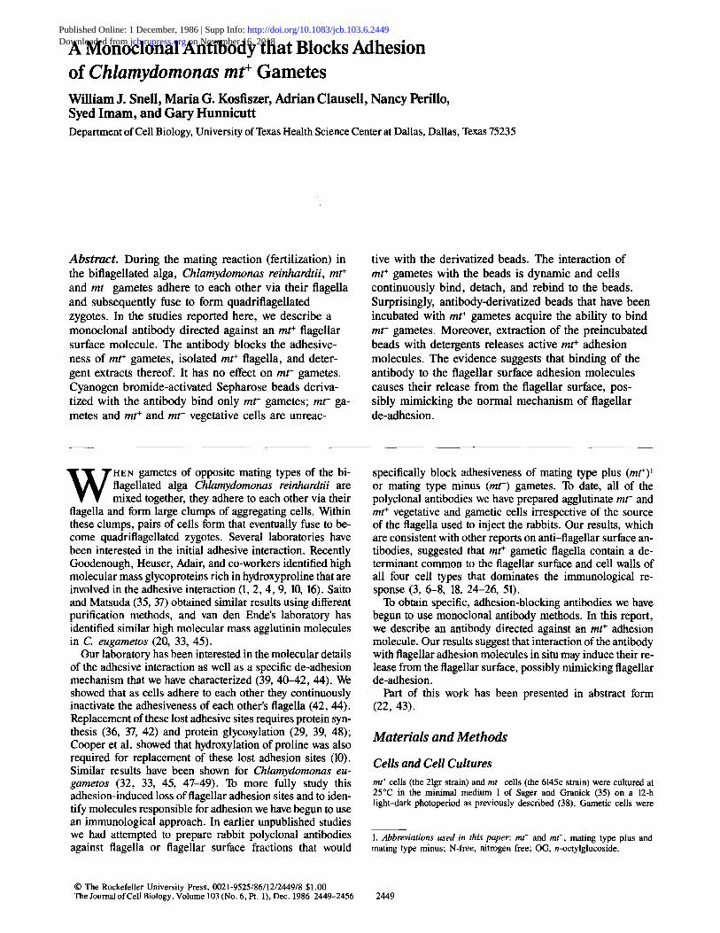
Figure 4. Only mt ÷ gametes bind to 2b40-derivatized Sepharose beads. 2b40 was covalently linked to Sepharose beads and incubated with mt- (a) and mt + (b) gametic and mt- (c) and mt ÷ (d) vegetative cells. Only mt ÷ gametes bound to the beads. The inset in b is a higher magnification image of mt ÷ gametes binding to the beads. In control experiments there was no binding of any of the four cell types to two preparations of beads derivatized with two different irrelevant antibodies.
Since 2b40 was directed against an antigen prepared from column fractions enriched with mt + adhesion molecules prepared from OG extracts, it was important to determine if the antibody could block the adhesiveness of these extracts. To do this, various dilutions of an OG extract of m t ÷ ga- metic flagella were adsorbed to nitrocellulose strips and ei- ther left untreated or treated with 2b40 or an irrelevant IgG1. The strips were then incubated with m t - gametes to test for m t ÷ adhesion molecule activity. The results, presented in Fig. 3, showed that the cells could detect very small amounts of the adsorbed adhesion molecules. Binding was observed in the control sample at 15 ng of protein per spot. Fig. 3 also shows that 2b40 completely blocked adhesion of cells to the adsorbed adhesion molecules, whereas the irrelevant IgG1 had little, if any, effect.
O n l y m t ~ G a m e t e s B o u n d t o 2 b 4 0 - d e r i v a t i z e d
S e p h a r o s e B e a d s
To further characterize the antibody, we covalently linked it to cyanogen bromide-activated Sepharose beads and the beads were mixed with m~ and m t - vegetative and gametic cells. The results shown in Fig. 4 indicated that only mt÷
gametes bound to the beads. In control experiments, we found that cells did not bind to beads derivatized with an ir- relevant IgG1 antibody. These results showed that the anti- body is directed against a flagellar surface determinant pres-
ent only on m r gametes. In addition, close examination of t h e m t ÷ gametes adhering to the 2b40-coated beads showed that cell walls were released by these adhering cells. That is, t h e m t ÷ gametes behaved as if the antibody-coated beads were mr- gametes and appeared to undergo normal sexual signaling.
Since these results directly indicated that the beads con- tained active antibody, we attempted to use the beads to affinity purify the antigen. To do this, preparations contain- ing adhesion molecules (OG extracts and Biogel A-15M col- umn fractions) were passed over a column containing the 2b40-derivatized beads. The run-through was collected, and the column was eluted with a buffer containing 0.1 M glycine, and 1% dioxane, pH 2.5. The eluted fractions were immedi- ately neutralized with K2PO4. The run-through and eluted samples were then assayed for their adhesive activity using the dried spot assay. The result was that adhesive activity was recovered in the run-through fraction and no adhesive activ- ity was found in the eluted fraction (not shown). Control experiments showed that the elution buffer alone did not destroy the activity of adhesion molecules. These results suggested either that the antibody has a low affinity for the antigen in solution, or that the soluble molecule is not in a conformation that enables it to bind to the antibody. In sepa- rate immunoblot experiments (not shown) in which 2b40 was used to probe OG extracts of m t ÷ gametic flagella, none of
The Journal of Cell Biology, Volume 103, 1986 2452

Figure 5. mt + gametes transfer adhesion molecules to 2b40-de- rivatized beads. 2b40-derivatized beads were incubated overnight with mt÷ gametes or mt + vegetative cells and then washed free of the cells by several centrifugations. To determine if transfer of adhe- sion molecules had occurred, the preincubated beads were mixed with rot- gametes. Only beads that had been preincubated with mt + gametes (A) had acquired the ability to bind mt- gametes. The control beads (B) were unreactive with the mt- gametes.
the proteins transferred from the running gel reacted with the antibody.
m t + G a m e t e s T r a n s f e r A d h e s i o n M o l e c u l e s to
A n t i b o d y - d e r i v a t i z e d B e a d s
In the course of the experiments with the bead-cell mixtures we observed by phase-contrast microscopy that the mt + ga- metes were in a dynamic interaction with the 2b40 beads. The cells continuously adhered to and de-adhered from the beads. This was similar to gamete-gamete interactions that occur during the mating reaction as well as to the gamete- flagella adhesion and de-adhesion that we have described
Figure 6. SDS PAGE analysis of proteins transferred to 2b40- derivatized beads. 2b40-derivatized beads (1.2-1.5 ml) were in- cubated with mt÷ gametes (+G), rot- gametes ( -G), and mt ÷ vegetative cells (+V) (1 liter of cells, 2 x 10⁷ cells/ml in N-free medium) overnight, washed free of cells, and eluted with 0.1% sar- kosyl in 10 mM Tris, 20 mM NaCI, pH 7.2. The vegetative sample was eluted with 30 mM OG in 10 mM Tris, 20 mM NaC1, pH 7.2. (Similar results were obtained if the mt ÷ gametic samples were eluted with OG instead of sarkosyl). The eluates were dialyzed, lyophilized, and tested for adhesion molecule activity. Equal por- tions of the eluates also were run on SDS PAGE and the gels were stained with the periodate-silver method. The arrow points to the high molecular mass band in the stacking gel that was consistently seen only in mt ÷ preparations. The unlabeled lane contained high molecular weight standards (Bio-Rad Laboratories).
previously (40, 44). To investigate the possibility that the cells might transfer active adhesion molecules to the beads, beads that had been incubated with m t + gametes were washed free of the cells by differential centrifugation and were mixed with m t - gametes. Fig. 5 shows that m t - ga- metes bound to beads previously incubated with m t ÷ ga- metes (Fig. 5 A) but failed to adhere to beads that had been incubated with m t + vegetative cells (Fig. 5 B ). m t + gametes were also still able to bind to the beads previously incubated with m t ÷ gametes, indicating that unreacted antibodies re- mained accessible on the beads (not shown). These results suggested that m t + gametes transferred adhesion molecules to the beads.
Snell et al. Antibody Inhibition of Chlamydomonas Fertilization 2453

We decided, because of these observations, to do a mod- ified affinity purification of the antigen by incubating m t ÷
gametes with the 2b40-derivatized beads and then eluting the antigen from the beads. To do this, m t ÷ gametes were in- cubated overnight with 2640 beads. As a control, m r - ga- metes and m t ÷ vegetative cells were also incubated with the beads under the same experimental conditions, except that the incubation with the vegetative cells was done in N-con- taining vegetative medium. In some experiments, we ob- served that the vegetative cells bound to the beads. It is likely that this was a nonspecific binding because it did not occur routinely. Also, the vegetative cells that bound to the beads were not removed by addition of soluble 2b40, whereas m t ÷
gametes were immediately removed by this treatment. At the end of the incubation the beads were collected on
a sintered glass filter, washed extensively with N-free medium, and tested for their ability to bind m t ÷ and m t - ga- metes. Only the beads previously incubated with m t ÷ ga- metes supported adhesion of m t - gametes. The beads that had been incubated with m t ÷ vegetative cells or m t - gametes did not bind m t - gametes. Beads from all three incubations, however, were still able to bind m t ÷ gametes. The beads were eluted with detergent-containing solutions, the eluates assayed for adhesion molecule activity in the dried spot assay and their protein composition was analyzed by SDS PAGE.
The bioassay showed that the eluate from the beads in- cubated with m t ÷ gametes had a titer of 16,000, whereas the eluates from the controls showed little if any activity above background. Equal proportions of the eluates from each bead preparation were analyzed by SDS PAGE and the results are shown in Fig. 6. The m t - sample had primarily one protein with an Mr of ~270,000, whereas the m t ÷
vegetative and m t ÷ gametic eluates both had polypeptides with M~'s of ~350,000 corresponding to the Mr of the major membrane protein of the flagellum. The amounts of these polypeptides present in the eluates varied from experiment to experiment. On the other hand, the m t ÷ gametic eluates showed a band in the stacking gel that was found consistently only in m t ÷ gametic eluates. Since this band was in the stacking gel it was not possible to determine its molecular mass or even to establish if it was a single molecule or a com- plex or aggregate of several molecules. Nevertheless, this band becomes a prime candidate for the 2b40 antigen, and appears to have properties similar to m t ÷ agglutinin mole- cules first identified by Adair et al. (4).
D i s c u s s i o n
Antibodies that block adhesion have been instrumental in identifying cell surface molecules involved in specific cell- cell recognition in many systems (11, 13, 14, 24, 27, 34, 46). In the present report we describe the results of our fii:st steps in using this method to identify adhesion molecules in C. r e i n h a r d t i i . We have used detergent extracts of m t ÷ gametic flagella enriched in adhesion molecules as immunogens to prepare monoclonal antibodies that block adhesion of m t ÷
gametes. The data presented here indicate that monoclonal antibody 2b40 is directed against a flagellar surface deter- minant intimately involved in the adhesive interaction and present only on m t + gametes. The antibody did not block the adhesiveness of r o t - gametes nor did ro t - gametes bind to beads coated with the antibody. In addition, m t ÷ and ro t -
vegetative cells were not specifically reactive with 2b40- derivatized beads. The simplest explanation for the anti- body's effects are that it binds to a flagellar surface adhesion molecule at or near the active site of the molecule and thereby renders the molecule unable to interact with adhe- sion molecules on the surface of m r - gametic flagella.
To our knowledge, this is the first report of a monoclonal antibody that specifically blocks adhesion of C. r e i n h a r d t i i
gametes. The van den Ende laboratory has reported on an ad- hesion-blocking rabbit polyclonal antibody directed against a C. e u g a m e t o s cell surface determinant (24, 25, 26, 45). This antiserum reacted with several flagellar surface glyco- proteins, and its action could be blocked by disaccharide moieties (25). Adair, Goodenough, Heuser, and co-workers have obtained monoclonal antibodies directed against flagel- lar surface molecules found only on gametic flagella (1, 3, 15). These antibodies, designated class II, react with both m t ÷ and m t - gametes, as well as with nonagglutinating mu- tant gametes that contain molecules closely related to the ag- glutinin molecules used as immunogens for the preparations of the monoclonal antibodies. The antibodies do not react with vegetative cells, and are directed against the agglutinin molecules.
T r a n s f e r o f A d h e s i o n M o l e c u l e s
Our observation that m t ÷ gametes could be separated from 2b40-derivatized beads by gentle washing procedures indi- cated that the interaction between the beads and the cells was a dynamic one. This is quite similar to the interaction of live gametes with each other or with flagella isolated from ga- metes of the opposite mating type. The results presented here, that m t ÷ gametes transferred active adhesion mole- cules to the 2b40 beads, suggest that binding of 2b40 anti- bodies to adhesion molecules might induce their release from the flagellar surface. Apparently the released adhesion molecules were in a multivalent form, because adhesion sites not interacting with the antibody were available to be bound by m t - gametes. Possibly a similar release mechanism is responsible both for loss of flagellar adhesiveness after cell fusion, and for the flagellar de-adhesion process we de- scribed previously. We have shown that as a consequence of adhesion, flagellar adhesion molecule activity is lost from the flagellar surface (42, 44). On live cells, the inactivated adhesion molecules are rapidly replaced by protein synthe- sis. With isolated flagella or with cells incubated in cyclohex- imide, the inactivated adhesion molecules are not replaced, and the cells de-adhere (42).
Several mechanisms can be envisioned that could lead to inactivation of flagellar adhesion molecules during adhesion. For example, the molecules themselves or nearby molecules might modify the adhesion molecules by adding or removing a critical component. Possibly a protease is activated that partially degrades adhesion molecules and renders them in- active. Alternatively, carbohydrate moeities could be added or disulfides formed or reduced. Another explanation, which is supported by our antibody data, would be that the adhesion molecules are released from the surface of flagella of live cells during adhesion. These released molecules might still be active, but since they were no longer tethered to the fla- gellum they could not support cell-cell or cell-flagella ad- hesion.
The similarity in the consequences of adhesion of m t ÷ ga-
The Journal of Cell Biology, Volume 103, 1986 2454
metes to 2b40-derivatized beads and to m t - flagella suggests that m t + gametes might transfer active adhesion molecules to m t - flagella. Although we showed that the adhesiveness of m t - flagella is lost as a consequence of their adhering to m t ÷ gametes, we never determined if the m t - flagella had acquired m t + adhesion molecules (44). It may be that loss of flagellar adhesiveness in this situation occurs because adhesion molecules on the live cells form a complex with the adhesion molecules on the surface of the isolated flagella. This event could cause release of the adhesion molecules from the flagellum of the live cell, possibly by an active re- lease mechanism. If this mechanism were not active in iso- lated flagella, the complex would remain on the surface of the isolated flagella and could be detected, because mole- cules transferred from the live cell had excess adhesion sites available. When two live cells interact, these putative com- plexes would form; but because both cells would have an in- tact release mechanism, the complex would be released from both flagella. Experiments to test these hypotheses are cur- rently in progress in our laboratory.
We would like to thank Mr. David Mahaffey for stimulating discussions about the interactions of cells with 2b40-derivatized beads. We are indebted to Dr. Pam Witte and Ms. Barbara Border for their invaluable help in the initial stages of monoclonal antibody production, Dr. Wayne Streilein for the use of his tissue culture facilities, and Dr. Fred Grinell for helpful dis- cussions and for reading the manuscript. In addition we are grateful to Dr. Ursula Goodenough of Washington University in St. Louis for lively and stimulating telephone conversations about adhesion and de-adhesion in Chlamydomonas.
This work was supported by National Institutes of Health grant GM- 25661 and National Science Foundation Biological Instrumentation grant PCM-8314034. W. J. Snell is the recipient of Research Career Development Award HD-00454 from the National Institutes of Health.
Received for publication 27 May 1986, and in revised form 19 August 1986.
References
1. Adair, W. S. 1985. Characterization of Chlamydomonas sexual aggluti- nins. J. Cell Sci. (Suppl. 2)233-360.
2. Adair, W. S., C. Hwang, and U. W. Goodenough. 1983. Identification and visualization of the sexual agglutinin from the mating type plus flagellar membrane of Chlamydomonas reinhardtii. Cell. 33:183-193.
3. Adair, W. S., J. Long, W. B. Mehard, J. E. Heuser, and U. W. Good- enough. 1983. Monoclonal antibodies directed against the sexual agglutinins of Chlamydomonas reinhardi. J. Cell Biol. 97 (5, Pt. 2):93a. (Abstr.)
4. Adair, W. S., B. C. Monk, R. Cohen, C. Hwang, and U. W. Good- enough. 1983. Sexual agglutinins from the Chlamydomonas flagellar mem- brane. Partial purification and characterization. J. Biol. Chem. 257:4593-4602.
5. Bavister, B. D. 1981. Substitution of a synthetic polymer for protein in a mammalian gamete culture system. J. Exp. Zool. 217:45-51.
6. Bloodgood, R. A., and G. S. May. 1982. Functional modification of the Chlamydomonas flagellar surface. J. Cell. Biol. 93:88-96.
7. Bloodgood, R. A., M. P. Woodward, and N. L. Salomonsky. 1986. Redistribution and shedding of flagellar membrane glycoproteins visualized using an anti-carbohydrate monoclonal antibody and concanavalin A. J. Cell Biol. 102:1797-1812.
8. Claes, H. 1977. Non-specific stimulation of the autolytic system in ga- metes from Chlamydomonas reinhardtii. Exp. Cell Res. 108:221-229.
9. Collin-Osdoby, P., and W. S. Adair. 1985. Characterization of the purified Chlamydomonas minus agglutinin. J. Cell Biol. 101:1144-1152.
10. Cooper, J. B., W. S. Adair, R. P. Mecham, J. E. Heuser, and U. W. Goodenough. 1983. Chlamydomonas agglutinin is a hydroxyproline-rich glyco- protein. Proc. Natl. Acad. Sci. USA. 80:5898-5901.
11. Damsky, C. H., J. Richa, D. Solter, K. Knudsen, and C. A. Buck. 1983. Identification and purification of a cell surface glycoprotein mediating intercel- lular adhesion in embryonic and adult tissue. Cell. 34:455-466.
12. Dubray, G., and G. Bezard. 1982. A highly sensitive acid-silver stain for 1,2 diol groups of glycoproteins and polysaccharides in polyacrylamide gels. Anal. Biochem. 119:325-329.
13. Edelman, G. M. 1985. Cell adhesion and molecular processes of morpho- genesis. Annu. Rev. Biochem. 54:135-169.
14. Gerisch, G. 1983. Univalent antibody fragments as tools for analysis of
cell-cell interactions in Dictyostelium. Curr. Top. Dev. BioL 140:243-270. 15. Goodenough, U. W. 1985. An essay on the origins and evolution ofeu-
karyotic sex. In Origin and Evolution of Sex. H. O. Haivorson and A. Monroy, editors. Alan R. Liss, Inc., New York. 123-140.
16. Goodenough, U. W., W. S. Adair, P. Collin-Osdoby, and J. E. Heuser. 1985. Structure of the Chlamydomonas agglutinin and related flagellar surface proteins in vitro and in situ. J. Cell Biol. 101:924-941.
17. Goodenough, U. W., P. A. Detmers, and C. Hwang. 1982. Activation for cell fusion in Chlamydomonas: analysis of wild-type and non-fusing mu- tams. J. Cell Biol. 92:378-386.
18. Goodenough, U. W., and D. Jurivich. 1978. Tipping and mating struc- ture activation induced in Chlamydomonas gametes by flagellar membrane an- tisera. J. Cell Biol. 79:680-693.
19. Jarvik, J. W., and J. L. Rosenbaum. 1980. Oversized flagellar membrane protein in paralyzed mutants of Chlamydomonas reinhardtii. J. Cell Biol. 85:258-272.
20. Klis, F. M., M. R. Samson, E. Touw, A. Musgrave, and H. van den Ende. 1985. Sexual agglutination in the unicellular green alga Chlamydomonas eugametos. Identification and properties of the mating type plus agglutination factor. Plant Physiol. (Bethesda). 79:740-745.
21. Kohler, G., and C. Milstein. 1975. Continuous culture of fused cells secreting antibody of predefined specificity. Nature (Lond.). 256:495--497.
22. Kosfiszer, M., A. Clausell, S. Imam, B. Lavery, Jr., B. Border, P. Witte, and W. J. Snell. 1984. A monoclonal antibody that blocks adhesion of rot+ ga- metes of Chlamydomonas. J. Cell Biol. 99 (4, Pt. 2):71a. (Abstr.)
23. Laemmli, U. K. 1970. Cleavage of structural proteins during the assem- bly of the head of bacteriophage T4. Nature (Lond.). 227:680-685.
24. Lens, P. F., F. Olofsen, A. Nederbragt, A. Musgrave, and H. van den Ende. 1982. An antiserum against a glycoprotein functional in flagellar adhe- sion between Chlamydomonas eugametos gametes. Arch. Microbiol. 131:241- 246.
25. Lens, P. F., F. Olofsen, P. van Egmond, A. Musgrave, and H. van den Ende. 1983. Isolation of an antigenic determinant from flagellar glycoproteins of Chlamydomonas eugametos. Arch. Microbiol. 135:311-314.
26. Lens, P. F., W. van den Briel, A. Musgrave, and H. van den Ende. 1980. Sex-specific glycoproteins in Chlamydomonas flagella. An immunological study. Arch. Microbiol. 126:77-81.
27. Loomis, W. F., S. A. Wheeler, W. A. Springer, and S. H. Barondes. 1983. Adhesion mutants of Dictyostelium discoideum lacking the saccharide de- terminant recognized by two adhesion-blocking monoclonal antibodies. Dev. Biol, 109:111-117.
28. Lowry, O. H., H. J. Rosebrough, A. L. Farr, and R. J. Randall. 1951. Protein measurement with the folin reagent. J. Biol. Chem. 193:265-275.
29. Matsuda, Y., K. Sakamoto, N. Kiuchi, T. Mizuochi, Y. Tsubo, and A. Kobata. 1982. Two tunicamycin-sensitive components involved in agglutina- tion and fusion of Chlamydomonas reinhardtii gametes. Arch. Microbiol. 131:87-90.
30. McKearu, T. J. 1980. Fusion of cells in an adherent monolayer. In Monoclonal Antibodies. Hybridomas: A New Dimension in Biological Analy- sis. R. H. Kennet, T. J, McKearn, K. B. Bechtol, editors. Plenum Publishing Corp., New York. 368-369.
31. Merril, C. R., D. Goldman, S. A. Sedman, and M. H. Ebert. 1981. Ultrasensitive stain for proteins in polyacrylamide gels shows regional variation in cerebrospinal fluid proteins. Science (Wash. DC). 211:1437-1438.
32. Musgrave, A., P. de Wildt, F. Schuring, K. Crabbendam, and H. van den Ende. 1985. Sexual agglutination in Chlamydomonas eugametos before and after cell fusion. Planta. 166:234-243.
33. Pijst, H. L. A., F. A. Ossendorp, P. Egmond, A. M. I. E. van Kemps, A. Musgrave, and H. van den Ende. 1984. Sex-specific binding and inactivation of agglutination factor in Chlamydomonas eugametos. Planta. 160:529-535.
34. Rasmussen, R. A., Y.-H. Chin, J. J. Woodruff, and T. G. Easton.1985. Lymphocyte recognition of lymph node high endothelium. VII. Cell surface proteins involved in adhesion defined by monoclonal anti-HEBF LN (A. 1 I) an- tibody. J. lmmunol. 135:19-24.
35. Sager, R., and S. Granick. 1954. Nutritional control of sexuality in Chlamydomonas reinhardtii. J. Gen. Physiol. 3:729-742.
36. Saito, T., and Y. Matsuda. 1984. Sexual agglutinin of mating type minus gametes in Chlamydomonas reinhardtii I. Loss and recovery of agglutinibility of gametes treated with EDTA. Exp. Cell. Res. 152:322-330.
37. Saito,.T., Y. Tsubo, and Y. Matsuda. 1985. Synthesis and turnover of cell body-agglutinin as a pool of flagellar surface-agglutinin in Chlamydomonas reinhardtii gamete. Arch. Microbiol. 142:207-210.
38. Snell, W. J. 1976. Mating in Chlamydomonas: a system for the study of specific cell adhesion. I. Ultrastructural and electrophoretic analysis of flagellar surface components involved in adhesion. J. Cell Biol. 68:48-69.
39. Snell, W. J. 1981. Flagellar adhesion and deadhesion in Chlamydomonas gametes: effects of Tunicamycin and observations on flagellar tip morphology. J. Supramol. Struct. Cell. Biochem. 16:371-376.
40. Snell, W. J. 1985. Cell-cell interactions in Chlamydomonas. Annu. Rev. Plant Physiol. 36:287-315.
41. Snell, W. J., A. Clausell, and W. S. Moore. 1983. Flagellar adhesion in Chlamydomonas induces synthesis of two high molecular weight cell surface proteins. J. Cell Biol. 96:589-597.
42. Snell, W. J., and W. S. Moore. 1980. Aggregation-dependent turnover of flagellar adhesion molecules in Chlamydomonas gametes. J. Cell Biol.
Snell et al. Antibody Inhibition of Chlamydomonas Fertilization 2455

84:203-210. 43. Snell, W. J., N. L. Perillo, G. R. Hunnicutt, D. T. Mahaffey, and M.
G. Kosflszer. 1985. A monoclonal antibody that binds to mt + flagellar adhe- sion molecules of Chlamydomonas. J. Cell Biol. 101 (5, Pt. 2):379a (Abstr.)
44. Snell, W. J., and S. Roseman. 1979. Kinetics of adhesion and de- adhesion of Chlamydomonas gametes. J. Biol. Chem. 254:10820-10829.
45. van den Ende, H. 1984. Sexual agglutination in Chlamydomonas. Adv. Microb. Physiol. 26:89-123.
46. Vollmers, H. P., and W. Birchmeier. 1983. Monoclonal antibodies that prevent adhesion of Bl6 melanoma cells and reduce metastasis in mice: connec- tion with human tumor cells. Proc. Natl. Acad. Sci. USA. 80:6863-6867.
47. Wiese, L., A. Hardcastle, and M. Stewart. 1984. Inactivation of the (+) gamete agglutinin during the mating-type reaction in Chlamydomonas. Gamete
Res. 9:441-449. 48. Wiese, L., and R. A. Mayer. 1982. Unilateral tunicamycin sensitivity of
gametogenesis in dioecious isogamous Chlamydomonas species. Gamete Res. 5:1-9.
49. Wiese, L., and W. Wiese. 1978. Sex cell contact in Chlamydomonas: a model for cell recognition. Syrup. Soc. Exp. Biol. 32:83-104.
50. Witman, G., K. Carlson, J. Berliner, and J. L. Rosenbaum. 1972. Chlamydomonas flagella. I. Isolation and electrophoretic analysis of microtu- boles, matrix, membranes, and mastigonemes. J. Cell Biol. 54:507-539.
51. Woodward, M. P., W. W. Young, Jr., and R. A. Bloodgood. 1985. De- tection of monoclonal antibodies specific for carbohydrate epitopes using perio- date oxidation. J. lmmunol. Methods. 78:143-153.
The Journal of Cell Biology, Volume 103, 1986 2456