bcm 410a lecture 35 immunity immunoglobulin structure antibody classes monoclonal antibodies...
TRANSCRIPT
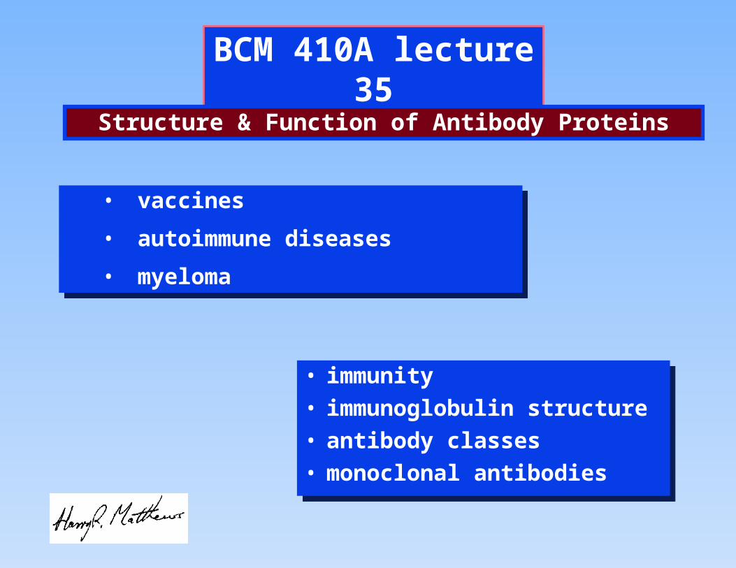
BCM 410A lecture 35
• immunity
• immunoglobulin structure
• antibody classes
• monoclonal antibodies
• immunity
• immunoglobulin structure
• antibody classes
• monoclonal antibodies
• vaccines
• autoimmune diseases
• myeloma
• vaccines
• autoimmune diseases
• myeloma
Structure & Function of Antibody Proteins
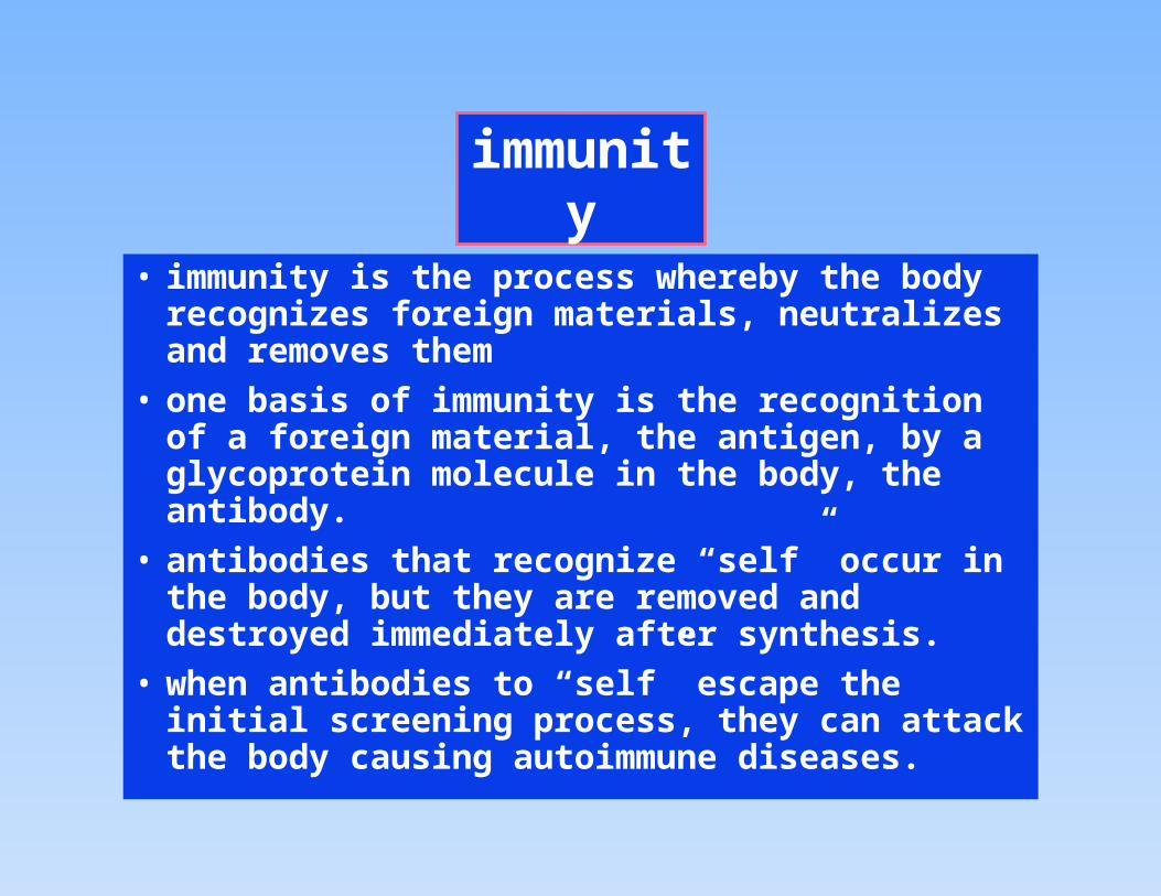
immunity
• immunity is the process whereby the body recognizes foreign materials, neutralizes and removes them
• one basis of immunity is the recognition of a foreign material, the antigen, by a glycoprotein molecule in the body, the antibody.
• antibodies that recognize “self” occur in the body, but they are removed and destroyed immediately after synthesis.
• when antibodies to “self” escape the initial screening process, they can attack the body causing autoimmune diseases.
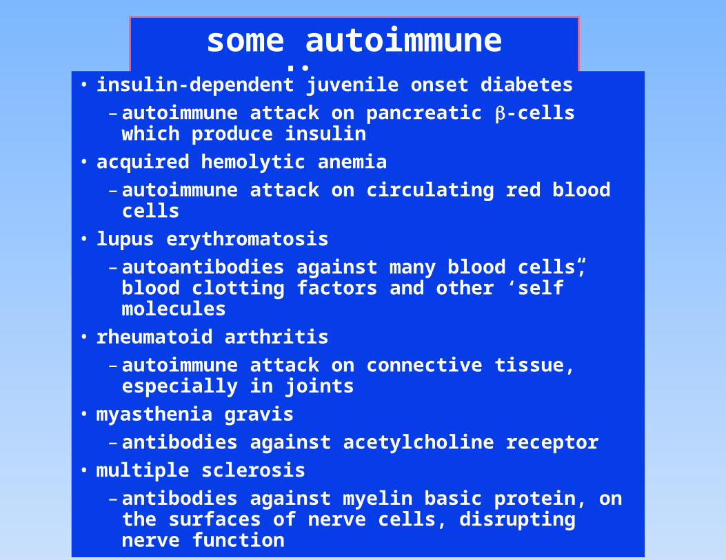
some autoimmune diseases• insulin-dependent juvenile onset diabetes
– autoimmune attack on pancreatic -cells which produce insulin
• acquired hemolytic anemia
– autoimmune attack on circulating red blood cells
• lupus erythromatosis
– autoantibodies against many blood cells, blood clotting factors and other ‘self” molecules
• rheumatoid arthritis
– autoimmune attack on connective tissue, especially in joints
• myasthenia gravis
– antibodies against acetylcholine receptor
• multiple sclerosis
– antibodies against myelin basic protein, on the surfaces of nerve cells, disrupting nerve function

The immune response
Unstimulated
IgM
IgG
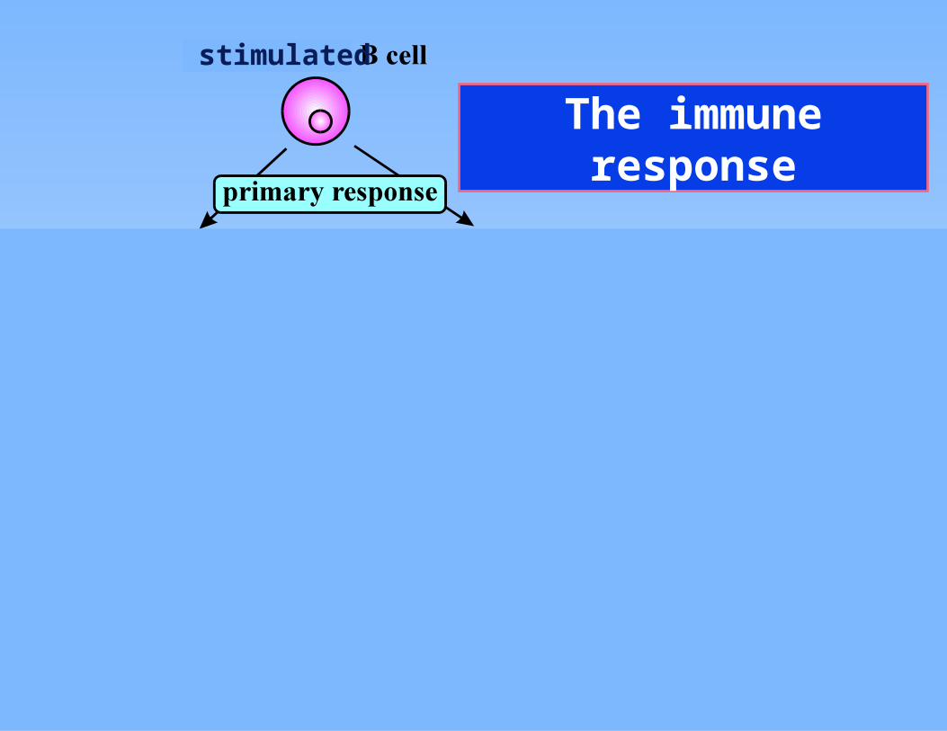
The immune response
stimulated
IgM
IgG
The immune response
IgM
IgG
stimulated
The immune response
IgM
IgG
stimulated
The immune response
IgM
IgG
stimulated
The immune response
IgM
IgG
stimulated
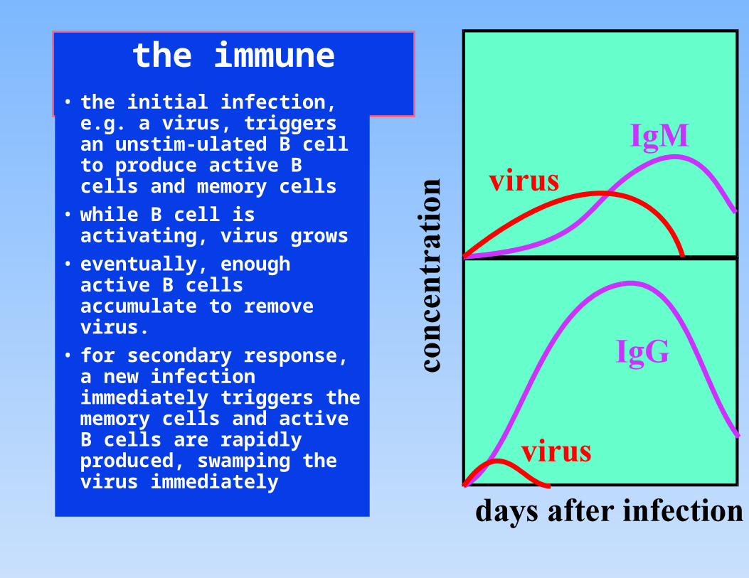
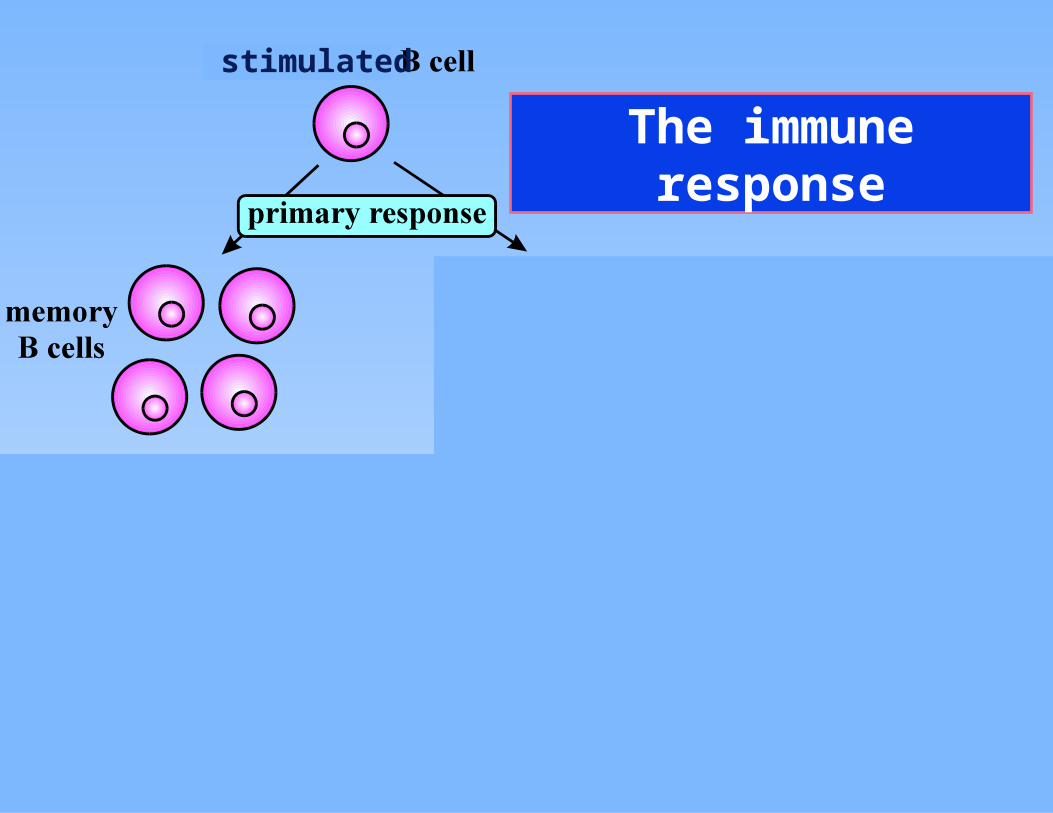
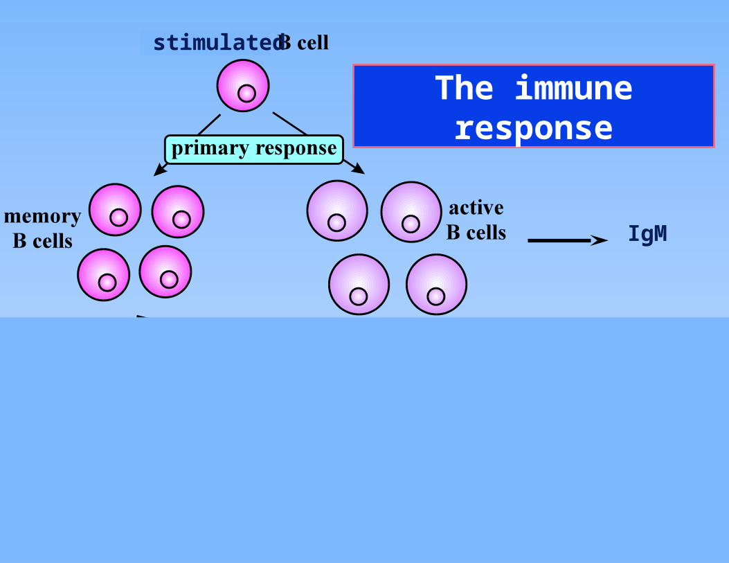
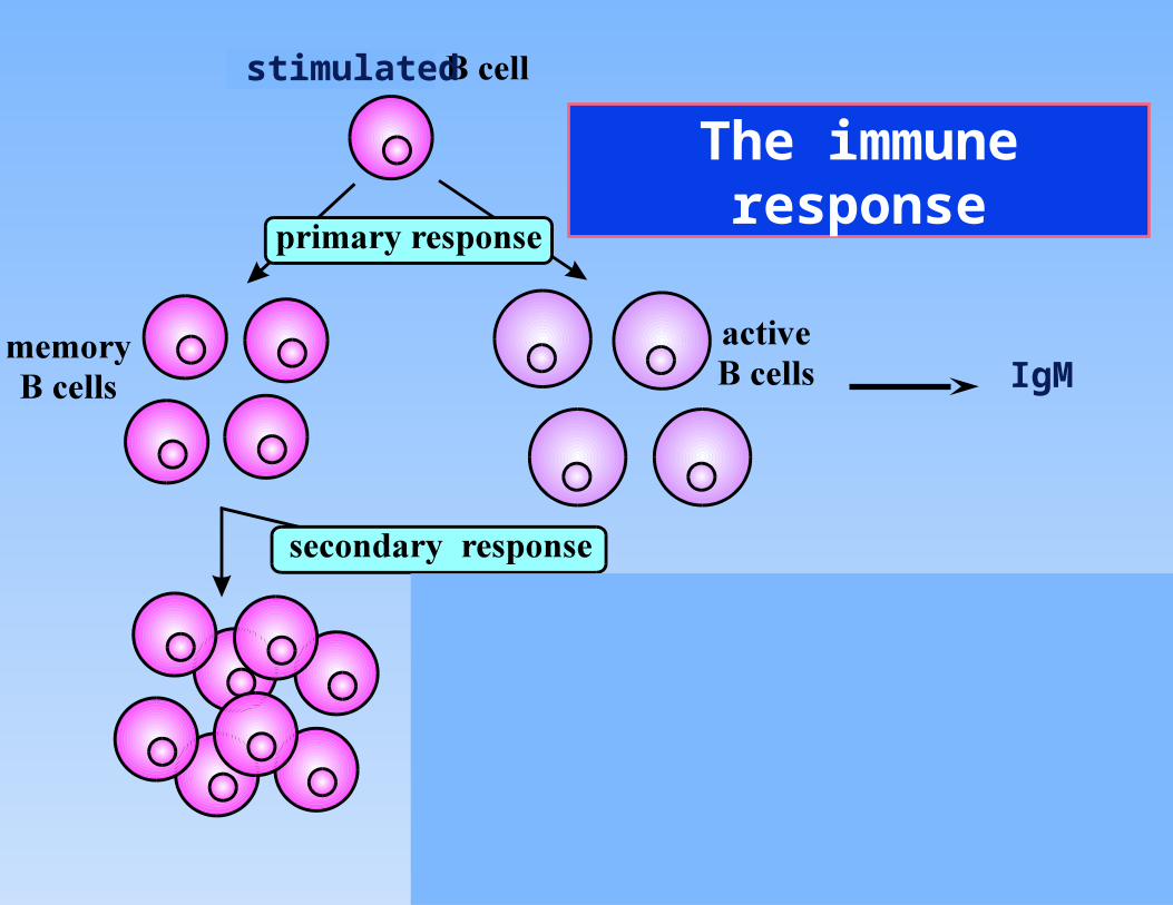
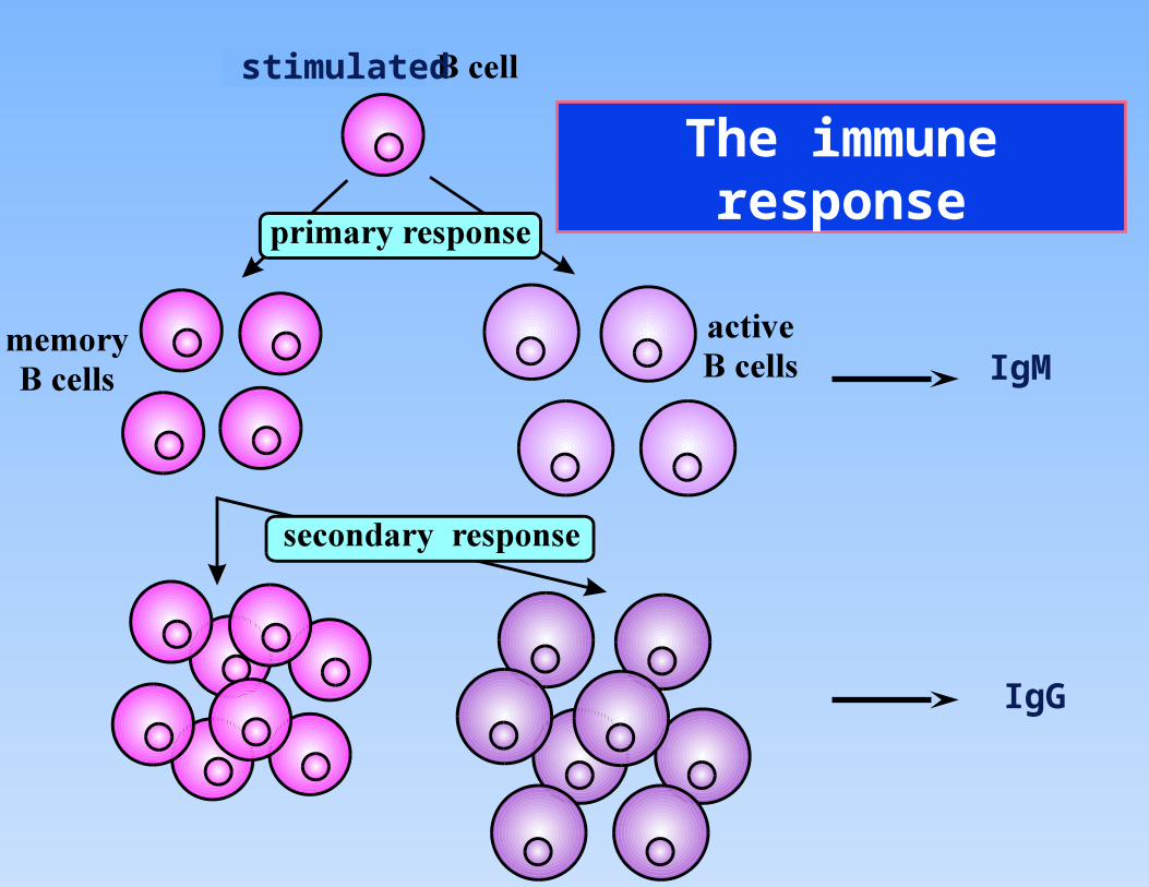
the immune response• the initial infection, e.g. a
virus, triggers an unstim-ulated B cell to produce active B cells and memory cells
• while B cell is activating, virus grows
• eventually, enough active B cells accumulate to remove virus.
• for secondary response, a new infection immediately triggers the memory cells and active B cells are rapidly produced, swamping the virus immediately
Monoclonal antibodies
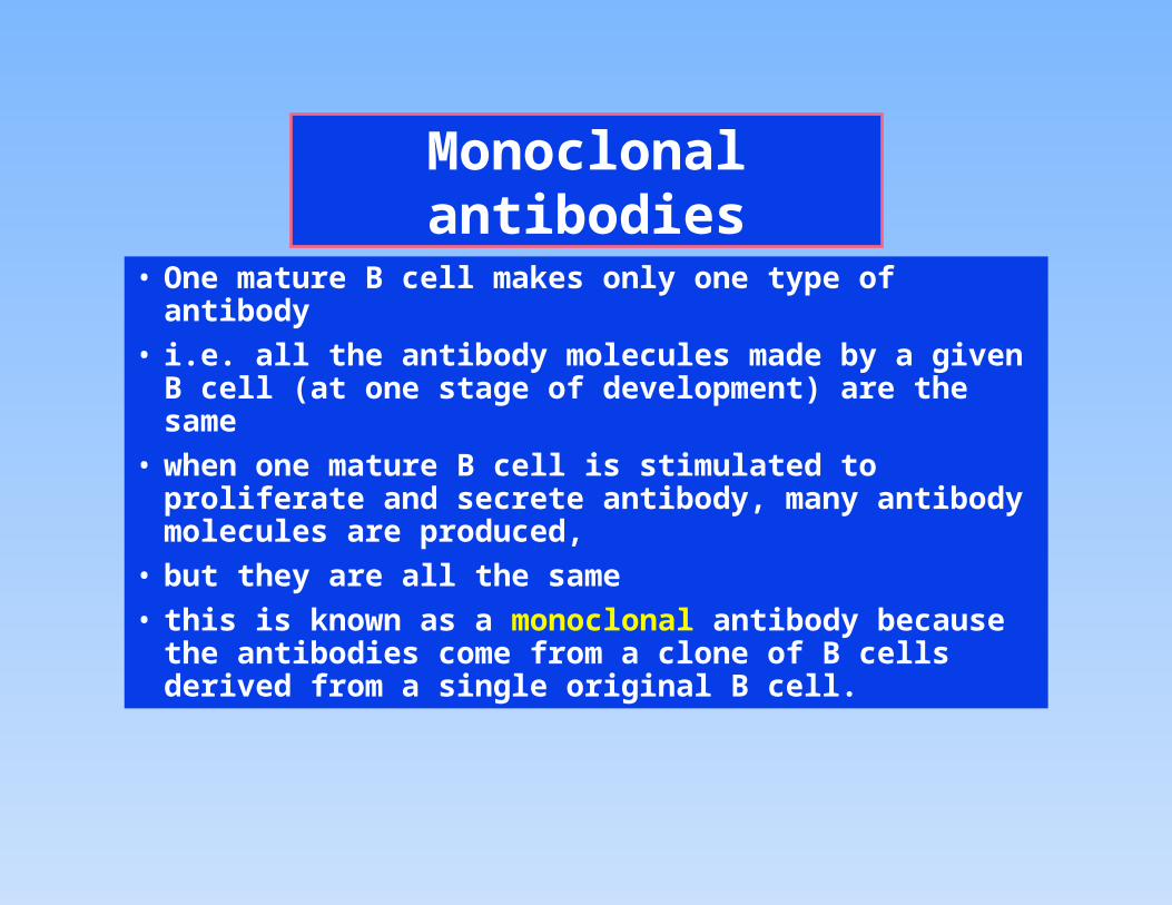
• One mature B cell makes only one type of antibody
• i.e. all the antibody molecules made by a given B cell (at one stage of development) are the same
• when one mature B cell is stimulated to proliferate and secrete antibody, many antibody molecules are produced,
• but they are all the same
• this is known as a monoclonal antibody because the antibodies come from a clone of B cells derived from a single original B cell.
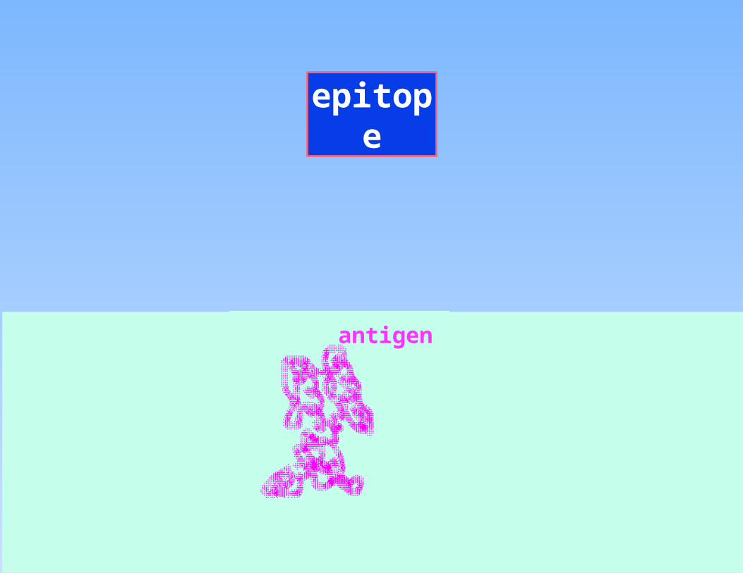
epitope
antigen
epitope
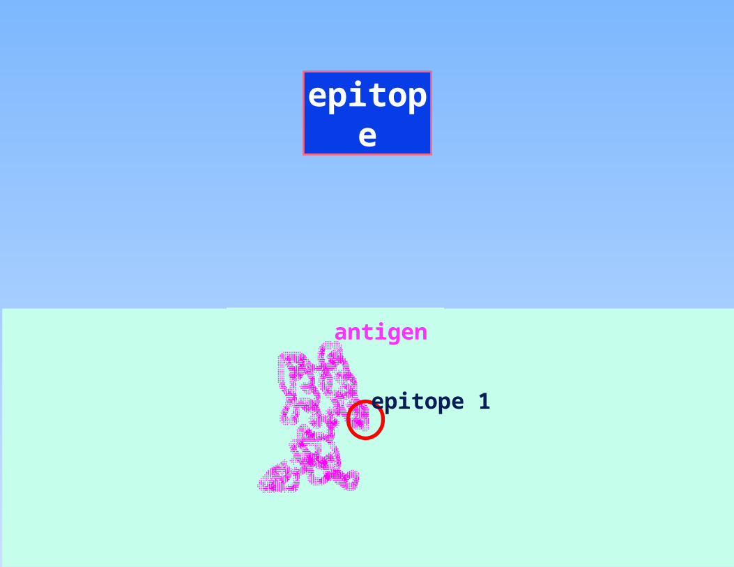
epitope 1
antigen
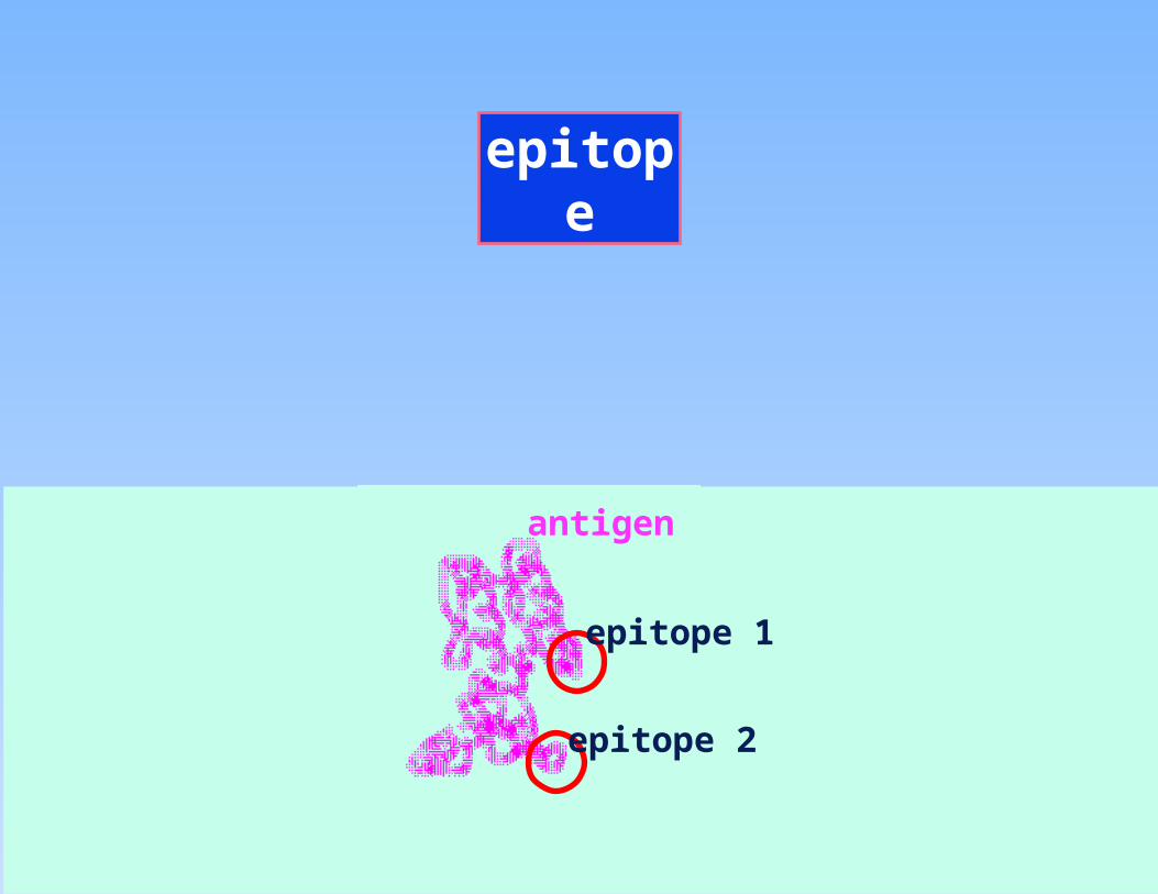
epitope
epitope 1
epitope 2
antigen
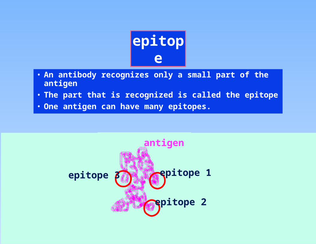
epitope
• An antibody recognizes only a small part of the antigen
• The part that is recognized is called the epitope
• One antigen can have many epitopes.
epitope 1
epitope 2
epitope 3
antigen
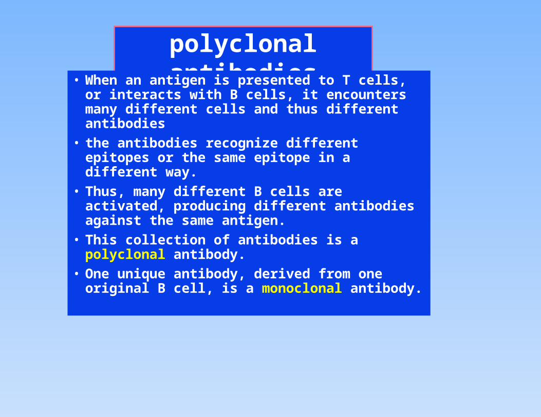
polyclonal antibodies
• When an antigen is presented to T cells, or interacts with B cells, it encounters many different cells and thus different antibodies
• the antibodies recognize different epitopes or the same epitope in a different way.
• Thus, many different B cells are activated, producing different antibodies against the same antigen.
• This collection of antibodies is a polyclonal antibody.
• One unique antibody, derived from one original B cell, is a monoclonal antibody.
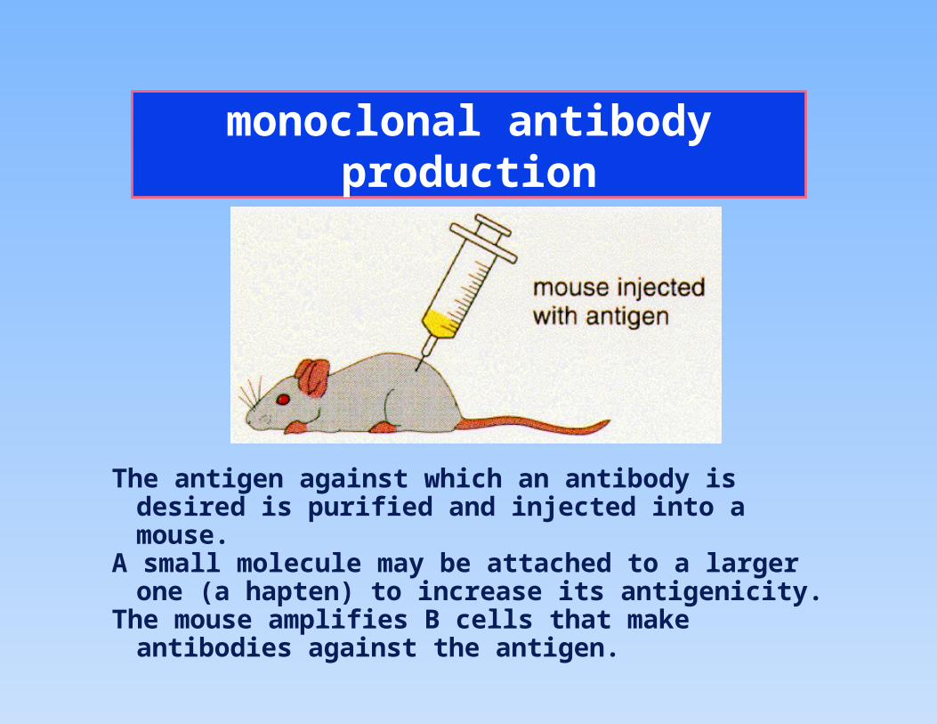
monoclonal antibody production
The antigen against which an antibody is desired is purified and injected into a mouse.
A small molecule may be attached to a larger one (a hapten) to increase its antigenicity.
The mouse amplifies B cells that make antibodies against the antigen.

monoclonal antibody production
B cells (including those making the antibodies) are removed and placed in cell culture in HAT medium.
The B cells survive but stop growing and dividing.
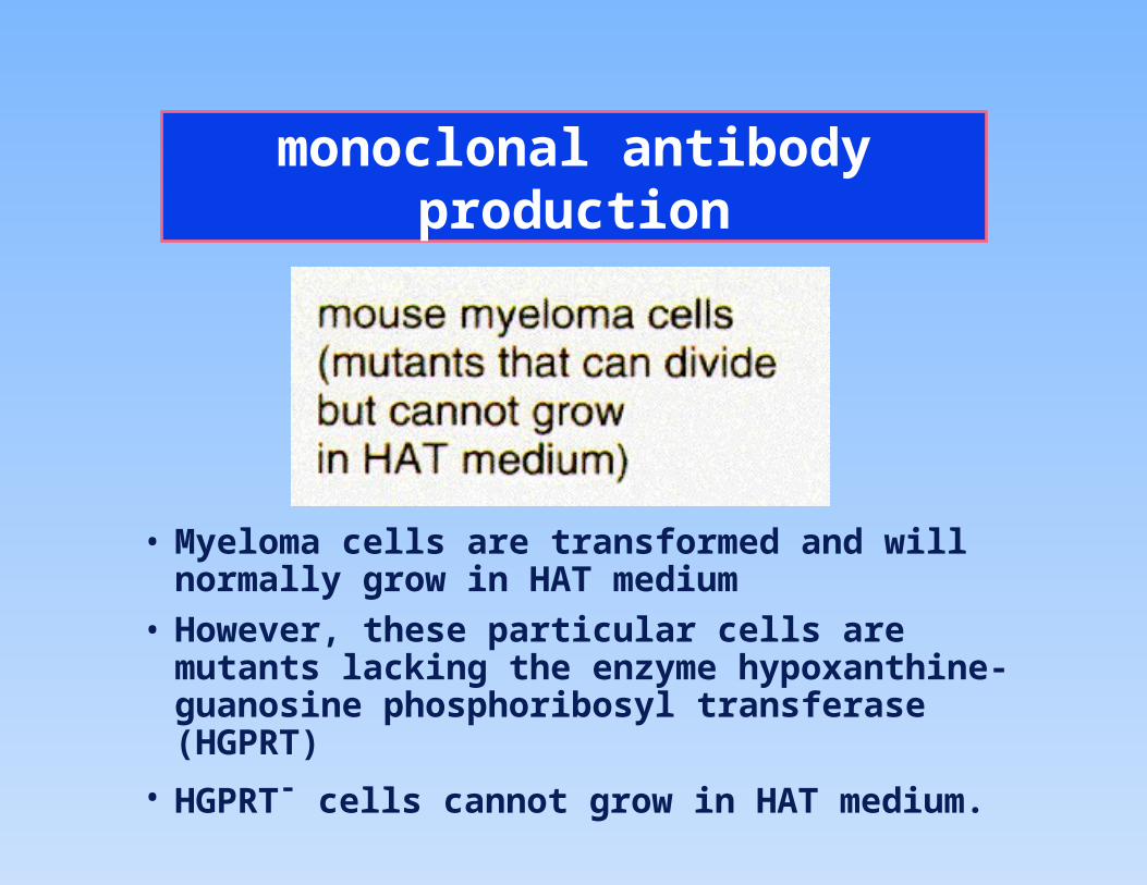
monoclonal antibody production
• Myeloma cells are transformed and will normally grow in HAT medium
• However, these particular cells are mutants lacking the enzyme hypoxanthine-guanosine phosphoribosyl transferase (HGPRT)
• HGPRT- cells cannot grow in HAT medium.
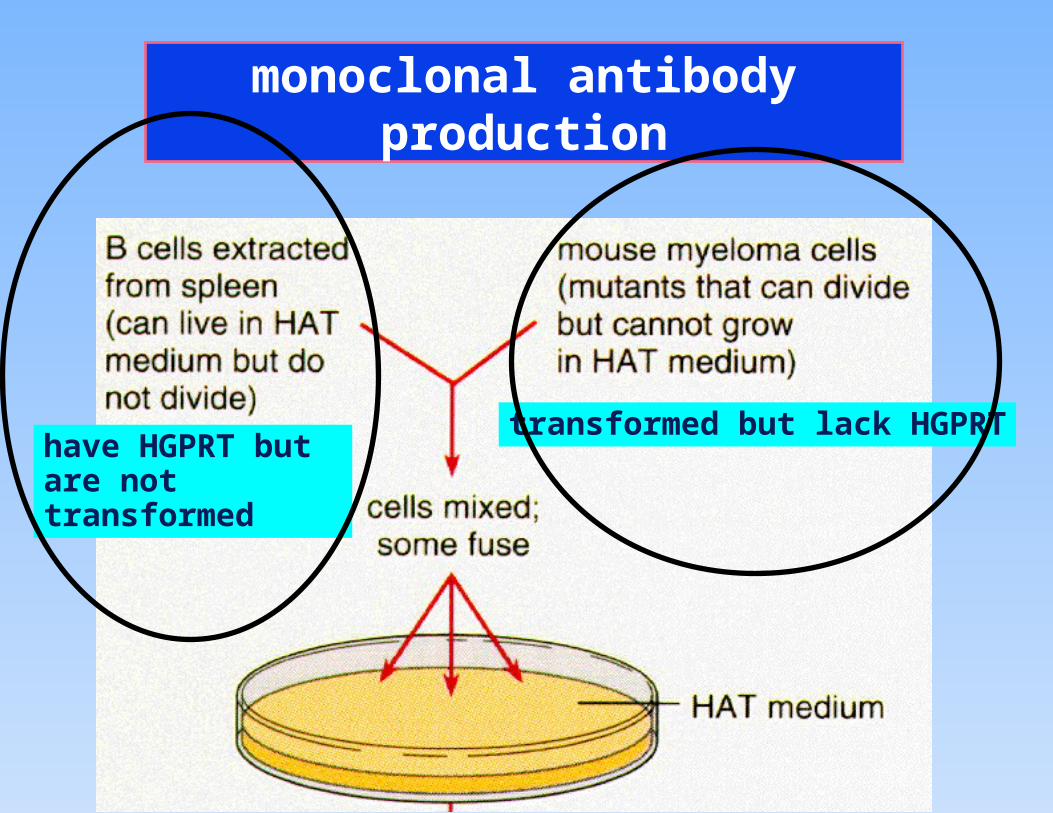
monoclonal antibody production
have HGPRT but are not transformed
transformed but lack HGPRT

monoclonal antibody production
In order to grow in HAT medium, cells must be both transformed and contain HGPRT
Only fused cells will grow
Fused cells will synthesize antibodies.
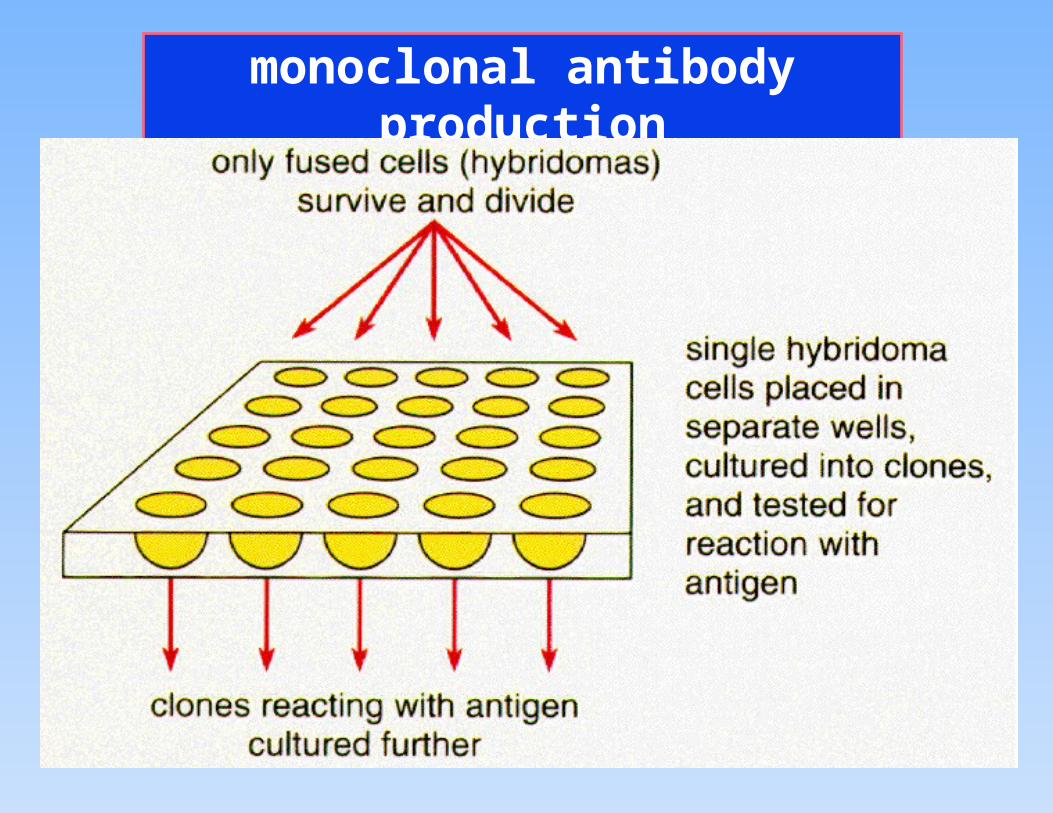
monoclonal antibody production
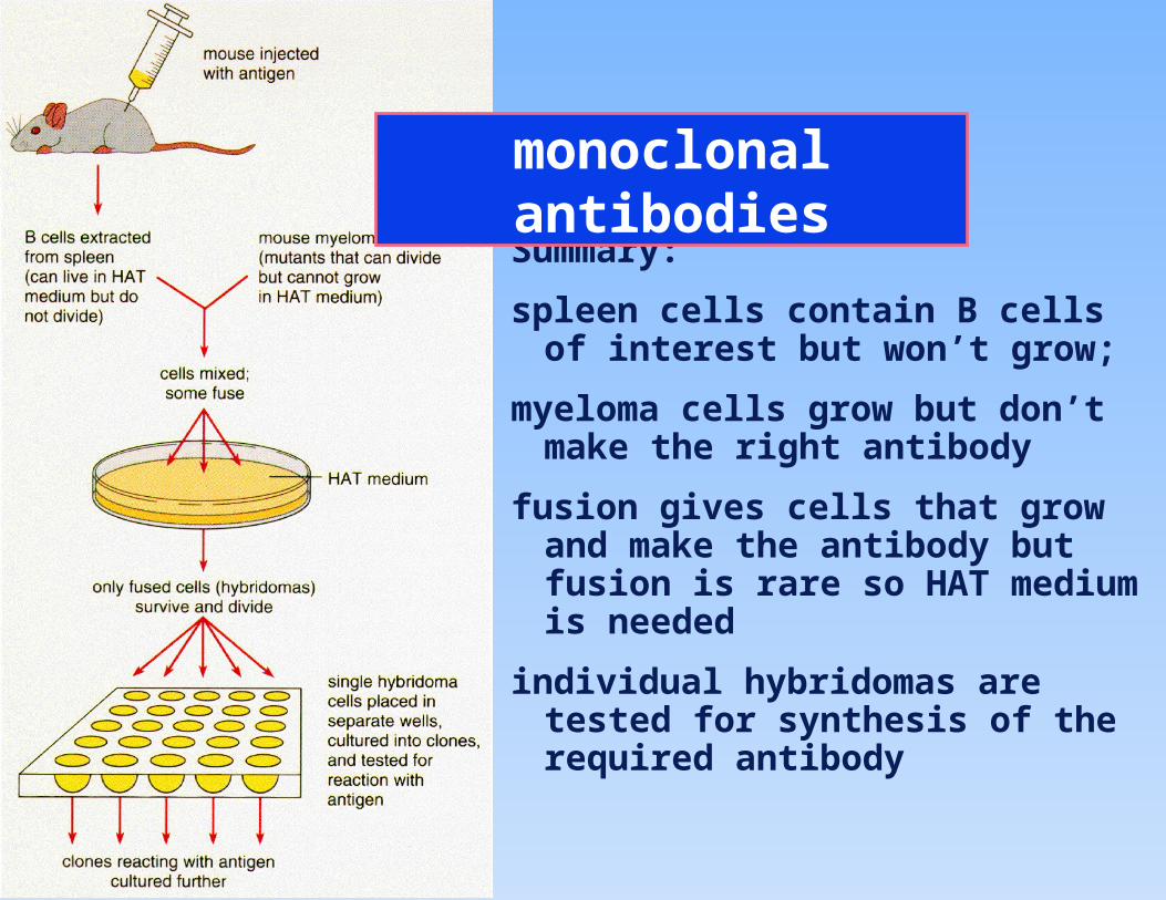
Summary:
spleen cells contain B cells of interest but won’t grow;
myeloma cells grow but don’t make the right antibody
fusion gives cells that grow and make the antibody but fusion is rare so HAT medium is needed
individual hybridomas are tested for synthesis of the required antibody
monoclonal antibodies
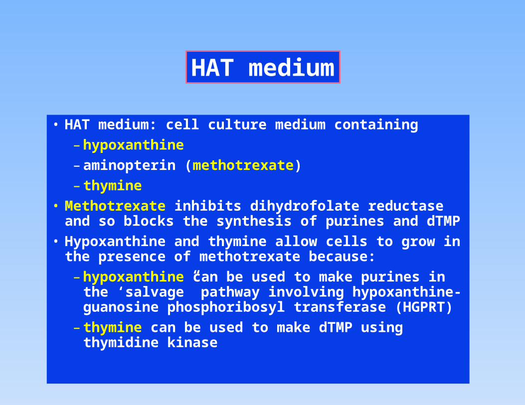
HAT medium
• HAT medium: cell culture medium containing
– hypoxanthine
– aminopterin (methotrexate)
– thymine
• Methotrexate inhibits dihydrofolate reductase and so blocks the synthesis of purines and dTMP
• Hypoxanthine and thymine allow cells to grow in the presence of methotrexate because:
– hypoxanthine can be used to make purines in the ‘salvage” pathway involving hypoxanthine-guanosine phosphoribosyl transferase (HGPRT)
– thymine can be used to make dTMP using thymidine kinase
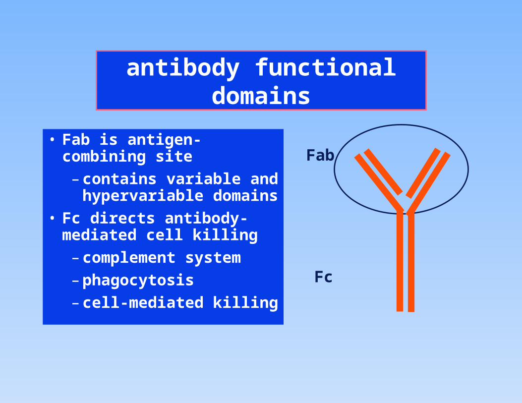
antibody functional domains
• Fab is antigen-combining site
– contains variable and hypervariable domains
• Fc directs antibody-mediated cell killing
– complement system
– phagocytosis
– cell-mediated killing
Fab
Fc
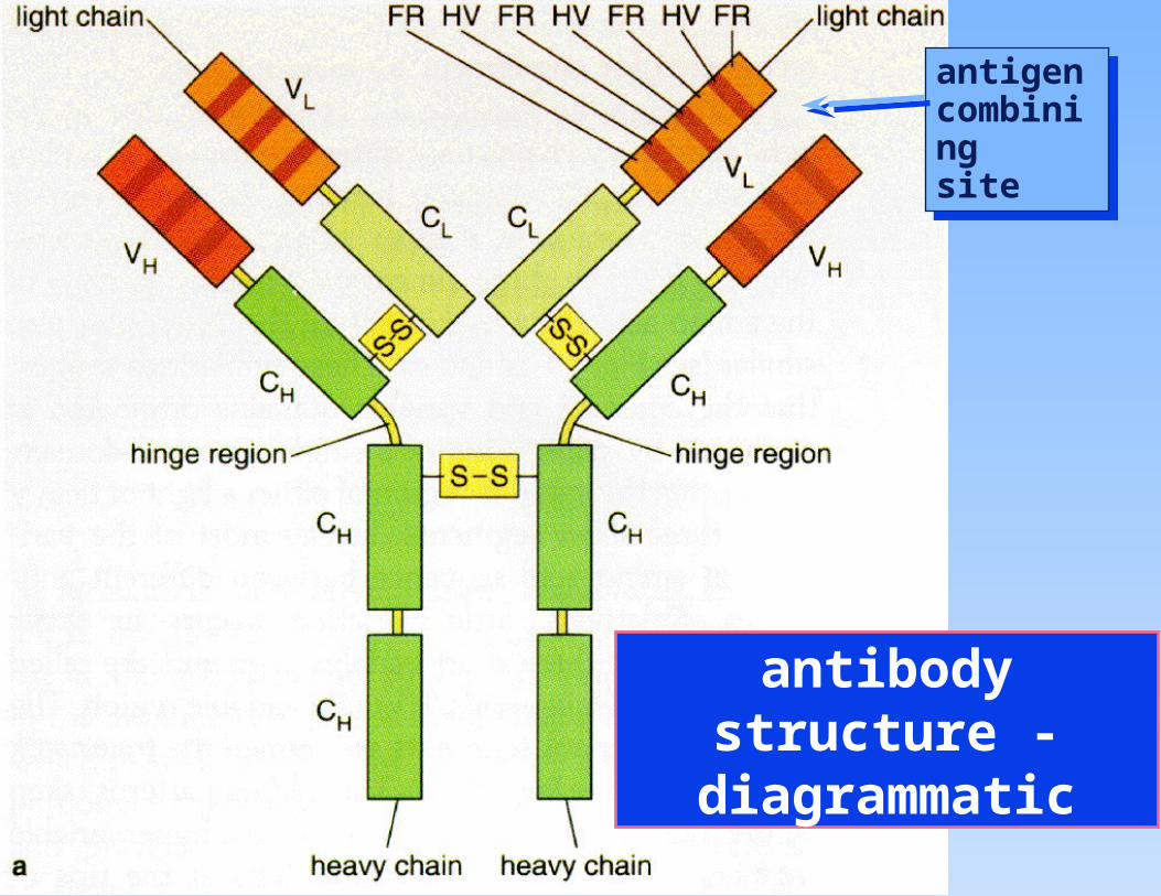
antibody structure - diagrammatic
antigen combiningsite
antigen combiningsite

antibody structure
• antibodies are glycoproteins
• each antibody contains 2 heavy chains, two light chains (polypeptides) and one carbohydrate chain
• the primary structure shows extensive regions of highly conserved sequence and other regions which are variable or highly variable
• the secondary structure is mostly -sheet
• the tertiary structure is a folded -sheet structure known as the immunoglobulin fold which occurs 12 times in one immunoglobulin
• the quaternary structure is maintained by multiple interactions, including S-S bonds.
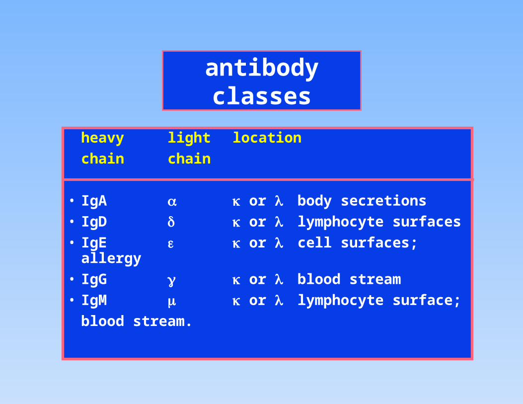
antibody classes
heavy light location
chain chain
• IgA or body secretions
• IgD or lymphocyte surfaces
• IgE or cell surfaces; allergy
• IgG or blood stream
• IgM or lymphocyte surface;
blood stream.
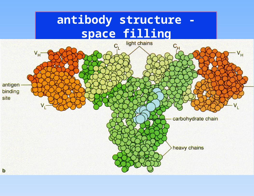
antibody structure - space filling
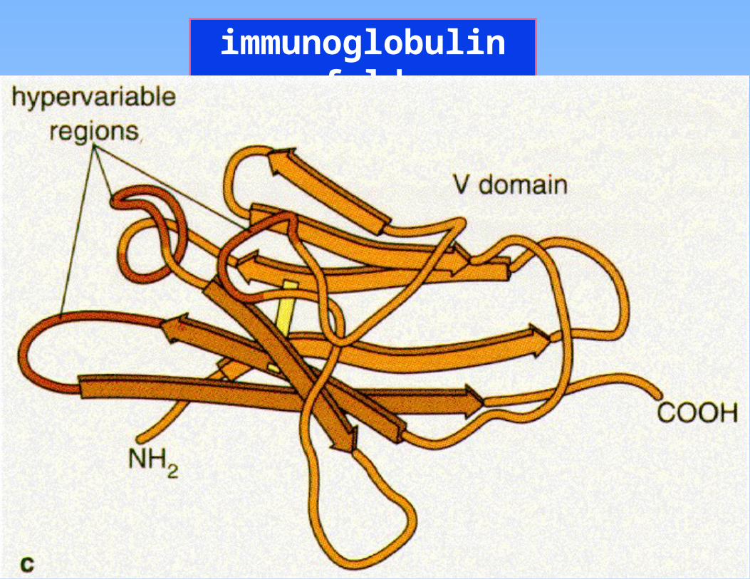
immunoglobulin fold
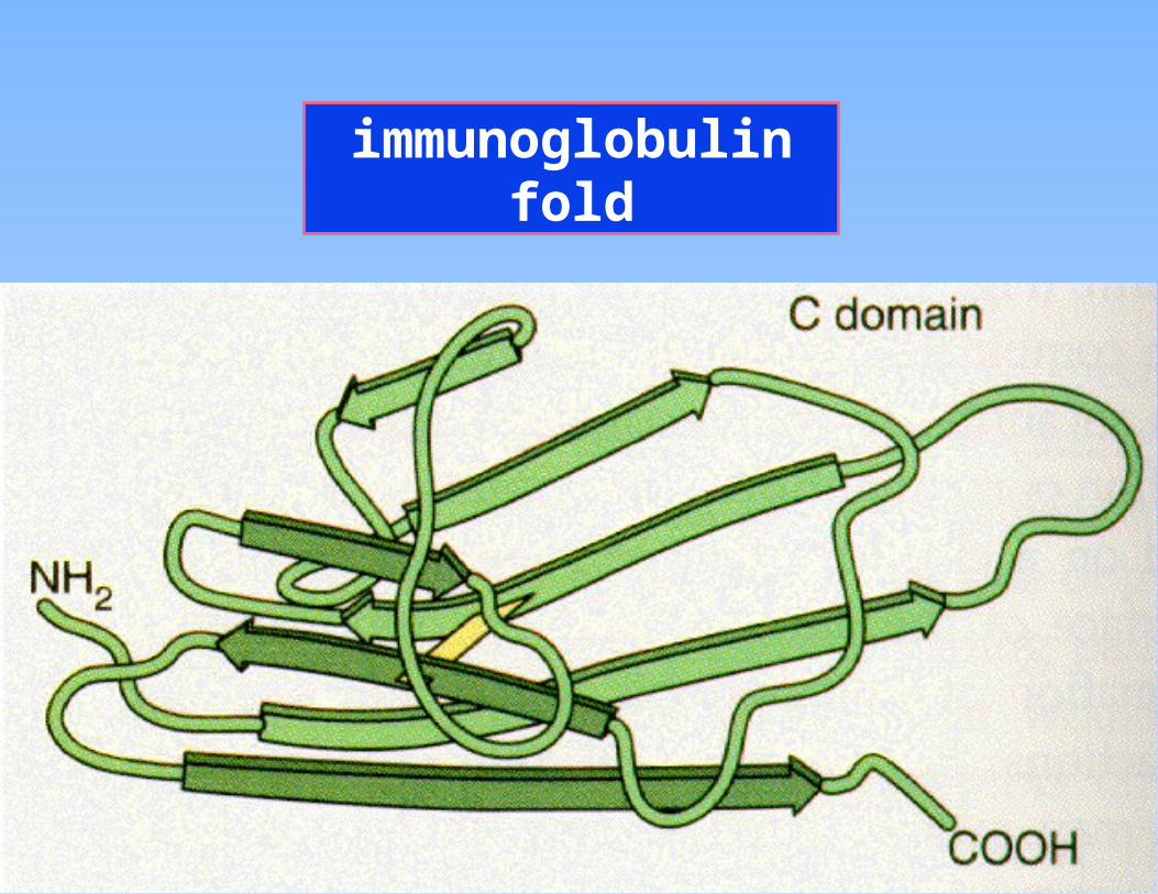
immunoglobulin fold
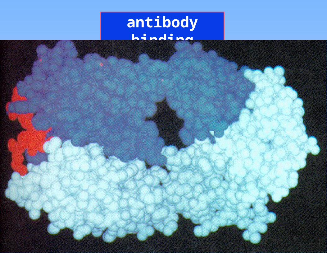
antibody binding

Office hours etc.
• For the rest of the quarter, normally noon to 12:45 and 4:30 to 5:30 every
weekday except Monday noon.
• Room 4125, MS1A
• email: [email protected]
• ‘phone 752-3570

BCM 410A lecture 35
• immunity
• immunoglobulin structure
• antibody classes
• monoclonal antibodies
• immunity
• immunoglobulin structure
• antibody classes
• monoclonal antibodies
• vaccines
• autoimmune diseases
• myeloma
• vaccines
• autoimmune diseases
• myeloma
Structure & Function of Antibody Proteins