monoclonal immunoglobulin deposition disease, : an update
TRANSCRIPT
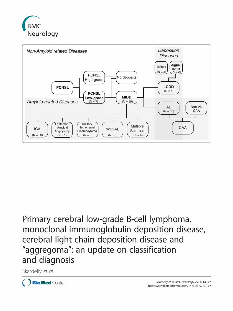
PCNSL
PCNSLHigh-grade
PCNSLLow-grade MIDD
LCDD
AL
No deposits
CAAMultiple Sclerosis
Leptomen. Amyloid
Angiopathy
Solitary Intracranial
Plasmocytoma WSVAL
Non-ALCAA
DepositionDiseases
Amyloid related Diseases
Non-Amyloid related Diseases
(N = 1)
(N = 2)(N = 3)
(N = 6)(N = 3)(N = 8)
(N = 50)
(N = 5)
ICA
(N = 30)
(N = 55)
Aggre-goma
Diffuse
(N = 7)
Primary cerebral low-grade B-cell lymphoma,monoclonal immunoglobulin deposition disease,cerebral light chain deposition disease and“aggregoma”: an update on classificationand diagnosisSkardelly et al.
Skardelly et al. BMC Neurology 2013, 13:107http://www.biomedcentral.com/1471-2377/13/107
Skardelly et al. BMC Neurology 2013, 13:107http://www.biomedcentral.com/1471-2377/13/107
CASE REPORT Open Access
Primary cerebral low-grade B-cell lymphoma,monoclonal immunoglobulin deposition disease,cerebral light chain deposition disease and“aggregoma”: an update on classificationand diagnosisMarco Skardelly1*, Georgios Pantazis2, Sotirios Bisdas3, Guenther C Feigl1, Martin U Schuhmann1,Marcos S Tatagiba1 and Rainer Ritz1
Abstract
Background: This work aims to add evidence and provide an update on the classification and diagnosis ofmonoclonal immunoglobulin deposition disease (MIDD) and primary central nervous system low-grade lymphomas.MIDD is characterized by the deposition of light and heavy chain proteins. Depending on the spatial arrangementof the secreted proteins, light chain-derived amyloidosis (AL) can be distinguished from non-amyloid light chaindeposition disease (LCDD). We present a case of an extremely rare tumoral presentation of LCDD (aggregoma) andreview the 3 previously published LCDD cases and discuss their presentation with respect to AL.
Case presentation: A 61-year-old woman presented with a 3½-year history of neurologic symptoms due to aprogressive white matter lesion of the left subcortical parieto-insular lobe and basal ganglia. 2 former stereotacticbiopsies conducted at different hospitals revealed no evidence of malignancy or inflammation; thus, no therapyhad been initiated. After performing physiological and functional magnetic resonance imaging (MRI), the tumorwas removed under intraoperative monitoring at our department. Histological analysis revealed large amorphousdeposits and small islands of lymphoid cells.
Conclusion: LCCD is a very rare and obscure manifestation of primary central nervous system low-grade lymphomasthat can be easily misdiagnosed by stereotactic biopsy sampling. If stereotactic biopsy does not reveal a definite result,a “wait-and-see” strategy can delay possible therapy for this disease. The impact of surgical removal, radiotherapy andchemotherapy in LCDD obviously remains controversial because of the low number of relevant cases.
Keywords: Aggregoma, Light chain deposition disease, Lymphoma, Monoclonal immunoglobulin deposition disease,Neurooncology, Primary central nervous system lymphoma, Stereotaxic surgery
* Correspondence: [email protected] of Neurosurgery, University Hospital Tuebingen,Hoppe-Seyler-Str. 3, 72076, Tuebingen, Baden-Wuerttemberg, GermanyFull list of author information is available at the end of the article
© 2013 Skardelly et al.; licensee BioMed Central Ltd. This is an Open Access article distributed under the terms of the CreativeCommons Attribution License (http://creativecommons.org/licenses/by/2.0), which permits unrestricted use, distribution, andreproduction in any medium, provided the original work is properly cited.
Skardelly et al. BMC Neurology 2013, 13:107 Page 2 of 9http://www.biomedcentral.com/1471-2377/13/107
BackgroundPrimary central nervous system lymphomas (PCNSL)are defined as non-Hodgkin’s (NHL) lymphomas thatprimarily arise in the central nervous system [1]. PCNSLaccount for approximately 1–2% of all primary cerebraltumors, and approximately 98–99% are classified as diffuseB-cell lymphoma (analogous to systemic B-cell NHL) [2,3].Intracerebral manifestations of T-cell lymphomas and sec-ondary lymphomas are extremely rare [3]. Low-gradePCNSL represents a less aggressive subgroup comparedwith systemic NHL and accounts for approximately 3–20%of all PCNSL [4,5]. Only a few low-grade PCNSL areassociated with the deposition of monoclonal light andheavy chain immunoglobulins (Ig).Monoclonal immunoglobulin deposition disease (MIDD)
is characterized by the deposition of monotypic light and/orheavy chain proteins in various tissues and organs. MIDDmainly affects the kidneys, but the involvement of otherorgans (e.g., the liver, heart and peripheral nerves) is notuncommon [6]. All forms of MIDD can be ascribed tomonoclonal expansion of an immunoglobulin (Ig) lightand/or heavy chain producing B-cells [7]. 2 subgroupsof MIDD can be differentiated histologically based onthe different spatial arrangement of the secreted proteins.In the more common subgroup, the light chain-derivedamyloidosis (AL) subgroup, proteins are aggregated infibrils to ß-pleated sheets that stain for Congo red anddisplay green birefringence under polarized light [8]. Thesecond subgroup is characterized by ultrastructural non-organized proteins, which aggregate in more amorphousCongo red-negative depositions. Randall and colleaguesinitially described 2 patients with the systemic depositionof non-amyloid Ig light chains and proposed the termlight chain deposition disease (LCDD) [9]. Subsequentreports confirmed the existence of systemic heavy chaindeposition disease (HCDD) as well as both light and heavychain deposition disease (LHCDD) [10].We provide an update regarding the diagnosis and
classification of primary cerebral low-grade B-celllymphomas and cerebral light chain deposit diseases.We present the case of a patient with a 3½-year progressivehemiparesis and hemi-hypoesthesia of the right side due toa delayed diagnosis and therapy of the extremely rare,tumor-presenting cerebral restricted LCDD, which can becalled “aggregoma” [11]. We further present a systematicoverview and discussion of the disease with respect to lightchain-derived amyloidosis.
Case presentationClinical presentationA 61-year-old woman was admitted to our departmentwith progressive brachiofacially accentuated hemiparesis,dysdiadochokinesia and hemi-hypoesthesia of the right sideof the body, which began 3½ years previous. She initially
presented with dyspraxia and fluctuating hypoesthesia ofthe right hand at the end of 2006; her cranial nerves werenot affected. She complained of increased fatigue butdid not present with weight loss, night sweats, fever orheadache. The woman had a history of hypothyroidismrelated to Hashimoto’s thyroiditis and suffered endocarditisand streptococcal sepsis in 1982. She was under a long-term medication treatment of 100 μg of thyroxin daily. AnMRI scan performed in May 2007 revealed a 2.8 × 2.0 ×2.4 cm lesion of the white matter at the level of theleft subcortical parieto-insular lobe and basal ganglia(Figure 1a). The lesion presented as hypointense onT1-weighted scans with some regions displaying slightenhancement after gadolinium administration and moder-ate inhomogeneous hyperintensity on T2-weighted scans.Blood serum inflammatory markers (leukocytes and CRP),cerebrospinal fluid protein, and the cell count revealed noabnormalities. A stereotactic serial biopsy was performedin June 2007. The histological analysis demonstratedcolloidal-bodied particles with scattered single cells withno proof of tumors or inflammatory cells. Based onthe obvious diagnosis of an atypical colloidal cyst, nofurther therapy was initiated. The first control MRI(3 months after biopsy) demonstrated no progression of thedisease. However, a clinical deterioration and progression ofthe lesion (3.6 × 2.3 × 2.6 cm) (Figure 1b & 1c) at thefollow-up MRI session in July 2008 warranted a stereotacticserial biopsy, which was conducted at a neurosurgical clinicthat specialized in stereotactic procedures. Prior to thestereotactic surgery, CSI (chemical shift imaging) wasperformed, which revealed a prominently reduced NAApeak, an elevation of lactate and only slight alterations ofcholine and creatine peaks (Figure 1d & 1e).The histological analysis of the biopsy that was performed
in June 2007 showed a glial cyst but exhibited no evidenceof malignancy or inflammation. In May 2010, the patientpresented for the first time at our department because ofclinical and radiological progression of the disease. Surgicalresection of the lesion with a subsequent histologicaldiagnosis was recommended At the day of admission, thepatient displayed poorer health and a worsened nutritionalstatus. The neurological examination demonstrated mod-erate hemiparesis of the right side and hemi-hypoesthesia,including the trigeminal nerve, with no other cranial nervedeficits. The woman had a high-grade disturbance of thefine motor skills of the right hand, dysdiadochokinesiaof the right side and displayed an instable standingposture and gait. Her reflexes exhibited increasedpositive Troemner’s and Babinski’s signs. On the dayof admission, a control MRI that included diffusiontensor imaging and a functional MRI were performed.The lesion progressed in volume to 4.2 × 3.2 × 5.3 cm(Figure 2a) with the same signal characteristics in T1- andT2-weighted imaging; displacement of the left pyramidal
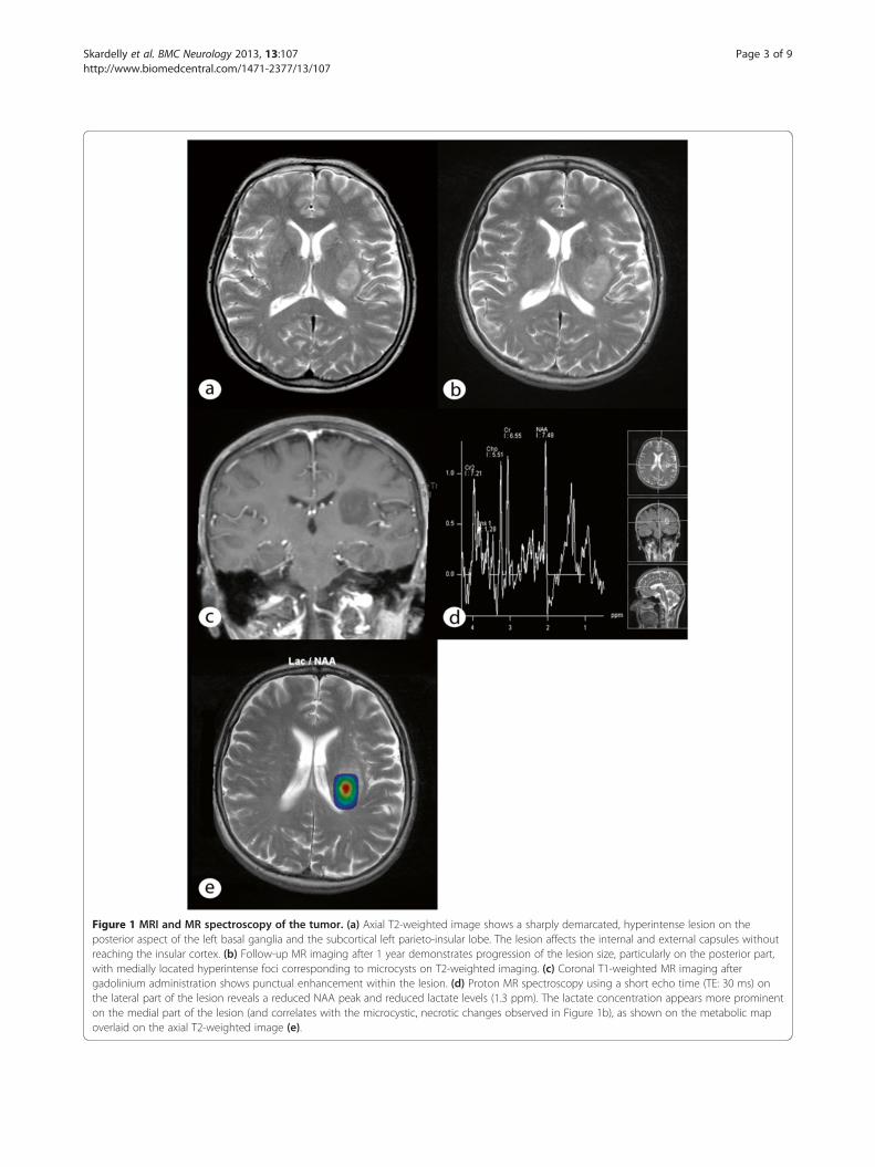
Figure 1 MRI and MR spectroscopy of the tumor. (a) Axial T2-weighted image shows a sharply demarcated, hyperintense lesion on theposterior aspect of the left basal ganglia and the subcortical left parieto-insular lobe. The lesion affects the internal and external capsules withoutreaching the insular cortex. (b) Follow-up MR imaging after 1 year demonstrates progression of the lesion size, particularly on the posterior part,with medially located hyperintense foci corresponding to microcysts on T2-weighted imaging. (c) Coronal T1-weighted MR imaging aftergadolinium administration shows punctual enhancement within the lesion. (d) Proton MR spectroscopy using a short echo time (TE: 30 ms) onthe lateral part of the lesion reveals a reduced NAA peak and reduced lactate levels (1.3 ppm). The lactate concentration appears more prominenton the medial part of the lesion (and correlates with the microcystic, necrotic changes observed in Figure 1b), as shown on the metabolic mapoverlaid on the axial T2-weighted image (e).
Skardelly et al. BMC Neurology 2013, 13:107 Page 3 of 9http://www.biomedcentral.com/1471-2377/13/107

Figure 2 Preoperative MRI and fiber tracking. (a) Coronal T1-weighted image after gadolinium administration shows lesion progression.(b) The fiber tracking imaging based on the preoperatively performed diffusion tensor imaging demonstrates displacement of the corticospinaland corticopontine tracts on the left side, whereas the body of the corpus callosum seems unaffected.
Skardelly et al. BMC Neurology 2013, 13:107 Page 4 of 9http://www.biomedcentral.com/1471-2377/13/107
and corticopontine tract was observed (Figure 2b) withsignificantly altered values of the fractional anisotropycompared with the healthy contralateral side.
Intervention and postoperative courseThe patient underwent a left-sided standard pterionalcraniotomy and a removal of the lesion via a transsylvian,transinsular approach (with respect to the displacement ofthe left pyramidal and corticopontine tracts) undercontinuous intraoperative monitoring with sensoryevoked and motor evoked potentials (SEPs and MEPs,respectively). The lesion appeared intraoperatively asan amorphous, gelatinous mass with some regionsappearing dull and lucid (Figure 3). The tumor mass couldbe almost completely removed with the preservation ofthe MEPs and SEPs. After the operation, the patientexhibited increased hemiparesis of the right side withhemiplegia of the right arm and partial facial palsy in
Figure 3 Intraoperative microscopic image. The image revealsthe amorphous, gelatinous mass of the tumor.
addition to a new expressive aphasia. A postoperative headCT revealed no signs of bleeding or infarction. By the timeof discharge, the patient had partially recovered fromaphasia and was mobilized with a Zimmer frame. In thefollow-up control in the outpatients’ department afterrehabilitation in October 2010, the patient demonstratedalmost complete recovery from expressive aphasia andhad slight palsy of the marginal mandibular branch of thefacial nerve but continued to display moderate brachiallyaccentuated hemiparesis of the right side. Subsequently,the department of hematooncology initiated diagnosticstaging, and no further manifestations of B-cell lymphomacould be identified. Whole-body CT, abdominal ultrasoundand bone marrow puncture revealed no suspicious findings.Electrophoresis showed a regular distribution and quantityof serum proteins (7.6 g/dl) and immunoglobulins.Additionally, there was no hint of monoclonality ofserum proteins in the immunofixation assay. Physiologicalalbuminuria in urine electrophoresis was observed.
NeuropathologyHistological examination of the resected tumor revealedlarge, amorphous, proteinaceous eosinophilic deposits andsmall islands of lymphoid cells between them (Figure 4a).The amorphous deposits were strongly PAS positive butnegative for Congo red staining and did not display apple-green birefringence under polarization (Figure 4b). Mostof the small lymphoid cells expressed the B-cell markerCD20 (Figure 4c). There were also few CD3-positiveT-cell lymphocytes (Figure 4d). Immunoreactivities forthe Ig-A heavy chain (Figure 4e) and λ-light chainimmunoglobulins (Figure 4f ) were partially observedin the deposits, plasmacytoid lymphocytes and matureplasma cells. The plasmacytic cells expressed CD38and CD138, whereas they were negative for C79a. Therewas a negative reaction for IgM, IgG and IgE heavy chains
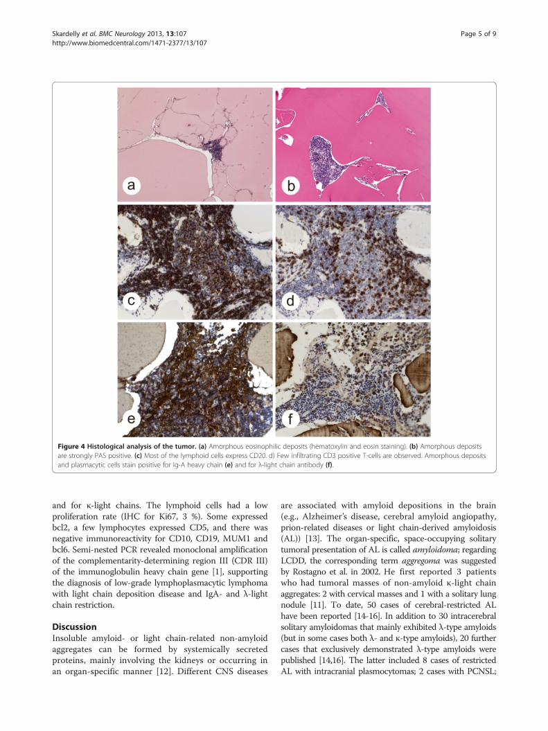
Figure 4 Histological analysis of the tumor. (a) Amorphous eosinophilic deposits (hematoxylin and eosin staining). (b) Amorphous depositsare strongly PAS positive. (c) Most of the lymphoid cells express CD20. d) Few infiltrating CD3 positive T-cells are observed. Amorphous depositsand plasmacytic cells stain positive for Ig-A heavy chain (e) and for λ-light chain antibody (f).
Skardelly et al. BMC Neurology 2013, 13:107 Page 5 of 9http://www.biomedcentral.com/1471-2377/13/107
and for κ-light chains. The lymphoid cells had a lowproliferation rate (IHC for Ki67, 3 %). Some expressedbcl2, a few lymphocytes expressed CD5, and there wasnegative immunoreactivity for CD10, CD19, MUM1 andbcl6. Semi-nested PCR revealed monoclonal amplificationof the complementarity-determining region III (CDR III)of the immunoglobulin heavy chain gene [1], supportingthe diagnosis of low-grade lymphoplasmacytic lymphomawith light chain deposition disease and IgA- and λ-lightchain restriction.
DiscussionInsoluble amyloid- or light chain-related non-amyloidaggregates can be formed by systemically secretedproteins, mainly involving the kidneys or occurring inan organ-specific manner [12]. Different CNS diseases
are associated with amyloid depositions in the brain(e.g., Alzheimer’s disease, cerebral amyloid angiopathy,prion-related diseases or light chain-derived amyloidosis(AL)) [13]. The organ-specific, space-occupying solitarytumoral presentation of AL is called amyloidoma; regardingLCDD, the corresponding term aggregoma was suggestedby Rostagno et al. in 2002. He first reported 3 patientswho had tumoral masses of non-amyloid κ-light chainaggregates: 2 with cervical masses and 1 with a solitary lungnodule [11]. To date, 50 cases of cerebral-restricted ALhave been reported [14-16]. In addition to 30 intracerebralsolitary amyloidomas that mainly exhibited λ-type amyloids(but in some cases both λ- and κ-type amyloids), 20 furthercases that exclusively demonstrated λ-type amyloids werepublished [14,16]. The latter included 8 cases of restrictedAL with intracranial plasmocytomas; 2 cases with PCNSL;

Skardelly et al. BMC Neurology 2013, 13:107 Page 6 of 9http://www.biomedcentral.com/1471-2377/13/107
1 case with leptomeningeal amyloid angiopathy; 6 caseswith multiple sclerosis; and 3 cases with widespreadsubcortical vascular amyloidosis with leukoencephalopathy(WSVAL). For further details, see the review by Schröderand colleagues [15]. By contrast, excluding our case, only 3intracerebral LCDD cases were previously reported: 2diffuse manifestations of λ-light chain aggregates[17,18] and 1 case of vascular presentation of non-amyloid λ-light chain aggregates, designated as cerebralLCDD vasculopathy (CLCDDV) [19]. For details aboutthese cases see “Additional file 1”.The case presented here is the first report on tumoral
presentation of a brain-restricted LCDD that can becalled an aggregoma. To create a better understanding ofthe relationships of the involved diseases they wereclassified in a pedigree (Figure 5); an overview of thedetails of the different manifestations of MIDD, ALand LCDD is provided in Table 1.In systemic amyloidosis or LCDD, the CNS is usually
not affected because of the blood–brain-barrier (BBB),which protects the brain sufficiently from circulating harm-ful metabolites (e.g., misfolded proteins) [20]. Nevertheless,54 cases of either brain-restricted amyloid or non-amyloiddeposition diseases, including our case, have been reportedthus far (Table 1). Most of the depositions were located inthe subcortical periventricular white matter, a preferredlocation for B-cell lymphoma [5]. Plasma cells wereassociated with deposits in many of the published caseswith proof of monoclonality in some cases, suggestingisolated intracerebral production of monoclonal immuno-globulins by monoclonal B-cell lymphocytes [15,19,21]. Itappears that the BBB exerts its function of controlling the
Figure 5 Schematic of classification of the disease. Pathological presentdiseases (MIDD), light chain-derived amyloidosis (AL) and light chain depositioof the reported case within the pedigree is highlighted in bold letters and linvasculopathy; PCNSL = Primary central nervous system lymphoma; ICA = Intraamyloidosis with leukoencephalopathy.
exchange of metabolites in both directions because innone of the 54 cases were other organ systems involved.In both isolated cerebral LCDD and AL, it has beenshown that brain deposits mainly consist of λ-typelight chains that form either amyloid or non-amyloidaggregations. Only in 5 cases of ICA were both κ- and λ-type amyloids demonstrated [16-19]. It is not clear whythe same proteins demonstrate different behaviors ofaggregation; however, there is evidence that both themicro-environmental conditions and intrinsic aminoacid sequence determine protein aggregation behavior[18]. To date, all 3 published aggregoma studies revealedκ-light chains, exclusively [11]. Hence, under micro-environmental conditions that are not associated with theformation of amyloids, tumoral aggregation of light chainscould be restricted to the sequence of κ-light chainamino acids. However, we provide evidence of a tumoralformation of λ-light chain deposits, which confoundsthis notion.As in other neurological diseases, the clinical presenta-
tion of MIDD depends mainly on the location ofprotein deposition and not on the histological finding.The major clinical signs of LCDD in the brain are epilepticseizures, cognitive impairments, headaches, and in thecase presented here, hemiparesis, all of which were themain neurological symptoms observed in intracerebralamyloidoma (Table 1; for details, see Additional file 1).Cerebral imaging techniques such as CT and MRI arerarely specific for the diagnosis of the underlyinghistopathology. In the case presented here, we observed atumor mass that was hypointense in T1-weighted scansand isointense to hyperintense in T2-weighted scans with
ation of both entities of monoclonal immunoglobulin depositionn disease (LCDD) in relation to the underlying disorders. The assignmentes. CAA = Cerebral amyloid angiopathy; CLCDDV = Cerebral LCDDcerebral amyloidoma, WSVAL = Widespread subcortical vascular
Table 1 Summary of findings of intracerebral light chain deposition diseases (No. 1–4) in comparison to intracerebralamyloidomas (No.5)
No. and Source 1 2 3 4 5*
Fischer et al. [18]2006
Popovic et al. [19]2007
Pantazis et al. [17]2010
Present case Foreid et al. [16] 2010
Fischer et al. [14] 2007- (1935–2010) -
Age 19 35 72 61 49 (15–71)
Sex M M M F 13F, 17M
Initial clinicalpresentation
Generalizedepileptic seizure
A) Progressive Focal epilepticseizure (hemiparesis)
Hemiparesis Epileptic seizures (12)
bulbar palsy Hemiparesis (6)
B) Putative Gait disturbance (2)
Paranoid Visual impairment (4)
schizophrenia Cognitive disturbance (5)
Hearing loss (1)
Duration ofsymptoms
No A) 1 ½ years 8 years 3 ½ years NA
B) 13 years
First location Subcortical Subcortical Subcortical Subcorticalparieto-insularlobe & basalganglia
Subcortical (21)
periventricularposterior horn
gyrus rectus & periventricularparietal lobe
Cortical (1)
periventricular Subcortical & cortical (8)
Diagnostic Stereotactic Biopsy Autopsy Stereotactic Biopsy Resection Resection (9)
Free-hand Biopsy (3)
Stereotactic Biopsy (13)
Autopsy (5)
CT and MRI-Findings T2: Diffusehyperintensemass withperifocal edema
NA in detail T1 + GL: T1: Hypointense CT: Hyperdense
Not enhanced T1+GL: Enhanced CT+CM: Enhanced
T2: Hyperintense T1: Hypointense
T2: Iso- to T1+GL: Enhanced
hyperintense T2: Iso- to
hyperintense
Histomorphology - Light chain depositswithin vessel wall
- Light chaindeposits
- Light chaindeposits aroundblood vessels
- Light chaindeposits
- Amyloid deposits
- B cell lymphomawith plasma-cellulardifferent
- around bloodvessels
- B cell lymphomawith abundantplasma cells
- Diffuse lympho-plasmacytic B-celllymphoma
- around blood vessels
- Infiltrating T-cells - within vessel wall - Infiltrating T-cells Infiltrating T-cells - within vessel wall
- MonoclonalB- cell proliferation
- In most cases NA, but insome cases lymphocytesand plasma cells
- Infiltrating T-cells
Immunohistochemistry - Congo red neg. - Congo red neg. - Congo red neg. - Congo red neg. - Congo red positive
- λ-Light chaindeposits
- λ-Light chaindeposits
- λ-Light chaindeposits
- λ-Light chaindeposits and IgA
- λ-Light chain deposits
- Plasma cells:λ-light chain>> κ-light chain
- Plasma cells: λ-lightchain >> κ-light chain
- Ki67: 2% - Ki67: 3% - 7–12-nm fibrils
- Ki67: 1-3% - CD3+, CD5+, CD20+,CD23-, Cyclin D-
- CD5+, CD38+, CD19+,CD10, CD79a-, CD138-
- CD20+, CD5+, CD3+
CD38+, CD19-, CD10-,MUM1-, bcl6-
- λ-light chain ++ 14/16
- κ-light chain + 2/16
* N = X/30, number of patients with the presented characteristics of all presented cases of all N=30 intracerebral amyloidomas.
Skardelly et al. BMC Neurology 2013, 13:107 Page 7 of 9http://www.biomedcentral.com/1471-2377/13/107
Skardelly et al. BMC Neurology 2013, 13:107 Page 8 of 9http://www.biomedcentral.com/1471-2377/13/107
a slight enhancement after an application of gadolinium.These MRI characteristics are also usually observed inICA (Table 1; for details, see Additional file 1). Otherpublished cases of brain-isolated LCDD showed similarMRI properties but a more diffuse protein depositionsimilar to other brain diseases such as low-grade astrocyto-mas, cerebral lymphomas and inflammatory diseases of thewhite matter.Because both, the clinical presentation and imaging
features of various cerebral disorders are not specific,histological analysis functions as a pivot point for furthertherapeutic strategy. Depending on the location of thedisease, the distinction of adjacent healthy brain tissueand the involvement of eloquent areas, a straightforwardsurgical removal, or an open alternative stereotacticalbiopsy would be the methods of choice. For ICA, it isknown that medical treatment is not successful, butsurgical removal has a good prognosis if the tumor canbe removed completely [22,23]. For cerebral-restrictedLCDD, only data from the therapy of 2 cases are availablethus far and only for a time period of 24 months at thelongest. Fischer et al. reported that after 3 cycles ofmethotrexate, the disease was steady for at least 24months after the onset of symptoms and even a slightdecrease in lesion size was observed by MRI after 6months (for details, see Additional file 1, Case I).Although Pantazis and colleagues also demonstratedstability of the disease by 20 months after chemotherapywith Rituximab (MabThera™, Rituxan™), Trofosfamide(Ixoten™) and steroids, the control MRI new lesions after16 months (for details, see Additional file 1, Case III). Thedata suggest that chemotherapy can provide at least atemporary benefit for disease progression but is mostlikely not sufficient to cure the disease possibly because ofthe drugs that cannot enter the brain across the intactBBB in sufficient levels to exert their full effect. Onewould assume an even weaker effect on aggregomasbecause penetration of the drugs across the BBB into thespace-occupying lesion is strongly impaired, as it was alsodemonstrated for ICA [22].
ConclusionMIDD is a very rare and obscure manifestation of primarycentral nervous system low-grade lymphoma. The diseaseis characterized by monoclonal production of immuno-globulins that can occur in an isolated manner in thebrain due to the BBB. The case on tumoral aggregation ofλ-light chain deposits presented here, confounds theformer assumption that tumoral aggregation of lightchains is restricted to κ-light chain amino acids. To date,only 53 cases of MIDD have been published, including 50cases of cerebral-restricted light chain-derived amyloidosisand 3 cases of the even less frequent non-amyloid lightchain deposition disease. In the case presented here, the
lesion was located in an eloquent area of the brain, whichnecessitated 2 stereotactic biopsies, both of which wereperformed at other hospitals (one in June 2007 and theother in July 2008) and demonstrated no evidence ofmalignancy or inflammation. However, the patient showedcontinuous tumor progression and clinical deterioration.After resection, histopathological examination determinedthat most of the tumor consisted of large, amorphous,proteinaceous, eosinophilic deposits with small islandsof lymphoid cells between the deposits. The lack ofawareness of LCDD and its unique histological presenta-tion contribute to the risk of misdiagnosis and underscorethe need for a careful description of this disease and thelimitations of biopsy. To date, the best therapeutic strategyfor this disease remains controversial because the impactof surgical removal, radiotherapy and chemotherapycannot be reliably evaluated due to the low numberof cases thus far.
Consent“Written informed consent was obtained from the patientfor publication of this case report and any accompanyingimages. A copy of the written consent is available forreview by the Series Editor of this journal”.
Additional file
Additional file 1: LCDD - case summary.
Competing interestsThe authors declare that they have no competing interests.
Authors’ contributionsMS composed the abstract, introduction and discussion, as well as theconclusion of the manuscript and created the illustration of the involveddiseases. GP did the histological analysis, created the histological figures andwrote the histological section. SB created the MRI figures, did the MRI imageanalysis and wrote the MRI section. GF treated the patient on the ward,helped with the draft of the clinical case and performed the literature search.MS performed the surgical intervention, prepared the intraoperative pictureand wrote the surgical section. MT did the surgical intervention, initiated thedraft of the case report and helped with the design of the manuscript. RRparticipated in the design of the case report and coordinated andsubstantially helped with the draft of the key manuscript sections. All authorsread and approved the final manuscript.
AcknowledgementWe acknowledge support by Deutsche Forschungsgemeinschaft and OpenAccess Publishing Fund of Tuebingen University.
Author details1Department of Neurosurgery, University Hospital Tuebingen,Hoppe-Seyler-Str. 3, 72076, Tuebingen, Baden-Wuerttemberg, Germany.2Institute of Brain Research, University Hospital Tuebingen, Hoppe-Seyler-Str.3, 72076, Tuebingen, Baden-Wuerttemberg, Germany. 3Department ofNeuroradiology, University Hospital Tuebingen, Hoppe-Seyler-Str. 3, 72076,Tuebingen, Baden-Wuerttemberg, Germany.
Received: 21 September 2012 Accepted: 6 August 2013Published: 15 August 2013
Skardelly et al. BMC Neurology 2013, 13:107 Page 9 of 9http://www.biomedcentral.com/1471-2377/13/107
References1. Plasswilm L, Herrlinger U, Korfel A, Weller M, Küker W, Kanz L, Thiel E,
Bamberg M: Primary central nervous system (CNS) lymphoma inimmunocompetent patients. Ann Hematol 2002, 81:415–423.
2. Jellinger KA, Paulus W: Primary central nervous system lymphomas–newpathological developments. J Neuro Oncol 1995, 24:33–36.
3. Reiche W, Hagen T, Schuchardt V, Billmann P: Diffusion-weighted MRimaging improves diagnosis of CNS lymphomas: A report of four caseswith common and uncommon imaging features. Clin Neurol Neurosurg2007, 109:92–101.
4. Paulus W: Classification, pathogenesis and molecular pathology ofprimary CNS lymphomas. J Neuro Oncol 1999, 43:203–208.
5. Jahnke K, Thiel E, Schilling A, Herrlinger U, Weller M, Coupland SE,Krümpelmann U, Stein H, Korfel A: Low-grade primary central nervoussystem lymphoma in immunocompetent patients. Br J Haematol 2005,128:616–624.
6. Pozzi C, Fogazzi GB, Banfi G, Strom EH, Ponticelli C, Locatelli F: Renaldisease and patient survival in light chain deposition disease. ClinNephrol 1995, 43:281–287.
7. Buxbaum J: Mechanisms of disease: monoclonal immunoglobulindeposition. Amyloidosis, light chain deposition disease, and light andheavy chain deposition disease. Hematol Oncol Clin North Am 1992,6:323–346.
8. Gallo GR, Feiner HD, Chuba JV, Beneck D, Marion P, Cohen DH:Characterization of tissue amyloid by immunofluorescence microscopy.Clin Immunol Immunopathol 1986, 39:479–490.
9. Randall RE, Williamson WC, Mullinax F, Tung MY, Still WJ: Manifestations ofsystemic light chain deposition. The Am J Me 1976, 60:293–299.
10. Preud'homme JL, Aucouturier P, Touchard G, Khamlichi AA, Rocca A,Denoroy L, Cogné M: Monoclonal immunoglobulin deposition disease: areview of immunoglobulin chain alterations. Int J Immunopharm 1994,16:425–431.
11. Rostagno A, Frizzera G, Ylagan L, Kumar A, Ghiso J, Gallo G: Tumoralnon-amyloidotic monoclonal immunoglobulin light chain deposits('aggregoma'): presenting feature of B-cell dyscrasia in 3 cases withimmunohistochemical and biochemical analyses. Br J Haematol 2002,119:62–69.
12. Tabatabai G, Baehring J, Hochberg FH: Primary amyloidoma of the brainparenchyma. Arch Neurol 2005, 62:477–480.
13. Gandhi D, Wee R, Goyal M: CT and MR imaging of intracerebralamyloidoma: case report and review of the literature. AJNR Am JNeuroradiol 2003, 24:519–522.
14. Fischer B, Palkovic S, Rickert C, Weckesser M, Wassmann H: Cerebral ALlambda-amyloidoma: clinical and pathomorphological characteristics.Review of the literature and of a patient. Amyloid 2007, 14:11–19.
15. Schröder R, Deckert M, Linke RP: Novel isolated cerebral ALlambdaamyloid angiopathy with widespread subcortical distribution andleukoencephalopathy due to atypical monoclonal plasma cellproliferation, and terminal systemic gammopathy. J Neuropathol ExpNeurol 2009, 68:286–299.
16. Foreid H, Barroso C, Evangelista T, Campos A, Pimentel J: Intracerebralamyloidoma: case report and review of the literature. Clin Neuropathol2010, 29:217–222.
17. Pantazis G, Psaras T, Krope K, von Coelln R, Fend F, Bock T, Schittenhelm J,Melms A, Meyermann R, Bornemann A: Cerebral low-grade lymphomaand light chain deposition disease: exceedingly high IgG levels in thecerebrospinal fluid as a diagnostic clue. Clin Neuropathol 2010,29:378–383.
18. Fischer L, Korfel A, Stoltenburg-Didinger G, Ransco C, Thiel E: A 19-year-oldmale with generalized seizures, unconsciousness and a deviation ofgaze. Brain pathol (Zurich, Switzerland) 2006, 16:185–187.
19. Popović M, Tavćar R, Glavac D, Volavsek M, Pirtosek Z, Vizjak A: Light chaindeposition disease restricted to the brain: The first case report.Hum Pathol 2007, 38:179–184.
20. Schröder R, Linke RP: Cerebrovascular involvement in systemic AA and ALamyloidosis: a clear haematogenic pattern. Virchows Arch 1999,434:551–560.
21. Laeng RH, Altermatt HJ, Scheithauer BW, Zimmermann DR: Amyloidomasof the nervous system: a monoclonal B-cell disorder with monotypicamyloid light chain lambda amyloid production. Cancer 1998, 82:362–374.
22. van Gameren II, Lokhorst H, Hazenberg BPC, Vellenga E: Therapeuticoptions in systemic AL amyloidosis. The Neth J Med 2004, 62:106–113.
23. Paccalin M, Hachulla E, Cazalet C, Tricot L, Carreiro M, Rubi M, Grateau G,Roblot P: Localized amyloidosis: a survey of 35 French cases.Amyloid 2005, 12:239–245.
doi:10.1186/1471-2377-13-107Cite this article as: Skardelly et al.: Primary cerebral low-grade B-celllymphoma, monoclonal immunoglobulin deposition disease, cerebrallight chain deposition disease and “aggregoma”: an update onclassification and diagnosis. BMC Neurology 2013 13:107.
Submit your next manuscript to BioMed Centraland take full advantage of:
• Convenient online submission
• Thorough peer review
• No space constraints or color figure charges
• Immediate publication on acceptance
• Inclusion in PubMed, CAS, Scopus and Google Scholar
• Research which is freely available for redistribution
Submit your manuscript at www.biomedcentral.com/submit