a novel heme-regulatory motif mediates ... - virginia tech · npas2, and the nuclear orphan...
TRANSCRIPT

MOLECULAR AND CELLULAR BIOLOGY, Aug. 2008, p. 4697–4711 Vol. 28, No. 150270-7306/08/$08.00�0 doi:10.1128/MCB.00236-08Copyright © 2008, American Society for Microbiology. All Rights Reserved.
A Novel Heme-Regulatory Motif Mediates Heme-DependentDegradation of the Circadian Factor Period 2�†
Jianhua Yang,1 Kevin D. Kim,1 Andrew Lucas,1 Karen E. Drahos,1 Carlo S. Santos,1 Sean P. Mury,1Daniel G. S. Capelluto,2 and Carla V. Finkielstein1*
Department of Biological Sciences1 and Department of Chemistry,2 Virginia Polytechnic Institute andState University, Blacksburg, Virginia 24061
Received 12 February 2008/Returned for modification 13 March 2008/Accepted 17 May 2008
Although efforts have been made to identify circadian-controlled genes regulating cell cycle progression andcell death, little is known about the metabolic signals modulating circadian regulation of gene expression. Weidentify heme, an iron-containing prosthetic group, as a regulatory ligand controlling human Period-2 (hPer2)stability. Furthermore, we define a novel heme-regulatory motif within the C terminus of hPer2 (SC841PA) asnecessary for heme binding and protein destabilization. Spectroscopy reveals that whereas the PAS domainbinds to both the ferric and ferrous forms of heme, SC841PA binds exclusively to ferric heme, thus acting asa redox sensor. Consequently, binding prevents hPer2 from interacting with its stabilizing counterpart cryp-tochrome. In vivo, hPer2 downregulation is suppressed by inhibitors of heme synthesis or proteasome activity,while SA841PA is sufficient to stabilize hPer2 in transfected cells. Moreover, heme binding to the SC841PA motifdirectly impacts circadian gene expression, resulting in altered period length. Overall, the data support amodel where heme-mediated oxidation triggers hPer2 degradation, thus controlling heterodimerization andultimately gene transcription.
Cellular homeostasis depends on a delicate balance betweenmetabolic activity and gene expression. Heme is a prostheticgroup essential for transport and storage of oxygen that isinvolved in the generation of cellular energy by respiration andsynthesis and degradation of lipids and in oxidative damage.Heme-based sensor proteins detect and respond to variationsin oxygen, carbon monoxide, and nitric oxide levels and cellu-lar redox state by acting on transcription, translation, proteintranslocation, and protein assembly (8, 25).
Heme binding to transcription factors is found in both pro-karyotes and lower eukaryotes; however, only three cases havebeen identified in higher eukaryotes: the basic leucine zippertranscription factor Bach1, the circadian transcription factorNPAS2, and the nuclear orphan receptor Rev-erb� (6, 23, 30,43). Although structurally unrelated, both NPAS2 and Bach1regulate the transcription of genes impacting heme synthesisand degradation (15, 36). Heme binds to NPAS2 through aregion of homology called PAS (Per-ARNT-Sim) to form agas-regulated sensor (6). Although DNA binding of NPAS2depends on its heterodimerization with another basic-helix-loop-helix-PAS protein termed Bmal1, heme is not requiredfor the dimer to bind DNA (6). Heme-bound NPAS2 acts as agas-sensing protein by binding carbon monoxide, causing inhi-bition of the DNA-binding capacity of NPAS2/Bmal1 (6). In-terestingly, heme binding to Rev-erb is mediated by a histidineresidue located in the carboxy tail of the ligand-binding do-main, but unlike NPAS2, Rev-erb activity is not responsive to
diatomic gases and is unlikely to sense redox conditions(30, 43).
Along with PAS, a second domain has been identified as aheme-binding site. The heme-regulatory motif (HRM) com-prises a stretch of residues where only a Cys-Pro core is abso-lutely conserved and a preferred hydrophobic residue is lo-cated in the fourth position (X-CP-�). This motif has beenidentified in functionally diverse proteins and is thought togovern the activity of a neighboring transmitter domain inresponse to heme binding (45). For example, heme binds to thetranscriptional repressor of the heme oxygenase-1 (HO-1)gene, Bach1, through multiple HRMs (37). Binding inhibitsBach1/MafK association with the HO-1 promoter, inducingsubcellular relocalization of Bach1 and degradation (37, 44). Inaddition to Bach1, various heme-mediated protein functionsrequire HRMs: the yeast transcriptional activator Hap1 thattranscribes genes encoding various cytochromes, catalase, andRox1, which represses anaerobic genes under high heme con-centration (see reference 12 and references within); the heme-regulated inhibitor kinase that controls the activity of the trans-lation initiator factor eIF-2� in stressed erythroid cells (4, 11);the erythroid 5-aminolevulinic acid synthase precursors whosetransport to the mitochondria is mediated by heme binding toHRMs (20); the heme lyase found in both Saccharomyces cer-evisiae and Neurospora crassa (35); the mammalian nuclearfactor erythroid 2 that plays a critical role in erythroid differ-entiation (22); the HO-2 that metabolizes heme (21); the ironregulatory protein 2 (IRP2), a regulator of iron metabolism inmammals (13), and the iron response regulator (Irr) in bacteriawhose turnover depends on the cellular iron availability(28, 42).
A second PAS-containing circadian molecule, Per2, has alsobeen implicated in heme binding and mediates per1 and per2transcription in vivo by a mechanism involving NPAS2 (15).
* Corresponding author. Mailing address: Department of BiologicalSciences, Virginia Polytechnic Institute and State University, 2119Derring Hall, Blacksburg, VA 24061. Phone: (540) 231-1159. Fax:(540) 231-9307. E-mail: [email protected].
† Supplemental material for this article may be found at http://mcb.asm.org/.
� Published ahead of print on 27 May 2008.
4697
by on July 15, 2008 m
cb.asm.org
Dow
nloaded from
Disruption of either the per1 or per2 gene in mice leads tocircadian deregulation of heme biosynthesis by altering theexpression levels of the rate-limiting enzymes Alas1 and Alas2(15, 46). Unlike NPAS2, Per2 does not contain a basic-helix-loop-helix domain, and it is hypothesized that heme control ofPer2-mediated gene transcription takes place indirectly bymodulating the expression of Bmal1. Consequently, while weknow much about how heme and Per2 signaling moleculesoperate in cell metabolism and circadian rhythms, we lack aclear understanding of how these two circuitries are integratedand operate to directly modulate gene expression.
Here we report the discovery of a previously uncharacter-ized heme-regulatory motif in Per2 with a functional link toprotein stability. We show that (i) heme binds to two distinctregions of human Period-2 (hPer2) and the oxidation state ofthe heme iron determines binding specificity and degradation;(ii) hPer2 stability is compromised when heme binds to theoutermost C-terminal domain of the protein, preventing hPer2from binding its heterodimeric counterpart human crypto-chrome 1 (hCry1); (iii) downregulation of hPer2 is suppressedin the presence of inhibitors of heme synthesis or proteasomeactivity; and (iv) a point mutation in the C-terminal HRM issufficient to stabilize hPer2 in vivo. Together, our data indicatethat an uncharacterized HRM functions as a binding site andtriggers heme-induced degradation of hPer2, likely regulatingcellular signaling by modulating the formation of hPer2/hCry1complex.
MATERIALS AND METHODS
Plasmid constructs and site-directed mutagenesis. Various hPer2 and hCry1cDNA fragments were cloned into the SalI and NotI sites of pGEX-4T-3. Frag-ments of hPer2 comprising residues 1 to 172, 173 to 355, 356 to 574, 173 to 574,822 to 872, and 822 to 1255 are referred to as hPer2(I), hPer2(II), hPer2(III),hPer2(II-III), hPer2(V4), and hPer2(V4-VII), respectively. The Cys residue ofeach putative HRM (Cys841 and Cys962) and Ser662 in hPer2 was mutated to Alaby site-directed mutagenesis using QuikChange (Stratagene). The hPer2,hPer2(SA841PA), hPer2(II-III), and hPer2(V4-VII) cDNAs were cloned intopCS2�myc-tag vector modified for ligation-independent cloning (Novagen).
Protein pull-down and hemin-agarose-binding assays. Glutathione S-trans-ferase (GST) fusion proteins were expressed in Escherichia coli strain Rosetta(Novagen) and purified by glutathione-Sepharose chromatography following themanufacturer’s instructions (GE HealthSciences). Untagged proteins were gen-erated by digestion of fusion proteins with thrombin followed by concentrationand buffer exchange (10 mM Tris-HCl [pH 8.0]). For pulldown assays, a total of5 �g of GST-hCry1-bound beads or an equivalent amount of glutathione beadswas washed in binding buffer A (20 mM Tris-HCl [pH 7.4], 100 mM NaCl, 5 mMEDTA, and 0.1% Triton X-100) and incubated with 2 �l of in vitro-transcribedand -translated 35S-labeled hPer2 or the indicated fragments at 4°C for 1 h. Afterthe beads were washed with low- and high-salt binding buffer A (with 100 mMand 1 M NaCl, respectively), bound proteins were eluted by boiling in Laemmlisample buffer and analyzed by sodium dodecyl sulfate-polyacrylamide gel elec-trophoresis (SDS-PAGE) and autoradiography. In other experiments, hemin[Fe(III)-heme, 10 �M] was added to either hPer2 or the preformed GST-hCry1/hPer2 complex and incubated at 4°C for 1 h. In the first scenario, hPer2/heminwas loaded onto GST-hCry1 beads, and binding proceeded at 4°C for an addi-tional hour. Samples were analyzed by autoradiography.
For hemin-agarose binding, 20 �l of hemin-agarose beads (Sigma) waswashed, resuspended in binding buffer B (10 mM sodium phosphate buffer [pH7.5], 500 mM NaCl, 5 mM EDTA, 1% Triton X-100) and incubated with 5 �g ofthe indicated recombinant proteins at 4°C for 1 h. Beads were washed with low-and high-salt binding buffer B (with 250 mM and 1 M NaCl, respectively), andproteins were analyzed by SDS-PAGE.
Spectroscopic analysis of heme-protein binding. Ferric heme binding wasdetermined by absorption spectra of 1 �M hemin in the absence or presence of1 �M of indicated proteins in 10 mM Tris-HCl, pH 8.0. The protein/hemin molarratio ranged from 0.25 to 8. Results were plotted as absorbance at the peak
versus the molar ratio of protein to hemin. To determine ferrous heme-bindingproperties, 30 mM sodium dithionite was added to reduce hemin to ferrousheme. Absorption spectra were recorded between 300 and 700 nm on a BeckmanDU-640 UV-visible spectrophotometer.
CD spectroscopy. Far-UV circular dichroism (CD) spectra were measured ona Jasco J-720 spectropolarimeter using a 1-mm-slit-width cuvette. The hPer2(V4-VII) protein (8.3 �M) was titrated against increasing concentrations of hemin(molar protein/hemin ratios of 1:1, 1:2, and 1:4) in 10 mM phosphate buffer (pH7.6) and 150 mM NaCl. Five accumulated scans for each sample were recordedfrom 190 to 240 nm with an increment of 0.5 nm, a scan rate of 50 nm min�1, aresponse time of 4 s, and a sensitivity of 50 millidegrees at room temperature. AllCD spectra were corrected by subtraction of the background from the spectrumobtained with either buffer alone or buffer containing hemin. Raw data wereconverted to mean residue ellipticity, �, in degrees cm2 dmol�1. A similar pro-cedure was followed for hPer2(II-III) and hPer2(V4-VII-SA841PA). Data wereanalyzed for protein secondary structure using DICHROWEB (38) and decon-voluted using CDSSTR (34).
In vitro degradation assays. For protein degradation experiments, Chinesehamster ovary (CHO) cell extracts were prepared in lysis buffer (Promega)containing 25 mM Tris-HCl (pH 7.8), 2 mM EDTA, 2 mM dithiothreitol (DTT),10% glycerol, and 1% Triton X-100. Alternatively, commercially available HeLacell extracts (fraction S100 from Biomol) were also used in these experiments.For in vitro degradation assays, 35S-labeled fragments of Cry1, Mdm2, hPer2,hPer2(S662A), and hPer2 proteins were incubated with cell extracts at 37°Csupplemented with ubiquitin (0.1 mg/ml) and an energy-regenerating system.Hemin was added to the mixture to a final concentration of 10, 25, 50, or 100 �M.Reactions were stopped by the addition of Laemmli sample buffer, resolved bySDS-PAGE, and visualized by autoradiography. Densitometric quantitation wascarried out using a FluoChem digital imaging system (Alpha Innotech).
Cell culture and analysis of endogenous Per2 protein. CHO cells were main-tained in F-12K medium (Invitrogen) supplemented with 10% fetal bovine serumand gentamicin (50 �g/ml). To detect endogenous levels of Per2, cells werecultured in serum-free medium containing 5 mM succinylacetone for 24 h priorto hemin addition (10 �M). Cells were harvested at the indicated times aftertreatment, and pellets were resuspended in lysis buffer (50 mM Tris-HCl [pH7.5], 10 mM MgCl2, 200 mM NaCl, 1% NP-40, 5% glycerol). For detection ofhCry1 levels, the procedure was essentially the same as the one described aboveexcept that cells were first transfected with pCS2�myc-hCry1 using Lipo-fectamine (Invitrogen) and the protein was allowed to express for 12 h before theaddition of succinylacetone. Endogenous Per2 and myc-hCry1 levels were de-tected by immunoblotting using specific antibodies (Santa Cruz).
Serum shock procedures and sample collection. Low-density CHO cells wereplated 4 days before the experiment, transfected with 0.5 �g of pCS2�myc-hPer2or -hPer2(SA841PA) using Lipofectamine and cultured for 12 h before synchro-nization (1). Briefly, at time zero, the medium was exchanged with 50% F-12Kmedium supplemented with 50% horse serum and gentamicin (50 �g/ml). After2 h of incubation, cells were washed twice with phosphate-buffered saline (PBS),and the medium was replaced with serum-free F-12K medium containing 5 mMsuccinylacetone. Hemin (10 �M) was added 24 h after serum shock, and the cellswere maintained for 6 h before the medium was replaced with serum-free F-12Kmedium containing 5 mM succinylacetone. At the indicated times, cells werewashed with PBS, frozen, and kept at �80°C until the extraction of whole-cellRNA. Reverse transcription-PCRs were performed using specific primers forRev-erb� and GAPDH (glyceraldehyde-3-phosphate dehydrogenase gene) (seesupplemental material for details).
Cell transfection and immunofluorescence assays. CHO cells were cultured oncoverslips for 24 h. Cells were then transfected with 0.5 �g of pCS2�myc-hPer2or -hPer2(SA841PA) using Lipofectamine (Invitrogen) and cultured for an ad-ditional 12 h. The effects of heme on myc-hPer2 and -hPer2(SA841PA) levelswere determined using transfected cells treated with either 10 �M hemin orsolvent for 2 h. After incubation, cells were maintained in serum-free medium foran additional 6 h and fixed in 3.7% formaldehyde–PBS–0.5% Triton X-100 atroom temperature. Fixed cells were washed with PBS containing 0.5% TritonX-100 and then 0.1% Triton X-100 and blocked with goat serum at roomtemperature for 30 min. Subcellular localization of myc fusion proteins wasdetected using an Cy3-conjugated anti-myc antibody (Sigma). Nuclei were de-tected by incubating fixed cells with 4�,6�-diamidino-2-phenylindole (DAPI)(Molecular Probes). Fluorescence was visualized using a DeltaVision Core mi-croscope equipped with a CoolSnap HQ2 camera (Applied Precision) at 457 nm,528 nm, and 617 nm. Signal intensities were measured using the profile plotanalysis.
4698 YANG ET AL. MOL. CELL. BIOL.
by on July 15, 2008 m
cb.asm.org
Dow
nloaded from
RESULTS
Heme regulates hPer2 stability. Like other cellular path-ways, the circadian clock relies on mechanisms of synthesis anddegradation of some of its components to sustain oscillations.Heme stimulates the expression of transcription factors thatregulate circadian rhythms by modulating the activity of theBmal1/NPAS2 complex, which transcriptionally controls theexpression of the mammalian period genes and of the alas1gene (6, 15). Because there is little evidence regarding themode by which heme acts on eukaryotic circadian transcriptionfactors, we aimed to elucidate the molecular basis by whichheme binding influences hPer2 function. First, we monitoredthe degradation of radiolabeled hPer2 in a cell-free system inresponse to hemin [Fe(III)-heme] treatment. 35S-labeledhPer2 was incubated with a cell extract as the source for ubiq-uitination enzymes and proteasome in the presence of variousconcentrations of hemin. Results show hPer2, but not a non-specific control protein (Mdm2 [see Fig. S1 in the supplemen-tal material]), is degraded shortly after the addition of heminin a dose-dependent manner (Fig. 1A). Incubation with 1, 10,and 100 �M of ligand resulted in a rapid reduction (�20, 60,and 90%, respectively) of hPer2 levels (Fig. 1A and data notshown). Importantly, this effect was inhibited when cell extractswere preincubated with the proteasome inhibitor MG-132,suggesting that heme-dependent degradation of hPer2 is me-diated by the ubiquitin-proteasome pathway (Fig. 1A). Next,we investigated whether heme binding to the hPer2 PASdomain mediates hPer2 turnover. Interestingly, 35S-labeledhPer2(II-III) (residues 173 to 574, comprises the PAS domain)remained stable in a cell-free assay even at high hemin con-centrations (Fig. 1B), suggesting that regions other than PAScontain heme-regulated instability elements mediating hPer2degradation.
Casein kinase I epsilon (CKIε), a central component of themammalian circadian clock, is the prime kinase involved inhPer2 downregulation by direct targeting of Ser662 for phos-phorylation (3, 7). To rule out any contribution of CKIε toheme-mediated degradation of hPer2, we analyzed 35S-labeledhPer2(S662A) in a cell-free system for its stability in the pres-ence of hemin (Fig. 1C). hPer2 levels remained stable in theabsence of hemin in cell extracts, ruling out the contribution ofother phosphorylation events in hPer2 stability. Results indi-cate that hPer2(S662A) levels remain sensitive to hemin addi-tion, supporting the existence of a novel mechanism for hPer2degradation that is independent of CKIε phosphorylation butdependent on the presence of heme.
Because hPer2 can be efficiently degraded in vitro, we wereprompted to look for evidence of heme-mediated degradationin vivo. First, endogenous Per2 levels were monitored in CHOcells after hemin addition. Time course experiments showedreduced levels of Per2 protein but not its mRNA upon incu-bation with hemin (Fig. 1D). This result excludes the possibilityof heme-mediated transcriptional effects on the per2 gene andpoints toward heme-mediated control of protein stability, sinceuntreated cells showed steady levels of Per2 (Fig. 1D). Tofurther explore the dependence of heme on Per2 stability,CHO cells were pretreated with succinylacetone, an inhibitorof -aminolevulinic acid dehydratase, to prevent de novo syn-thesis of endogenous heme (Fig. 1E). Consistent with our in
vitro data, downregulation of Per2 in CHO cells was inhibitedby succinylacetone but induced by further addition of exoge-nous hemin, indicating that heme synthesis is essential for Per2degradation.
Heme binds within the C-terminal domain of hPer2. Toidentify the region on hPer2 involved in heme targeting, puri-fied GST-hPer2 fragments [GST-hPer2(I), GST-hPer2(II),GST-hPer2(III), GST-hPer2(II-III), and GST-hPer2(V4-VII)(Fig. 2A)] were analyzed for heme-binding activity using he-min-agarose affinity chromatography (Fig. 2B). Direct interac-tions between hemin and PAS domain-containing fragmentsGST-hPer2(II), GST-hPer2(III), and GST-hPer2(II-III) weredetected, confirming both the role of the PAS domain in hemebinding and the reliability of the method to define heme-interacting domains (Fig. 2B). Based on this result, it seemstwo regions within the PAS might be involved in heme binding.This can be addressed based on the functional homologyamong the PAS domains of the circadian NPAS2 and Per2proteins. The PAS domain in NPAS2 typically encompasses�150 amino acids and contains two highly degenerate 50-residue subdomains termed A and B repeats, each of whichbinds one molecule of heme (for a review, see reference 10).Our results show that hemin is able to bind the truncated formsof PAS domain comprising either subdomain (Fig. 2 and seeFig. 4) (see Fig. S2 in the supplemental material) with equimo-lar stoichiometry suggesting that, like NPAS2, two indepen-dent regions within the hPer2-PAS domain are capable ofhemin binding. Interestingly, while the N-terminal fragment ofhPer2 comprising residues 1 to 172 [GST-hPer2(I)] did notexhibit any association with hemin-agarose beads (Fig. 2B,right panel), a distinct segment of the protein located withinthe C-terminal region, GST-hPer2(V4-VII), exhibited strongassociation with hemin, suggesting that a heme-binding motif islocated within this region.
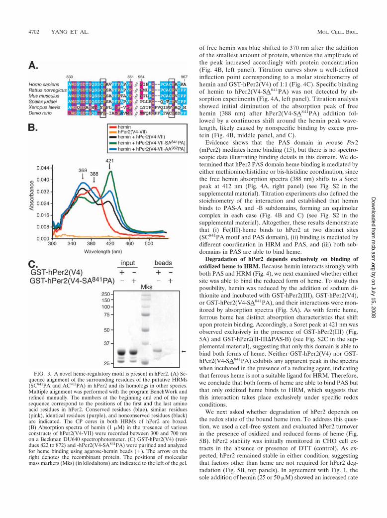
A novel heme-regulatory motif mediates hPer2-heme inter-action. Heme-protein interaction is alternatively mediated byevolutionary conserved heme-regulatory motifs where Cys-Proresidues are invariant and where there is a tendency for ahydrophobic amino acid to be in the fourth position. Inspec-tion of the hPer2 sequence determined the presence of twoputative HRMs (Fig. 3A). Interestingly, both HRMs were lo-cated within hPer2(V4-VII), a fragment that exhibits heme-binding capacity (Fig. 2). Comparative analysis of global mul-tiple Per2 sequence alignments exhibits conserved residuesclustered in the HRMs and surrounded by sequence elementsof high (for SC841PA) and low (for AC962PA) conservation(Fig. 3A). Phylogenetic analyses indicate that both putativeHRMs are highly conserved modules in Per2 proteins amongmetazoan lineages, especially in mammals, suggesting that thesequences under investigation have a comparatively youngmost recent common ancestor (Fig. 3A).
Unlike other heme-binding sites, HRMs establish bondingbetween the cysteine sulfur and the iron atom of heme (45).Accordingly, we tested whether any of the putative HRMsidentified in hPer2(V4-VII) were able to directly bind heme.Untagged hPer2(V4-VII) and its SA841PA and AA962PA mu-tant forms were analyzed after hemin addition by absorptionspectroscopy (Fig. 3B). The hPer2(V4-VII) protein fragmentshifted the peak of the strongest heme absorption band (388nm), the Soret band, toward a shorter wavelength by �19 nm
VOL. 28, 2008 HEME BINDING MEDIATES DEGRADATION OF Period-2 4699
by on July 15, 2008 m
cb.asm.org
Dow
nloaded from

FIG. 1. Heme modulates hPer2 stability in vitro and in vivo. (A, top) 35S-labeled hPer2 ([35S]-hPer2) [35S-hPer2(II-III) in panel B and35S-hPer2(S662A) in panel C] was added to CHO cell extracts in the absence or presence of hemin (10 �M and 100 �M) and incubated at 37°C.Aliquots were removed at 0, 1, and 2 h and resolved by SDS-PAGE and autoradiography. In other experiments, CHO extracts were preincubatedwith MG-132 before the addition of 35S-hPer2 and hemin (10 �M). Bands were quantified using an AlphaImager and normalized to the inputamount (at time zero in bottom panels). The figure shows data from a single experiment that was repeated three times with similar results. Thearrows on the right denote radiolabeled protein. The positions of molecular mass markers (in kilodaltons) are indicated to the left of the gels.(D) CHO cells were incubated with hemin (10 �M) for 2, 4, 6, 8, 10, or 12 h in serum-free medium. Samples were collected at the indicated times,and endogenous levels of hPer2 were analyzed by immunoblotting (top panel). Bands were quantified using an AlphaImager and normalized totubulin levels (bottom right panel). Total RNA was isolated from cells harvested at each time point (in hours) and converted to cDNA in reactionsthat contain equivalent amounts of total RNA. Gene-specific primers (Per2 and GAPDH) were used for PCR amplification. GAPDH was usedas internal control (bottom left panel). (E) CHO cells were treated with succinylacetone (SA) to deplete cells of endogenous heme. After removalof the medium, cells were incubated with either serum-free medium (control), SA, or SA plus hemin (10 �M). Extracts were subjected toSDS-PAGE and immunoblotting. Total protein levels were monitored by either tubulin or actin expression (bottom panels).
4700 YANG ET AL. MOL. CELL. BIOL.
by on July 15, 2008 m
cb.asm.org
Dow
nloaded from

(369 nm [Fig. 3B]), consistent with heme binding to HRMs(45). The SA841PA mutant form of hPer2(V4-VII), but not theAA962PA mutant form, abolished the protein’s ability to shiftthe hemin absorption spectrum to shorter wavelengths andconfirmed the essential role of Cys841 in heme binding (Fig.3B). A second slight shift in the Soret peak (421 nm) wasdetected in the wild-type fragment (Fig. 3B). Whereas an ad-ditional residual shoulder was observed at a shorter wave-length for the SA841PA protein, we believe this shoulder re-sults from excess amounts of free hemin in the sample. Ourstudies indicate that neither Cys841 nor Cys962 is responsiblefor the peak observed at 421 nm, suggesting the existence of asecondary component involved in heme binding that we latermapped between residues 1,121 and 1,255 of hPer2 (data notshown). Overall, our results pinpoint SC841PA as a novelheme-binding motif located at the C terminus of hPer2. As adirect test of the role of the SC841PA motif in heme binding,we examined whether a shorter fragment of hPer2 [GST-hPer2(V4), residues 822 to 872] and its HRM mutant form[GST-hPer2(V4-SA841PA)] were able to bind hemin by affinity
chromatography (Fig. 3C). As expected, GST-hPer2(V4) dis-played a strong interaction for hemin, and the mutation onCys841 completely abrogated binding. Collectively, these dataindicate that heme binds to hPer2 directly through Cys841.
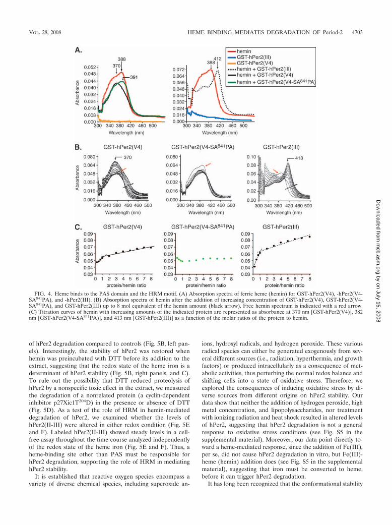
Binding of heme to both HRM and PAS follows a precisestoichiometry. Further evidence of direct binding of heme tohPer2(V4) and the PAS domain-containing fragmenthPer2(III) was obtained by absorption spectra and titrationexperiments. Among hPer2 PAS-containing fragments,hPer2(III) was chosen because of its signal intensity. Heminbinding to hPer2(V4) shifted the Soret band from 388 to 370nm, an event that was prevented by the mutation of Cys841
to Ala, confirming Cys841 as the axial heme ligand (Fig. 4A,left panel). Since hPer2(V4) does not have any appreciableabsorption between 300 and 700 nm, the observed spectralchanges on free hemin are due to alterations in the elec-tronic structure and coordination state of the heme ironcaused by its interaction with hPer2(V4). To examine thespecificity of heme binding, hemin was titrated with increas-ing amounts of GST-hPer2(V4). The absorption peak
FIG. 2. Heme binds hPer2 in two distinct regions. (A) Schematic representation of hPer2 (1,255 residues) architecture including the PASdomain (residues 186 to 434) and the PAS-A (residues 186 to 235) and PAS-B (residues 322 to 374) subdomains. Fragments hPer2(I) (residues1 to 172), hPer2(II) (residues 173 to 355), hPer2(III) (residues 356 to 574), hPer2(II-III) (residues 173 to 574), and hPer2(V4-VII) (residues 822to 1,255) are represented below. (B) GST-tagged hPer2 fragments were purified using affinity chromatography (left panel). Samples were analyzedfor hemin binding using hemin-agarose (right panel).
VOL. 28, 2008 HEME BINDING MEDIATES DEGRADATION OF Period-2 4701
by on July 15, 2008 m
cb.asm.org
Dow
nloaded from
of free hemin was blue shifted to 370 nm after the additionof the smallest amount of protein, whereas the amplitude ofthe peak increased accordingly with protein concentration(Fig. 4B, left panel). Titration curves show a well-definedinflection point corresponding to a molar stoichiometry ofhemin and GST-hPer2(V4) of 1:1 (Fig. 4C). Specific bindingof hemin to hPer2(V4-SA841PA) was not detected by ab-sorption experiments (Fig. 4A, left panel). Titration analysisshowed initial diminution of the absorption peak of freehemin (388 nm) after hPer2(V4-SA841PA) addition fol-lowed by a continuous shift around the hemin peak wave-length, likely caused by nonspecific binding by excess pro-tein (Fig. 4B, middle panel, and C).
Evidence shows that the PAS domain in mouse Per2(mPer2) mediates heme binding (15), but there is no spectro-scopic data illustrating binding details in this domain. We de-termined that hPer2 PAS domain heme binding is mediated byeither methionine/histidine or bis-histidine coordination, sincethe free hemin absorption spectra (388 nm) shifts to a Soretpeak at 412 nm (Fig. 4A, right panel) (see Fig. S2 in thesupplemental material). Titration experiments also defined thestoichiometry of the interaction and established that heminbinds to PAS-A and -B subdomains, forming an equimolarcomplex in each case (Fig. 4B and C) (see Fig. S2 in thesupplemental material). Altogether, these results demonstratethat (i) Fe(III)-heme binds to hPer2 at two distinct sites(SC841PA motif and PAS domain), (ii) binding is mediated bydifferent coordination in HRM and PAS, and (iii) both sub-domains in PAS are able to bind heme.
Degradation of hPer2 depends exclusively on binding ofoxidized heme to HRM. Because hemin interacts strongly withboth PAS and HRM (Fig. 4), we next examined whether eithersite was able to bind the reduced form of heme. To study thispossibility, hemin was reduced by the addition of sodium di-thionite and incubated with GST-hPer2(III), GST-hPer2(V4),or GST-hPer2(V4-SA841PA), and their interactions were mon-itored by absorption spectra (Fig. 5A). As with ferric heme,ferrous heme has distinct absorption characteristics that shiftupon protein binding. Accordingly, a Soret peak at 421 nm wasobserved exclusively in the presence of GST-hPer2(III) (Fig.5A) and GST-hPer2(II-IIIPAS-B) (see Fig. S2C in the sup-plemental material), suggesting that only this domain is able tobind both forms of heme. Neither GST-hPer2(V4) nor GST-hPer2(V4-SA841PA) exhibits any apparent peak in the spectrawhen incubated in the presence of a reducing agent, indicatingthat ferrous heme is not a suitable ligand for HRM. Therefore,we conclude that both forms of heme are able to bind PAS butthat only oxidized heme binds to HRM, which suggests thatthis interaction takes place exclusively under specific redoxconditions.
We next asked whether degradation of hPer2 depends onthe redox state of the bound heme iron. To address this ques-tion, we used a cell-free system and evaluated hPer2 turnoverin the presence of oxidized and reduced forms of heme (Fig.5B). hPer2 stability was initially monitored in CHO cell ex-tracts in the absence or presence of DTT (control). As ex-pected, hPer2 remained stable in either condition, suggestingthat factors other than heme are not required for hPer2 deg-radation (Fig. 5B, top panels). In agreement with Fig. 1, thesole addition of hemin (25 or 50 �M) showed an increased rate
FIG. 3. A novel heme-regulatory motif is present in hPer2. (A) Se-quence alignment of the surrounding residues of the putative HRMs(SC841PA and AC962PA) in hPer2 and its homologs in other species.Multiple alignment was performed with the program BenchWork andrefined manually. The numbers at the beginning and end of the topsequence correspond to the positions of the first and the last aminoacid residues in hPer2. Conserved residues (blue), similar residues(pink), identical residues (purple), and nonconserved residues (black)are indicated. The CP cores in both HRMs of hPer2 are boxed.(B) Absorption spectra of hemin (1 �M) in the presence of variousconstructs of hPer2(V4-VII) were recorded between 300 and 700 nmon a Beckman DU640 spectrophotometer. (C) GST-hPer2(V4) (resi-dues 822 to 872) and -hPer2(V4-SA841PA) were purified and analyzedfor heme binding using agarose-hemin beads (�). The arrow on theright denotes the recombinant protein. The positions of molecularmass markers (Mks) (in kilodaltons) are indicated to the left of the gel.
4702 YANG ET AL. MOL. CELL. BIOL.
by on July 15, 2008 m
cb.asm.org
Dow
nloaded from
of hPer2 degradation compared to controls (Fig. 5B, left pan-els). Interestingly, the stability of hPer2 was restored whenhemin was preincubated with DTT before its addition to theextract, suggesting that the redox state of the heme iron is adeterminant of hPer2 stability (Fig. 5B, right panels, and C).To rule out the possibility that DTT reduced proteolysis ofhPer2 by a nonspecific toxic effect in the extract, we measuredthe degradation of a nonrelated protein (a cyclin-dependentinhibitor p27Xic1T204D) in the presence or absence of DTT(Fig. 5D). As a test of the role of HRM in hemin-mediateddegradation of hPer2, we examined whether the levels ofhPer2(II-III) were altered in either redox condition (Fig. 5Eand F). Labeled hPer2(II-III) showed steady levels in a cell-free assay throughout the time course analyzed independentlyof the redox state of the heme iron (Fig. 5E and F). Thus, aheme-binding site other than PAS must be responsible forhPer2 degradation, supporting the role of HRM in mediatinghPer2 stability.
It is established that reactive oxygen species encompass avariety of diverse chemical species, including superoxide an-
ions, hydroxyl radicals, and hydrogen peroxide. These variousradical species can either be generated exogenously from sev-eral different sources (i.e., radiation, hyperthermia, and growthfactors) or produced intracellularly as a consequence of met-abolic activities, thus perturbing the normal redox balance andshifting cells into a state of oxidative stress. Therefore, weexplored the consequences of inducing oxidative stress by di-verse sources from different origins on hPer2 stability. Ourdata show that neither the addition of hydrogen peroxide, highmetal concentration, and lipopolysaccharides, nor treatmentwith ionizing radiation and heat shock resulted in altered levelsof hPer2, suggesting that hPer2 degradation is not a generalresponse to oxidative stress conditions (see Fig. S5 in thesupplemental material). Moreover, our data point directly to-ward a heme-mediated response, since the addition of Fe(III),per se, did not cause hPer2 degradation in vitro, but Fe(III)-heme (hemin) addition does (see Fig. S5 in the supplementalmaterial), suggesting that iron must be converted to heme,before it can trigger hPer2 degradation.
It has long been recognized that the conformational stability
FIG. 4. Heme binds to the PAS domain and the HRM motif. (A) Absorption spectra of ferric heme (hemin) for GST-hPer2(V4), -hPer2(V4-SA841PA), and -hPer2(III). (B) Absorption spectra of hemin after the addition of increasing concentration of GST-hPer2(V4), GST-hPer2(V4-SA841PA), and GST-hPer2(III) up to 8 mol equivalent of the hemin amount (black arrow). Free hemin spectrum is indicated with a red arrow.(C) Titration curves of hemin with increasing amounts of the indicated protein are represented as absorbance at 370 nm [GST-hPer2(V4)], 382nm [GST-hPer2(V4-SA841PA)], and 413 nm [GST-hPer2(III)] as a function of the molar ratios of the protein to hemin.
VOL. 28, 2008 HEME BINDING MEDIATES DEGRADATION OF Period-2 4703
by on July 15, 2008 m
cb.asm.org
Dow
nloaded from
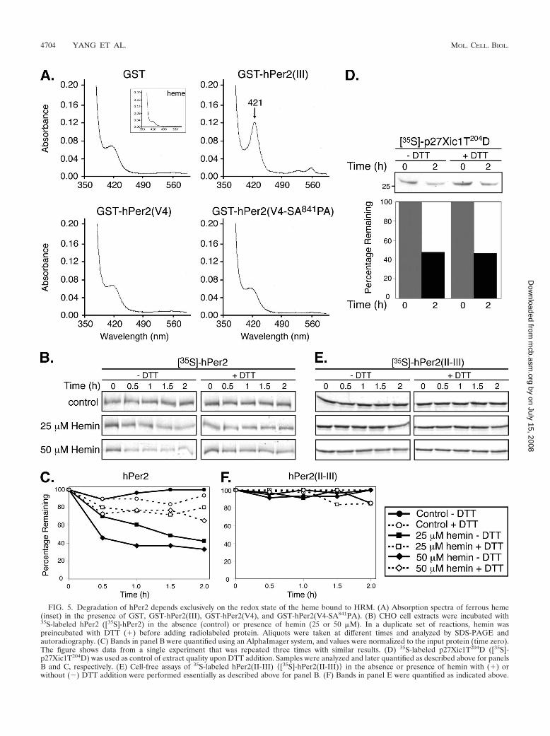
FIG. 5. Degradation of hPer2 depends exclusively on the redox state of the heme bound to HRM. (A) Absorption spectra of ferrous heme(inset) in the presence of GST, GST-hPer2(III), GST-hPer2(V4), and GST-hPer2(V4-SA841PA). (B) CHO cell extracts were incubated with35S-labeled hPer2 ([35S]-hPer2) in the absence (control) or presence of hemin (25 or 50 �M). In a duplicate set of reactions, hemin waspreincubated with DTT (�) before adding radiolabeled protein. Aliquots were taken at different times and analyzed by SDS-PAGE andautoradiography. (C) Bands in panel B were quantified using an AlphaImager system, and values were normalized to the input protein (time zero).The figure shows data from a single experiment that was repeated three times with similar results. (D) 35S-labeled p27Xic1T204D ([35S]-p27Xic1T204D) was used as control of extract quality upon DTT addition. Samples were analyzed and later quantified as described above for panelsB and C, respectively. (E) Cell-free assays of 35S-labeled hPer2(II-III) {[35S]-hPer2(II-III)} in the absence or presence of hemin with (�) orwithout (�) DTT addition were performed essentially as described above for panel B. (F) Bands in panel E were quantified as indicated above.
4704 YANG ET AL. MOL. CELL. BIOL.
by on July 15, 2008 m
cb.asm.org
Dow
nloaded from
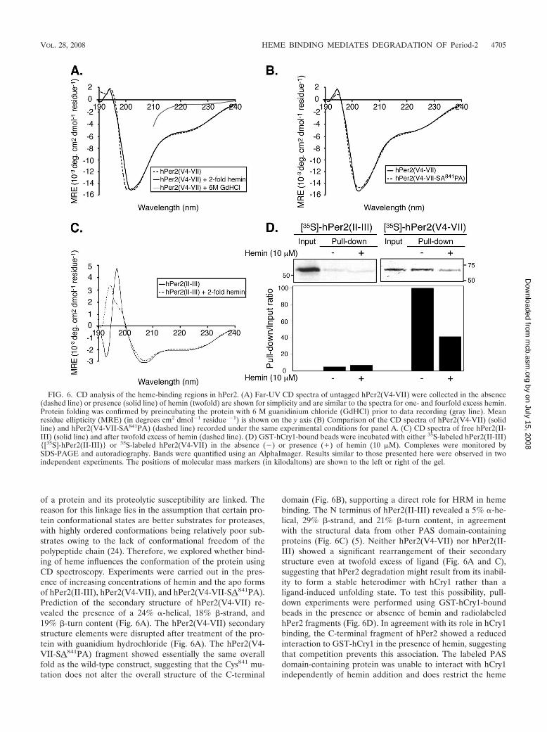
of a protein and its proteolytic susceptibility are linked. Thereason for this linkage lies in the assumption that certain pro-tein conformational states are better substrates for proteases,with highly ordered conformations being relatively poor sub-strates owing to the lack of conformational freedom of thepolypeptide chain (24). Therefore, we explored whether bind-ing of heme influences the conformation of the protein usingCD spectroscopy. Experiments were carried out in the pres-ence of increasing concentrations of hemin and the apo formsof hPer2(II-III), hPer2(V4-VII), and hPer2(V4-VII-SA841PA).Prediction of the secondary structure of hPer2(V4-VII) re-vealed the presence of a 24% �-helical, 18% �-strand, and19% �-turn content (Fig. 6A). The hPer2(V4-VII) secondarystructure elements were disrupted after treatment of the pro-tein with guanidium hydrochloride (Fig. 6A). The hPer2(V4-VII-SA841PA) fragment showed essentially the same overallfold as the wild-type construct, suggesting that the Cys841 mu-tation does not alter the overall structure of the C-terminal
domain (Fig. 6B), supporting a direct role for HRM in hemebinding. The N terminus of hPer2(II-III) revealed a 5% �-he-lical, 29% �-strand, and 21% �-turn content, in agreementwith the structural data from other PAS domain-containingproteins (Fig. 6C) (5). Neither hPer2(V4-VII) nor hPer2(II-III) showed a significant rearrangement of their secondarystructure even at twofold excess of ligand (Fig. 6A and C),suggesting that hPer2 degradation might result from its inabil-ity to form a stable heterodimer with hCry1 rather than aligand-induced unfolding state. To test this possibility, pull-down experiments were performed using GST-hCry1-boundbeads in the presence or absence of hemin and radiolabeledhPer2 fragments (Fig. 6D). In agreement with its role in hCry1binding, the C-terminal fragment of hPer2 showed a reducedinteraction to GST-hCry1 in the presence of hemin, suggestingthat competition prevents this association. The labeled PASdomain-containing protein was unable to interact with hCry1independently of hemin addition and does restrict the heme
FIG. 6. CD analysis of the heme-binding regions in hPer2. (A) Far-UV CD spectra of untagged hPer2(V4-VII) were collected in the absence(dashed line) or presence (solid line) of hemin (twofold) are shown for simplicity and are similar to the spectra for one- and fourfold excess hemin.Protein folding was confirmed by preincubating the protein with 6 M guanidinium chloride (GdHCl) prior to data recording (gray line). Meanresidue ellipticity (MRE) (in degrees cm2 dmol�1 residue �1) is shown on the y axis (B) Comparison of the CD spectra of hPer2(V4-VII) (solidline) and hPer2(V4-VII-SA841PA) (dashed line) recorded under the same experimental conditions for panel A. (C) CD spectra of free hPer2(II-III) (solid line) and after twofold excess of hemin (dashed line). (D) GST-hCry1-bound beads were incubated with either 35S-labeled hPer2(II-III){[35S]-hPer2(II-III)} or 35S-labeled hPer2(V4-VII) in the absence (�) or presence (�) of hemin (10 �M). Complexes were monitored bySDS-PAGE and autoradiography. Bands were quantified using an AlphaImager. Results similar to those presented here were observed in twoindependent experiments. The positions of molecular mass markers (in kilodaltons) are shown to the left or right of the gel.
VOL. 28, 2008 HEME BINDING MEDIATES DEGRADATION OF Period-2 4705
by on July 15, 2008 m
cb.asm.org
Dow
nloaded from
effect on hPer2/hCry1 formation to the C-terminal portion ofhPer2.
HRM is required for degradation of hPer2 in vivo. To gainfurther insight into the role of HRM in the degradation of Per2in vivo, we transiently transfected CHO cells with either myc-hPer2 or myc-hPer2-SA841PA and evaluated their subcellularlocalization and intracellular levels in response to hemin addi-tion by immunofluorescence microscopy (Fig. 7A). In agree-ment with previous observations (40), immunofluorescencestaining showed that myc-hPer2 was distributed in both nuclearand cytosolic compartments and that its accumulation was re-markably higher in the former (Fig. 7A). The cellular distri-bution of myc-hPer2-SA841PA mutant was similar to that of thewild-type protein, and thus, we conclude the Cys841Ala muta-tion does not alter hPer2 localization (Fig. 7A). Hemin addi-tion to myc-hPer2-transfected cells resulted in decreased levelsof the nuclear protein without increasing hPer2 levels in thecytosolic compartment, suggesting that degradation, ratherthan translocation, was triggered by hemin (Fig. 7A and B).Supporting the role of HRM in heme-mediated hPer2 degra-dation in vivo, the addition of hemin to myc-hPer2-SA841PA-transfected cells did not result in apparent changes in mutantprotein levels (Fig. 7A). Profile plotting of signal intensityalong cross sections of cells transfected with wild-type andhPer2 mutants confirmed hemin-induced nuclear degradationof hPer2 and unambiguously confirmed that this phenomenonis mediated by HRM (Fig. 7B).
To further support the concept that HRM is sufficient to pro-mote heme-mediated hPer2 degradation and that heme bindingto PAS domain plays a distinct role (15), we transfected cells withmyc-hPer2, myc-hPer2-SA841PA, or myc-hPer2(II-III) and evalu-ated their total protein levels in response to hemin addition (Fig.7C). The remarkable stability of myc-hPer2(II-III) observed inthe presence of hemin contrasted greatly with the levels of myc-hPer2 detected under the same condition, suggesting that bindingof heme to the PAS domain does not alter its stability in vivo (Fig.7C). The presence of equivalent levels of myc-hPer2-SA841PA inthe absence or presence of hemin further supports our model.
Binding of heme to HRM prevents the formation of thehPer2/hCry1 complex. The C terminus of Per2 physically as-sociates with Cry proteins (9, 19), and the complex translocatesto the nucleus where it acts as a negative regulator by directlyinteracting with Clock/Bmal1 (31). Thus, we first askedwhether heme treatment of cells alters the intracellular levelsof the hPer2/hCry1 complex (Fig. 8). Immunoprecipitation as-says of hemin-treated myc-hPer2/FLAG-hCry1 cells were an-alyzed for the presence of heterodimers by immunoblotting(Fig. 8A). Results show reduced levels of bound hCry1 inhemin-treated samples, indicating that heme alters hPer2/hCry1 levels in cells (Fig. 8A). To rule out the possibility thatheme can cause hCry1 degradation and disrupt hPer2/hCry1interaction, transfected CHO cells were incubated with hemin,and hCry1 levels were monitored at different times. As shownin Fig. S3A in the supplemental material, hCry1 levels re-mained invariable throughout the time course analyzed, sug-gesting that hCry1 stability is independent of the presence ofheme. A similar result was obtained when hCry1 stability wastested in the presence of hemin in a cell-free assay (see Fig.S3B in the supplemental material).
We then examined whether heme binding to hPer2 prevents
the formation of hPer2/hCry1 or disrupts an already preformedcomplex instead. To evaluate either model, we first recapitu-lated the cellular events leading to heme-dependent reductionof hPer2/hCry1 levels in vitro (Fig. 8B). Recombinant GST-hCry1, 35S-labeled hPer2, and hemin were simultaneously in-cubated, and the amount of 35S-labeled hPer2 present in thecomplex was analyzed by pull-down experiments (Fig. 8B and6D). As was the case with transfected cell extracts, our in vitroassay showed lower levels of 35S-labeled hPer2 associated withGST-hCry1 in the presence of hemin, supporting a modelwhere ligand binding compromises hPer2/hCry1 complex for-mation. Because of the nature of our in vitro assay, only twoproteins and hemin were present, which also suggests thatheme binding to the C terminus of hPer2 prevents or disruptsits association with hCry1 and that heme-mediated degrada-tion of hPer2 might be a consequence of lack of association.
Next, we established which event of the complex formationis inhibited by heme binding. In the first scenario, hPer2/GST-hCry1 complex was allowed to form and later incubated withhemin (Fig. 8C). In a parallel experiment, hPer2 was preincu-bated with hemin, added to GST-hCry1, and analyzed by pull-down experiments (Fig. 8C). Results demonstrate that morehPer2 is bound to GST-hCry1 when the complex is preformed,suggesting that hemin is unable to disrupt a stable het-erodimer.
Expression of a non-heme-responsive HRM form of hPer2alters the pattern of circadian gene expression. The observa-tion that circadian gene expression can persist for several daysin serum-free medium after an initial serum shock (1, 2)prompted us to test the effects of hPer2 and hPer2(SA841PA)mutant on the mRNA accumulation profile of circadian genes.We investigated one of the known downstream effectors ofPer2 signaling, Rev-erb�, a transcript that is lowest at timeswhen Per2 expression peaks in the nucleus (26). CircadianRev-erb� expression is controlled by components of the generalfeedback loop, thus influencing the period length and phase-shifting properties of the clock (26). In agreement with ourmodel, cells transfected with hPer2 exhibited reduced levels ofRev-erb� compared with nontransfected cells (Fig. 9A, rightpanel), an effect that is reversed when cells were pretreatedwith hemin (Fig. 9A, bottom left panel). Moreover, transfec-tion with hPer2(SA841PA) resulted in sustained downregula-tion of Rev-erb� transcription throughout the analyzed timecourse (Fig. 9A, bottom right panel). As predicted, the addi-tion of hemin to hPer2(SA841PA)-transfected cells did notresult in altered levels of Rev-erb�, since the ligand can nolonger bind the mutant protein and is therefore unable to acton its stability.
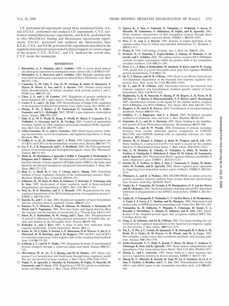
Overall, our observations favor a scenario where heme plays anessential role in controlling hPer2 cellular levels by targetinghPer2 for degradation and preventing hPer2/hCry1 complex ac-cumulation (Fig. 9B). Our tryptophan fluorescence spectroscopydata show that both PAS and HRM bind heme with roughlyequal affinity in the nanomolar range {Kd hPer2(V4-VII) [disso-ciation constant], 9.20 � 0.94 nM; Kd hPer2(II-III), 12.31 � 0.43nM; see Fig. S4 in the supplemental material}. Interestingly,whereas ferrous heme will bind only to the PAS domain (Fig.5A), its ferric form could, in principle, target either bindingsite. At this point, we hypothesize that binding of ferric hemeto either PAS or HRM might depend on their availability. For
4706 YANG ET AL. MOL. CELL. BIOL.
by on July 15, 2008 m
cb.asm.org
Dow
nloaded from
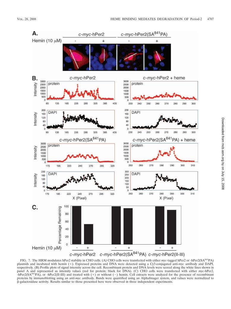
FIG. 7. The HRM modulates hPer2 stability in CHO cells. (A) CHO cells were transfected with either myc-tagged hPer2 or -hPer2(SA841PA)plasmids and incubated with hemin (�). Expressed proteins and DNA were detected using a Cy3-conjugated anti-myc antibody and DAPI,respectively. (B) Profile plots of signal intensity across the cell. Recombinant protein and DNA levels were scored along the white lines shown inpanel A and represented as intensity values (red for protein; black for DNA). (C) CHO cells were transfected with either myc-hPer2,-hPer2(SA841PA), or -hPer2(II-III) and treated with (�) or without (�) hemin. Cell extracts were analyzed for the presence of recombinantproteins by immunoblotting using an anti-myc antibody. Bands were quantified using an AlphaImager system, and values were normalized to�-galactosidase activity. Results similar to those presented here were observed in three independent experiments.
VOL. 28, 2008 HEME BINDING MEDIATES DEGRADATION OF Period-2 4707
by on July 15, 2008 m
cb.asm.org
Dow
nloaded from

example, preassociation of hPer2 to hCry1 prevents the accessof heme to HRM but not PAS (Fig. 8 and 9B) and thus affectssignaling downstream. Accordingly, binding of heme to PAS inthe mPer2/hCry1 complex regulates the transcriptional activityof Bmal1/NPAS2 and the expression of the alas1 gene (15).Conversely, the absence of hCry1 will allow heme to bindHRM (or both HRM and PAS simultaneously) and promoteinstability of hPer2 (this study), an event that is exclusivelymediated by HRM, since heme binding to PAS does not alterhPer2 instability (Fig. 1B). In this scenario, downregulation ofhPer2 directly impacts the oscillatory expression of circadiangenes. Thus, this novel pathway ensures an alternative mech-anism to physiologically controlling the circadian clock by act-ing on gene expression.
DISCUSSION
The mammalian circadian system influences most physiologicalactivities, including sleep/wake cycles, cardiovascular activity,
body temperature, blood pressure, glucose and fat metabolism,renal plasma flow, liver metabolism and detoxification, and hor-monal secretion (32). Cross talk between the body’s circadianrhythm and metabolic systems has been identified within both thegluconeogenic and lipogenic pathways and in organisms as di-verse as flies and mammals. Examples include the circadian os-cillatory expression of the sterol-regulatory element-binding pro-teins 1a and 1c, a group of transcription factors that bind to thesterol regulatory element to control the hepatic transcriptomeand thus the hepatic physiology. In addition, the orphan nuclearreceptor Rev-erb�, a negative regulator of the circadian coregene bmal1, is expressed according to a robust circadian patternand is induced during normal adipogenesis. Conversely, the reti-noic acid-related orphan receptors also modulate bmal1 expres-sion while regulating lipid flux, lipogenesis, and lipid storage inskeletal muscle, providing an additional nodal point interrelatingmetabolic and circadian physiology. Further studies linked carbo-hydrate metabolism and circadian rhythms in fruit flies, andstrong evidence supports a cross talk mechanism between nuclear
FIG. 8. Binding of heme prevents hPer2/hCry1 heterodimerization. (A) CHO cells were cotransfected with either myc-hPer2 or pCS2�(myc)6empty vector and FLAG-hCry1 and treated with hemin (10 �M) (�) as shown in Fig. 7. Extracts were incubated with anti-myc-tagged beads, andsamples were analyzed by SDS-PAGE and immunoblotting using anti-myc and anti-FLAG antibodies (Ab). IP, immunoprecipitation; WB,Western blotting. (B) GST-hCry1-bound beads were incubated with 35S-labeled hPer2 ([35S]-hPer2) in the absence (�) and presence (�) of hemin(10 �M), and binding was monitored by SDS-PAGE and autoradiography. Bands were quantified using an AlphaImager. (C) GST-hCry1-boundbeads were incubated with 35S-labeled hPer2 (�) before or after hemin (10 �M) addition (�). In all cases, beads were exhaustively washed, andsamples were analyzed by SDS-PAGE and autoradiography. The figure shows data from a single experiment that was repeated three times withsimilar results.
4708 YANG ET AL. MOL. CELL. BIOL.
by on July 15, 2008 m
cb.asm.org
Dow
nloaded from
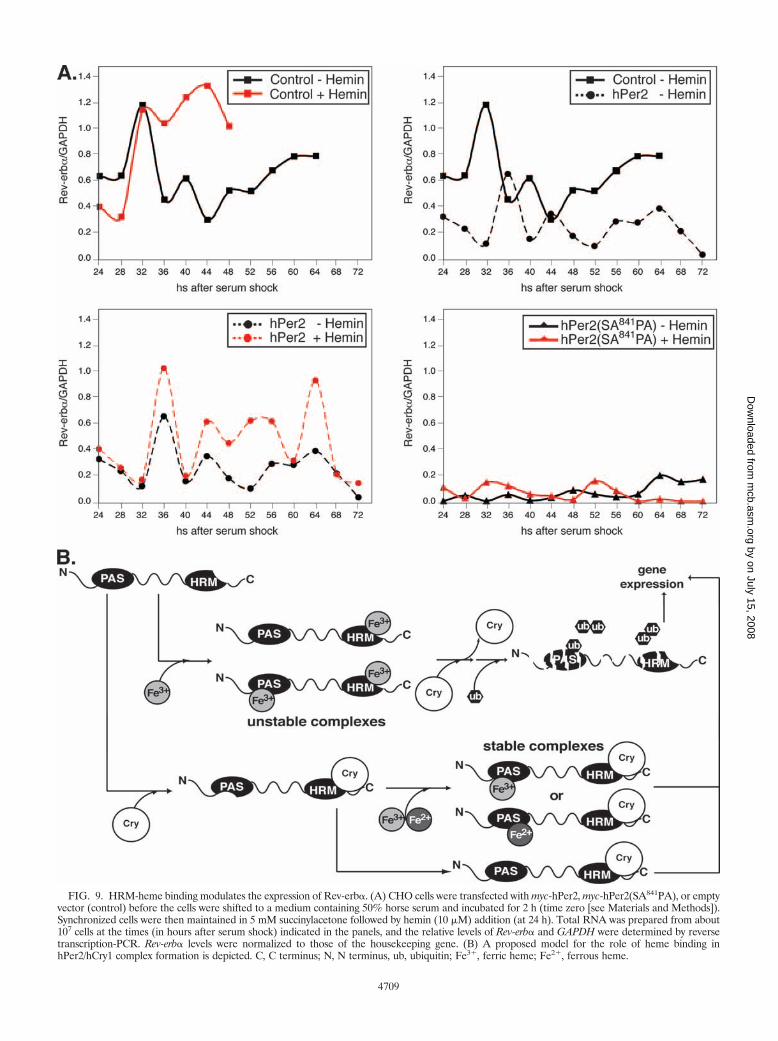
FIG. 9. HRM-heme binding modulates the expression of Rev-erb�. (A) CHO cells were transfected with myc-hPer2, myc-hPer2(SA841PA), or emptyvector (control) before the cells were shifted to a medium containing 50% horse serum and incubated for 2 h (time zero [see Materials and Methods]).Synchronized cells were then maintained in 5 mM succinylacetone followed by hemin (10 �M) addition (at 24 h). Total RNA was prepared from about107 cells at the times (in hours after serum shock) indicated in the panels, and the relative levels of Rev-erb� and GAPDH were determined by reversetranscription-PCR. Rev-erb� levels were normalized to those of the housekeeping gene. (B) A proposed model for the role of heme binding inhPer2/hCry1 complex formation is depicted. C, C terminus; N, N terminus, ub, ubiquitin; Fe3�, ferric heme; Fe2�, ferrous heme.
4709
by on July 15, 2008 m
cb.asm.org
Dow
nloaded from

hormone receptors and the core circadian complex Clock/Bmal1in adipogenesis (for a review, see reference 18).
An additional level of complexity arises from experimentsshowing that many heme-containing molecules regulate cellu-lar homeostasis which, consistent with the circadian oscillatorynature of heme levels, led us to propose heme as a candidatebridge molecule for the circadian and metabolic mechanisms.We and other groups have reported that heme directly targetscircadian clock components modulating both gene transcrip-tion and protein stability (6, 15, 43; this article). We establishedthat heme directly binds to a novel regulatory motif in hPer2 ina redox-dependent manner, resulting in hPer2 instability andaltered hPer2/hCry1 formation. Therefore, we propose thathPer2 acts as a heme sensor-transducer molecule, couplingmetabolic signals to the circadian oscillator.
Control of Per2 stability plays a key role in driving circadianrhythmicity (31). During the transcription-translational feedbackloop, Per2 is rapidly degraded as a result of phosphorylation bythe double-time kinase in Drosophila (27) or CKIε in mammals(3), altering the levels of Per2 available for heterodimerizationand nuclear translocation. Although phosphorylation remains theprimary mechanism responsible for Per2 degradation, alternativemechanisms to control its stability might exist. We tested thesimplest model in which binding of heme to hPer2 induces pro-tein instability in a phosphorylation-independent fashion. Indeed,heme favors hPer2 degradation both in vitro and in vivo. Moreimportantly, this event is independent of both phosphorylation byCKIε and binding of heme to the PAS domain, indicating thatdegradation of hPer2 can occur by alternative mechanisms. Pe-riod protein turnover is mediated by ubiquitination and furtherdegradation by the proteasome pathway (39). Our data agreed,showing that inhibitors of proteasome function restore hPer2levels, supporting a model where heme-mediated degradation ofhPer2 depends on ubiquitination. Similarly, heme-mediated ubiq-uitination and degradation exist in iron regulatory proteins inother systems (13). Specifically, IRP2 oxidation, which is medi-ated by heme binding to its regulatory domain, triggers IRP2ubiquitination-dependent degradation regulating the expressionof genes involved in iron metabolism (13, 14, 41). In addition, theDNA-binding activity of the transcriptional repressor Bach1 dra-matically decreases upon heme binding through multiple HRMs(23, 36), inducing nuclear export of Bach1 (37), polyubiquitina-tion, and degradation of the repressor (44). Heme also binds tothe bacterial iron response regulator through two distinct regionsincluding an HRM, a necessary interaction for normal degrada-tion (28, 29, 42). In this scenario, both redox states are requiredfor rapid turnover of Irr, although its stability is independent ofubiquitination and likely mediated by an unknown specific pro-tease (42). Like IRP2 and Irr, heme-dependent degradation ofhPer2 is mediated by a CP core of a HRM. Unlike IRP2, wherethe Cys and His residues within the HRM participate in coordi-nation and are responsible for axial ligand of ferric and ferrousheme (13), the HRM of hPer2 lacks the His component found inthe HRM of IRP2 and exclusively binds ferric heme. More im-portantly, whereas oxidized heme binds to both HRM and PAS ofhPer2, it is only its interaction with the former that is responsiblefor hPer2 degradation. This is the first demonstration of ligand-induced instability of a clock gene product and is a novel mode ofregulation of the circadian feedback loop.
To understand the mechanism underlying heme-hPer2 rec-
ognition, we studied whether conformational changes are as-sociated with ligand binding and heterodimeric complex for-mation. It is not known whether or to what extent hemebinding to hPer2 plays a role in hPer2/hCry1 complex forma-tion. Examples show slight secondary structural changes inhelicity in the electron transport protein cytochrome b562 uponheme binding (16), whereas large changes in secondary struc-ture are revealed when the His-rich protein II is compared tothe apoprotein after ferric heme addition (33). Our secondarystructural studies of the C-terminal domain of hPer2 show thatheme binding does not induce major conformational changesin the protein, suggesting that degradation of hPer2 does notresult from unfolding upon ligand binding but is most likelymediated by an unknown, specific ubiquitin ligase enzyme.Much has been done to identify the molecules responsible forselective recognition of oxidized target proteins, including therecent characterization of the heme-oxidized IRP2 ubiquitinligase-1 responsible for IRP2 turnover (13, 41). Interestingly,mPer2 ubiquitination is reduced by its interaction with Cry andis mediated by the Cry-binding domain residing in the C-terminal portion of mPer2 (9, 19), a mode of regulation closelyresembling the organization of the Per/Tim loop in Drosophila(17).
All of these findings raise the question of whether hemebinding to the C terminus of hPer2 prevents the formation ofthe hPer2/hCry1 complex or rather perturbs the stability of analready preformed heterodimer. Here, we provide evidencethat heme acts by preventing hPer2 from binding to hCry1when bound to HRM, whereas heme-PAS binding neitherpromotes hPer2 degradation nor affects hCry1 association.Heme binding to PAS plays a role in mPer2 interaction withthe Bmal1/NPAS2 complex and in its transcriptional activity(15). Accordingly, cyanocobalamin, a vitamin B12 analoguewith a similar porphyrin ring structure to heme, greatly de-creases the binding of NPAS2 and mPer2 to a heme-agarosematrix (15). The overall data are reconciled in a model whereheme binding to either HRM or PAS in hPer2 targets differentcircadian complexes for regulation, likely connecting the cel-lular response to changes in heme levels. Furthermore, wepropose that selectivity of binding is dictated by the redox stateof the iron core in the heme molecule. Last, we demonstratethat transcription of the orphan nuclear receptor Rev-erb�, amajor regulator of the circadian oscillator that influences pe-riod length and affects the phase-shifting properties of theclock, is responsive to heme binding to the HRM of hPer2.These experiments add a new level of regulation in circadiangene expression by directly coupling metabolic sensing to thetranscriptional control of the molecular oscillator.
ACKNOWLEDGMENTS
We thank William Huckle (Virginia Tech) and Mark O’Brian(SUNY, Buffalo) for critical reading of the manuscript and all mem-bers of the Finkielstein laboratory for help and discussions. We aregrateful to Steven L. McKnight (University of Texas SouthwesternMedical Center) for providing us with the hPer2 and hCry1 cDNAs.
This work was supported by the Jeffress Memorial Trust, AmericanHeart Association, and Susan G. Komen Foundation (C.V.F.) andWendy Will Case and Concern Foundations (D.G.S.C.). J.Y. is par-tially funded by an AdvanceVT postdoctoral fellowship (NSF SBE-0244916). A.L. is a Wilkins-Fralin Research Fellow and Sigma XiScholar. K.D.K. and K.E.D. are Sigma Xi Scholars.
4710 YANG ET AL. MOL. CELL. BIOL.
by on July 15, 2008 m
cb.asm.org
Dow
nloaded from
J.Y. performed all experiments except those mentioned below. A.L.and D.G.S.C. performed and analyzed CD experiments, C.V.F. per-formed immunofluorescence experiments, and K.D.K. performed thein vitro hPer2/hCry1 binding and fluorescence spectroscopy experi-ments. D.G.S.C. processed and analyzed the fluorescence data.K.E.D., C.S.S., and S.P.M. performed the experiments described in thesupplemental material and provided technical support at various stagesof the project. C.V.F., D.G.S.C., and J.Y. analyzed the overall data.C.V.F. wrote the manuscript.
REFERENCES
1. Balsalobre, A., F. Damiola, and U. Schibler. 1998. A serum shock inducescircadian gene expression in mammalian tissue culture cells. Cell 93:929–937.
2. Balsalobre, A., L. Marcacci, and U. Schibler. 2000. Multiple signaling path-ways elicit circadian gene expression in cultured Rat-1 fibroblasts. Curr. Biol.10:1291–1294.
3. Camacho, F., M. Cilio, Y. Guo, D. M. Virshup, K. Patel, O. Khorkova, S.Styren, B. Morse, Z. Yao, and G. A. Keesler. 2001. Human casein kinaseIdelta phosphorylation of human circadian clock proteins period 1 and 2.FEBS Lett. 489:159–165.
4. Chen, J. J., and I. M. London. 1995. Regulation of protein synthesis byheme-regulated eIF-2 alpha kinase. Trends Biochem. Sci. 20:105–108.
5. Crews, S. T., and C. M. Fan. 1999. Remembrance of things PAS: regulationof development by bHLH-PAS proteins. Curr. Opin. Genet. Dev. 9:580–587.
6. Dioum, E. M., J. Rutter, J. R. Tuckerman, G. Gonzalez, M. A. Gilles-Gonzalez, and S. L. McKnight. 2002. NPAS2: a gas-responsive transcriptionfactor. Science 298:2385–2387.
7. Eide, E. J., M. F. Woolf, H. Kang, P. Woolf, W. Hurst, F. Camacho, E. L.Vielhaber, A. Giovanni, and D. M. Virshup. 2005. Control of mammaliancircadian rhythm by CKIε-regulated proteasome-mediated PER2 degrada-tion. Mol. Cell. Biol. 25:2795–2807.
8. Gilles-Gonzalez, M. A., and G. Gonzalez. 2005. Heme-based sensors: defin-ing characteristics, recent developments, and regulatory hypotheses. J. Inorg.Biochem. 99:1–22.
9. Griffin, E. A., Jr., D. Staknis, and C. J. Weitz. 1999. Light-independent roleof CRY1 and CRY2 in the mammalian circadian clock. Science 286:768–771.
10. Gu, Y. Z., J. B. Hogenesch, and C. A. Bradfield. 2000. The PAS superfamily:sensors of environmental and developmental signals. Annu. Rev. Pharmacol.Toxicol. 40:519–561.
11. Hirai, K., M. Martinkova, J. Igarashi, I. Saiful, S. Yamauchi, S. El-Mashtoly, T.Kitagawa, and T. Shimizu. 2007. Identification of Cys385 in the isolated kinaseinsertion domain of heme-regulated eIF2alpha kinase (HRI) as the heme axialligand by site-directed mutagenesis and spectral characterization. J. Inorg. Bio-chem. 101:1172–1179.
12. Hon, T., A. Hach, H. C. Lee, T. Cheng, and L. Zhang. 2000. Functionalanalysis of heme regulatory elements of the transcriptional activator Hap1.Biochem. Biophys. Res. Commun. 273:584–591.
13. Ishikawa, H., M. Kato, H. Hori, K. Ishimori, T. Kirisako, F. Tokunaga, andK. Iwai. 2005. Involvement of heme regulatory motif in heme-mediatedubiquitination and degradation of IRP2. Mol. Cell 19:171–181.
14. Iwai, K., R. D. Klausner, and T. A. Rouault. 1995. Requirements for iron-regulated degradation of the RNA binding protein, iron regulatory protein 2.EMBO J. 14:5350–5357.
15. Kaasik, K., and C. C. Lee. 2004. Reciprocal regulation of haem biosynthesisand the circadian clock in mammals. Nature 430:467–471.
16. Kamiya, N., Y. Okimoto, Z. Ding, H. Ohtomo, M. Shimizu, A. Kitayama, H.Morii, and T. Nagamune. 2001. How does heme axial ligand deletion affectthe structure and the function of cytochrome b562? Protein Eng. 14:415–419.
17. Kloss, B., A. Rothenfluh, M. W. Young, and L. Saez. 2001. Phosphorylationof period is influenced by cycling physical associations of double-time, pe-riod, and timeless in the Drosophila clock. Neuron 30:699–706.
18. Kohsaka, A., and J. Bass. 2007. A sense of time: how molecular clocksorganize metabolism. Trends Endocrinol. Metab. 18:4–11.
19. Kume, K., M. J. Zylka, S. Sriram, L. P. Shearman, D. R. Weaver, X. Jin, E. S.Maywood, M. H. Hastings, and S. M. Reppert. 1999. mCRY1 and mCRY2are essential components of the negative limb of the circadian clock feedbackloop. Cell 98:193–205.
20. Lathrop, J. T., and M. P. Timko. 1993. Regulation by heme of mitochondrialprotein transport through a conserved amino acid motif. Science 259:522–525.
21. McCoubrey, W. K., Jr., T. J. Huang, and M. D. Maines. 1997. Heme oxy-genase-2 is a hemoprotein and binds heme through heme regulatory motifsthat are not involved in heme catalysis. J. Biol. Chem. 272:12568–12574.
22. Nagai, T., K. Igarashi, J. Akasaka, K. Furuyama, H. Fujita, N. Hayashi, M.Yamamoto, and S. Sassa. 1998. Regulation of NF-E2 activity in erythroleu-kemia cell differentiation. J. Biol. Chem. 273:5358–5365.
23. Ogawa, K., J. Sun, S. Taketani, O. Nakajima, C. Nishitani, S. Sassa, N.Hayashi, M. Yamamoto, S. Shibahara, H. Fujita, and K. Igarashi. 2001.Heme mediates derepression of Maf recognition element through directbinding to transcription repressor Bach1. EMBO J. 20:2835–2843.
24. Pace, C. N., and A. J. Barrett. 1984. Kinetics of tryptic hydrolysis of thearginine-valine bond in folded and unfolded ribonuclease T1. Biochem. J.219:411–417.
25. Ponka, P. 1999. Cell biology of heme. Am. J. Med. Sci. 318:241–256.26. Preitner, N., F. Damiola, L. Lopez-Molina, J. Zakany, D. Duboule, U. Al-
brecht, and U. Schibler. 2002. The orphan nuclear receptor REV-ERBalphacontrols circadian transcription within the positive limb of the mammaliancircadian oscillator. Cell 110:251–260.
27. Price, J. L., J. Blau, A. Rothenfluh, M. Abodeely, B. Kloss, and M. W. Young.1998. double-time is a novel Drosophila clock gene that regulates PERIODprotein accumulation. Cell 94:83–95.
28. Qi, Z., I. Hamza, and M. R. O’Brian. 1999. Heme is an effector molecule foriron-dependent degradation of the bacterial iron response regulator (Irr)protein. Proc. Natl. Acad. Sci. USA 96:13056–13061.
29. Qi, Z., and M. R. O’Brian. 2002. Interaction between the bacterial ironresponse regulator and ferrochelatase mediates genetic control of hemebiosynthesis. Mol. Cell 9:155–162.
30. Raghuram, S., K. R. Stayrook, P. Huang, P. M. Rogers, A. K. Nosie, D. B.McClure, L. L. Burris, S. Khorasanizadeh, T. P. Burris, and F. Rastinejad.2007. Identification of heme as the ligand for the orphan nuclear receptorsREV-ERBalpha and REV-ERBbeta. Nat. Struct. Mol. Biol. 14:1207–1213.
31. Reppert, S. M., and D. R. Weaver. 2002. Coordination of circadian timing inmammals. Nature 418:935–941.
32. Schibler, U., J. Ripperger, and S. A. Brown. 2003. Peripheral circadianoscillators in mammals: time and food. J. Biol. Rhythms 18:250–260.
33. Schneider, E. L., and M. A. Marletta. 2005. Heme binding to the histidine-rich protein II from Plasmodium falciparum. Biochemistry 44:979–986.
34. Sreerama, N., and R. W. Woody. 2000. Estimation of protein secondarystructure from circular dichroism spectra: comparison of CONTIN,SELCON, and CDSSTR methods with an expanded reference set. Anal.Biochem. 287:252–260.
35. Steiner, H., G. Kispal, A. Zollner, A. Haid, W. Neupert, and R. Lill. 1996.Heme binding to a conserved Cys-Pro-Val motif is crucial for the catalyticfunction of mitochondrial heme lyases. J. Biol. Chem. 271:32605–32611.
36. Sun, J., H. Hoshino, K. Takaku, O. Nakajima, A. Muto, H. Suzuki, S.Tashiro, S. Takahashi, S. Shibahara, J. Alam, M. M. Taketo, M. Yamamoto,and K. Igarashi. 2002. Hemoprotein Bach1 regulates enhancer availability ofheme oxygenase-1 gene. EMBO J. 21:5216–5224.
37. Suzuki, H., S. Tashiro, S. Hira, J. Sun, C. Yamazaki, Y. Zenke, M. Ikeda-Saito, M. Yoshida, and K. Igarashi. 2004. Heme regulates gene expressionby triggering Crm1-dependent nuclear export of Bach1. EMBO J. 23:2544–2553.
38. Whitmore, L., and B. A. Wallace. 2004. DICHROWEB, an online server forprotein secondary structure analyses from circular dichroism spectroscopicdata. Nucleic Acids Res. 32:W668–W673.
39. Yagita, K., F. Tamanini, M. Yasuda, J. H. Hoeijmakers, G. T. van der Horst,and H. Okamura. 2002. Nucleocytoplasmic shuttling and mCRY-dependentinhibition of ubiquitylation of the mPER2 clock protein. EMBO J. 21:1301–1314.
40. Yagita, K., S. Yamaguchi, F. Tamanini, G. T. van Der Horst, J. H. Hoeijmakers,A. Yasui, J. J. Loros, J. C. Dunlap, and H. Okamura. 2000. Dimerization andnuclear entry of mPER proteins in mammalian cells. Genes Dev. 14:1353–1363.
41. Yamanaka, K., H. Ishikawa, Y. Megumi, F. Tokunaga, M. Kanie, T. A.Rouault, I. Morishima, N. Minato, K. Ishimori, and K. Iwai. 2003. Identi-fication of the ubiquitin-protein ligase that recognizes oxidized IRP2. Nat.Cell Biol. 5:336–340.
42. Yang, J., K. Ishimori, and M. R. O’Brian. 2005. Two heme binding sites areinvolved in the regulated degradation of the bacterial iron response regula-tor (Irr) protein. J. Biol. Chem. 280:7671–7676.
43. Yin, L., N. Wu, J. C. Curtin, M. Qatanani, N. R. Szwergold, R. A. Reid, G. M.Waitt, D. J. Parks, K. H. Pearce, G. B. Wisely, and M. A. Lazar. 2007.Rev-erbalpha, a heme sensor that coordinates metabolic and circadian path-ways. Science 318:1786–1789.
44. Zenke-Kawasaki, Y., Y. Dohi, Y. Katoh, T. Ikura, M. Ikura, T. Asahara, F.Tokunaga, K. Iwai, and K. Igarashi. 2007. Heme induces ubiquitination anddegradation of the transcription factor Bach1. Mol. Cell. Biol. 27:6962–6971.
45. Zhang, L., and L. Guarente. 1995. Heme binds to a short sequence thatserves a regulatory function in diverse proteins. EMBO J. 14:313–320.
46. Zheng, B., U. Albrecht, K. Kaasik, M. Sage, W. Lu, S. Vaishnav, Q. Li, Z. S.Sun, G. Eichele, A. Bradley, and C. C. Lee. 2001. Nonredundant roles of themPer1 and mPer2 genes in the mammalian circadian clock. Cell 105:683–694.
VOL. 28, 2008 HEME BINDING MEDIATES DEGRADATION OF Period-2 4711
by on July 15, 2008 m
cb.asm.org
Dow
nloaded from