a novel method of preparing rat-monoclonal antibody ... · key words: rat monoclonal...
TRANSCRIPT

CELL STRUCTURE AND FUNCTION 20: 151-156 (1995)
© 1995 by Japan Society for Cell Biology
A Novel Method of Preparing Rat-Monoclonal Antibody-Producing Hybridomasby Using Rat Medial Iliac Lymph Node Cells
Yumiko Kishiro1, Megumi Kagawa1, Ichiro Naito2 and Yoshikazu Sado1*Divisions of l Immunologyand 2 Ultrastructure Research, Shigei Medical Research Institute, 2117 Yamada,
Okayama, Japan
Key words: rat monoclonal antibody/medial iliac lymph node cells/rat-mouse hybridomas/cell fusion
ABSTRACT.A novel method of preparing hybridomas producing rat monoclonal antibodies was estab-lished. The enlarged medial iliac lymph nodes from rats injected via hind footpads with an emulsion of antigenand Freund's adjuvant were used for cell fusion. Ovalbuminwas used as a representative antigen. The incidence
of hybridomas producing antibody of interest with this method was about 10 times higher than that of hybrid-omas with the conventional method using mouse or rat spleen cells. The average percentages of hybridomas pro-
ducing IgGl, IgG2a, IgG2b and IgG2c were 37.1%, 47.0%, 15.9% and 0.0%, respectively. A single injectionwith antigen was sufficient for immunization in this method.
In 1975, Kohler and Milstein first reported the cell fu-sion technique that made it possible to generate hybridcell lines producing monoclonal antibodies (7). Sincethen, monoclonal antibodies have been used increas-ingly so as to be applied in almost all areas of lifescience. Reagents and myelomas for cell fusion havebeen improved (2, 3, 4, 5, 16), and hybridoma technol-ogy is now firmly established. It is still difficult, how-ever, to obtain desired monoclonal antibodies becauseof low incidence of positive hybridomas (i.e., hybrido-mas producing antibody of interest) when spleen cellsare used as a source of B cells.In the past, we have established several experimentalmodels of anti-glomerular basement membrane anti-body-induced glomerulonephritis in rats (ll, 12, 13,14). Wefound in the early stage of our research thatfootpad injection as well as intraperitoneal injection ofthe nephritogenic antigen could induce severe nephritisin rats and that the rats showed hypertrophic medial il-iac lymph nodes. We speculated that the medial iliaclymph nodes could be applied as a reliable source of Bcells for producing hybridomas yielding monoclonal an-tibodies.
In this paper, we propose a novel method of produc-ing monoclonal antibody comparing it with convention-al methods. Our method is termed the rat lymph nodemethod which consists of hind footpad injection of rat
* To whomcorrespondence should be addressed.Abbreviations: BSA, bovine serum albumin; ELISA, enzyme-
linked immunosorbent assay; GBM, glomerular basement mem-brane.
and subsequent cell fusion with the enlarged lymphnodes. The conventional methods are termed mouseand rat spleen methods which consist of intraperitonealimmunization of an animal and subsequent cell fusionwith the spleen. Ovalbuminwas used as a representativeantigen.The first half of this paper deals with comparison ofthe novel method and conventional methods. We foundthat the rat lymph node method was about 10 timesmoreeffective as the mouseand rat spleen methods.The latter half of this paper describes factors that mayaffect the incidence of positive hybridomas in the ratlymph node method. The effect of booster injection, thetiming of the cell fusion, and the use of the inguinallymph nodes as a fusion partner were examined. Weconclusively found that the booster injection was notnecessary, that the timing of fusion was effective 2weeks or later after the injection although the rate ofpositive wells decreased as time passed, and that themedial iliac lymph nodes were more suitable for the fu-sion than the inguinal lymph nodes.
MATERIALS AND METHODS
Immunization of animals. Mouse and rat spleen methods :Eight-week-old female BALB/c mice and WKY/NCrjrats(Charles River Japan, Inc., Yokohama) were injected intraper-itoneally with an emulsion (0.2 ml) containing 50 fig chickenovalbumin (Grade VII, Sigma, St. Louis, U.S.A.) and
Freund's complete adjuvant. After 2 weeks, a booster injec-tion with an emulsion (0.2 ml) containing 50 fig ovalbuminand Freund's incomplete adjuvant was given intraperitoneal-
151

Y. Kishiro et al.
ly. The last booster injection with 50 fig ovalbumin in phos-phate-buffered saline (50 ^1) was given intravenously 4 weeksafter the initial injection.Rat lymph node method: Eight-week-old female WKY/NCrj rats were injected via hind footpads with an emulsion(0.2 ml) containing 50 fig ovalbumin and Freund's completeadjuvant. A booster injection with 50 fig ovalbumin in phos-phate-buffered saline (50 fA) was given into the hind footpads4 weeks after the initial injection. Rats were kept in polycarbo-nate plastic cages containing woodshavings as bedding.Cell fusion. Mouse myeloma cell line SP2/O-Agl4 (16)was used for cell fusion. The animals were sacrificed 3 daysafter the last booster injection. The cells (1 x 10⁸) from thespleen or the lymph nodes were washed in serum-freeDulbecco's modified eagle medium (D-MEM) (GIBCO,U.S.A.) and mixed with myeloma cells (2 x 10⁷) in aratio of5 :1 and centrifuged. Fifty percent (wt/vol) polyethylene glycol4000 was added drop by drop over 1 min to the cell pellet andthe cells were spun down (5). The polyethylene glycol was di-luted out by the dropwise addition of serum-free D-MEMandthe cells were spun down.The cells were resuspended in HATselection medium. The HATselection mediumwas GIT medi-um (Wako Pure Chemical Industries, Osaka, Japan) contain-ing \Q% fetal bovine serum (Whittaker Bioproducts, Walker-sville, MD, U.S.A.), 10% BM-condimed HI (BoehringerMannheim Biochemica, Germany), \% non-essential aminoacids (Whittaker Bioproducts), 10 mMhypoxanthin, 0.4 mMaminopterin, and 1.6 mMthymidine. The cells were plated infour 96-well tissue culture plates (Becton Dickinson Labware,U.S.A.). HAT selection medium (100 /^I/well) was added toeach well 4 days after the fusion. Culture medium waschanged with fresh HATselection medium 8 days after the fu-sion. At day 1 1, supernatants were collected and screened.Screening assay. The supernatants were screened for pro-duction of anti-ovalbumin antibodies by solid-phase enzyme-linked immunosorbent assay (ELISA). ELISA plates (Nunc,Roskilde, Denmark) were coated with ovalbumin (3 jug/ml; inphosphate-buffered saline) for 2 h at 37°C. The plates werewashed and blocked with \% bovine serum albumin (BSA)for 1 h at 37°C. The plates were washed and the supernatantwas added to each well. After 1 h incubation at 37°C, theplates were washed and peroxidase-conjugated rabbit anti-body to rat immunoglobulins (Dako A/S) was added to eachwell (1:1000 dilution). After 1 h incubation at 37°C, the plateswere washed and o-phenylenediamine (Wako Pure ChemicalIndustries, Osaka, Japan) was added to each well and incu-bated for 1 h at room temperature. The colorimetric reactionwas stopped with 3 MH2SO4.Color changes were measuredby a Bio-Rad model 450 microplate reader at 492 nm. Positivewells were defined as wells that showed 0.2 or more higher ab-sorbance unit than the maximumvalue of the modewhenthewells were classified in 20 ranks produced by difference of 0. 1absorbanceunit.Screening oflgG subclasses. IgG subclass of antibodies inthe rat lymph node method was determined by ELISA. Peroxi-
dase-conjugated sheep antibodies to rat IgGl, IgG2a, IgG2band IgG2c (The Binding Site, Birmingham, England) were
used as secondary antibodies (1 : 1000 dilution).Effect of booster injection given 3 or 4 days before cell fu-sion. Eight-week-old female WKY/NCrjrats were injectedwith an emulsion (0.2 ml) containing 50 fig ovalbumin andFreund's complete adjuvant into the hind footpads. Cell fu-sion was performed without booster injection 4 weeks plus 3or 4 days after the injection.Timing ofcellfusion in lymph node method. Rats were in-jected with the antigen emulsion as described above. The medi-al iliac lymph nodes were taken out from 3 rats per week 2, 3,4, 5 and 7 weeks after the initial injection and the inguinallymph nodes were taken out from the same rats 3 and 4 weeksafter immunization. Cell fusion was performed with pooledcells (1 x 10⁸) and the screening assay was performed as de-scribed above.
RESULTS
Rat medial iliac lymph nodes. The hind footpad in-jection of an emulsion containing antigen and adjuvantinduced hypertrophy of the popliteal, inguinal and
Fig. 1. Enlarged medial iliac lymph nodes (arrows) of the rat in-jected with antigen emulsion. C, colon; K, kidney; L, liver; S, spleen;U, uterus.
152

Hybridoma Preparation Using Rat LymphNode Cells
T a b le I . S e r u m a n t i b o dy t i t e rs o f m ic e a n d r a t s i mm u n i z e d v i a di f f e r e nt r o u t es .
Ro u te o fim m u n iz a tio n
S e r u m t i t e r s
A m m al D ay s a fte r im m u n iz atio n nu mb er
l l 2 1 2 8 3 5
Mouseintr aperitoneal
Ratintraperitoneal
Ratfootpad
320
1280
320
80
160
160
640
1280
320
2 5 6 0 1 02 400 1 0240 0
2 5 6 0 5 12 0 0 5 1 2 00
12 80 0 5 1 200 2 0 4 8 00
1 0240 2 5 6 0 0 2 5 6 0 0
1 02 40 12 8 0 0 2 5 6 0 0
1 0240 12 8 0 0 12 8 0 0
2 5 6 0 0 5 12 0 0 5 12 0 0
1 2 8 0 0 2 5 6 0 0 5 1 200
2 5 6 0 0 5 1200 5 12 0 0
Serum titer was defined as the reciprocal of the highest dilution giving the value which was 0.1 absorbance unithigher than the control value by ELISA.
medial iliac lymph nodes in rats. The medial iliac lymphnodes were about 20 times as heavy as normal ones andcontained about 1 x 10⁸ cells 3 weeks after the injection(Fig. 1).
Serum antibody titers. To confirm that the animalswere immunized sufficiently, the titer of serum antibodywas measured on days ll, 21, 28 and 35 from the initialimmunization (Table I). The titers increased sufficientlyin both mice and rats 4 weeks after the initial immuniza-tion. Titers of the sera from rats immunized via hindfootpads were slightly higher than those of the serafrom rats immunized intraperitoneally.Frequency offusion. Fig. 2 shows histograms of pri-mary screening of the spleen methods and of the lymphnode method. These were the results of 1 of 3 independ-ent experiments. In the spleen methods, absorbance ofnegative wells, i.e., background values of absorbancewere in the range of 0 to 0.1. In the lymph node meth-od, absorbance of negative wells was in the range of 0.3to 0.5 and was higher than that of the rat spleen meth-od. This suggests that the medial iliac lymph nodes had
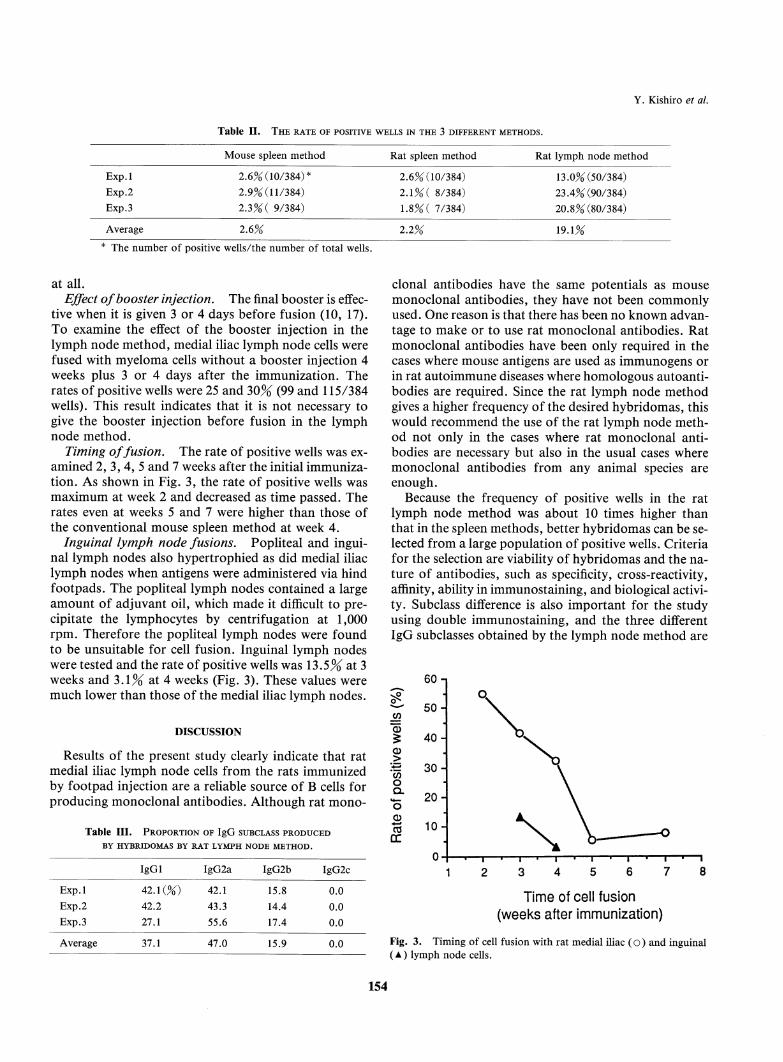
more B cells producing ovalbumin-specific antibodythan the spleen, although many B cells were not fusedwith myeloma cells. When the definition of positivewells is applied, 10 out of 384 wells were positive inboth mouse and rat spleen methods and 90 wells in therat lymph node method. Wells having absorbance over1.0 were 8 in the mouse spleen method, 1 in the ratspleen method and 60 in the rat lymph node method.Table II shows the rate of positive wells in the 3 inde-pendent experiments. The average rate was about 2 to3% in the spleen methods, whereas in the lymph nodemethod the average rate was about 20%. Administra-tion of antigen via hind footpads with subsequent fu-sion with medial iliac lymph node induced about 10times higher incidence of positive hybridomas than thatof the spleen method.IgG subclasses. IgG subclass of antibodies resultingfrom rat lymph node cell fusion was determined (TableIII). On average 47.0% of positive hybridomas for oval-bumin produced IgG2a and 37.1% of IgGl and 15.9%of IgG2b. There were no IgG2c-producing hybridomas
( 3 5 5 ) ( A ) ( 2 8 7 ) ( B ) ( 1 0 5
」 5 0 5 0
(6 9 )
5 0CD* 4 0 4 0 4 0
ofe 3 0 3 0 3 0
_oE 2 0 2 0 2 0
1 0
0 -0 .
1 0 1 0
蝣蝣蝣 _ _ 蝣 - o * サ
0 . 5 1 . 0 1 . 5 2 . 0 0 . 0 0 . 5 1 . 0 1 . 5 2 . 0 0 . 0.5 1 .0 1 .5 2 .0
O D 4 9 2O D 4 9 2 O D 4 9 2
Fig. 2. Histogram of screening of primary hybridoma supernatants by ELISA ll days after cell fusion. A, mouse spleen method; B, rat spleenmethod; C, rat lymph node method
153
Y. Kishiro et al.
Table II. The rate of positive wells in the 3 different methods.
M o u s e s p le e n m e t h o d Ra t sp l e e n m e t ho d R a t l y m p h n o d e m e t h o d
E x p . l 2. 6 % (1 0 / 38 4 ) * 2. 6 % ( 1 0 /3 8 4) 13 . 0% ( 50 / 38 4 )
E x p .2 2 . 9 ^ ( l l / 3 8 4 ) 2. 1 % ( 8 / 3 8 4 ) 2 3. 4 % ( 9 0 /3 8 4 )
E x p .3 2.3 % ( 9 / 3 8 4 ) 1. 8 % ( 7 / 3 8 4 ) 2 0. 8 % ( 8 0 /3 8 4 )
A v era g e 2 . 6 % 2 . 2 % 19. 1 %
The number of positive wells/the number of total wells.
atall.Effect of booster injection. The final booster is effec-tive when it is given 3 or 4 days before fusion (10, 17).To examinethe effect of the booster injection in thelymph node method, medial iliac lymph node cells werefused with myeloma cells without a booster injection 4weeks plus 3 or 4 days after the immunization. Therates of positive wells were 25 and 30% (99 and 115/384wells). This result indicates that it is not necessary togive the booster injection before fusion in the lymphnode method.Timingoffusion. The rate of positive wells was ex-amined 2, 3, 4, 5 and 7 weeks after the initial immuniza-tion. As shown in Fig. 3, the rate of positive wells wasmaximumat week 2 and decreased as time passed. Therates even at weeks 5 and 7 were higher than those ofthe conventional mousespleen methodat week 4.Inguinal lymph node fusions. Popliteal and ingui-nal lymph nodes also hypertrophied as did medial iliaclymph nodes whenantigens were administered via hindfootpads. The popliteal lymph nodes contained a largeamount of adjuvant oil, which made it difficult to pre-cipitate the lymphocytes by centrifugation at 1,000rpm. Therefore the popliteal lymph nodes were foundto be unsuitable for cell fusion. Inguinal lymph nodeswere tested and the rate of positive wells was 13.5% at 3weeks and 3.1% at 4 weeks (Fig. 3). These values weremuch lower than those of the medial iliac lymph nodes.
DISCUSSION
Results of the present study clearly indicate that ratmedial iliac lymph node cells from the rats immunizedby footpad injection are a reliable source of B cells forproducing monoclonal antibodies. Although rat mono-
T a b l e I I I . P r o p o r t i o n o f I g G s u b c l a s s p r o d u c e d
B Y H Y B R I D O M A S B Y R AT L Y M P H N O D E M E T H O D .
I g G I I g G 2 a I g G 2 b I g G 2 c
E x p . 1 4 2 A ( % ) 4 2 . 1 1 5 . 8 0 . 0
E x p .2 4 2 .2 4 3 .3 14 .4 0 .0
E x p .3 2 7 .1 5 5 .6 17 .4 0 .0
A v er a g e 3 7 . 1 4 7 .0 15 .9 0 .0
clonal antibodies have the same potentials as mousemonoclonal antibodies, they have not been commonlyused. One reason is that there has been no knownadvan-tage to makeor to use rat monoclonalantibodies. Ratmonoclonal antibodies have been only required in thecases wheremouseantigens are used as immunogensorin rat autoimmunediseases wherehomologousautoanti-bodies are required. Since the rat lymph node methodgives a higher frequency of the desired hybridomas, thiswould recommendthe use of the rat lymph node meth-od not only in the cases where rat monoclonal anti-bodies are necessary but also in the usual cases wheremonoclonal antibodies from any animal species areenough.
Because the frequency of positive wells in the ratlymph node method was about 10 times higher thanthat in the spleen methods, better hybridomas can be se-lected from a large population of positive wells. Criteriafor the selection are viability of hybridomas and the na-ture of antibodies, such as specificity, cross-reactivity,affinity, ability in immunostaining, and biological activi-ty. Subclass difference is also important for the studyusing double immunostaining, and the three differentIgG subclasses obtained by the lymph node method are
6 0 -
<」 E l
5 0 -CO
CD E lg 4 0 -
CD E>4-サ 3 0 -ino EQ _
2 0 -o
CD E0 3 1 0 -
D CE
Fig.
Time of cell fusion(weeks after immunization)
3. Timing of cell fusion with rat medial iliac (o) and inguinallymph node cells.
154

Hybridoma Preparation Using Rat Lymph Node Cells
convenient for such study.Another advantage of the lymph node method is thata single injection of antigen emulsion is sufficient for im-munization. This advantage saves not only time andeffort but also amounts of immunogenswhich are oftenvery valuable or difficult to obtain. Cell fusion wouldcertainly be possible at any time 2 weeks after the injec-tion.Mouse SP2/O myelomacells were used as a fusionpartner in the present study, but there was absolutely notrouble encountered at all. The rat x mouse hybridomasproliferated as well as mouse x mouse hybridomas did.One practical disadvantage of the rat x mouse hybrido-mas might be mandatoryuse of nude mice or nude ratsfor obtaining ascites. Because reliable rat myelomacellshave already been established (4), if those cells are usedas a fusion partner, there might be no disadvantage com-pared to the mouse conventional method.Mirza et al. reported that administration of antigenvia mouse footpads with subsequent fusion of poplitealand inguinal lymph node lymphocytes induces a higherfrequency of hybridomas secreting specific antibodythan either intradermal immunization and lymph nodelymphocyte fusion or conventional subcutaneous immu-nization and intraperitoneal boost followed by spleniclymphocyte fusion (8). The main differences betweenthe method of Mirza et al. and the rat lymph node meth-od are the animals used and the lymph nodes used. Ratfootpad is naturally larger than that of mouse and islarge enough to accept at least 0.2 ml of antigen emul-sion. Whena rat was injected with 0.1 ml antigen emul-sion, a small amount of injected antigen still remainedin spots in the footpad 4 weeks after the injection. Thisindicated that draining lymph nodes had been exposedto antigens for a long time. It was also found in the pre-sent study that inguinal lymph nodes were less suitablefor cell fusion because of their lower rates of positive hy-bridomas. The use of medial iliac lymph nodes from asingle rat is enough for a single fusion, therefore the useof inguinal lymph nodes is not recommended.A certain stage of B cells is thought to be suitable forcell fusion to produce hybridomas which secrete anti-bodies (1, 6). In the rat lymph node method, the rate ofpositive wells was the highest at 2 weeks aftet the injec-tion and it decreased as time passed. Because the size ofthe medial iliac lymph nodes was almost the same in 2to 7 weeks after the injection, and because the numberof clonies per well was almost the same from fusion tofusion, the decrease of the rate of positive wells may beexplained by the shifting of the stage of B cells into un-suitable stage.Ovalbumin is a protein from chicken eggs that has amolecular weight of 42,699 daltons (9). It is antigenic torats and is often used as a carrier protein for producingantibodies to haptens. To demonstrate reliability of the
lymph node method to other types of antigens, the fol-lowing successful results are discussed below.Wehave recently produced a monoclonal antibody totype IV collagen a5 chain by using a 13-amino-acid-resi-due synthetic peptide as antigen in the form conjugatedto keyhole limpet hemocyanin (18). In general, the prob-ability that monoclonal antibody produced to a synthet-ic peptide reacts with a native antigen is low. Usually,repeated cell fusions are required to obtain the desiredmonoclonal antibody. When we applied our novellymph node method to type IV collagen a5 chain, 34wells were positive to the synthetic peptide by ELISA,but only 3 of them were positive to the native antigen.One of the 3 wells was used for subcloning and a clonewas established. The antibody produced by the clonecould be used in western blotting and in staining of tis-sue sections. The use of the rat lymph node method isstrongly recommended for this kind of work using syn-thetic peptides as antigens.While studying anti-glomerular basement membrane(GBM)nephritis, which is an autoimmune disease in-duced by autoantibodies against type IV collagen inglomerular basement membrane,we were able to estab-lish 2 hybridoma clones which produced nephritogenicautoantibodies belonging to IgG2a (14). These cloneswere obtained from 3 separate fusions by rat spleenmethod. Wehave since applied the rat lymph nodemethod to obtain other types of nephritogenic anti-bodies of different IgG subclasses. Wewere able to ob-tain 122 positive wells against rat GBMin indirect im-munofluorescence. Weestablished 2 clones of hybrido-masproducing IgGl and 2 clones producing IgG2bfrom the positive wells (15). Antibodies from theseclones were definitely nephritogenic although the severi-ty of nephritis varied from antibody to antibody. Theywere autoantibodies with biological activities. We there-fore believe that our novel method using rat medial iliaclymph node is certainly reliable and applicable for pro-duction of new monoclonal antibodies.
Acknowledgments. Wethank Dr. H. Shigei, Chairman of theBoard for financial support and Dr. T. Okigaki, Director of the in-stitute for helpful advice on our research. Wealso thank Mrs. C.Takahashi for taking care of the animals.
REFEREN CES
1. Andersson, J. and Melchers, F. 1978. The antibody reper-toire of hybrid cell lines obtained by fusion of X63-AG8 myelo-ma cells with mitogen-activated B-cell blasts. Curr. Top. Micro-
biol. Immunol, 81: 130-139.Cote, R.J., Morrissey, D.M., Houghton, A.N., Beattie,
E.J., Oettgen, H.F., and Old, L.J. 1983. Generation ofhu-manmonoclonal antibodies reactive with cellular antigens.
Proc. Natl Acad. Sci. USA, 80: 2026-2030.Galfre, G., Howe, S.C., Milstein, C, Butcher, G.W., and
155

Howard, J.C. 1977. Antibodies to major histocompatibilityantigens produced by hybrid cell lines. Nature, 266: 550-552.Galfre, G., Milstein, C., andWright, B. 1979. Ratxrathy-brid myelomas and a monoclonal anti-Fd portion of mouse
IgG. Nature, 277: 131-133.Gefter, M.L., Margulies, D.H., and Scharff, M.D. 1977.
A simple method for polyethylene glycol-promoted hybridiza-tion of mouse myeloma cells. Somat. Cell Genet., 3: 231-236.Goding, J.W. 1980. Antibody production by hybridomas. /.Immunol. Methods, 39: 285-308.Kohler, G. and Milstein, C. 1975. Continuous cultures of
fused cells secreting antibody of predefined specificity. Nature,256: 495-497.
Mirza, I.H., Wilkin, T.J., Cantarini, M., and Moore, K.1987. A comparison of spleen and lymph node cells as fusionpartners for the raising of monoclonal antibodies after differentroutes of immunisation. /. Immunol. Methods, 105: 235-243.
Nisbet, A.D., Saundry, R.H., Moir, A.J.G., Fothergill,
L.A., and Fothergill, J.E. 1981. The complete amino-acid se-quence of hen ovalbumin. Eur. J. Biochem., 115: 335-345.
Oi, V.T., Jones, P.P., Goding, J.W., Herzenberg, L.A., andHerzenberg, L.A. 1978. Properties of monoclonal antibodiesto mouse Ig allotypes, H-2, and la antigens. Curr. Top. Micro-
biol Immunol, 81: 115-129.
Sado, Y., Okigaki, T., Takamiya, H., and Seno, S. 1984. Ex-perimental autoimmuneglomerulonephritis with pulmonary
hemorrhage in rats. The dose-effect relationship of the nephrito-genic antigen from bovine glomerular basement membrane. /.
Clin. Lab. Immunol, 15: 199-204.
Y. Kishiro et al.
12. Sado, Y. and Naito, I. 1987. Experimental autoimmune glo-merulonephritis in rats by soluble isologous or homologousanti-gens from glomerular and tubular basement membrane. Br. J.
Exp. PathoL, 68: 695-704.
13. Sado, Y., Naito, I., and Okigaki, T. 1989. Transfer ofanti-glomerular basement membrane antibody-induced glomerulone-
phritis in inbred rats with isologous antibodies from the urine ofnephritic rats. /. PathoL, 158: 325-332.
14. Sado, Y., Kagawa, M., Rauf, S., Naito, I., Moritoh, C, andOkigaki, T. 1992. Isologous monoclonal antibodies can in-duce anti-GBM glomerulonephritis in rats. /. PathoL, 168: 221-
227.
15. Sado, Y., Kagawa, M. Naito, I., and Okigaki, T. 1993. Animproved method of preparation of rat nephritogenic monoclon-al antibodies. In Abstracts of Xllth International Congress of
Nephrology, (Eliahou, H.E., Iaina, A. and Bar-Khayim, Y.eds.). p. 65.
16. Shulman, M., Wilde, CD., and Kohler, G. 1978. A bettercell line for making hybridomas secreting specific antibodies. Na-
ture, 276: 269-270.
17. Stahli, C, Staehelin, T., Miggiano, V., Schmidt, J., and
Haring, P. 1980. High frequencies of antigen-specific hybrid-omas: dependence on immunization parameters and predictionby spleen cell analysis. /. Immunol. Methods, 32: 297-304.
18. Yoshioka, K., Hino, S., Takemura, T., Maki, S.,
WlESLANDER, J., TAKEKOSHI, Y., MAKINO, H., KAGAWA, M.,Sado, Y., and Kashtan, C.E. 1994. Type IV collagen a5
chain: Normal distribution and abnormalities in X-linked Al-port syndrome revealed by monoclonal antibody. Am. J. Pa-
thoL, 144: 986-996.
{Received for publication, January 12, 1995and in revised form, February 17, 1995)
156