a role for nephrin, a renal protein, in vertebrate ... · a role for nephrin, a renal protein, in...
TRANSCRIPT
A role for nephrin, a renal protein, in vertebrateskeletal muscle cell fusionRegina Lee Sohna,1,2, Ping Huanga,1, Genri Kawaharaa, Matthew Mitchella, Jeffrey Guyona,3, Raghu Kallurib,Louis M. Kunkela,c, and Emanuela Gussonia,4
aDivision of Genetics and Program in Genomics, Children’s Hospital Boston, 300 Longwood Avenue, Boston, MA 02115; cThe Manton Center for OrphanDisease Research, Children’s Hospital Boston and the Howard Hughes Medical Institute, Boston, MA 02115; and bDivision of Matrix Biology, Beth IsraelDeaconess Hospital and Department of Biological Chemistry and Molecular Pharmacology, Harvard Medical School and Harvard-MIT Division of HealthSciences and Technology, Boston, MA 02115
Contributed by Louis M. Kunkel, April 22, 2009 (sent for review February 10, 2009)
Skeletal muscle is formed via fusion of myoblasts, a well-studiedprocess in Drosophila. In vertebrates however, this process is lesswell understood, and whether there is evolutionary conservationwith the proteins studied in flies is under investigation. Sticks andstones (Sns), a cell surface protein found on Drosophila myoblasts,has structural homology to nephrin. Nephrin is a protein expressedin kidney that is part of the filtration barrier formed by podocytes.No previous study has established any role for nephrin in skeletalmuscle. We show, using two models, zebrafish and mice, that theabsence of nephrin results in poorly developed muscles and in-completely fused myotubes, respectively. Although nephrin-knockout (nephrinKO) myoblasts exhibit prolonged activation ofMAPK/ERK pathway during myogenic differentiation, expressionof myogenin does not seem to be altered. Nevertheless, MAPKpathway blockade does not rescue myoblast fusion. Co-cultures ofunaffected human fetal myoblasts with nephrinKO myoblasts ormyotubes restore the formation of mature myotubes; however,the contribution of nephrinKO myoblasts is minimal. These studiessuggest that nephrin plays a role in secondary fusion of myoblastsinto nascent myotubes, thus establishing a possible functionalconservation with Drosophila Sns.
myoblast fusion � sticks and stones
Vertebrate skeletal muscle is a syncytium formed via twophases of myoblast fusion. During the first phase, myoblasts
fuse to generate nascent myotubes that serve as scaffolds forfurther growth; in the second phase, more myocytes fuse intothese nascent myotubes to form mature myotubes (1, 2). Myo-blast fusion is a highly regulated process in Drosophila, wherefounder cells serve as the ‘‘seeds’’ in muscle formation andexpress the membrane protein Kirre, also known as Dumb-founded (Duf ) (3, 4). Fusion-competent myoblasts (FCM) ex-press the transmembrane protein Sticks and stones (Sns) (5)which interacts with Kirre/Duf (6), bringing the two muscle celltypes together, to initiate a complex intra-cellular program thatleads to the fusion of the FCM into the myotubes. There has beensparse evidence demonstrating whether the orthologues of theseproteins are conserved in vertebrate myoblasts (7), althoughrecent studies reported a role for Kirrel (Kirre-like) in developingzebrafish skeletal muscle (8).
Nephrin is a protein the function of which has been associatedwith maintenance of the kidney filtration barrier (9). Mutationsin nephrin cause the congenital nephrotic syndrome of theFinnish type (10). Similarly, nephrin-knockout mice (nephrinKO)die at day 2 after birth of severe proteinuria (11–13). Despite thestructural similarities with Drosophila Sns, nephrin expression orfunction in vertebrate skeletal muscle has not yet been reported.In the current study, nephrin was found expressed in developingmouse skeletal muscle and in human fetal muscle cells under-going fusion. Nephrin expression is also up-regulated in theskeletal muscle of two murine models of human musculardystrophies, when myogenic repair is needed. Downregulation of
nephrin expression in developing zebrafish results in fish unableto swim with abnormal myosepta length. Accordingly, musclecells isolated from nephrinKO mice exhibit a secondary fusiondefect and are unable to form mature myotubes. NephrinKO
myoblast cultures maintain MAPK/ERK pathway activity duringdifferentiation, but this activation does not affect expression ofmyogenin, which appears normal. Pharmacological inhibition ofthe MAPK/ERK pathway does not restore normal myotubeformation, suggesting that this pathway is likely not causative ofthe observed fusion deficiency. In cell mixing experiments,human myoblasts are able to fuse to nephrinKO nascent myotubesto form hybrid mature myotubes. Conversely, nephrinKO myo-blasts provide little or no contribution to developing humanmyotubes. These studies suggest a new role for nephrin inskeletal muscle, where its expression appears to be necessary formononucleated myoblasts to fuse into myotubes. These findingsalso highlight the possible functional conservation betweennephrin and Drosophila Sns.
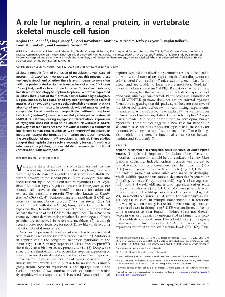
ResultsNephrin Is Expressed in Embryonic, Adult Diseased, or Adult InjuredMuscle. If nephrin is important for fusion of myoblasts intomyotubes, its expression should be up-regulated when myoblastfusion is occurring. Indeed, nephrin message was present bynested reverse transcription–polymerase chain reaction (RT-PCR) in embryonic murine skeletal muscle (Fig. 1A, E15.5), inthe skeletal muscle of young mice with muscular dystrophy,which exhibit spontaneous muscle degeneration/regeneration(14) (Fig. 1 A, mdx Y (dystrophin null); Scg Y (�-sarcoglycan-null), both 3–4 weeks old) and in wild-type muscle after acuteinjury with cardiotoxin (Fig. 1 A, Ctx). No message was detectedin uninjured adult wild-type mouse skeletal muscle (Fig. 1 A,Wt), in 4-month old mdx (Fig. 1 A, mdx O) or �-sarcoglycan (Fig.1A, Scg O) muscles. In multiple independent PCR reactionsfollowed by sequence analysis, the full nephrin message, includ-ing most of exon 1a through the 3�UTR was confirmed to be thesame transcript as that found in kidney (data not shown).Nephrin was also transiently up-regulated in human fetal skel-etal myoblasts (isolated from 17-week-old fetus) undergoingfusion in culture for 2 days (Fig. 1 C–E), after which nephrinexpression returned to the low baseline levels (Fig. 1D). Thus,
Author contributions: R.L.S., P.H., and E.G. designed research; R.L.S., P.H., G.K., M.M., andJ.G. performed research; G.K., R.K., and L.M.K. contributed new reagents/analytic tools;R.L.S., P.H., G.K., L.M.K., and E.G. analyzed data; and R.L.S., P.H., and E.G. wrote the paper.
The authors declare no conflict of interest.
1R.L.S. and P.H. contributed equally to this work.
2Present address: PAREXEL, International, 200 West Street, Waltham, MA 02451.
3Present address: National Marine Fisheries Service, Auke Bay Laboratories, Ted StevensMarine Research Institute, 17109 Pt Lena Loop Road, Juneau, AK 99801.
4To whom correspondence should be addressed. E-mail: [email protected].
This article contains supporting information online at www.pnas.org/cgi/content/full/0904398106/DCSupplemental.
9274–9279 � PNAS � June 9, 2009 � vol. 106 � no. 23 www.pnas.org�cgi�doi�10.1073�pnas.0904398106
nephrin expression is exquisitely controlled and is present whenmyoblast fusion is expected to occur. It was also confirmed thatone nephrin-associated protein, Neph1 (kirrel), is ubiquitouslypresent in all of the muscles tested, regardless of age, disease, orinjury (Fig. 1B).
Downregulation of Nephrin Expression in Developing Zebrafish. Ze-brafish nephrin has been localized to the pronephros as inmammalian kidneys (15). Loss of nephrin by morpholino knock-down results in altered podocyte morphology and nephrosis at96 hpf. Whether the muscles were affected was not indicated.Nephrin morpholino experiments were therefore conducted.Two morpholinos, one directed against nephrin (MO1) and amismatched morpholino (MIS-MO1), were injected into fertil-ized eggs. The morpholino MO1 (kindly provided by I. Drum-mond) is directed against the transmembrane domain of nephrin
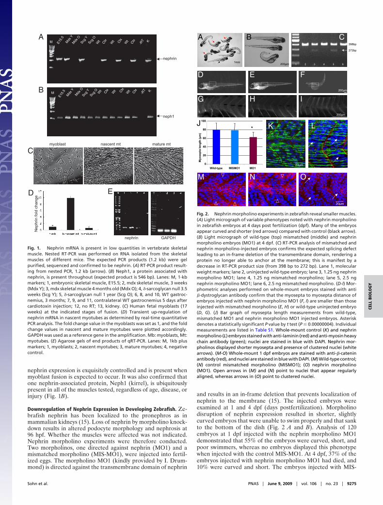
and results in an in-frame deletion that prevents localization ofnephrin to the membrane (15). The injected embryos wereexamined at 1 and 4 dpf (days postfertilization). Morpholinodisruption of nephrin expression resulted in shorter, slightlycurved embryos that were unable to swim properly and that sankto the bottom of the dish (Fig. 2 A and B). Analysis of 120embryos at 1 dpf injected with the nephrin morpholino MO1demonstrated that 55% of the embryos were curved, short, andpoor swimmers, whereas no embryos displayed this phenotypewhen injected with the control MIS-MO1. At 4 dpf, 37% of theembryos injected with nephrin morpholino MO1 had died, and10% were curved and short. The embryos injected with MIS-
A
nephrin
M E15.5
Mdx Y
Wt Ctx
-RT KMdx
OScg
YScg
O
Wt Ctx
Wt Ctx
B
neph1
M
E15.5
Mdx Y
Wt Ctx
-RT K
Mdx O
Scg Y
Scg O Wt Ctx
Wt Ctx
myoblast nascent mt mature mt
C
GAPDHnephrin
Nep
hrin
fold
cha
ngeD E
Fig. 1. Nephrin mRNA is present in low quantities in vertebrate skeletalmuscle. Nested RT-PCR was performed on RNA isolated from the skeletalmuscles of different mice. The expected PCR products (1.2 kb) were gelpurified, sequenced and confirmed to be nephrin. (A) RT-PCR product result-ing from nested PCR, 1.2 kb (arrow). (B) Neph1, a protein associated withnephrin, is present throughout (expected product is 546 bp). Lanes: M, 1-kbmarkers; 1, embryonic skeletal muscle, E15.5; 2, mdx skeletal muscle, 3 weeks(Mdx Y); 3, mdx skeletal muscle 4 months old (Mdx O); 4, �-sarcoglycan null 3.5weeks (Scg Y); 5, �-sarcoglycan null 1 year (Scg O); 6, 8, and 10, WT gastroc-nemius, 3 months; 7, 9, and 11, contralateral WT gastrocnemius 5 days aftercardiotoxin injection; 12, no RT; 13, kidney. (C) Human fetal myoblasts (17weeks) at the indicated stages of fusion. (D) Transient up-regulation ofnephrin mRNA in nascent myotubes as determined by real-time quantitativePCR analysis. The fold change value in the myoblasts was set as 1, and the foldchange values in nascent and mature myotubes were plotted accordingly.GAPDH was used as a reference gene in the amplification. Mb: myoblasts, Mt:myotubes. (E) Agarose gels of end products of qRT-PCR. Lanes: M, 1kb plusmarkers; 1, myoblasts; 2, nascent myotubes; 3, mature myotubes; 4, negativecontrol.
272bp
B
200 m1mm
A C
D E
H
F
398bp
2 3 4 5 61
200 m
G I
200 m
0
20
40
60
80
100
Wild-type MISMO1 MO1
Myo
sept
ale
ngth
(m
)
*J K L
M N O
20 m
20 m
Fig. 2. Nephrin morpholino experiments in zebrafish reveal smaller muscles.(A) Light micrograph of variable phenotypes noted with nephrin morpholinoin zebrafish embryos at 4 days post fertilization (dpf). Many of the embryosappear curved and shorter (red arrows) compared with control (black arrow).(B) Light micrograph of wild-type (top) mismatched (middle) and nephrinmorpholino embryos (MO1) at 4 dpf. (C) RT-PCR analysis of mismatched andnephrin morpholino-injected embryos confirms the expected splicing defectleading to an in-frame deletion of the transmembrane domain, rendering aprotein no longer able to anchor at the membrane; this is manifest by adecrease in RT-PCR product size (from 398 bp to 272 bp). Lane 1, molecularweight markers; lane 2, uninjected wild-type embryo; lane 3, 1.25 ng nephrinmorpholino MO1; lane 4, 1.25 ng mismatched morpholino; lane 5, 2.5 ngnephrin morpholino MO1; lane 6, 2.5 ng mismatched morpholino. (D-I) Mor-phometric analyses performed on whole-mount embryos stained with anti�-dystroglycan antibody confirm that the myosepta to myosepta distance ofembryos injected with nephrin morpholino MO1 (F, I) are smaller than thoseinjected with mismatched morpholino (E, H) or wild-type uninjected embryo(D, G). (J) Bar graph of myosepta length measurements from wild-type,mismatched MO1 and nephrin morpholino MO1 injected embryos. Asteriskdenotes a statistically significant P value by t test (P � 0.00000004). Individualmeasurements are listed in Table S1. Whole-mount control (K) and nephrinmorpholino (L) embryos stained with anti-laminin (red) and anti-myosin heavychain antibody (green); nuclei are stained in blue with DAPI. Nephrin mor-pholinos displayed shorter myosepta and presence of clustered nuclei (whitearrows). (M-O) Whole-mount 1 dpf embryos are stained with anti-�-cateninantibody (red), and nuclei are stained in blue with DAPI. (M) Wild-type control;(N) control mismatched morpholino (MISMO1); (O) nephrin morpholino(MO1). Open arrows in (M) and (N) point to nuclei that appear regularlyaligned, whereas arrows in (O) point to clustered nuclei.
Sohn et al. PNAS � June 9, 2009 � vol. 106 � no. 23 � 9275
CELL
BIO
LOG
Y

MO1 appeared to be normal (wild-type) (Fig. 2B). RT-PCRanalysis confirmed the splicing defect in the embryos injectedwith the nephrin morpholino MO1 (Fig. 2C). To study whetherthe abnormal length of MO1-injected embryos was caused byshorter individual myosepta, the distances between myoseptawas determined by microscopic analysis on whole-mount em-bryos at 96 hpf stained for alpha-dystroglycan (Fig. 2 D–I). Tenembryos each were analyzed for myosepta length in non-injectedcontrol, MIS-MO1, and nephrin MO1-injected embryos. Withineach embryo, five myosepta were measured starting from thesame anatomical position (Fig. 2 D–I, [supporting information(SI) Table S1]. Compared with MIS-MO1–injected embryos, thenephrin MO1-injected zebrafish displayed shorter myoseptum tomyoseptum distance, consistent with a muscle defect (Fig. 2 J;P � 4E-08 via t test). Comparison using a t test of the myoseptumto myoseptum distance in wild-type compared with MO1-injected embryos was also significant (P � 2.08E-08), whereas nosignificant difference was observed by comparing wild-type andMIS-MO1–injected embryos (P � 0.75). Finally, whole-mountcontrol, MIS-MO1–injected, or MO1-injected zebrafish em-bryos were stained at 1dpf using antibodies directed to lamininand myosin heavy chain (MHC, F59) (Fig. 2 K and L) or withanti-�-catenin (Fig. 2 M–O). Wild-type (Fig. 2 K and M) andMIS-MO1-injected embryos (Fig. 2N) displayed regularlyaligned nuclei (Fig. 2 M and N open arrowheads) and compactedmyofibers (Fig. 2K). In contrast, zebrafish embryos injected withnephin morpholino MO1 exhibited a less organized structure inseveral (but not all) myosepta, with compacted clusters of nuclei(Fig. 2L and O, arrows) that were not observed in wild-type orMIS-MO1–injected embryos. Therefore, downregulation ofnephrin expression in developing zebrafish results in a pheno-type consistent with muscle defects.
Assessment of Myocyte Fusion in Nephrin-Knockout Myoblast Cul-tures. To assess whether nephrin plays a role in myoblast fusionand determine when its expression is necessary, we turned to amurine model. Methods for propagation and differentiation ofmuscle cells are well established for mouse compared withzebrafish tissue (16–19). Skeletal myoblasts were isolated fromneonate nephrinKO and wild-type mice, as nephrinKO mice diewithin the first 2 days of life because of kidney failure (11). Thefusion ability of nephrinKO and wild-type cultures were assessedin vitro. For each culture, equal numbers of mononuclear cellswere plated, allowed to differentiate and the fusion indices (FI;number of nuclei in myotubes divided by total number of nuclei)were calculated over 4 successive days. Wild-type and nephrinKO
myoblasts began to fuse from day 1 (Fig. 3 A and D), but by day2 clear differences arose: more of the knockout myocytesremained mononuclear and the overall myotube size was small(Fig. 3 E and F), whereas myotubes in the wild-type cultures werebranching and twitching (Fig. 3 B and C, twitching not shown).The nephrinKO myocytes had lower fusion indices (Fig. 3G) andwere also inefficient in forming large myotubes (containing fiveor more nuclei) compared with wild-type myocytes (Fig. 3H).Immunohistochemistry with an antibody against desmin, a myo-genic specific marker, demonstrated that the mononuclear cellsin the knockout cultures were indeed myogenic, even if they werenot fusing (Fig. 3F, arrows). Moreover, the number of desmin-positive mononuclear cells was significantly higher in knockoutcultures compared with wild-type cultures at days 2, 3 and 4 indifferentiation medium (Fig. 3I). These findings raised thequestion as to whether nephrinKO mononuclear cells had im-paired differentiation potential because of persistent prolifera-tive activity.
NephrinKO Myocytes Have Constitutively Activated MAPK/ERK Path-way, but This Does Not Seem to Affect Myogenin Expression. Wild-type and knockout myoblasts were plated at equal numbers and
cultured under proliferation conditions. Cell numbers werecounted daily for 4 days and although there was no significantdifference during the first 2 days, nephrinKO cells subsequentlygrew much faster than wild-type cells (Fig. S1 A). Samples fromthe cultures were immunostained at day 4 for desmin and thepercentage of positive cells was similar in both cultures (Fig.S1B). Cell cycle analyses were also performed using flow cy-tometry, and control cultures were found to contain half thenumber of cells in S phase compared with nephrinKO cultures(Fig. S1 C and D).
Studies by others have suggested that the p42/44 (Erk1/Erk2)MAPK pathway is necessary for the activation and proliferationof satellite cells upon injury (20, 21). To address whether thedecreased fusion ability of nephrinKO myoblasts could be causedby a persistent activation of Erk1/Erk2, wild-type and nephrinKO
myoblasts differentiated cultures were analyzed by Western blotusing antibodies directed against total and phospho-p42/44MAPK (Fig. S1 E–G). Whereas the p42/44 MAPK pathwayappeared nearly inactive during differentiation in control cul-tures, it was persistently active in the nephrinKO samples(Fig. S1E).
To address whether the constitutive activation of the MAPKpathway could result in slowed myogenic differentiation and,consequently, fusion of nephrinKO myocytes, wild-type andnephrinKO myoblast cultures were differentiated over the courseof 4 days, and myogenin expression was monitored (Fig. S2). Itwas found that myogenin expression was similar in wild-type andnephrinKO cultures, suggesting that early myogenic differentia-tion is not altered by the persistent activation of MAPK/ERK innephrinKO myoblasts. Next, the MAPK inhibitor PD98059 wasapplied during myoblast differentiation. Treatment of nephrinKO
cells with 50 �M PD98059 markedly blocked the phosphoryla-tion of ERK1/2; however nephrinKO myotubes treated with theinhibitor remained small (Fig. S3), suggesting that the persistent
A
D
B
E
C
F
WT
KO
00.10.20.30.40.50.60.70.80.9
Day1
Fusi
on In
dex>
2
Day2 Day3 Day4
***
GWT
KO
% D
esm
in+ c
ells
0
10
20
30
40
50
60
70
80
90
Day1 Day2 Day3 Day4
** *
00.10.20.30.40.50.60.70.8
Fusi
on In
dex>
5
***
Day1 Day2 Day3 Day4
H I
Fig. 3. Myoblasts isolated from nephrinKO neonatal mice are unable to formmature myotubes. Light micrographs of wild-type (A, B) and knockout (D, E)myocytes after 1 day (A, D) and 4 days (B, E) in differentiation medium.Immunofluorescence staining for desmin of wild-type (C) and knockout (F)cultures demonstrates that the mononuclear cells in the nephrinKO culturesare myogenic (white arrows). (G, H) Quantification of the fusion indices forcontrol and nephrinKO myoblasts. The fusion index (FI) is determined bydividing the number of nuclei found within myotubes by the total number ofnuclei in a microscopic field (three to four microscopic fields per sample; threeindependent experiments). (G) Overall FI, counting nuclei in myotubes con-taining two or more nuclei. (H) FI for myotubes containing five or more nuclei.(I) Percentage of desmin-positive mononuclear cells decreases in wild-type asthe FI increases, whereas it remains the same in the nephrinKO cultures.Asterisk indicates significant differences between wild-type (lilac) and knock-out (crimson), P � 0.05.
9276 � www.pnas.org�cgi�doi�10.1073�pnas.0904398106 Sohn et al.

activation of the MAPK pathway is likely not associated with thefusion defect observed in nephrinKO myoblast cultures.
Nephrin Must Be Present on Myoblasts for Secondary Fusion to Occur.Studies in Drosophila suggest that Sns must be present infusion-competent myoblasts (FCM), but not in nascent myo-tubes, for myoblast fusion to occur. Our initial studies did notclarify whether nephrin expression is required on mononuclearcells, nascent myotubes, or both, for myotubes to form. Toaddress this question, coculture experiments were performed bymixing murine myoblasts (wild-type or nephrinKO) with humanmyoblasts. First, murine wild-type and nephrinKO myoblastslabeled with the green fluorescent dye CellTracker CMFDA (22,23) were co-cultured with human myoblasts. Fusion was allowedto occur for 1 and 4 days, after which the presence of mouse- andhuman-derived nuclei to the formed myotubes was quantified(diagrammed in Fig. 4A and Fig. S4). Coculture of wild-typemouse myoblasts with human myoblasts resulted in hybridmyotubes containing a similar proportion of mouse and human-derived nuclei after 1 day (Fig. 4 B and C) and day 4 (Fig. 4 F,
G, and J). In contrast, nephrinKO myoblasts initially formedhybrid myotubes with human cells 1 day after differentiation(Fig. 4 D, E, and K), but by day 4 the contribution of human-derived cells was more predominant (Fig. 4 H, I, and K). Thus,consistent with our previous findings, nascent myotube forma-tion is not affected by nephrin depletion, whereas lack of nephrinexpression results in little additional contribution of murineknockout myoblasts to hybrid myotubes.
To address whether nephrin expression is necessary in nascentmyotubes to allow fusion of myoblasts into them, wild-type ornephrinKO nascent myotubes were co-cultured with humanmononuclear cells (Fig. 5A). Both co-cultures gave rise to hybridmyotubes (Fig. 5 B–E), demonstrating efficient cell fusionbetween murine and human myocytes. The number of totalnuclei in the myotubes, regardless of their origin (murine orhuman), revealed no significant difference between wild-typeand knockout co-cultures (Fig. 5F). However, there were morenuclei of human origin in knockout hybrid myotubes than inwild-type myotubes (37.4% vs. 17.5%, Fig. 5G). Thus, wild-typemyotubes recruit both murine and human myoblasts, whilenephrinKO myotubes recruit primarily human myoblasts. Theconverse experiment was also performed to confirm whethernephrin must be present on myoblasts to complete fusion (Fig.5H). Here, wild-type (Fig. 5 I and J) or nephrinKO myoblasts (Fig.5 K and L) were co-cultured with human nascent myotubesresulting in chimeric myotubes of similar size in both co-cultures(Fig. 5M). However, knockout myoblasts fused with humanmyotubes less efficiently. Only 39% of nuclei in knockoutchimeric myotubes were from murine myotubes compared with56% in wild-type chimeric myotubes (Fig. 5N), and these per-centages were statistically significantly different by t test (P �0.01). These findings support the conclusion that absence ofnephrin in myoblasts results in inefficient formation of second-ary (mature) myotubes.
DiscussionNephrin is a cell surface protein expressed in the kidneyglomerulus at the epithelial podocytes. Podocytes interdigitatewith one another to form a filtration barrier that allows waterand ionic salts, but not proteins, to leave the bloodstream (9).Although nephrin function in the kidney is still largely unknown,it seems to be involved in the ‘‘outside-in’’ signaling that main-tains the communication between podocyte foot processes andsustains the podocyte barrier function (24, 25). Despite struc-tural similarities to Drosophila Sns, a cell-surface protein ex-pressed by fusion-competent myoblasts, nephrin has been re-ported as ‘‘absent’’ in skeletal muscle (26). The results reportedhere indicate that process of myoblast fusion in Drosophila maybe replicated by homologous players in mammalian cells.
Nephrin is expressed in skeletal muscle when cell fusion isoccurring, during development and during injury or disease thatrequire myofiber regeneration. Overall, the level of nephrinexpression is low, and it is present in a narrow window of time.Nephrin is not detected in wild-type adult muscle, whereas it ispresent in murine embryonic muscle and in the muscle of young,but not old, mice with muscular dystrophy, which undergo aspontaneous phase of myofiber regeneration at �2–3 weeks ofage (14). Myofiber regeneration requires fusion of mononuclearcells into nascent myofibers, supporting the need for nephrinexpression. Consistently, young patients (�1 year of age) af-fected by Duchenne Muscular Dystrophy (DMD), express higherlevels of nephrin mRNA in their muscle than unaffected indi-viduals or older DMD patients (27, 28). With age, both mice andhumans with muscular dystrophy exhibit impaired muscle re-generation capacity accompanied by increased fibrosis, partlybecause of exhaustion of resident satellite cells (29–31). There-fore, a decline of nephrin expression in aged mice is consistent
Human myoblastsMouse WT or KO myoblasts
+
Allow cells to fuse for 1 and 4 days, stain with anti-human nuclei Ab
WT KO
A
B
C
D
E
F H
G I0
0.10.20.30.40.50.60.70.80.9
Day1 Day4
% H
uman
nuc
lei i
n hy
brid
myo
tube
s
WTKO
*
K
0
0.1
0.2
0.3
0.4
0.5
0.6
0.7
0.8
Day1 Day4
Fusi
on in
dex
WTKOJ
Fig. 4. NephrinKO myoblasts fuse poorly to human myoblasts compared withcontrol mouse myoblasts. (A) Wild-type or nephrinKO myoblasts were labeledwith a green fluorescent dye and mixed with human myoblasts for 1 and 4 daysin DM. Myotubes were then fixed and stained with anti-human nuclei anti-body (red). Nuclei were stained with DAPI. The myotubes with dual labelingwere analyzed for contribution of human and mouse nuclei. Representativeimages showing the fusion of wild-type (B, C, F, G) or nephrinKO (D, E, H, I)myoblasts to human myoblasts after coculture for 1 day (B-E) and 4 days (F-I).The fusion index (number of fused nuclei, irrespective of their species/totalnumber nuclei) (J), was similar at both days 1 and 4. The contribution of humanversus mouse-derived nuclei to the myotubes was also assessed (K). At day 1,there was no statistical difference between the number of wild-type ornephrinKO mouse myoblasts that had fused to human myoblasts; however, atday 4, many more human-derived nuclei were detected within the myotubesof the nephrinKO:human myoblast hybrid cultures compared with wild-typemouse myoblasts:human myoblasts (P � 8.68204 E-11 by t test). White arrowsin (B) through (I) point to nuclei of murine origin.
Sohn et al. PNAS � June 9, 2009 � vol. 106 � no. 23 � 9277
CELL
BIO
LOG
Y

with the hypothesis of a decline in the number of ‘fusion-competent‘‘ myoblasts in the chronic disease state.
Lack of nephrin in myogenic cells unveils a defect in recruit-ment of fusion-competent myoblasts to nascent myotubes. Myo-blasts from nephrinKO mice are unable to form large myotubesand to maintain a constitutively active MAPK/ERK1/2 pathway,even when in differentiation medium. However, the activatedMAPK/ERK1/2 pathway that has been associated with theactivation of satellite cells (20, 21) does not seem to interferewith early differentiation of nephrinKO myoblasts or to be thecause of inefficient myoblast fusion. In support of this, it wasfound that expression of myogenin did not differ in nephrinKO
cultures compared with control, and that pharmacological inhi-bition of MAPK/ERK pathway failed to rescue normal myotubeformation in nephrinKO cultures. Our results suggest that nephrinmay have a role in vertebrate myoblasts similar to Drosophila Sns.For secondary myotubes to form, nephrin must be present in the
mononucleated myoblasts and not in the nascent myotubes.Conversely, human myoblasts fuse efficiently to nephrinKO nas-cent myotubes, suggesting that nephrin expression on nascentmyotubes is not necessary for wild-type myoblasts to fuse.
Nephrin may act in concert with other transmembrane pro-teins with similar motifs likely to be important for vertebratemuscle fusion. For example, CDO and BOC are members of theIg superfamily and have been found to regulate fusion (32–34).Mannose receptors have been shown to regulate myogenicmotility preceding myoblast fusion (35). Whether these or othermembrane-associated molecules interact with nephrin to resultin myoblast fusion is currently unknown.
Nephrin in the vertebrate kidney is part of a complex networkof proteins that are necessary for the structural and signalingintegrity of the podocyte slit diaphragm (9). Future work mustbe done to determine other structural partners for nephrin inskeletal muscle and factors that modulate the expression of
Allow cells to fuse for 24 hours, stain with anti-human nuclei Ab
Mouse WT or KO myotubes
WT KO
+
Human myoblasts
Allow cells to fuse for 24 hours, stain with anti-human nuclei Ab
+
Mouse WT or KO myoblasts Human myotubes
WT KO
0
2
4
6
8
10
12
14
WT KO
Nuc
lei/
myo
tube
mousehuman
%Sp
ecie
s-sp
ecifi
cnu
clei
inm
yotu
bes
0
10
20
30
40
50
60
70
WT KO
*
02468
10121416182022
WT KO
Nuc
lei/
myo
tube
%Sp
ecie
s-sp
ecifi
cnu
clei
inm
yotu
bes
0102030405060708090
WT KO
*mouse
human
A
B
C
D
E
F G
H
I
J
K
L
M N
Fig. 5. Human fetal myoblasts are able to fuse into nephrinKO nascent myotubes, whereas nephrinKO myoblasts fuse less efficiently into nascent humanmyotubes as compared with wild-type murine myoblasts. (A) Schematic: wild-type or nephrinKO nascent myotubes were labeled with a green fluorescent dyeand mixed with human mononuclear myoblasts for 24 hours in DM. Myotubes were then fixed and stained with anti-human nuclei antibody. Myotubes withdual labeling were analyzed for contribution of human and mouse nuclei. (B-E) Representative confocal images showing fusion of human myoblasts (red nuclei,white arrows) with wild-type (B, C) or nephrinKO (D, E) mouse myotubes (green). Nuclei were stained with DAPI (blue). (F) The total number of nuclei in myotubes,regardless of their origin (mouse or human), does not differ in wild-type and knockout myotubes co-cultures. (G) The percentage of human nuclei in myotubeswith dual labels was calculated and was found to be significantly increased when human myoblasts were mixed with knockout nascent myotubes compared withwild-type nascent myotubes. *P � 0.01. (H) Schematic representation of mixing of prelabeled mouse myoblasts (wild-type or knockout) with human nascentmyotubes. (I-L) Representative confocal images showing the fusion of murine myoblasts (green, white arrowheads) with human myotubes (red nuclei). Nucleiwere stained with DAPI (blue). Coculture of wild-type (I, J) or nephrinKO (K, L) murine myoblasts with human nascent myotubes. The total number of nuclei inmyotubes, regardless of their origin, does not differ in the co-cultures (M); however, the percentage of mouse nuclei in myotubes was significantly decreasedwhen knockout myoblasts were mixed with human myotubes compared with wild-type myoblasts (N). *P � 0.01 via t test.
9278 � www.pnas.org�cgi�doi�10.1073�pnas.0904398106 Sohn et al.

nephrin. Studies suggest that nephrin expression can be in-creased in the kidney by PPAR-� agonists, such as pioglitazone,a drug for diabetes (36). Perhaps similar treatment with myo-blasts can also increase nephrin expression and thereby increasemyoblast fusion. Thus, with more knowledge of the regulation ofnephrin in the skeletal muscle, it may become possible toimprove the efficiency of cell-based therapies for skeletal musclediseases such as Duchenne Muscular Dystrophy.
Materials and MethodsRNA Isolation and RT-PCR Amplification of Nephrin mRNA. Total RNA wasisolated from primary myocyte cultures using Qiagen RNeasy kit (Qiagen) andfrom gastrocnemius muscles of C57Bl6, mdx5cv, delta-sarcoglycan null mice aspreviously described (37). Detailed methods, including cardiotoxin injury,primer sequences, and RT/PCR amplification conditions can be found in the SIMaterials and Methods.
Zebrafish Morpholino Experiments. Zebrafish experiments were performedusing previously described procedures (38), with 1.25 and 2.5 ng of nephrinmorpholino or mismatched morpholino oligonucleotides. Morpholino se-quences, RT-PCR, and whole-mount immunostaining analyses are detailed inSI Materials and Methods.
Primary Muscle Cell Culture and Fusion Experiments. Primary muscle cultureswere derived from the limb muscles of 1–2-day-old wild-type and nephrinKO
neonates as described (18). Differentiation experiments were performed byplating the myocytes at a density of 8 � 104 cells/well in 12-well plates.Detailed information on culture conditions and determination of the fusionindices is described in SI Materials and Methods.
Cell Cycle Analysis. Flow-cytometry analyses of the cell cycle were performedaccording to a previously described protocol (39). Additional details including
Western blot analyses of ERK1/2 proteins can be found in SI Materials andMethods.
Co-Culturing Experiments of Murine and Human Myoblasts. Murine myoblastswere stained with 5 �M CellTracker green CMFDA (Molecular Probes). Labeledmouse and human myoblasts were plated together in equal numbers andallowed to differentiate for 1 and 4 days in differentiation medium. Cells werethen stained with anti-human nuclei antibody (Millipore, clone 235–1, MAB1281). Additional information can be found in SI Materials and Methods.
Co-Cultures of Myoblasts with Nascent Myotubes. To form nascent myotubes,mouse or human myoblasts at �70% confluency were switched to differen-tiation medium (DM) for 24 hours. Meanwhile, 20–30% confluent myoblastswere cultured in DM for 24 hours to produce differentiated, mononucleatedcells. After 24 hours, murine myoblasts or myotubes were labeled with 5 �MCellTracker green CMFDA. Murine and human cells were trypsinized, plated in12-well plates at equal cell numbers, and co-cultured for another 24 hours.Cells were then fixed, permeabilized, and stained with anti-human nucleiantibody (Millipore) as detailed in SI Materials and Methods.
ACKNOWLEDGMENTS. The authors thank Ian Drummond for the nephrinmorpholino (MO1). The authors also thank the MRDDRC flow cytometryfacility, the MRDDRC sequencing facility and MDDRC Imaging Core for con-focal microscopy, all supported by a grant from the National Institute ofHealth (5P30HD018655). The authors express their gratitude to the membersof the Gussoni and Kunkel laboratories for helpful discussions and criticalreview of this manuscript. This work was supported by funding from theMuscular Dystrophy Association to E.G. (MDA 3589 and MDA 4146), theNational Institutes of Health (RO1NS047727 to E.G. and 5P50NS040828 toL.M.K.). L.M.K. is an Investigator supported by the Howard Hughes MedicalInstitute (HHMI). R.L.S. was supported by a Mentored Clinical Scientist Devel-opment Award from the National Heart, Lung, and Blood Institute (NIH-K08HLL04216). P.H. is supported by a Scientist Development Grant fromAmerican Heart Association (0730285N) and by the Joshua Frase Foundation.R.K. is supported by the NIH grant DK 55001. The authors have no conflicts ofinterest to declare.
1. Horsley V, Pavlath GK (2004) Forming a multinucleated cell: Molecules that regulatemyoblast fusion. Cells Tissues Organs 176:67–78.
2. Richardson BE, Nowak SJ, Baylies MK (2008) Myoblast fusion in fly and vertebrates:New genes, new processes and new perspectives. Traffic 9:1050–1059.
3. Abmayr SM, Balagopalan L, Galletta BJ, Hong SJ (2003) Cell and molecular biology ofmyoblast fusion. Int Rev Cytol 225:33–89.
4. Chen EH, Olson EN (2005) Unveiling the mechanisms of cell-cell fusion. Science308:369–373.
5. Bour BA, Chakravarti M, West JM, Abmayr SM (2000) Drosophila SNS, a member of theimmunoglobulin superfamily that is essential for myoblast fusion. Genes Dev 14:1498–1511.
6. Galletta BJ, Chakravarti M, Banerjee R, Abmayr SM (2004) SNS: Adhesive properties,localization requirements and ectodomain dependence in S2 cells and embryonicmyoblasts. Mech Dev 121:1455–1468.
7. Krauss RS (2007) Evolutionary conservation in myoblast fusion. Nat Genet 39:704–705.8. Srinivas BP, Woo J, Leong WY, Roy S (2007) A conserved molecular pathway mediates
myoblast fusion in insects and vertebrates. Nat Genet 39:781–786.9. Tryggvason K (2001) Nephrin: Role in normal kidney and in disease. Adv Nephrol
Necker Hosp 31:221–234.10. Kestila M, et al. (1998) Positionally cloned gene for a novel glomerular protein—
nephrin—is mutated in congenital nephrotic syndrome. Mol Cell 1:575–582.11. Hamano Y, et al. (2002) Determinants of vascular permeability in the kidney glomer-
ulus. J Biol Chem 277:31154–31162.12. Putaala H, et al. (2001) The murine nephrin gene is specifically expressed in kidney,
brain and pancreas: Inactivation of the gene leads to massive proteinuria and neonataldeath. Hum Mol Genet 10:1–8.
13. Rantanen M, et al. (2002) Nephrin TRAP mice lack slit diaphragms and show fibroticglomeruli and cystic tubular lesions. J Am Soc Nephrol 13:1586–1594.
14. Watchko JF, O’Day TL, Hoffman EP (2002) Functional characteristics of dystrophicskeletal muscle: Insights from animal models. J Appl Physiol 93:407–417.
15. Kramer-Zucker AG, Wiessner S, Jensen AM, Drummond IA (2005) Organization of thepronephric filtration apparatus in zebrafish requires Nephrin, Podocin and the FERMdomain protein Mosaic eyes. Dev Biol 285:316–329.
16. Yaffe D, Saxel O (1977) Serial passaging and differentiation of myogenic cells isolatedfrom dystrophic mouse muscle. Nature 270:725–727.
17. Yablonka-Reuveni Z, et al. (1999) The transition from proliferation to differentiationis delayed in satellite cells from mice lacking MyoD. Dev Biol 210:440–455.
18. Rando TA, Blau HM (1994) Primary mouse myoblast purification, characterization, andtransplantation for cell-mediated gene therapy. J Cell Biol 125:1275–1287.
19. Neville C, et al. (1997) Skeletal muscle cultures. Methods Cell Biol 52:85–116.20. Shefer G, et al. (2001) Skeletal muscle cell activation by low-energy laser irradiation: A
role for the MAPK/ERK pathway. J Cell Physiol 187:73–80.21. Jones NC, et al. (2005) The p38alpha/beta MAPK functions as a molecular switch to
activate the quiescent satellite cell. J Cell Biol 169:105–116.
22. Munoz-Barroso I, et al. (1998) Dilation of the human immunodeficiency virus-1 enve-lope glycoprotein fusion pore revealed by the inhibitory action of a synthetic peptidefrom gp41. J Cell Biol 140:315–323.
23. Jaroszeski MJ, Gilbert R, Heller R (1994) Detection and quantitation of cell-cell elec-trofusion products by flow cytometry. Anal Biochem 216:271–275.
24. Lehtonen S (2008) Connecting the interpodocyte slit diaphragm and actin dynamics:Emerging role for the nephrin signaling complex. Kidney Int 73:903–905.
25. Uchida K, et al. (2008) Decreased tyrosine phosphorylation of nephrin in rat and humannephrosis. Kidney Int 73:926–932.
26. Kuusniemi AM, et al. (2004) Tissue expression of nephrin in human and pig. Pediatr Res55:774–781.
27. Haslett JN, et al. (2002) Gene expression comparison of biopsies from Duchennemuscular dystrophy (DMD) and normal skeletal muscle. Proc Natl Acad Sci USA99:15000–15005.
28. Haslett JN, et al. (2003) Gene expression profiling of Duchenne muscular dystrophyskeletal muscle. Neurogenetics 4:163–171.
29. Yablonka-Reuveni Z, Anderson JE (2006) Satellite cells from dystrophic (mdx) micedisplay accelerated differentiation in primary cultures and in isolated myofibers. DevDyn 235:203–212.
30. Webster C, Blau HM (1990) Accelerated age-related decline in replicative life-span ofDuchenne Muscular Dystrophy myoblasts: Implications for cell and gene therapy.Somatic Cell Molec Genet 16:557–565.
31. Pagel CN, Partridge TA (1999) Covert persistence of mdx mouse myopathy is revealedby acute and chronic effects of irradiation. J Neurol Sci 164:103–116.
32. Kang JS, et al. (2002) BOC, an Ig superfamily member, associates with CDO to positivelyregulate myogenic differentiation. EMBO J 21:114–124.
33. Wegorzewska M, Krauss RS, Kang JS (2003) Overexpression of the immunoglobulinsuperfamily members CDO and BOC enhances differentiation of the human rhabdo-myosarcoma cell line RD. Mol Carcinog 37:1–4.
34. Cole F, et al. (2004) Positive regulation of myogenic bHLH factors and skeletal muscledevelopment by the cell surface receptor CDO. Dev Cell 7:843–854.
35. Jansen KM, Pavlath GK (2006) Mannose receptor regulates myoblast motility andmuscle growth. J Cell Biol 174:403–413.
36. Benigni A, et al. (2006) Transcriptional regulation of nephrin gene by peroxisomeproliferator-activated receptor-gamma agonist: Molecular mechanism of the antipro-teinuric effect of pioglitazone. J Am Soc Nephrol 17:1624–1632.
37. Eisenberg I, et al. (2007) Distinctive patterns of microRNA expression in primarymuscular disorders. Proc Natl Acad Sci USA 104:17016–17021.
38. Guyon JR, et al. (2003) The dystrophin associated protein complex in zebrafish. HumMol Genet 12:601–615.
39. Darzynkiewicz Z, Juan G (1997) in Current Protocols in Cytometry, ed. Robinson J.(Wiley, New York), Vol. 1, pp. 7.5.1–7.5.24.
Sohn et al. PNAS � June 9, 2009 � vol. 106 � no. 23 � 9279
CELL
BIO
LOG
Y