a tour of the cell. discovery of cells needed microscopes 1665 – robert hooke observes “boxes”...
TRANSCRIPT

A Tour of the Cell

Discovery of cells needed microscopes
• 1665– Robert Hooke observes “boxes” in cork bark; he
calls them cells.
• 1674– Anton van Leeuwenhoek observes single celled
organisms in well water


Three types of microscope images (Figure 4.1)
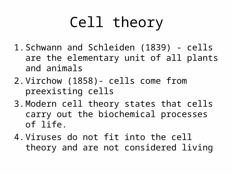
Cell theory
1. Schwann and Schleiden (1839) - cells are the elementary unit of all plants and animals
2. Virchow (1858)- cells come from preexisting cells
3. Modern cell theory states that cells carry out the biochemical processes of life.
4. Viruses do not fit into the cell theory and are not considered living

Relative Sizes (Fig. 4.3)
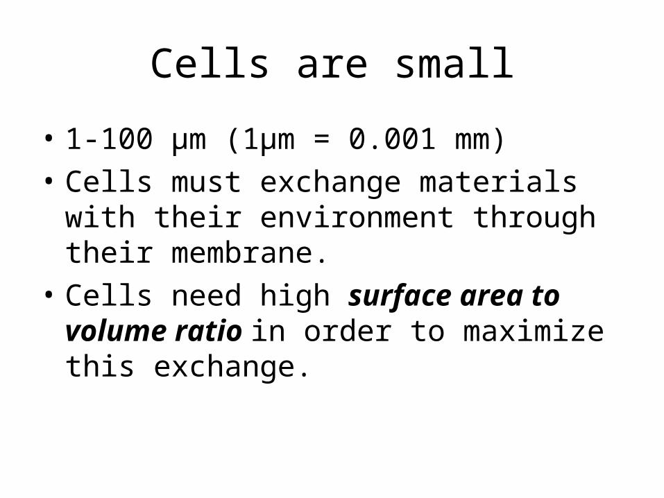
Cells are small
• 1-100 µm (1µm = 0.001 mm)• Cells must exchange materials with their
environment through their membrane. • Cells need high surface area to volume ratio
in order to maximize this exchange.

As size increases, surface area to volume ratio decreases
Length SA = 6 * length2 V = Length3 SA/V
1 6 1 6
2 24 8 3
3 54 27 2
4 96 64 1.5
5 150 125 1.2

Two types of cells: Prokaryotes and Eukaryotes

Prokaryotes are bacterial cells
• Archaea, Eubacteria– Most primitive organisms
• No inner structure • DNA floats in cytoplasm• External structures
– Cell wall– Flagella - move– Pili – for attachment and transfer DNA
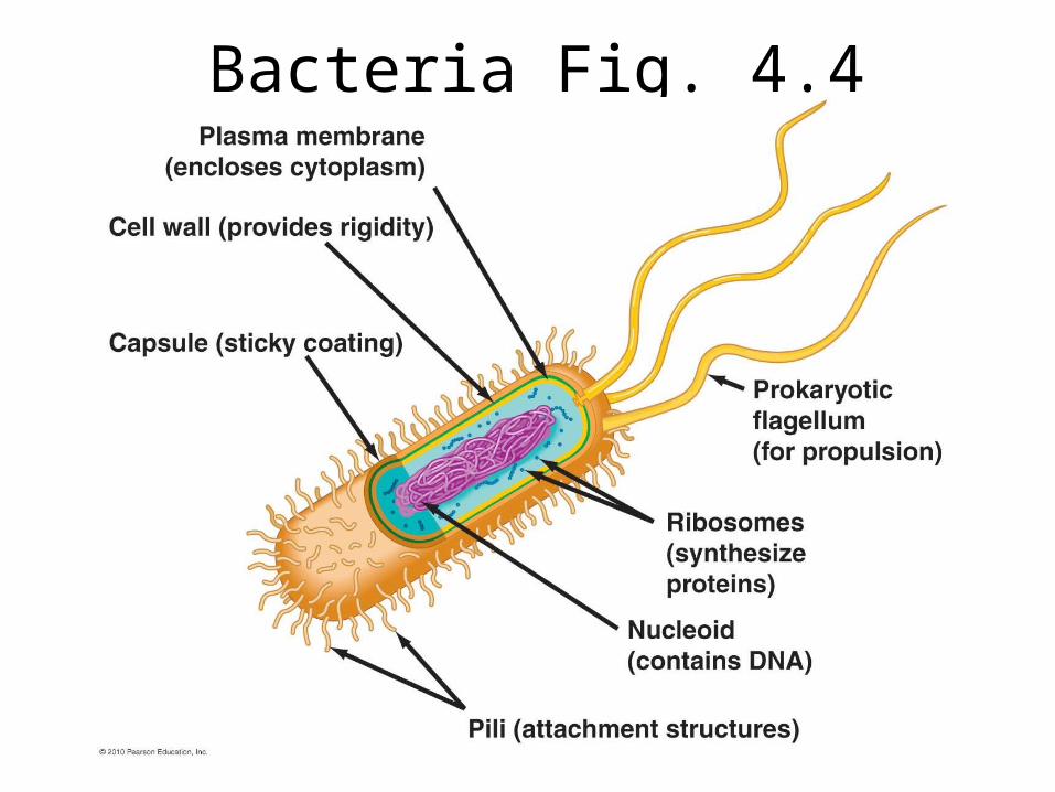
Bacteria Fig. 4.4
Eukaryotes• All other kingdoms
– Animalia– Plantae– Fungi– Protist kingdom(s)

Animal Cell (Fig. 4.5)
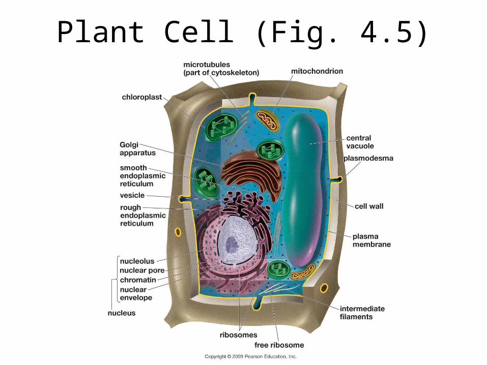
Plant Cell (Fig. 4.5)

External Eukaryote Structure
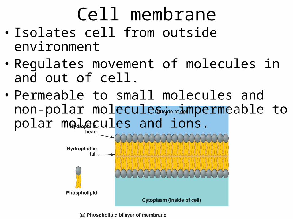
Cell membrane• Isolates cell from outside environment• Regulates movement of molecules in and out of
cell.• Permeable to small molecules and non-polar
molecules; impermeable to polar molecules and ions.
Three main components of cell membrane
• Phospholipid bi-layer: two layers of phospholipids situated with hydrophilic ends facing out ward and hydrophobic tails facing inward
• Cholesterols – Four ringed lipids, regulates the fluidity of membrane
• Proteins – Hydrophobic a.a within membrane with hydrophilic a.a outside
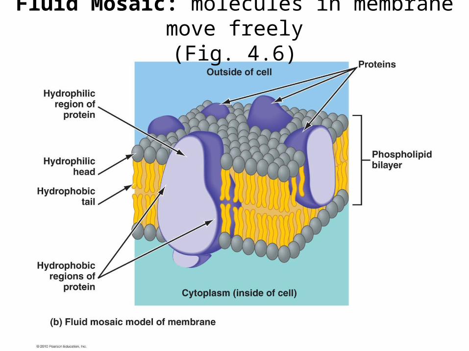
Fluid Mosaic: molecules in membrane move freely(Fig. 4.6)

Three Types of Membrane Proteins
• Transport proteins: allow specific molecules to enter/exit cell
• Receptor proteins: bind to molecules ( i.e. hormones, nutrients)
• Recognition proteins: cell specific proteins that identify cell.
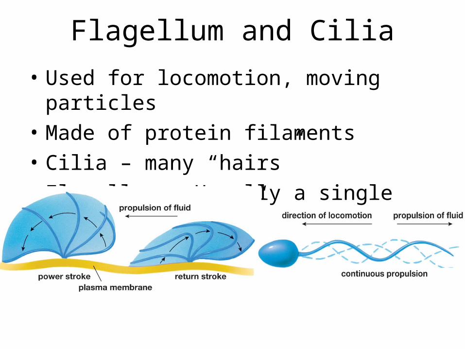
Flagellum and Cilia
• Used for locomotion, moving particles• Made of protein filaments• Cilia – many “hairs”• Flagellum – Usually a single undulating “tail”

Flagellum and Cilia (Fig. 4.22)

Inside a Eukaryote
• Cytoplasm• Organelles made of phospholipid bilayer• Nucleus containing chromosomes• Mitochondria – in most eukaryotes• Chloroplast – in plants and some protists

Cytoplasm
• Viscous liquid• Dissolved molecules• Organelles• High concentration of proteins
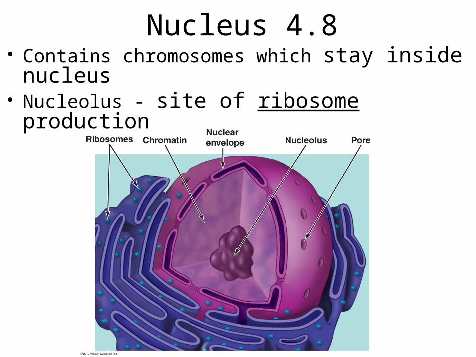
Nucleus 4.8• Contains chromosomes which stay inside nucleus• Nucleolus - site of ribosome production

Ribosomes (Fig. 4.10)
• Small protein subunits (Large and small)• Site of protein synthesis
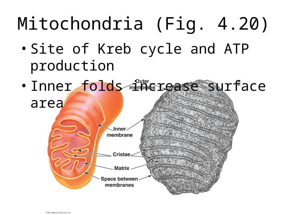
Mitochondria (Fig. 4.20)• Site of Kreb cycle and ATP production• Inner folds increase surface area


Chloroplasts Fig. 4.19• Contain chlorophyll (green pigment)• Site of photosynthesis

Endoplasmic Reticulum
• Membrane extension of the nucleus• Many folds = lots surface area• Rough ER – contain ribosomes which make
proteins• Smooth ER – makes phospholipids• Makes new membrane• Break down toxins

Endoplasmic Reticulum (Fig. 4.13)
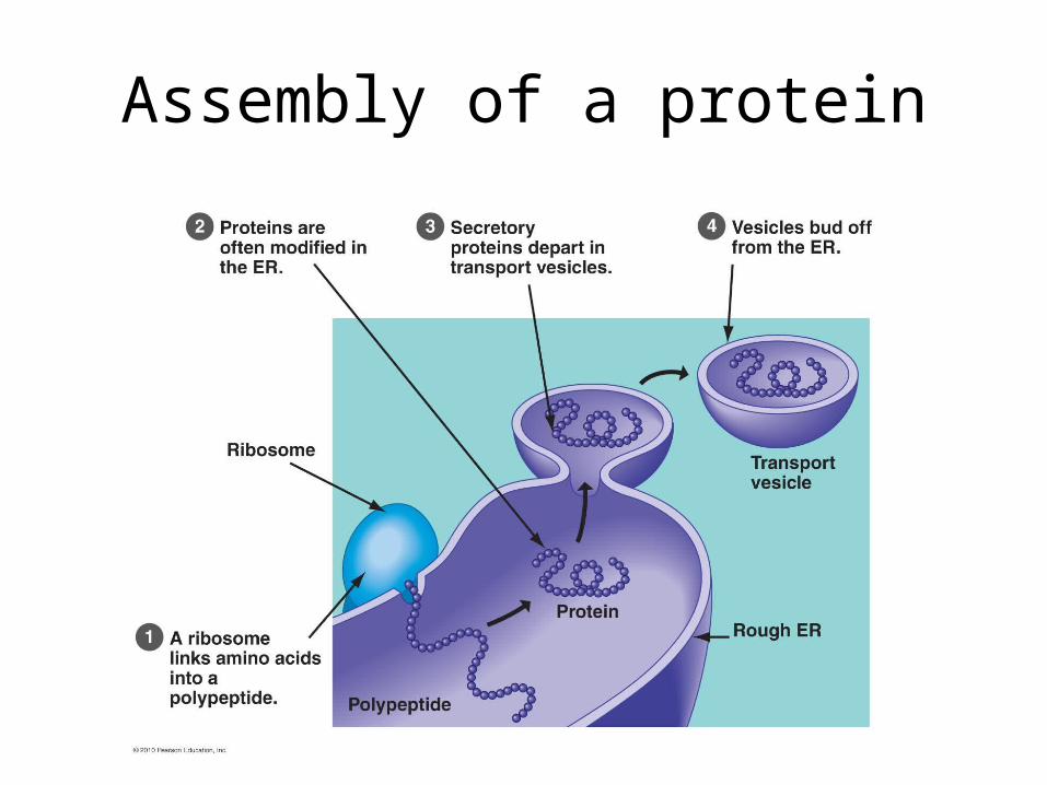
Assembly of a protein
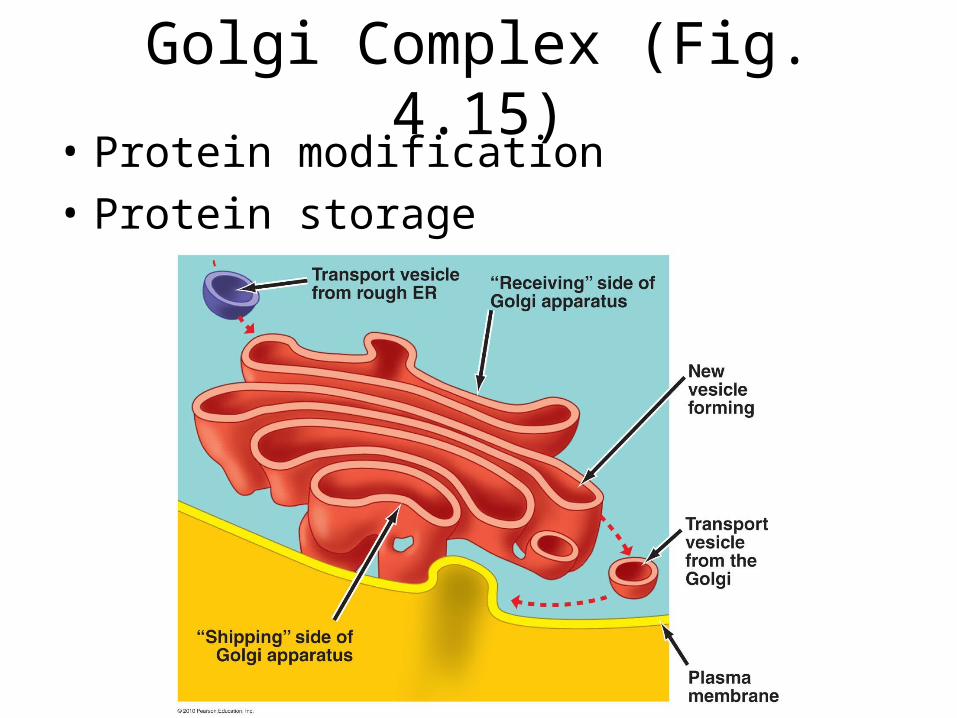
Golgi Complex (Fig. 4.15)• Protein modification • Protein storage

Vacuoles (Fig. 4.17)• Membrane bound storage structures• Contractile vacuoles regulate water content
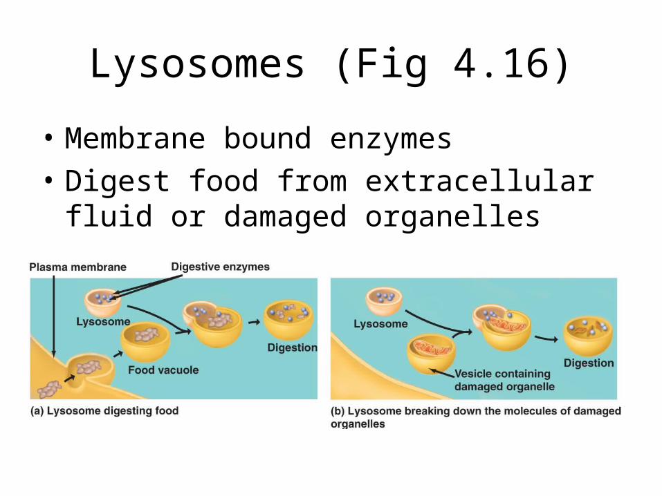
Lysosomes (Fig 4.16)
• Membrane bound enzymes• Digest food from extracellular fluid or
damaged organelles

Cytoskeleton provides structure and support (Fig. 4.21)
• Interconnected proteins• Can change arrangement and location within a cell

(Fig. 4.21)