a two-step mechanism for epigenetic specification of ...cmm.ucsd.edu/cleveland/dwc journal...
TRANSCRIPT

ART I C L E S
A two-step mechanism for epigenetic specification ofcentromere identity and functionDaniele Fachinetti1, H. Diego Folco1,4, Yael Nechemia-Arbely1, Luis P. Valente2, Kristen Nguyen1, Alex J. Wong1,Quan Zhu3, Andrew J. Holland1,4, Arshad Desai1, Lars E. T. Jansen2 and DonW. Cleveland1,5
The basic determinant of chromosome inheritance, the centromere, is specified in many eukaryotes by an epigenetic mark. Usinggene targeting in human cells and fission yeast, chromatin containing the centromere-specific histone H3 variant CENP-A isdemonstrated to be the epigenetic mark that acts through a two-step mechanism to identify, maintain and propagate centromerefunction indefinitely. Initially, centromere position is replicated and maintained by chromatin assembled with thecentromere-targeting domain (CATD) of CENP-A substituted into H3. Subsequently, nucleation of kinetochore assembly ontoCATD-containing chromatin is shown to require either the amino- or carboxy-terminal tail of CENP-A for recruitment of innerkinetochore proteins, including stabilizing CENP-B binding to human centromeres or direct recruitment of CENP-C, respectively.
The centromere is the fundamental unit for ensuring chromosomeinheritance. Centromeres have a distinct type of chromatin in which hi-stoneH3 is replaced by a conserved homologue initially identified in hu-mans and named CENP-A (refs 1,2). Although specific chromosomalregions containing α-satellite repeats3 are the sites of centromereformation and CENP-A association in humans, α-satellite DNAsequences are neither sufficient nor essential for centromere identity4.Rather, except for budding yeast, it is well accepted that in almost allother organisms, including fission yeast, centromeres depend on oneor more as yet unidentified epigenetic marks5. Among the strongestevidence for an epigenetically defined centromere was the discoveryin humans of migration of a functional centromere from an initiallocation to a new site on the same chromosome6–9. These loci, referredto as neocentromeres, are stably loaded with CENP-A (ref. 8) and format previous euchromaticDNA sites withoutα-satellite repeats6,10,11.Epigenetic inheritance implies that the mark must be self-templating
and be stably maintained across the cell cycle. Centromere-boundCENP-A molecules are indeed quantitatively redistributed to bothsister centromeres during centromeric DNA replication, yielding eachdaughter centromere with half the level of the initially loaded CENP-A(ref. 12). Furthermore, in human12 and Drosophila13,14 cells, loading ofnew CENP-A into centromeric chromatin occurs once per cell cycleonly during or on exit frommitosis.
1Ludwig Institute for Cancer Research and Department of Cellular and Molecular Medicine, University of California at San Diego, La Jolla, California 92093, USA.2Instituto Gulbenkian de Ciência, Rua da Quinta Grande 6, P-2780-156, Oeiras, Portugal. 3Laboratory of Genetics, The Salk Institute for Biological Studies, La Jolla,California 92037, USA. 4Present addresses: Laboratory of Biochemistry and Molecular Biology, National Cancer Institute, National Institutes of Health, Bethesda,Maryland 20892, USA (H.D.F.); Department of Molecular Biology and Genetics, Johns Hopkins University School of Medicine, Baltimore, Maryland 21205, USA(A.J.H.).5Correspondence should be addressed to D.W.C. (e-mail: [email protected])
Received 7 February 2013; accepted 11 June 2013; published online 21 July 2013; DOI: 10.1038/ncb2805
Although the precise nature of centromeric chromatin is highlycontroversial15–24, CENP-A is a central component in all models.Reduction or mutation in CENP-A (refs 25–28) has proved it to beessential for continuing centromere function27. Indeed, forced loadingof CENP-A by tethering it or components that bind it to specific DNAloci has produced partial ectopic centromere formation29–32 and inone case with full kinetochore function after deletion of the authenticcentromere33. These reports reinforce the sufficiency of CENP-Afor recruitment of some centromere and kinetochore components,but offer no insight into how centromere identity and function areepigenetically specified and maintained. Approaches25–28 relying onRNA interference (RNAi)-mediated messenger RNA degradation toidentify the epigenetic mark have suffered from incomplete silencing,especially for proteins such as CENP-A with long half-lives. Toovercome these limitations, we now use gene targeting in humandiploid cells and in fission yeast to demonstrate that CENP-A-containing chromatin is the epigenetic mark that can identify, maintainand propagate human or yeast centromere function indefinitely.
RESULTSConditional CENP-A gene deletion in diploid human cellsUsing a single-stranded adeno-associated virus 2 (AAV2) vector toenhance homologous recombination34,35, both human CENP-A alleles
1056 NATURE CELL BIOLOGY VOLUME 15 | NUMBER 9 | SEPTEMBER 2013
© 2013 Macmillan Publishers Limited. All rights reserved.
ART I C L E S
CENP-A–/F 5 days after Ad-Cre transduction
Perc
en
tag
e o
f
ob
serv
ed
even
ts
MicronucleiLagging chromosomesChromosomes mis-aligned
50
75
100
125
Tim
e in
mito
sis
(m
in)
25
Day:
17
Mr (K)
55
Allele 2
X XX X X XX X
1 2 3 4
1
4
5_3′UTR
5_3′UTR
ATG
ATG
STOP
STOP
CENP-A+/+
Allele 2
X X X XX X 5_3′UTR
STOP
Neomycin 5_3′UTR
2 5_3′UTR1
Flox allele
3 NeomycinX X X X X X4 X 5_3′UTR1
CENP-A–/F
X = polymorphismN
TA
d-C
re
ACACENP-A Merge
CE
NP
-A in
ten
sity (%
)
Day:
Days after Ad-Cre infection
CENP-AF/+
1 X
X
2 3
2 3 41
ATG
FRT
FRT FRT
FRT
FRTATG
ATG
ATG
1 2 3 4
STOP
2
STOP
+/+ F/+ –/F
F/+ –/F
F/+ –/F
0 1 3 5 7 0 1 3 5 7
0Day 0 3 5 0 3 5 7
Day 0 3 5 0 3 5 7
10
20
30
9′ 27′ 48′ 51′
54′ 57′ 60′ 105′
0
25
50
75
100
0 1 3 5 7 9
e
CATD
CATD
CATD
LoxP
LoxP
LoxP
LoxP
LoxP
CATD
CATD
4
CENP-A
α-tubulin
a d
0
1
3
5
7
9
Days after F/+
Ad-Cre transduction
–/F
Allele 1
Flox allele
KO allele
f
c
b
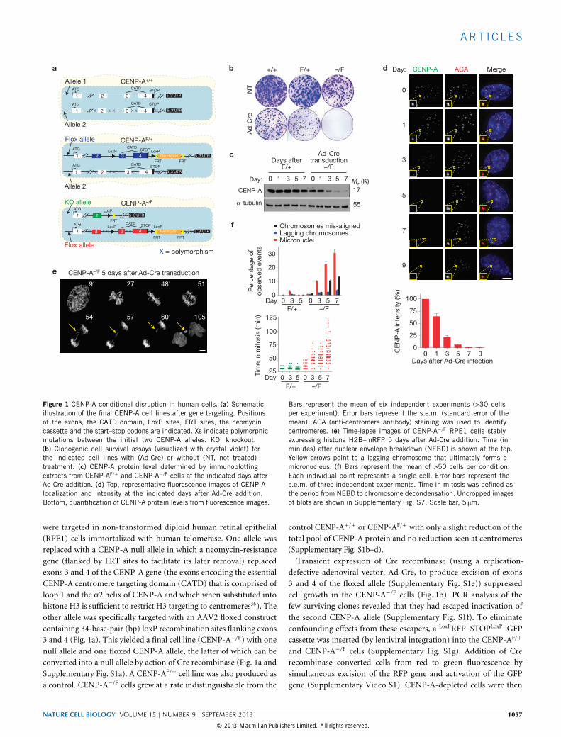
Figure 1 CENP-A conditional disruption in human cells. (a) Schematicillustration of the final CENP-A cell lines after gene targeting. Positionsof the exons, the CATD domain, LoxP sites, FRT sites, the neomycincassette and the start–stop codons are indicated. Xs indicate polymorphicmutations between the initial two CENP-A alleles. KO, knockout.(b) Clonogenic cell survival assays (visualized with crystal violet) forthe indicated cell lines with (Ad-Cre) or without (NT, not treated)treatment. (c) CENP-A protein level determined by immunoblottingextracts from CENP-AF/+ and CENP-A−/F cells at the indicated days afterAd-Cre addition. (d) Top, representative fluorescence images of CENP-Alocalization and intensity at the indicated days after Ad-Cre addition.Bottom, quantification of CENP-A protein levels from fluorescence images.
Bars represent the mean of six independent experiments (>30 cellsper experiment). Error bars represent the s.e.m. (standard error of themean). ACA (anti-centromere antibody) staining was used to identifycentromeres. (e) Time-lapse images of CENP-A−/F RPE1 cells stablyexpressing histone H2B–mRFP 5 days after Ad-Cre addition. Time (inminutes) after nuclear envelope breakdown (NEBD) is shown at the top.Yellow arrows point to a lagging chromosome that ultimately forms amicronucleus. (f) Bars represent the mean of >50 cells per condition.Each individual point represents a single cell. Error bars represent thes.e.m. of three independent experiments. Time in mitosis was defined asthe period from NEBD to chromosome decondensation. Uncropped imagesof blots are shown in Supplementary Fig. S7. Scale bar, 5 µm.
were targeted in non-transformed diploid human retinal epithelial(RPE1) cells immortalized with human telomerase. One allele wasreplaced with a CENP-A null allele in which a neomycin-resistancegene (flanked by FRT sites to facilitate its later removal) replacedexons 3 and 4 of the CENP-A gene (the exons encoding the essentialCENP-A centromere targeting domain (CATD) that is comprised ofloop 1 and the α2 helix of CENP-A and which when substituted intohistone H3 is sufficient to restrict H3 targeting to centromeres36). Theother allele was specifically targeted with an AAV2 floxed constructcontaining 34-base-pair (bp) loxP recombination sites flanking exons3 and 4 (Fig. 1a). This yielded a final cell line (CENP-A−/F) with onenull allele and one floxed CENP-A allele, the latter of which can beconverted into a null allele by action of Cre recombinase (Fig. 1a andSupplementary Fig. S1a). A CENP-AF/+ cell line was also produced asa control. CENP-A−/F cells grew at a rate indistinguishable from the
control CENP-A+/+ or CENP-AF/+ with only a slight reduction of thetotal pool of CENP-A protein and no reduction seen at centromeres(Supplementary Fig. S1b–d).Transient expression of Cre recombinase (using a replication-
defective adenoviral vector, Ad-Cre, to produce excision of exons3 and 4 of the floxed allele (Supplementary Fig. S1e)) suppressedcell growth in the CENP-A−/F cells (Fig. 1b). PCR analysis of thefew surviving clones revealed that they had escaped inactivation ofthe second CENP-A allele (Supplementary Fig. S1f). To eliminateconfounding effects from these escapers, a LoxPRFP–STOPLoxP–GFPcassette was inserted (by lentiviral integration) into the CENP-AF/+
and CENP-A−/F cells (Supplementary Fig. S1g). Addition of Crerecombinase converted cells from red to green fluorescence bysimultaneous excision of the RFP gene and activation of the GFPgene (Supplementary Video S1). CENP-A-depleted cells were then
NATURE CELL BIOLOGY VOLUME 15 | NUMBER 9 | SEPTEMBER 2013 1057
© 2013 Macmillan Publishers Limited. All rights reserved.

ART I C L E S
Day 9
Day 0
CE
NP
-C inte
nsity (%
)
Days after Ad-Cre infection
0 1 3 5 7 9
Days after Ad-Cre infection
0 1 3 5 7 9
Days after Ad-Cre infection
0 1 3 5 7 9
CE
NP
-A inte
nsity (%
)
Day 0
Day 7
CE
NP
-T inte
nsity (%
)
CE
NP
-A inte
nsity (%
)
Day 0
Day 9
CE
NP
-N inte
nsity (%
)
CE
NP
-A inte
nsity (%
)
Pro
tein
inte
nsity (%
)
CENP-A
CENP-N
CENP-C
CENP-P
1 3 5 7 9
Days after Ad-Cre infection
Days after Ad-Cre infection
Days after Ad-Cre infection
0
25
50
75
100
Pro
tein
inte
nsity (%
)
CENP-I Ndc80
1 3 5 7 9
Pro
tein
inte
nsity (%
)
CENP-A CENP-B
1 3 5 7 9
0
25
50
75
100
0
25
50
75
100
0
25
50
75
100
CENP-C ACACENP-A Merge
CENP-NSNAP ACACENP-A Merge
CENP-T ACACENP-A Merge
0
25
50
75
100
0
25
50
75
100
d
e
f
a
b
c
CENP-A CENP-T
Figure 2 Disrupted centromere positioning and kinetochore nucleationrequires almost complete loss of CENP-A. (a–c) Left, representative fluores-cence images show the localization and intensity of CENP-A and CENP-C(a), CENP-N (b) and CENP-T (c) at day 0 and 7 or 9 after Ad-Cre addition toinactivate the remaining CENP-A allele in CENP-A−/F cells. Right, bar graphs(the mean of four independent experiments; >30 cells per experiment) showCENP-A (red), CENP-C (green), CENP-N (orange) and CENP-T (blue) levels 0to 9 days after Ad-Cre addition. Error bars represent the s.e.m. ACA staining
was used to identify centromeres. CENP-N was tracked by covalent labellingwith rhodamine-benzyl guanine after stable expression of a gene encodingSNAP-tagged CENP-N. (d–f) Kinetics of loss from centromeres as CENP-Alevels diminish defines three groups with similar kinetics of loss: proteinslost in proportion to CENP-A (d), those lost more slowly and whose depletionrequires complete loss of CENP-A (e), and CENP-B (f), whose binding isreduced by half by complete loss of CENP-A. Statistics source data are inSupplementary Table S2. Scale bar, 5 µm.
collected by fluorescence-activated cell sorting (FACS) to recovergreen fluorescent cells. After depletion of both alleles of CENP-A, cellsduplicated at a similar rate as control cells for the first 5 days (∼6divisions), then exhibited slowed cycling from extended mitosis andstopped dividing only 9–11 days (∼8–9 divisions) after initial CENP-Agene inactivation (Supplementary Fig. S1h).CENP-A excision led to progressive loss of accumulated CENP-A
and its levels decreased by approximately half each cell cycle (Fig. 1c,d),consistent with the expected redistribution of centromere-boundCENP-A to sister centromeres during DNA replication but withoutaddition of new CENP-A (ref. 12). Only 1% of the initial CENP-Alevel was detectable within 7 days (Fig. 1d; as expected for the1/27dilution = 0.8%). No centromere-bound CENP-A could bedetected 9 days following excision (Fig. 1d). Lowered levels of CENP-Acorresponded with increased rates of chromosome segregation defectsand increased mitotic duration, accompanied by a significant increaseinmicronuclei formation from failure in initial kinetochore attachmentto spindle microtubules or initially aligned chromosomes lagging inanaphase (Fig. 1e,f and Supplementary Video S2).
Disrupted kinetochore nucleation requires almost completeloss of CENP-ACENP-A loading has been proposed to be at the foundation ofrecruitment (direct and indirect) of all of the components of thecore centromere26,37–39. Three patterns of centromere/kinetochoreprotein loss after inactivation of the conditional CENP-A allele in theCENP-A−/FRPE1 cells were identified in asynchronous cycling cells.The first group included: CENP-C and CENP-N, two primary
components of the constitutive centromere-associated network(CCAN) that have been shown to directly interact with CENP-Achromatin37,38 and CENP-P, a more distal kinetochore protein39. Lossof centromere-bound CENP-C, CENP-N and CENP-P was initiallyin proportion to loss of CENP-A (Fig. 2a,b,d and SupplementaryFig. S2a), consistent with direct binding to CENP-A chromatin or, inthe case of CENP-P, through a CENP-N/CENP-C-dependent complex.Surprisingly, a substantial proportion (∼30%) of CENP-C remainedeven after CENP-A depletion to <1%of its initial level (Fig. 2a,d).A second pattern included: CENP-T, a component of the CENP-
T/W/S/X complex and whose histone fold domains bind to chromatin
1058 NATURE CELL BIOLOGY VOLUME 15 | NUMBER 9 | SEPTEMBER 2013
© 2013 Macmillan Publishers Limited. All rights reserved.
ART I C L E S
NH2 CATD
1 44 52
αN α1 α2 α3
75 114 135–14029
Untreated UntreatedAd-Cre Ad-Cre
16
43/44
43/44
Mr (K)
55
F/+ –/F
–/F
CENP–A
Ad-Cre:
–/F
H3CATD
- +- - - +
H3 C
ATD
H3CATD
H3 C
-A
H3C-A
NH2H3 C
ATD
NH2H3CATD
H3 C
ATD+C
H3CATD+C
CEN
P-A C-H
3
CENP-AC-H3
H3-N
H2CEN
P-A
H3-NH2CENP-A
αNH3 C
ATD
αNH3CATD
A-NH2H3
A-NH2H3
H3
H3
Co
lonie
s
(perc
enta
ge r
ela
tive t
o u
ntr
eate
d)
NH2(1-29)H
3 CATD
NH2(1-29)H3CATD
Short-term viability assay
CENP-A +Rescue
–/F
+Retrovirus
EYFP–rescue
CENP-A–/F
+Ad-Cre
Plate
500 cellspuro or blast selection
14 days
Short-term
viability
14 days
F/+–/F
CEN
P-A
CENP-A
C
0
20
40
60
80
a d
e
b
c
GFP
CENP-A
α-tubulin
NH
2H
3C
AT
DH
3C
AT
D+
CH
3
F/+
H3
CA
TD
CE
NP
-A
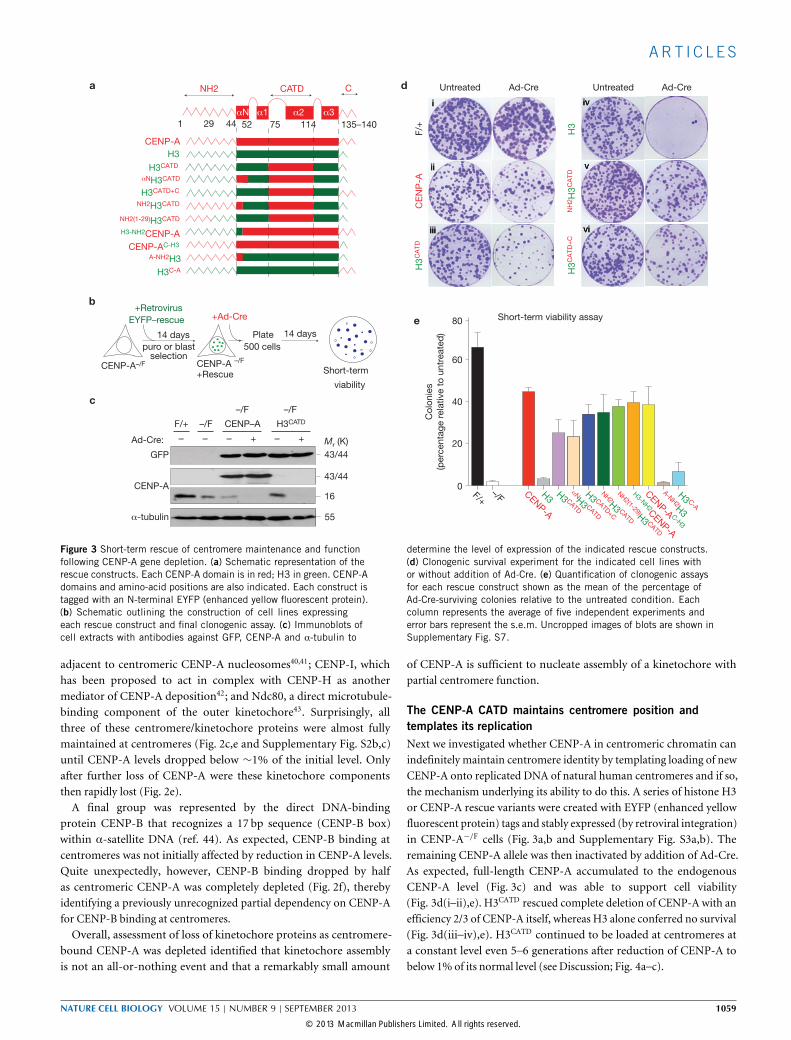
Figure 3 Short-term rescue of centromere maintenance and functionfollowing CENP-A gene depletion. (a) Schematic representation of therescue constructs. Each CENP-A domain is in red; H3 in green. CENP-Adomains and amino-acid positions are also indicated. Each construct istagged with an N-terminal EYFP (enhanced yellow fluorescent protein).(b) Schematic outlining the construction of cell lines expressingeach rescue construct and final clonogenic assay. (c) Immunoblots ofcell extracts with antibodies against GFP, CENP-A and α-tubulin to
determine the level of expression of the indicated rescue constructs.(d) Clonogenic survival experiment for the indicated cell lines withor without addition of Ad-Cre. (e) Quantification of clonogenic assaysfor each rescue construct shown as the mean of the percentage ofAd-Cre-surviving colonies relative to the untreated condition. Eachcolumn represents the average of five independent experiments anderror bars represent the s.e.m. Uncropped images of blots are shown inSupplementary Fig. S7.
adjacent to centromeric CENP-A nucleosomes40,41; CENP-I, whichhas been proposed to act in complex with CENP-H as anothermediator of CENP-A deposition42; and Ndc80, a direct microtubule-binding component of the outer kinetochore43. Surprisingly, allthree of these centromere/kinetochore proteins were almost fullymaintained at centromeres (Fig. 2c,e and Supplementary Fig. S2b,c)until CENP-A levels dropped below ∼1% of the initial level. Onlyafter further loss of CENP-A were these kinetochore componentsthen rapidly lost (Fig. 2e).A final group was represented by the direct DNA-binding
protein CENP-B that recognizes a 17 bp sequence (CENP-B box)within α-satellite DNA (ref. 44). As expected, CENP-B binding atcentromeres was not initially affected by reduction in CENP-A levels.Quite unexpectedly, however, CENP-B binding dropped by halfas centromeric CENP-A was completely depleted (Fig. 2f), therebyidentifying a previously unrecognized partial dependency on CENP-Afor CENP-B binding at centromeres.Overall, assessment of loss of kinetochore proteins as centromere-
bound CENP-A was depleted identified that kinetochore assemblyis not an all-or-nothing event and that a remarkably small amount
of CENP-A is sufficient to nucleate assembly of a kinetochore withpartial centromere function.
The CENP-A CATD maintains centromere position andtemplates its replicationNext we investigated whether CENP-A in centromeric chromatin canindefinitely maintain centromere identity by templating loading of newCENP-A onto replicated DNA of natural human centromeres and if so,the mechanism underlying its ability to do this. A series of histone H3or CENP-A rescue variants were created with EYFP (enhanced yellowfluorescent protein) tags and stably expressed (by retroviral integration)in CENP-A−/F cells (Fig. 3a,b and Supplementary Fig. S3a,b). Theremaining CENP-A allele was then inactivated by addition of Ad-Cre.As expected, full-length CENP-A accumulated to the endogenousCENP-A level (Fig. 3c) and was able to support cell viability(Fig. 3d(i–ii),e). H3CATD rescued complete deletion of CENP-A with anefficiency 2/3 of CENP-A itself, whereas H3 alone conferred no survival(Fig. 3d(iii–iv),e). H3CATD continued to be loaded at centromeres ata constant level even 5–6 generations after reduction of CENP-A tobelow 1% of its normal level (see Discussion; Fig. 4a–c).
NATURE CELL BIOLOGY VOLUME 15 | NUMBER 9 | SEPTEMBER 2013 1059
© 2013 Macmillan Publishers Limited. All rights reserved.

ART I C L E S
GAPDH
α-tubulin
HJURP
HJU
RP si
RNA
GAPD
H si
RNA
55
37
84
Mr (K)Paren
tal c
ells
Days after CENP-A depletion
Pro
tein
inte
nsity (%
)
CENP-CCENP-T CENP-I CENP-N
EYFP–H3CATD
Perc
enta
ge o
f m
icro
nucle
i
Days after CENP-A depletion
CENP-A–/F
+ H3CATD
+Ad-Cre
Immuno-fluorescence
0, 6, 9, 12,16
DaysC
Day 0
Day 1
2D
ay 9
siR
NA
GA
PD
H
siR
NA
HJU
RP
CENP-A–/F + SNAP–H3CATD
+Ad-Cre
6 days
+siRNA
3 days
+ BTP(SNAP block)
7 h
CENP-A–/–
+ SNAP–H3CATD
+TMR-star(SNAP pulse)
CENP-Adepletion depletion
15 min
Label newH3CATD
Quench H3CATD andde novo H3CATD
synthesis
GAPDH/HJURP
EYFP–H3CATD ACACENP-A Merge
0
25
50
75
100
125
0 6 12 169
0 6 12 169
20
40
60
CENP-A– /F + EYFP–H3CATD
0
a
ef g
d
b c
BTP: bromothenylpteridine
(non-fluorescent)
TMR-star: tetramethylrhodamine
(red fluorescent)
CENP-A SNAP–H3CATD
SNAP DAPI
α-tubulin
Figure 4 H3CATD is sufficient for centromere identity and to templateits chromatin replication. (a) Representative schematic of experimentaldesign in b. (b) Representative fluorescence images of the localizationand intensity of endogenous CENP-A and EYFP–H3CATD at days 0,9 and 12 after Ad-Cre addition. ACA staining was used to identifycentromeres. (c) Bar graphs indicate protein intensity at centromeresof CENP-C, CENP-T, CENP-I, CENP-N and EYFP–H3CATD (in b) at theindicated days after Ad-Cre treatment. Columns represent the mean offive independent experiments (>30 cells per experiment) and error barsrepresent the s.e.m. (d) HJURP and GAPDH levels in cells with or withoutsiRNA treatment for GAPDH or HJURP, determined by immunoblotting.
α-tubulin was used as a loading control. (e) Representative schematicof experimental design in f. (f) Representative fluorescence images fromthe experiment in e to determine the localization and intensity of CENP-Aand SNAP-H3CATD after siRNA of GAPDH or HJURP. α-tubulin stainingwas used to identify telophase-early G1 cells. (g) Columns are the meanof the percentage of micronuclei formation in CENP-A−/F cells rescuedwith EYFP–H3CATD at the indicated days after Ad-Cre addition. Errorbars represent the s.e.m. of three independent experiments (>100 cellsper experiment). Statistics source data are in Supplementary Table S2.Uncropped images of blots are shown in Supplementary Fig. S7. Scalebars, 5 µm.
To determine whether CATD-dependent centromere identity wasmaintained through replication of centromeric chromatin at mitoticexit12 and in an HJURP-dependent manner45,46 in the absence ofendogenous CENP-A, a SNAP-tagged H3CATD construct was stablyexpressed in CENP-A−/F cells. In cells treated with a control GAPDHshort interfering RNA (siRNA), assembly of new SNAP-H3CATD
molecules at centromereswas observed only atmitotic exit. This loadingwas completely dependent on HJURP, as its reduction abolished newSNAP−H3CATD assembly (Fig. 4d–f). Furthermore, in agreement withrecent findings47, the continued HJURP-dependent loading of H3CATD
missing Ser 68 demonstrated that it is not necessary for CENP-A recog-nition and loading byHJURP, in contrast with a previous hypothesis48.Whereas cell-cycle-dependent loading of newH3CATD at centromeres
continued after CENP-A depletion, other centromeric proteinsincluding CENP-I, CENP-N, CENP-C and CENP-T were inefficientlyassembled at the CENP-A-depleted H3CATD-containing centromeres,although all but CENP-C were maintained longer when comparedwith cells with no rescue construct (Fig. 4c and SupplementaryFig. S3d). Failure to maintain or recruit kinetochore proteins causeda marked increase in chromosome segregation defects (Fig. 4g) thatprevented long-term survival (>100 generations; Fig. 5a,b). Thus,substituting the CATD into histone H3 enables it to mediate epigeneticinheritance of centromere position through a mechanism requiring its
cell-cycle-dependent recruitment by the chaperone/loader HJURP, butH3CATD-containing centromeric chromatin is not sufficient to nucleateassembly of a functional kinetochore.
N- or C-terminal tail of centromere-bound CENP-A enableslong-term centromere rescueWe then investigated how CENP-A confers long-term centromerefunction. The αN helix of CENP-A has been shown to beimportant for contacting DNA as it exits a CENP-A-containingnucleosome19,49 and the six-amino-acid C-terminal tail of CENP-A can directly recruit CENP-C and drive aspects of kinetochoreassembly in in vitro extracts29. We therefore created stable celllines expressing H3CATD with the addition of the CENP-A αNhelix (αNH3CATD), C-terminal tail (H3CATD+C) or all or part ofits N-terminal tail (NH2H3CATD or NH2(1−29)H3CATD; Fig. 3a andSupplementary Fig. S3a). Addition to H3CATD of either CENP-A tail rescued not only growth in the short-term assay—similarto the full-length CENP-A (Fig. 3d(v–vi),e and SupplementaryFig. S3c)—but also supported growth indefinitely (Fig. 5b), with onlya mild slow growth phenotype relative to cells still containing theendogenous CENP-A (Supplementary Fig. S4a). Centromere functioncontinued in the complete absence of CENP-A, as deletion of bothendogenous CENP-A genes was confirmed by immunoblotting, PCR
1060 NATURE CELL BIOLOGY VOLUME 15 | NUMBER 9 | SEPTEMBER 2013
© 2013 Macmillan Publishers Limited. All rights reserved.

ART I C L E S
viability# viability ∗
GFP
α-tubulin
CENP-A
(3–19 a.a.) 16
43/44
43/44
Mr (K)
55
+/+ F/+ –/F –/F –/– –/F –/– –/F –/–
+EYFP
CEN
P-A
+EYFP
NH2 H
3CAT
D
H3CAT
D+C
+EYFP
FACSsort
+Ad-CRE
Plate
500 cells
14 days Coloniespicked
1 monthLong-term
viability
CENP-A–/–
+EYFP–rescue
CENP-A–/F
+EYFP–rescue
CENP-C CENP-T CENP-I
Ndc80
CEN
P-A
NH2H3 C
ATD
H3 C
ATD+C
H3-N
H2CEN
P-A
CEN
P-A C-H
3
Dsn1
Centr
om
ere
pro
tein
inte
nsity (%
)
CENP-A–/–+EYFP-
Short-term Long-term
+
+
+
+
+
–
–
–
–
+
–
CENP-A
CENP-AC-H3
H3-NH2CENP-A
NH2H3CATD
H3CATD+C
H3C-A
A-NH2H3
H3CATD
αNH3CATD
H3
NH2(1-29)H3CATD
+
+
+
+
+
+
–
–
–
+
+
0
25
50
75
100
a d
b
∗Survival > 100 generations
#Survival > 12, < 20 generations
CENP-C Merge CENP-C Merge CENP-C Merge
CENP-T Merge CENP-T Merge CENP-T Merge
CENP-A–/– + EYFP–H3CATD+CCENP-A–/– + EYFP–NH2H3CATDCENP-A–/– + EYFP-CENP-A
c e
Figure 5 N- or C-terminal tails of CENP-A nucleate kinetochore assembly.(a) Schematic of the long-term viability assay used to isolate survivalclones after endogenous CENP-A depletion. (b) Table of viability ofvarious CENP-A–histone H3 chimaera for conferring short- (12–19generations) and long-term (>100 generations) survival. (c) Immunoblotto quantify levels of each EYFP-tagged rescue variant or endogenousCENP-A, visualized with GFP or CENP-A antibodies. α-tubulin was usedas a loading control. (d) Representative fluorescence images show the
localization and intensity of CENP-C or CENP-T in the indicated celllines. ACA was used to mark centromeres. (e) Column graphs quantifyingcentromere intensities from experiments such as those in d to measureCENP-C, CENP-T, CENP-I, Ndc80 and Dsn1 protein intensity in theindicated cell lines. Columns represent the mean of three independentexperiments (>30 cells per experiment). Error bars represent the s.e.m.Uncropped images of blots are shown in Supplementary Fig. S7. Scalebar, 5 µm.
and immunofluorescence microscopy (Fig. 5c and SupplementaryFig. S4b,c). As expected, loading of the rescue constructs occurredonly after mitotic exit and was dependent on HJURP (SupplementaryFig. S4d–f). Redundancy of the N- and C-terminal tails of CENP-Ain supporting long-term viability was further confirmed by full rescuewith CENP-A constructs deleted in either domain (H3−NH2CENP-Aand CENP-AC−H3; Fig. 5b).
The CENP-A C-terminal tail directs CENP-C binding, but is notessential for kinetochore assemblyOur findings suggested that full kinetochore assembly was nucleated byrecruitment by either tail domain of one or more components. Indeed,consistent with direct CENP-C recruitment by the CENP-A C-terminaltail29, centromere-associated CENP-C was reduced in cells lackingthe CENP-A C terminus (CENP-AC−H3 and NH2H3CATD; Fig. 5d,e).Nevertheless, a fraction ofCENP-C remained centromere bound in cellsrescued long-term in the absence of the C-terminal tail, demonstratingthe existence of CENP-A C-terminal-independent loading of CENP-C,possibly through an existing CENP-C that templates new CENP-C loading. This remaining CENP-C was essential for centromerefunction, as depletion of it with siRNA produced catastrophicchromosome mis-segregation (Supplementary Fig. S4g). Similarreduction was also observed for CENP-I, suggesting its stabilizationby CENP-C (Fig. 5e and Supplementary Fig. S5a). In any event,centromeres rescued with CENP-A variants missing its C-terminaltail (CENP-AH3−C and NH2H3CATD) were sufficient for long-term
kinetochore function, with normal assembly of associated centromereproteins thereby maintaining centromere function (Fig. 5d,e andSupplementary Fig. S5a).
The CENP-A N-terminal tail controls CENP-B levels atcentromeresCENP-C, CENP-T, CENP-N, Ndc80 andDsn1 were fullymaintained atcentromeres in the absence of CENP-A following long-term rescue witheach variant missing the N-terminal CENP-A tail (H3−NH2CENP-A andH3CATD+C; Fig. 5d,e). Surprisingly, however, the CENP-A N-terminaltail was required for proper loading of the direct DNA-bindingprotein CENP-B. Without that N-terminal tail, only 50% of thenormal centromeric amount of CENP-B was loaded at centromeres(Fig. 6a and Supplementary Fig. S5b). This CENP-A-dependentCENP-B binding was essential for centromere function. Indeed, stablereduction of total CENP-B by half in CENP-A-deleted cells rescuedby NH2H3CATD markedly increased chromosome mis-segregationand nearly eliminated colony survival (Fig. 6b,c and SupplementaryFig. S5c). Similar reduction in CENP-B did not affect rescue withfull-length CENP-A, presumably because kinetochore functionalitywas still maintained through recruitment of CENP-C mediated bythe C-terminal tail (Fig. 6b,c). Indeed, further reduction of CENP-Bdid not affect kinetochore function and survival of the H3CATD+C
cells (Fig. 6c). This finding implicates CENP-B stabilization as oneimportant component of kinetochore assembly nucleated in a mannerdependent on the CENP-A N-terminal tail.
NATURE CELL BIOLOGY VOLUME 15 | NUMBER 9 | SEPTEMBER 2013 1061
© 2013 Macmillan Publishers Limited. All rights reserved.

ART I C L E S
CE
NP
-B inte
nsity (%
)
∗∗
∗∗
Control CENP-B
CE
NP
-A–/–
+C
EN
P-A
CE
NP
-A–/–
+N
H2H
3C
AT
D
shRNA
Control
shRNA
CENP-B
CENP-B 65
55
Chro
mo
so
me
seg
reg
atio
n e
rro
rs (%
)
Control
CEN
P-B
Control
CEN
P-B
Control
CEN
P-BsiRNA:
CENP-A
CENP-A
0
10
2060
80
100
0
10
2060
80
100Chromosome II
Chr. II
Chr. VII
Diploid Aneuploid
<2n >2n2n
Chromosome VII
<2n 2n>2n <2n 2n>2n <2n 2n>2n <2n 2n>2n <2n 2n>2n <2n 2n>2n
Genotype
EYFP rescue constructs
CEN
P-A
CEN
P-A
CEN
P-A
NH2H3 C
ATD
NH2H3CATD H3CATD+C
H3CATD+CNH2H3CATD
NH2H3 C
ATD
NH2H3 C
ATD
H3 C
ATD+C
H3-N
H2CEN
P-A
CEN
P-A C-H
3
0
50
100
CENP-A–/–+EYFP-rescue constructs
CENP-A–/–+EYFP-rescue constructs
CENP-A–/–
+CENP-A
CENP-A–/F:
CENP-A–/–:
CENP-A–/–
+NH2H3CATD
CE
NP
-A–/–
+N
H2H
3C
AT
D
CENP-A–/–
+H3CATD+C
α-tubulin
Mr (K)
0
20
40
60
a
c d
bshRNA shRNA
Figure 6 The CENP-A N-terminal tail controls CENP-B levels at centromeres.(a) Column graphs represent the means of the percentage of CENP-Bintensity in the indicated cell lines. Error bars represent the s.e.m. ofthree independent experiments (>30 cells per experiment. ∗∗P < 0.006).(b) Left, clonogenic survival assays after stable insertion to express acontrol shRNA (scramble) or against CENP-B. Right, immunoblot of cellextracts to measure total CENP-B levels. α-tubulin was used as a loadingcontrol. (c) Percentage of cells that undergo chromosome mis-segregationafter GAPDH or CENP-B depletion by siRNA in the indicated cell line.Column graphs represent the means of three independent experiments and
error bars represent the s.e.m. (d) Faithfulness of chromosome segregationafter growth for 30 generations measured with FISH for centromereregions of chromosomes 2 and 7 (CEN2 and CEN7, respectively) in thepresence (CENP-A−/F) or absence (CENP-A−/−) of endogenous CENP-A.Bars represent the mean of duplicate experiments in the indicated cellline (>100 cells per experiment). Bottom, diploid or aneuploid cellsidentified by localization of CEN2 (red) and CEN7 (green) after growth of theCENP-A−/−+NH2H3CATD cells. Statistics source data are in SupplementaryTable S2. Uncropped images of blots are shown in Supplementary Fig. S7a.Scale bar, 5 µm.
Accurate chromosome segregation with centromere-targetedhistone H3 carrying either CENP-A tail domainThe degree of complementation of normal chromosome segregationin cells deleted in CENP-A and rescued with CENP-A or H3CATD
including either CENP-A tail domain was measured using fluorescencein situ hybridization (FISH) to visualize chromosomes 2 and 7. Noincrease in aneuploidy was observed in CENP-A- or H3CATD+C-rescuedcells and only a minor increase was found in NH2H3CATD-rescuedcells (Fig. 6d). Direct visualization of chromosome segregation usingtime-lapse microscopy confirmed almost normal segregation in allrescued lines (Supplementary Fig. S5d,e). Overall, long-term, faithfulcentromere replication and kinetochore function was mediated byCATD-containing centromeric chromatinwith either tail of CENP-A.
The principles of epigenetic centromere inheritance andfunction are conserved in fission yeastWe next tested the generality of our findings by examiningwhether the CATD and tail domains of CENP-A similarly specifycentromere identity and kinetochore assembly in the fission yeast
Schizosaccharomyces pombe, an organism whose epigenetically definedcentromeres contain repeat motifs that are reminiscent of the repetitivearrays found at most higher eukaryotic centromeres50. Varioushistone H3− CENP-ACnp1 rescue genes were constructed to testthe contribution of the fission yeast CATD (which lies within theCENP-ACnp1 histone fold domain (HFD)), the short CENP-ACnp1
N-terminal tail (19 amino acids) and the remaining regions ofthe CENP-ACnp1 HFD that were not included in the CATD (forsimplicity we refer to these as N-HFD and C-HFD; Fig. 7a). GFP-tagged chimaeric CENP-ACnp1 variants were expressed in a yeaststrain bearing a tetO array inserted at centromere 2, as well asexpressing a tetR–Tomato fusion protein that allows visualizationof centromere 2 (Fig. 7c and Supplementary Table S1 and Fig. S6).H3CATD co-localized with the centromere, whereas a Cnp1 whoseCATD was exchanged with the corresponding domain of histoneH3 (Cnp1CATDswap) was found scattered throughout the nucleus(Fig. 7b,c). Chromatin immunoprecipitation confirmed enrichment ofH3CATD, but not Cnp1CATDswap, specifically at centromeres relative tonon-centromeric regions (Fig. 7b,d).
1062 NATURE CELL BIOLOGY VOLUME 15 | NUMBER 9 | SEPTEMBER 2013
© 2013 Macmillan Publishers Limited. All rights reserved.

ART I C L E S
NH2 tail CATD C-HFD
1 20
αN α1 α2 α3
57 102 120
N-HFD
H3CATD
H3CATD
N-HFDH3CATD
N-HFDH3CATD
NH2H3CATD
NH2H3CATD
NH2H3CATD
H3
H3CATD+C-HFD
H3CATD+C-HFD
Cnp1
Cnp1
Cnp1CATDswap
Cnp1CATDswap
–LEU
selection
LEU+
GFP rescueChIP
semi-quantitativePCR
Chr I Chr II
Chr III
cc1
cc3
cc2fbp1
Live-cell imaging
cc1/3
fbp1
cc2
fbp1
Input IP Input IP Input IP Input IP
cnp1-76 (ts)
Cnp1+
URA+
LEU+
Rescue
URA+ LEU–
–LEU–URA –LEU+FOA –LEU 25 °C –LEU 36 °C
URA+ LEU+
cnp1 Δ
cnp1Δ
Cnp1+
URA+ LEU+
Rescue
FOA
selection
cnp1Δ
LEU+
Rescue
URA- LEU+
Cnp1+
URA+
Empty vector
PMGwild-type
clr4Δ
cnp1 tscnp1Δ + Cnp1
cnp1Δ +
NH2H3CATD
wild-type
clr4Δ
cnp1 tscnp1Δ + Cnp1
cnp1Δ +
PMG + TBZ
a
f g
e
b d
h
c
IP/input: 0.8 4.5 4.5 1.5
0.1 13.3 3.8 0.4IP/input:
DIC
GFP
cen2
Merge
cnp1Δ
H3CATDVector Cnp1 Cnp1CATDswap
H3CATDCnp1 Cnp1CATDswapEmpty vector
Figure 7 The fission yeast CATD is necessary and sufficient for centromereidentity, but requires addition of the CENP-ACnp1 N terminus to providelong-term centromere function. (a,b) Schematic of the CENP-Acnp1-histoneH3 gene constructs (a) and the experimental approach used to testmaintenance of centromere position and function in the fission yeast S.pombe (b). CENP-ACnp1 domains are red; H3 in green. ChIP, chromatinimmunoprecipitation. (c) Representative live-cell imaging of GFP-taggedconstructs as outlined in b. The position of the centromere of chromosome2 was marked by binding of a tetR-tomato fusion protein to a tetOarray inserted at the central core of cen2 (ref. 63). (d) Chromatinimmunoprecipitation using GFP antibody of cells from b. Wild-type cellswere transformed with GFP-tagged constructs as indicated. Enrichment
at centromeric central cores (cc1/3 and cc2 products) was compared withinput DNA relative to the control non-centromeric locus fbp1. The depictionof fission yeast chromosomes is not drawn to scale. (e) Schematic ofplasmid-shuffling assay for testing CENP-ACnp1 rescue constructs in cnp11cells. (f,g) Rescue experiment of cnp11 cells using the plasmid-shufflingassay outlined in e or of cnp1-76 cells. (h) Serial dilution of cnp11 cellscontaining NH2H3CATD or CENP-ACnp1 as the unique source of CENP-ACnp1.Growth was assayed in pombe minimal medium with glutamate (PMG)with or without the addition of a spindle poison drug thiabendazole (TBZ;15 µgml−1). Wild-type, clr41 (heterochromatin defective mutant) andcnp1-ts controls are also shown. In f–h, a tenfold dilution series is shownfor each strain.
The ability of the various Cnp1 rescue constructs to complementthe complete absence of CENP-A was tested by employing a plasmid-shuffling assay (Fig. 7e). H3CATD was unable to confer cell viability, butaddition of the Cnp1 N-terminal tail to H3CATD did sustain long-termviability (Fig. 7f). Similar results were observed when the rescueconstructs were then introduced in cells harbouring a CENP-ACnp1
temperature-sensitive mutant allele (cnp1-76 ; ref. 51) as the uniquesource of CENP-A (Fig. 7g). There was no noticeable difference ingrowth rates among control cells expressing full-lengthCnp1 orH3CATD
with the Cnp1 N-terminal tail. Only a slight sensitivity was seen forcells rescued with the latter construct and exposed to drugs thatdrive spindle disassembly, indicating that centromere function islargely restored (Fig. 7h).In contrast with human, CENP-ACnp1 has only one amino-acid
C-terminal extension beyond the end of the HFD (ref. 52). Thecorresponding rescue construct (H3CATD+C−HFD) did not maintaingrowth in the complete absence of CENP-ACnp1, although it didrestore growth of the cnp1-76 mutant at the restrictive temperature(Fig. 7f,g), probably facilitating the recruitment of the thermo-sensitive
cnp1-76 mutant to centromeres that in turn may recruit kinetochorecomponents through its N-terminal tail. Thus, in fission yeast theCATD requires the addition of the Cnp1 N-terminal tail to providelong-term viability, as in human cells.
DISCUSSIONA long-standing question in chromosome inheritance is the epigeneticmark of centromere identity. By artificially targeting components tochromatin29–31,33, several groups have demonstrated that large artificialarrays of CENP-A-containing chromatin are sufficient to generatepartially functional centromeres, but only after completely abrogatingany epigenetic component. One earlier effort had reduced CENP-Alevels using siRNA (ref. 27). Such approaches fail to test the epigeneticquestion as they are plagued by partial suppression that precludestesting epigenetic sufficiency. Indeed, we now demonstrate that as littleas 1% of the original CENP-A level is sufficient for retention of at leastpartial centromere function and assembly of kinetochore proteins.Our use of conditional gene inactivation in human cells and in
fission yeast has overcome the previous technical limitations and
NATURE CELL BIOLOGY VOLUME 15 | NUMBER 9 | SEPTEMBER 2013 1063
© 2013 Macmillan Publishers Limited. All rights reserved.

ART I C L E S
Mitotic exit
Step 1
Centromere identity mediated by CATD templating centromere
chromatin replication
HJURP
A
H3
Interphase
CATD
NT/W X/S
C
B
Carboxy tail directs CENP-A- dependent CENP-C assembly
Amino tail stabilizes CENP-B
New CENP-A deposition
Ndc80
CENP-T/W/X/S
A
Step 2
Nucleation of kinetochore assembly on CATD chromatin
A H3A H3
CENP-C
CENP-A
CENP-B
HJURP
CENP-N
H3
CENP-I
CENP-P
Figure 8 Model for centromeric identity, maintenance and functionthrough a two-step mechanism. In the first step, centromere identityis achieved by the CATD-containing chromatin through its loadingmediated by HJURP at mitotic exit. Subsequently, new and old CENP-A
can nucleate kinetochore assembly on CATD-containing chromatinthrough the action of the CENP-A N-terminal tail that stabilizesCENP-B or by the direct recruitment of CENP-C by the C-terminal tailof CENP-A.
has now established that the CATD of CENP-A when substitutedinto histone H3 can template its cell-cycle-dependent, HJURP/Scm3-dependent centromeric loading at a constant level in the completeabsence of CENP-A. Consideration of the number of molecules ofCENP-A at the normal centromere offers strong support for thisconclusion. The number of CENP-A molecules at the 40–500 kilobasechicken centromeres has been reported to be between 25 and 40(ref. 53). Increasing that by tenfold to account for the increasedcentromere size in humans yields a maximal estimate of ∼250–400CENP-A molecules per centromere, with a corresponding predictionof <1 molecule remaining per centromere within 9 divisions afterCENP-A gene inactivation (400 molecules per centromere×1/29=0.8molecules per centromere). Therefore, our evidence demonstratesthat H3CATD continues to be loaded at its initial level at eachcentromere for 4–5 generations after the CENP-A level has fallenbelow ∼1 molecule per centromere. It is important to note thatinitial H3CATD assembly occurs in the presence of endogenousCENP-A; therefore, our evidence offers no insight into de novocentromere formation.
Despite its necessity and sufficiency for maintaining centromereidentity in the absence of CENP-A, we have shown that H3CATD isnot sufficient for long-term centromere function and cell viabilitybecause it does not nucleate kinetochore assembly. Rather, we haveidentified two redundant pathways that function to initiate kinetochoreassembly onto an epigenetically defined chromatin core containing theCATD. Our evidence establishes that CENP-A-containing chromatinis the epigenetic mark that can identify, maintain and propagatehuman centromere function indefinitely through a conserved two-stepmechanism in which it templates its own CATD-dependent replicationand nucleates subsequent kinetochore assembly (Fig. 8). Furthermore,we demonstrate that the principles of epigenetic centromere inheritanceand function are conserved from human to fission yeast.The most plausible model is that the CATD establishes the epigenetic
mark by physically modifying centromeric chromatin54 mediated byits interaction with histone H4 (ref. 21) and by the exposure of thepositively charged Loop1 of the CATD (refs 21,22). Conformationallyconstrained CENP-A-containing chromatin and the exposed Loop1can then attract centromeric components such as CENP-N or the
1064 NATURE CELL BIOLOGY VOLUME 15 | NUMBER 9 | SEPTEMBER 2013
© 2013 Macmillan Publishers Limited. All rights reserved.

ART I C L E S
nucleosome-like CENP-T/W/S/X complex. Indeed, recognition ofthese chromatin features offers an explanation for the slow loss ofCENP-N or CENP-T fromH3CATD-defined centromeres.Further, earlier evidence had shown that the C-terminal CENP-A
tail can recruit CENP-C in vitro and that this is sufficient to initiateassembly of kinetochore components that are capable of microtubulecapture29. Adding to that earlier work, our evidence has establishedthat the human CENP-A C-terminal tail not only recruits CENP-C,but also is sufficient to nucleate functional kinetochore assemblyrequired for high-fidelity chromosome segregation indefinitely whenlinked to a histone H3 variant with the CATD. Nevertheless, ourevidence has also demonstrated that the CENP-A rescue constructlacking the CENP-AC-terminal tail still supports kinetochore assembly.Indeed, a fraction of CENP-C remains bound to centromeres evenin the absence of the CENP-A C terminus, possibly through itsproposed DNA-binding domain or interaction with histone H3 orCENP-B (refs 55–57). This underscores that the C terminus of CENP-Ais not essential for centromere function as previously suggested29,although in its absence kinetochore function is slightly impaired,as observed by an increase in chromosome segregation errors andmodestly increased aneuploidy.Regarding a previously unrealized influence of CENP-B on
kinetochore assembly, binding of CENP-B had previously beenreported to affect the formation of heterochromatin58. Added tothis, use of chromatin fibres and super-resolution microscopy hadrevealed CENP-A nucleosomes to be interspersed with chromatincontaining H3K4me2 (ref. 59) or H3K9me3 (ref. 53) histone H3modifications. In addition, evidence for CENP-B binding to theCENP-B box was demonstrated to include an effect on positioningof CENP-A nucleosomes on alphoid DNA (refs 60,61). Our evidencehas uncovered that at least a fraction of the centromeric CENP-B thatis dependent on the first 29 amino acids of the CENP-A N terminusis required for accurate chromosome segregation (in the absence ofthe parallel kinetochore formation pathway mediated by the CENP-AC-terminal tail). This provides direct evidence in support of a role forthe N-terminal tail of CENP-A in centromere function and for CENP-Bin kinetochore assembly.Finally, we note that an essential impact of CENP-B on reinforcing
CENP-A chromatin offers an explanation for the previous finding thatthe density of CENP-B boxes influences the assembly and maintenanceof CENP-A chromatin in human cells62. Our findings, coupled withevidence that direct targeting of CENP-C to a specific DNA site issufficient to recruit CENP-A to that site in chicken cells33 (but not inhuman cells32, at least when the endogenous centromere is still present),are consistent with epigenetic centromere identity that is stabilized byCENP-C and CENP-B. �
METHODSMethods and any associated references are available in the onlineversion of the paper.
Note: Supplementary Information is available in the online version of the paper
ACKNOWLEDGEMENTSThe authors would like to thank P. Jallepalli (Sloan-Kettering, New York, USA)for helpful suggestions, J. F. Mata and M. C. C. Silva (Gulbenkian Institute,Oeiras, Portugal), T. Panchenko (University of Pennsylvania, Philadelphia, USA)
for technical help, D. Foltz (University of Virginia, Charlottesville, USA), A. Straight(Stanford University, USA), P. Maddox (Université de Montréal, Canada), S-T. Liu(University of Toledo, USE), A. Miyawaki (RIKEN, Japan), R. Allshire (WTCCB,Edinburgh, UK), Y. Watanabe (University of Tokyo, Japan) and O. Limbo andP. Russell (TSRI, La Jolla, USA) for providing reagents.We also thank B. E. Black andC. Bartocci (TSRI, La Jolla) for helpful comments on the manuscript, E. Khaliullinafor drawing the model in Fig. 8, the Neuroscience Microscopy Shared Facility (P30NS047101,University of California, SanDiego) and the FACS facility in theHESCCF(Sanford Consortium for Regenerative Medicine, La Jolla, CA). This work wassupported by a grant (GM074150) from theNational Institutes of Health to D.W.C.,who receives salary support from the Ludwig Institute for Cancer Research. D.F.was supported by a European Molecular Biology Organization (EMBO) long-termfellowship.
AUTHOR CONTRIBUTIONSH.D.F. and A.D. designed and performed yeast experiments and contributedto manuscript writing. L.P.V. and L.E.T.J. performed FISH experiments. L.E.T.J.targeted the first CENP-A allele. D.F., Y.N-A., K.N., A.J.W., Q.Z., A.J.H. performedthe experiments. D.F. analysed the data. D.F. and D.W.C. conceived theexperimental design and wrote the manuscript.
COMPETING FINANCIAL INTERESTSThe authors declare no competing financial interests.
Published online at www.nature.com/doifinder/10.1038/ncb2805Reprints and permissions information is available online at www.nature.com/reprints
1. Palmer, D. K., O’Day, K., Wener, M. H., Andrews, B. S. & Margolis, R. L. A 17-kDcentromere protein (CENP-A) copurifies with nucleosome core particles and withhistones. J. Cell Biol. 104, 805–815 (1987).
2. Earnshaw, W. C. & Rothfield, N. Identification of a family of human centromereproteins using autoimmune sera from patients with scleroderma. Chromosoma 91,313–321 (1985).
3. Cleveland, D. W., Mao, Y. & Sullivan, K. F. Centromeres and kinetochores: fromepigenetics to mitotic checkpoint signaling. Cell 112, 407–421 (2003).
4. Karpen, G. H. & Allshire, R. C. The case for epigenetic effects on centromere identityand function. Trends Genet. 13, 489–496 (1997).
5. Ekwall, K. Epigenetic control of centromere behavior. Annu. Rev. Genet. 41,63–81 (2007).
6. Du Sart, D. et al. A functional neo-centromere formed through activation of alatent human centromere and consisting of non-alpha-satellite DNA. Nat. Genet.16, 144–153 (1997).
7. Ventura, M. et al. Recurrent sites for new centromere seeding. Genome Res. 14,1696–1703 (2004).
8. Amor, D. J. et al. Human centromere repositioning ‘in progress’. Proc. Natl Acad.Sci. USA 101, 6542–6547 (2004).
9. Warburton, P. E. Chromosomal dynamics of human neocentromere formation.Chromosome Res. 12, 617–626 (2004).
10. Depinet, T. W. et al. Characterization of neo-centromeres in marker chromosomeslacking detectable alpha-satellite DNA. Hum. Mol. Genet. 6, 1195–1204 (1997).
11. Warburton, P. E. et al. Immunolocalization of CENP-A suggests a distinct nucleosomestructure at the inner kinetochore plate of active centromeres. Curr. Biol. 7,901–904 (1997).
12. Jansen, L. E., Black, B. E., Foltz, D. R. & Cleveland, D. W. Propagationof centromeric chromatin requires exit from mitosis. J. Cell Biol. 176,795–805 (2007).
13. Schuh, M., Lehner, C. F. & Heidmann, S. Incorporation of Drosophila CID/CENP-Aand CENP-C into centromeres during early embryonic anaphase. Curr. Biol. 17,237–243 (2007).
14. Mellone, B. G. et al. Assembly of Drosophila centromeric chromatin proteins duringmitosis. PLoS Genet. 7, e1002068 (2011).
15. Dimitriadis, E. K., Weber, C., Gill, R. K., Diekmann, S. & Dalal, Y. Tetramericorganization of vertebrate centromeric nucleosomes. Proc. Natl Acad. Sci. USA 107,20317–20322 (2010).
16. Furuyama, T. & Henikoff, S. Centromeric nucleosomes induce positive DNAsupercoils. Cell 138, 104–113 (2009).
17. Krassovsky, K., Henikoff, J. G. & Henikoff, S. Tripartite organization of centromericchromatin in budding yeast. Proc. Natl Acad. Sci. USA 109, 243–248 (2012).
18. Mizuguchi, G., Xiao, H., Wisniewski, J., Smith, M. M. & Wu, C. Nonhistone Scm3and histones CenH3-H4 assemble the core of centromere-specific nucleosomes. Cell129, 1153–1164 (2007).
19. Conde e Silva, N. et al. CENP-A-containing nucleosomes: easier disassembly versusexclusive centromeric localization. J. Mol. Biol. 370, 555–573 (2007).
20. Camahort, R. et al. Cse4 is part of an octameric nucleosome in budding yeast. Mol.Cell 35, 794–805 (2009).
NATURE CELL BIOLOGY VOLUME 15 | NUMBER 9 | SEPTEMBER 2013 1065
© 2013 Macmillan Publishers Limited. All rights reserved.
ART I C L E S
21. Sekulic, N., Bassett, E. A., Rogers, D. J. & Black, B. E. The structure of(CENP-A-H4)(2) reveals physical features that mark centromeres. Nature 467,347–351 (2010).
22. Tachiwana, H. et al. Crystal structure of the human centromeric nucleosomecontaining CENP-A. Nature 476, 232–235 (2011).
23. Zhang, W., Colmenares, S. U. & Karpen, G. H. Assembly of Drosophila centromericnucleosomes requires CID dimerization. Mol. Cell 45, 263–269 (2012).
24. Black, B. E. & Cleveland, D. W. Epigenetic centromere propagation and the natureof CENP-a nucleosomes. Cell 144, 471–479 (2011).
25. Regnier, V. et al. CENP-A is required for accurate chromosome segregation and sus-tained kinetochore association of BubR1. Mol. Cell Biol. 25, 3967–3981 (2005).
26. Liu, S. T., Rattner, J. B., Jablonski, S. A. & Yen, T. J. Mapping the assembly pathwaysthat specify formation of the trilaminar kinetochore plates in human cells. J. Cell Biol.175, 41–53 (2006).
27. Black, B. E. et al. Centromere identity maintained by nucleosomes assembledwith histone H3 containing the CENP-A targeting domain. Mol. Cell 25,309–322 (2007).
28. Fujita, Y. et al. Priming of centromere for CENP-A recruitment by humanhMis18alpha, hMis18beta, and M18BP1. Dev. Cell 12, 17–30 (2007).
29. Guse, A., Carroll, C. W., Moree, B., Fuller, C. J. & Straight, A. F. In vitrocentromere and kinetochore assembly on defined chromatin templates. Nature 477,354–358 (2011).
30. Barnhart, M. C. et al. HJURP is a CENP-A chromatin assembly factor sufficient toform a functional de novo kinetochore. J. Cell Biol. 194, 229–243 (2011).
31. Mendiburo, M. J., Padeken, J., Fulop, S., Schepers, A. & Heun, P. Drosophila CENH3is sufficient for centromere formation. Science 334, 686–690 (2011).
32. Gascoigne, K. E. et al. Induced ectopic kinetochore assembly bypasses therequirement for CENP-A nucleosomes. Cell 145, 410–422 (2011).
33. Hori, T., Shang, W. H., Takeuchi, K. & Fukagawa, T. The CCAN recruits CENP-A tothe centromere and forms the structural core for kinetochore assembly. J. Cell Biol.200, 45–60 (2013).
34. Russell, D. W. & Hirata, R. K. Human gene targeting by viral vectors. Nat. Genet. 18,325–330 (1998).
35. Berdougo, E., Terret, M. E. & Jallepalli, P. V. Functional dissection of mitoticregulators through gene targeting in human somatic cells. Methods Mol. Biol. 545,21–37 (2009).
36. Black, B. E. et al. Structural determinants for generating centromeric chromatin.Nature 430, 578–582 (2004).
37. Carroll, C. W., Milks, K. J. & Straight, A. F. Dual recognition of CENP-A nucleosomesis required for centromere assembly. J. Cell Biol. 189, 1143–1155 (2010).
38. Carroll, C. W., Silva, M. C., Godek, K. M., Jansen, L. E. & Straight, A. F. Centromereassembly requires the direct recognition of CENP-A nucleosomes by CENP-N. Nat.Cell Biol. 11, 896–902 (2009).
39. Foltz, D. R. et al. The human CENP-A centromeric nucleosome-associated complex.Nat. Cell Biol. 8, 458–469 (2006).
40. Nishino, T. et al. CENP-T-W-S-X forms a unique centromeric chromatin structurewith a histone-like fold. Cell 148, 487–501 (2012).
41. Hori, T. et al. CCAN makes multiple contacts with centromeric DNA to providedistinct pathways to the outer kinetochore. Cell 135, 1039–1052 (2008).
42. Okada, M. et al. The CENP-H-I complex is required for the efficient incorporation ofnewly synthesized CENP-A into centromeres. Nat. Cell Biol. 8, 446–457 (2006).
43. Cheeseman, I. M., Chappie, J. S., Wilson-Kubalek, E. M. & Desai, A. The conservedKMN network constitutes the core microtubule-binding site of the kinetochore. Cell127, 983–997 (2006).
44. Masumoto, H., Masukata, H., Muro, Y., Nozaki, N. & Okazaki, T. A human centromereantigen (CENP-B) interacts with a short specific sequence in alphoid DNA, a humancentromeric satellite. J. Cell Biol. 109, 1963–1973 (1989).
45. Dunleavy, E. M. et al. HJURP is a cell-cycle-dependent maintenance and depositionfactor of CENP-A at centromeres. Cell 137, 485–497 (2009).
46. Foltz, D. R. et al. Centromere-specific assembly of CENP-a nucleosomes is mediatedby HJURP. Cell 137, 472–484 (2009).
47. Bassett, E. A. et al. HJURP uses distinct CENP-A surfaces to recognizeand to stabilize CENP-A/histone H4 for centromere assembly. Dev. Cell 22,749–762 (2012).
48. Hu, H. et al. Structure of a CENP-A-histone H4 heterodimer in complex withchaperone HJURP. Genes Dev. 25, 901–906 (2011).
49. Panchenko, T. et al. Replacement of histone H3 with CENP-A directs globalnucleosome array condensation and loosening of nucleosome superhelical termini.Proc. Natl Acad. Sci. USA 108, 16588–16593 (2011).
50. Allshire, R. C. & Karpen, G. H. Epigenetic regulation of centromeric chromatin: olddogs, new tricks? Nat. Rev. Genet. 9, 923–937 (2008).
51. Castillo, A. G. et al. Plasticity of fission yeast CENP-A chromatin driven by relativelevels of histone H3 and H4. PLoS Genet. 3, 1264–1274 (2007).
52. Torras-Llort, M., Moreno-Moreno, O. & Azorin, F. Focus on the centre: the roleof chromatin on the regulation of centromere identity and function. EMBO J. 28,2337–2348 (2009).
53. Ribeiro, S. A. et al. A super-resolution map of the vertebrate kinetochore. Proc. NatlAcad. Sci. USA 107, 10484–10489 (2010).
54. Black, B. E., Brock, M. A., Bedard, S., Woods, V. L. Jr & Cleveland, D. W. Anepigenetic mark generated by the incorporation of CENP-A into centromericnucleosomes. Proc. Natl Acad. Sci. USA 104, 5008–5013 (2007).
55. Trazzi, S. et al. The C-terminal domain of CENP-C displays multiple and criticalfunctions for mammalian centromere formation. PLoS ONE 4, e5832 (2009).
56. Sugimoto, K., Yata, H., Muro, Y. & Himeno, M. Human centromere protein C(CENP-C) is a DNA-binding protein which possesses a novel DNA-binding motif. J.Biochem. 116, 877–881 (1994).
57. Suzuki, N. et al. CENP-B interacts with CENP-C domains containing Mif2 regionsresponsible for centromere localization. J. Biol. Chem. 279, 5934–5946 (2004).
58. Okada, T. et al. CENP-B controls centromere formation depending on the chromatincontext. Cell 131, 1287–1300 (2007).
59. Sullivan, B. A. & Karpen, G. H. Centromeric chromatin exhibits a histonemodification pattern that is distinct from both euchromatin and heterochromatin.Nat. Struct. Mol. Biol. 11, 1076–1083 (2004).
60. Hasson, D. et al. The octamer is the major form of CENP-A nucleosomes at humancentromeres. Nat. Struct. Mol. Biol. 20, 687–695 (2013).
61. Tanaka, Y. et al. Human centromere protein B induces translational positioning of nu-cleosomes on alpha-satellite sequences. J. Biol. Chem. 280, 41609–41618 (2005).
62. Okamoto, Y., Nakano, M., Ohzeki, J., Larionov, V. & Masumoto, H. A minimal CENP-Acore is required for nucleation and maintenance of a functional human centromere.EMBO J. 26, 1279–1291 (2007).
63. Sakuno, T., Tada, K. & Watanabe, Y. Kinetochore geometry defined by cohesion withinthe centromere. Nature 458, 852–858 (2009).
1066 NATURE CELL BIOLOGY VOLUME 15 | NUMBER 9 | SEPTEMBER 2013
© 2013 Macmillan Publishers Limited. All rights reserved.

DOI: 10.1038/ncb2805 METHODS
METHODSCell culture. hTERT RPE-1 (ATCC) cells were maintained at 37 ◦C in a 5%CO2 atmosphere with 21% oxygen. hTERT RPE-1 cells were maintained inDMEM:F12 medium containing 10% fetal bovine serum (Clontech), 0.348%sodium bicarbonate, 100Uml−1 penicillin, 100Uml−1 streptomycin and 2mMl-glutamine. Monastarol was used at 100 µM for 10 h.
CENP-A gene targeting. To generate the CENP-A−/F cell line, two rounds ofgene targeting were performed. To generate the first conditional allele the lefthomology arm (encompassing a total of 1,373 bp of the genomic CENP-A locusfrom 43 bp upstream of CENP-A exon 2 to 134 bp downstream of exon 4) wasPCR amplified fromBAC clone RP11-106G13, carrying the entire genomic CENP-Alocus (Invitrogen) and cloned into the BamHI/SpeI sites of the Bluscript derivativepNY (ref. 35). A loxP site was inserted 102 bp upstream of exon 3, such thatexons 3 and 4 were flanked between this loxP and a loxP present in pNY. The1,042 bp right homology arm was amplified from the same BAC clone and runsfrom 351 bp upstream of exon 5 to 691 bp downstream of the beginning of exon5 and was subcloned as an XhoI/KpnI fragment into the corresponding sites ofpNY. The whole cassette encompassing the left arm, loxP, FRT, Neo cassette,FRT, right arm was cloned into the NotI sites of pAAV-LacZ, replacing the lacZgene. The CENP-A knockout targeting construct was derived from this floxedconstruct by transformation into Cre-expressing Escherichia coli, thereby resultingin site-specific removal of exons 3 and 4. A second floxed allele was created bygene synthesis (GenScript) to create 9 polymorphic variants and inserted into thepBluescript derivative pNY using the same approaches as described above. Theentire insert was then excised through NotI digestion and ligated to a pAAV vectorbackbone. Procedures for preparation of infectious AAV particles, transductionof hTERT-RPE1 cells and isolation of properly targeted clones were performed asdescribed previously35.
Generation of stable cell lines, siRNA and SNAP-tag. The differenttransgenes used in this study were introduced by retroviral delivery as de-scribed previously64. Stable integrates were selected in 5 µgml−1 puromycin or10 µgml−1 blasticidin S and single clones isolated using fluorescence activatedcell sorting (FACS Vantage; Becton Dickinson). An RFP–STOP–GFP wasintegrated by lentiviral infection. siRNAs were introduced using LipofectamineRNAiMax (Invitrogen). A pool of four siRNAs directed against CENP-C(5′-GCGAAUAGAUUAUCAAGGA-3′, 5′-GAACAGAAUCCAUCACAAA-3′, 5′-CGAAGUUGAUAGAGGAUGA-3′, 5′-UCAGGAGGAUUCGTGAUUA-3′), CENP-B (5′-CCAACAAGCUGUCUCCCUA-3′, 5′-GGACAUCAAAGCUGAGUCA-3′,5′-GGAGGGUGAUGUUGAUAGU-3′, 5′-GGCGGGAGUUCGAGGUCUU-3′)or HJURP (targeting nucleotides 1,135–1,153, 1,225–1,243, 1,815–1,833 and2,017–2,033 of the HJURP open reading frame) and a single siRNA directed againstGAPDH (5′-UGGUUUACAUGAUCCAAUA-3′) were purchased fromDharmacon.Short hairpin RNA (shRNA) against CENP-B and 2 negative controls (an emptyand a non-effective 29-oligonucleotide scrambled sequence) were obtained fromOriGene. SNAP labelling was conducted as described previously12.
Clonogenic colony assay and Adeno-Cre treatment. Cells were plated ina 12-well plate at 4 × 10⁴. The next day, cells were washed three times inDMEM:F12 medium containing 2% fetal bovine serum. Ad-Cre virus was addedat a multiplicity of infection of 250 in 400 µl of DMEM:F12 medium containing2% fetal bovine serum. After 3.5 h, cells were washed three times with DMEM:F12medium containing 10% fetal bovine serum. After 2 days, 500 cells were platedin triplicate on a 10 cm2 dish. After a further 14 days, colonies were fixed for10min in methanol and stained for 10min using a crystal violet staining solution(1% crystal violet and 20% ethanol). The percentage of clonogenic survival wasdetermined by dividing the number of colonies formed in the Ad-Cre-treatedcondition versus the untreated cells. For live-cell microscopy, immunoblot analysisor immunofluorescence staining, cells were plated at 4×10⁵ on a 10 cm2 dish.
Immunoblotting. For immunoblot analysis protein samples were separated bySDS–PAGE, transferred onto nitrocellulose membranes (BioRad) and then probedwith DM1A (α-tubulin, 1:5,000), CENP-A (Cell Signaling, 1:1,000), GFP (Roche,1:500), HJURP (Covance, 1:1,000; ref. 46), CENP-B (Abcam and Upstate, 1:1,000)or GAPDH (Abcam, 1:10,000). Proteins for fission yeast were extracted with0.7N NaOH solution and probed with GFP (Roche, 1:500) and β-actin (ACTB,Proteintech, 1:4,000).
Immunofluorescence and live-cell microscopy. Cells were fixed in 4%formaldehyde at room temperature or in methanol at 20 ◦C for 10min. Incubationswith primary antibodies were conducted in blocking buffer for 1 h at room temper-ature using the following antibodies: CENP-A (Abcam, 1:1,500), CENP-T (1:5,000),CENP-C (Covance, 1:1,000), CENP-B (Abcam, 1:1,000), ACA (Antibodies Inc,1:500), Ndc80 (Abcam, 1:1,000), CENP-I (a gift from S-T. Liu, University of Toledo,USA), Dsn1 (1:1,000) and α-tubulin (1:2,000). Immunofluorescence images werecollected using a Deltavision Core system (Applied Precision). For quantification ofcentromere signal intensity, un-deconvolved two-dimensional maximum intensityprojections were saved as un-scaled 16-bit TIFF images and signal intensitiesdetermined usingMetaMorph (Molecular Devices). A 15×15 pixel circle was drawnaround a centromere (marked by ACA staining) and an identical circle drawnadjacent to the structure (background). The integrated signal intensity of eachindividual centromere was calculated by subtracting the fluorescence intensity of thebackground from the intensity of the adjacent centromere. About 20 centromereswere averaged to provide the average fluorescence intensity for each individualcell. Centromere signal intensity was also quantified using an automated system65.Aliquots treated with Ad-Cre were compared with untreated conditions (Day 0).For CENP-N and CENP-P–SNAP, untreated controls were processed at the sametime of the Ad-Cre treatment (day 1 to day 9). For high-resolution spinning-discfluorescence microscopy, cells were imaged using a ×60 1.4NA PlanApochromatoil lens on a spinning-disc confocal mounted on a Nikon TE2000-E invertedmicroscope equipped with a solid-state laser combiner (ALC)—491 nm and 561 nmlines—a Yokogawa CSU10 head and a CCD (charge-coupled device) Clara camera(Andor Technology). Acquisition parameters, shutters and focus were controlledby iQ 1.10.0 software (Andor Technology). Z sections of 5× 2mM were acquiredat 5min time intervals for RFP and/or EYFP and maximum intensity projectioncreated using MetaMorph. Movies were assembled and analysed using QuickTime(Apple).
Fluorescence in situ hybridization. Fluorescence in situ hybridization wasperformed as previously described28 with the following alterations: centromere-specific probes for chromosomes 2 and 7 were labelled with tetramethyl-rhodamine-5-dUTP andFluorescein-12-dUTP (Roche), respectively and coverslipswerewashedin 2× SSC containing 60% formamide. More than 100 cells were analysed for eachdata point.
Yeast strain and plasmid construction. Standard procedures were used forbacterial and fission yeast growth, genetics and manipulations66. CENP-Acnp1/H3chimaeric constructs were obtained by a combination of DNA synthesis (GenScript),PCR and subsequent subcloning into standard LEU2-marked pREP81 plasmids.The codon usage of all constructs was optimized to the initial CENP-Acnp1 genecodon usage (Leto software). S. pombe strains used in this study are described inSupplementary Table S1. Cells were grown at 30 ◦C in all the experiments unlessindicated otherwise. To create a cnp11 strain, a standard auxotrophic strain P013was transformed with an ura4+-marked plasmid pREP82-cnp1+, thereby producingP162. P162 was subsequently transformed with a NAT cassette replacing the entirecnp1ORF and thus obtaining a ura+ NATr strain, P164, which was checked by DNAblot and transformed with the aforementioned pREP81-based plasmids harbouringchimaeric constructs. Finally, transformants were employed in plasmid-shufflingassays by plating serial dilution in PMG−LEU+FOA (containing 50 µgml−1 uracil)and PMG −LEU−URA as control plates. For the cnp1-76 (ts) complementationassay, P482 strain was transformed with the mentioned pREP81-based plasmids.leu+ transformants were isolated and analysed by serial dilution assays on PMG−LEU at 25 ◦C and 36 ◦C. To isolate cnp11-harbouring pREP81-based plasmids asa unique source providing CENP-ACnp1, FOA-resistant colonies were picked directlyfrom the plasmid-shuffling assay plate and subjected to two rounds of streaking ontoPMG−LEU+FOA. Loss of the ura+marker (that is, cnp1+ plasmid) was confirmed.Subsequently, newly isolated strains were assessed by dilution serial assays on PMGcontaining thiabendazole (TBZ) at 15 µgml−1 for 4 days at 30 ◦C.
S. pombe live-imaging. Cells were grown overnight until logarithmic phase inminimalmediumPMG−LEU at 30 ◦C and thenmounted in PMG2%agarose. Cellswere imaged on a spinning-disc microscope with a ×100 1.4NA PlanApochromatoil lens on a spinning-disc confocal mounted on a Nikon TE2000-E invertedmicroscope equipped with a solid-state laser combiner (ALC)—491 nm and 561 nmlines—a Yokogawa CSU10 head and a CCD Clara camera (Andor Technology).Fourteen 0.5-µm z sections were acquired and further image processing, including
NATURE CELL BIOLOGY
© 2013 Macmillan Publishers Limited. All rights reserved.
METHODS DOI: 10.1038/ncb2805
maximum intensity projections, was performed using ImageJ (National Institutes ofHealth).
Chromatin immunoprecipitation. Cells were grown at 30 ◦C in PMG −LEU.For each immunoprecipitation, 1 µl of anti-GFP (A11122, Invitrogen) was used.Chromatin immunoprecipitation was performed as described previously67 with thefollowing modifications. After incubating the samples (that is, crude extracts andbeads) with 250 µl of TES for 6 h at 65 ◦C, 220 µl of TE and 30 µl of proteinaseK (10mgml−1) were added for subsequent overnight incubation at 37 ◦C. Thefollowing day, 3 volumes of PB buffer (Qiagen) were added and samples werepurified with Qiagen PCR purification kit as indicated by the manufacturer.
64. Shah, J. V. et al. Dynamics of centromere and kinetochore proteins;implications for checkpoint signaling and silencing. Curr. Biol. 14,942–952 (2004).
65. Bodor, D. L., Rodriguez, M. G., Moreno, N. & Jansen, L. E. Analysis of proteinturnover by quantitative SNAP-based pulse-chase imaging. Curr. Protoc. Cell Biol.Chapter 8, Unit8 8 (2012).
66. Moreno, S., Klar, A. & Nurse, P. Molecular genetic analysis offission yeast Schizosaccharomyces pombe. Methods Enzymol. 194,795–823 (1991).
67. Folco, H. D., Pidoux, A. L., Urano, T. & Allshire, R. C. Heterochromatin andRNAi are required to establish CENP-A chromatin at centromeres. Science 319,94–97 (2008).
NATURE CELL BIOLOGY
© 2013 Macmillan Publishers Limited. All rights reserved.
S U P P L E M E N TA RY I N F O R M AT I O N
WWW.NATURE.COM/NATURECELLBIOLOGY 1
DOI: 10.1038/ncb2805
h
2 3LoxP LoxP
4 STOP
excisedregion
controlregion
g
F/+
-/F
NT CRE
Count cells andimmunofluorescence analysis
GFPRFPSTOP
CAGLoxP LoxP
3 5 7 9 11 13 151
10
100
1000
F/+ +Ad-CRE-/F+Ad-CREF/+ NT-/FNT
fold
cha
nge
in c
ell n
umbe
r
Days after Ad-CRE infection
+/+ F/+ -/F0
255075
100
% E
xcis
ion
e
f
a
+/+ F/+ -/F
CENP-A
α tubulin
c
0 2 4 6 81
10
100
1000
Days
+/+ +/- -/F -/- H2O
2Kb
1Kb
0.5Kb
b
3 4 Neomycin
R2
F1 R15_3’UTR3 4
F1 R15_3’UTR
F1
fold
cha
nge
in c
ell n
umbe
r
+/+ F/+ -/F
retroviralintegration
24h
Ad-CRE
-/F
-/F-/F
-/F
-/F
-/F-/F
-/F-/-
-/F-/--/-
-/--/--/-
FACS sortfor green cells
262 201
250 3
77%
1.2%
-/- 1 2 3Escapers
Flox allele
Flox alleleafter Cre
d
CEN
P-A
AC
AM
ERG
E
CENP-AF/+ CENP-A-/F2Kb
1.6Kb 0.8Kb
0.8Kb
1.6Kb2Kb
30
20
10
0F/+ -/FC
ENP-
A in
tens
ity(A
U)
CENP-A
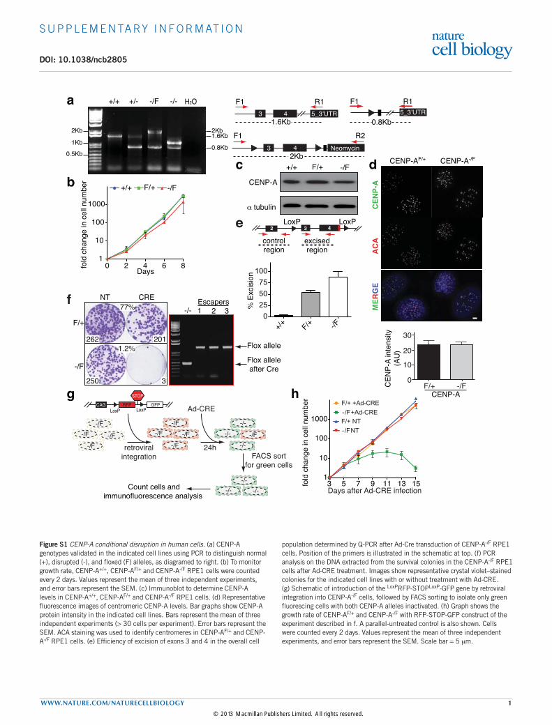
Supplementary figure S1Figure S1 CENP-A conditional disruption in human cells. (a) CENP-A genotypes validated in the indicated cell lines using PCR to distinguish normal (+), disrupted (-), and floxed (F) alleles, as diagramed to right. (b) To monitor growth rate, CENP-A+/+, CENP-AF/+ and CENP-A-/F RPE1 cells were counted every 2 days. Values represent the mean of three independent experiments, and error bars represent the SEM. (c) Immunoblot to determine CENP-A levels in CENP-A+/+, CENP-AF/+ and CENP-A-/F RPE1 cells. (d) Representative fluorescence images of centromeric CENP-A levels. Bar graphs show CENP-A protein intensity in the indicated cell lines. Bars represent the mean of three independent experiments (> 30 cells per experiment). Error bars represent the SEM. ACA staining was used to identify centromeres in CENP-AF/+ and CENP-A-/F RPE1 cells. (e) Efficiency of excision of exons 3 and 4 in the overall cell
population determined by Q-PCR after Ad-Cre transduction of CENP-A-/F RPE1 cells. Position of the primers is illustrated in the schematic at top. (f) PCR analysis on the DNA extracted from the survival colonies in the CENP-A-/F RPE1 cells after Ad-CRE treatment. Images show representative crystal violet–stained colonies for the indicated cell lines with or without treatment with Ad-CRE. (g) Schematic of introduction of the LoxPRFP-STOPLoxP-GFP gene by retroviral integration into CENP-A-/F cells, followed by FACS sorting to isolate only green fluorescing cells with both CENP-A alleles inactivated. (h) Graph shows the growth rate of CENP-AF/+ and CENP-A-/F with RFP-STOP-GFP construct of the experiment described in f. A parallel-untreated control is also shown. Cells were counted every 2 days. Values represent the mean of three independent experiments, and error bars represent the SEM. Scale bar = 5 mm.
© 2013 Macmillan Publishers Limited. All rights reserved.

S U P P L E M E N TA RY I N F O R M AT I O N
2 WWW.NATURE.COM/NATURECELLBIOLOGY
c CENP-I ACACENP-A MERGE
Day 0
Day 9 0255075
100
CEN
P-I i
nten
sity
(%)
Days after Ad-Cre infection0 1 3 5 7 9
[CEN
P-A
inte
nsity
(%)]
b Ndc80 ACACENP-C MERGE
Day 0
Day 7 0
25
50
75
100
Ndc
80 in
tens
ity (%
)Days after Ad-Cre infection
0 1 3 5 7[C
ENP-
A in
tens
ity (%
)]
aACACENP-A MERGECENP-PSNAP
Day 0
Day 9 0
25
50
75
100
CEN
P-P
inte
nsity
(%)
Days after Ad-Cre infection0 1 3 5 7 9
[CEN
P-A
inte
nsity
(%)]
Supplementary figure S2
Figure S2 Centromeric proteins dissociate from centromeres with different kinetics during CENP-A depletion. (a) Representative fluorescence images show the localization and intensity of CENP-P (purple bars). ACA staining was used to identify centromeres. Red bars show CENP-A intensity from Figure 1E. Bars represent the mean of three independent experiments (> 30 cells per experiment). Error bars represent the SEM. CENP-P was tracked by covalent labeling with rhodamine-benzyl guanine after stable expression
of a gene encoding SNAP-tagged CENP-P (b) As in A, except that CENP-C and Ndc80 at day 0 and 7 are shown after Ad-Cre infection. Cells were arrested in monostarol for 12 hours. Yellow bars show Ndc80 intensity of the fluorescence images at the indicated days after Ad-Cre treatment. (c) As in a, but for CENP-A and CENP-I levels at day 0 and 9 after Ad-Cre infection. Interphase cells were quantified. Statistics source data are in supplementary table S2. Scale bar = 5 mm.
© 2013 Macmillan Publishers Limited. All rights reserved.

S U P P L E M E N TA RY I N F O R M AT I O N
WWW.NATURE.COM/NATURECELLBIOLOGY 3
CEN
P-A
EYFP ACA MERGE
H3
CEN
P-AC
-H3
H3-
NH
2 CEN
P-A
NH
2 H3C
ATD
H3C
ATD
+CH
3C-A
A-N
H2 H
3a
H3C
ATD
αNH
3CAT
D
A-N
H2 H
3
H3C
-A
H3-
NH
2 CEN
P-A
CEN
P-AC
-H3
αNH
3CAT
D
NH
2(1-
29) H
3CAT
D
c Untreated Ad-Cre
MGPRRRSRKPEAPRRRSPSPTPTPGPSRRGPSLGASSHQHSRRRQGWLKEIRKLQKSTHLLIRKLPFSRLA
αNN-terminal
α2 α3Loop1 Loop2 C-terminal
REICVKFTRGVDFNWQAQALLALQEAAEAFLVHLFEDAYLLTLHAGRVTLFPKDVQLARRIRGLEEGLG
α1
29EYFP ACA MERGEb
Untreated Ad-Cre
-/F
Day
0D
ay 1
2D
ay 1
6
CENP-A CENP-T
CEN
P-A-/F
+H
3CAT
D
EYFP-H3CATD MERGE
NH
2(1-
29) H
3CAT
D
d
Supplementary figure S3
Figure S3 Clonogenic and immunofluorescence localization assays determine that the CATD is sufficient for centromere identity and for templating its replication. (a) CENP-A amino acid sequence and the domains, including the amino terminal tail (red), the aN helix (green), the CATD (light blue), and the carboxyl terminal tail (orange). (b) Representative fluorescence images of the localization of EYFP-rescue
constructs of experiment outlined in Figure 3. (c) Clongenic survival assays (as in Figure 3d) for the CENP-A-/F cells with and without incubation in Ad-Cre and with the indicated rescue constructs. (d) Representative fluorescence images of the localization and intensity of CENP-T, CENP-A and EYFP-H3CATD at the indicated days after Ad-Cre treatment. Scale bar = 5 mm.
© 2013 Macmillan Publishers Limited. All rights reserved.

S U P P L E M E N TA RY I N F O R M AT I O N
4 WWW.NATURE.COM/NATURECELLBIOLOGY
+NH
2 H3C
ATD
+CEN
P-A
CENP-A-/F
+H3C
ATD
+C
Days
-/F +NH2H3CATD
-/- +NH2H3CATD-/F +CENP-A
-/- +CENP-A
0 2 4 6 8 101
10
100
1000
fold
cha
nge
in c
ell
num
ber
0 2 4 6 8 101
10
100
1000
fold
cha
nge
in c
ell
num
ber
1
10
100
1000
fold
cha
nge
in c
ell
num
ber
0 2 4 6 8 10
-/F +H3CATD+C
-/- +H3CATD+C
-/F +CENP-A
-/- +CENP-A
a
Days Days
F/+ -/- +C
ENP-A
-/F -/- +H
3-NH2 CENP-A
-/- +C
ENP-AC-H
3
H 2O
-/- +N
H2 H3CAT
D
-/- +H
3CATD+C
Intact
excised
3 4
CATDNeomycin
F1 R3Intact
Excised
Neomycin
F1 R3
b
0
50
100
-/F -/- -/F -/-+H3-NH2CENP-A +H3CATD+C
CEN
P-A
inte
nsity
(%)
CENP-A ACA
CEN
P-A-/-
+H3C
ATD
+C
c
g%
of d
ivid
ing
cells
0
10
20
30
CEN
P-C
inte
sity
(%)
0
25
50
75
100
CENP-A-/- CENP-A-/F CENP-A-/- CENP-A-/F CENP-A-/-
GAPDH CENP-C GAPDH CENP-C
siRNA: GAPDH CENP-C
DAPICENP-A
1.5kb
0.5kb
CENP-A-/-
+EYFP-NH2H3CATDCENP-A-/-
+EYFP-H3CATD-C
C
CEN
P-A-/-
SN
APN
H2 H
3CAT
DTu
bulin
ACA
DAP
I
G2 pro-metaphase metaphase anaphase early G1
NT
GAPDH
HJURP NT
GAPDH
HJURP NT
GAPDH
HJURP
0
25
50
75
100
EYFP
inte
nsity
(%)
CENP-ANH2H3CATD
H3CATD+Ce
siR
NA
GAP
DH
siR
NA
HJU
RP
EYFP-H3CATD+C ACA DAPIEYFPf
d
CENP-A-/-+EYFP-NH2H3CATD
Supplementary figure S4
siRNA:
Figure S4 H3CATD combined with either the amino or carboxyl tails of human CENP-A confer long term centromere identify and kinetochore function. (a) (Top) Clonogenic survival assays for RPE1 cells with the indicated CENP-A genotypes and rescue constructs. (Bottom) Corresponding growth rates determined by counting cell numbers every 2 days for CENP-A-/F cells and CENP-A-/- cells with CENP-A, NH2H3CATD, or H3CATD-C rescue genes. Values represent the mean of three independent experiments, and error bars represent the SEM. (b) Confirmation of excision of endogenous CENP-A alleles in the indicated survival rescue construct cells. A schematic representation of the position of the primers is also shown. (c) (Top) Representative fluorescence images show the loss of endogenous CENP-A at centromeres only for cells lacking the amino-terminus of CENP-A epitopes recognized by our CENP-A antibody. ACA staining was used to identify centromeres. (Bottom) Quantitation of the fluorescence images in the CENP-A-/- + H3CATD-C and CENP-A-/- + NH2CENP-A cells. (d) Representative
fluorescence images showing localization of the NH2H3CATDSNAP in CENP-A-/- cells during different phases of the cell cycle. ACA staining was used to identify centromeres and a-tubulin to identify telophase/early G1 cells. (e) Indirect immuno-fluorescence images of localization and intensity of EYFP-H3CATD-C at centromeres after GAPDH or HJURP siRNA, respectively. ACA staining was used to identify centromeres. (f) Quantitation of EYFP intensity at centromeres in images from (e) after GAPDH, HJURP siRNA, or no siRNA. Error bars represent the SEM of two independent experiments (> 30 cells per experiment). ACA staining was used to identify centromeres. (g) (Left) CENP-C protein intensity after GAPDH or CENP-C siRNA in the indicated cell lines, respectively. Bars represent the mean of two independent experiments (> 30 cells per experiment). (Right) Percentage of cells that undergo division after 72 hours of CENP-C depletion, determined by live cell microscopy. Error bars represent the SEM. Statistics source data are in supplementary table S2. Scale bar = 5 mm.
© 2013 Macmillan Publishers Limited. All rights reserved.
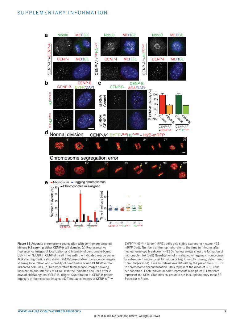
S U P P L E M E N TA RY I N F O R M AT I O N
WWW.NATURE.COM/NATURECELLBIOLOGY 5
b
d Normal division
Chromosome segregation error
CENP-A-/- EYFP-NH2H3CATD + H2B-mRFP36’27’24’18’15’9’6’
39’ 63’6’ 15’ 27’ 33’ 36’
0
40
80
Tim
e in
mito
sis
(min
)
-/FH3CATD+C
-/-CENP-A
-/F -/- -/F -/-NH2H3CATD
0
5
10
15
20
Micronuclei Lagging chromosomesChromosomes mis-aligned
Num
ber o
f eve
nts
(%)
-/F -/- -/F -/- -/F -/-
25
H3CATD+CCENP-A NH2H3CATD
e
Control
0
25
50
75
100
CENP-BCENP-A-/-
+CENP-ACENP-A-/-
+NH2H3CATD
CENP-BCENP-B
ACA/DAPI
shR
NA
Con
trol
shR
NA
CEN
P-B
c
CEN
P-B
inte
nsity
(%)
Control
CENP-BshRNA:
CENP-B
H3C
ATD
+CN
H2 H
3CAT
D
CEN
P-A-/-
+H3C
ATD
+C
CEN
P-A-/-
+NH
2 H3C
ATD
Ndc80 MERGENdc80 MERGENdc80 MERGE
CENP-I MERGE CENP-I MERGECENP-I MERGE
CEN
P-A-/-
+CEN
P-A
a
CENP-BEYFP/DAPI
Supplementary figure S5Figure S5 Accurate chromosome segregation with centromere-targeted histone H3 carrying either CENP-A tail domain. (a) Representative fluorescence images of localization and intensity of centromere-bound CENP-I or Ndc80 in CENP-A-/- cell lines with the indicated rescue genes. ACA staining (red) is also shown. (b) Representative fluorescence images showing localization and intensity of centromere bound CENP-B in the indicated cell lines. (c) Representative fluorescence images showing localization and intensity of CENP-B in the indicated cell lines after 2 days of shRNA against CENP-B. (Right) Quantitation of CENP-B protein intensity of fluorescence images. (d) Time-lapse images of CENP-A-/-+
EYFPNH2H3CATD (green) RPE1 cells also stably expressing histone H2B-mRFP (red). Numbers at the top right refer to the time in minutes after nuclear envelope breakdown (NEBD). Yellow arrows show the formation of micronuclei. (e) (Left) Quantitation of misaligned or lagging chromosomes or subsequent micronuclei formation or (right) mitotic timing, determined from images in (d). Time in mitosis was defined by the period from NEBD to chromosome decondensation. Bars represent the mean of > 50 cells per condition. Each individual point represents a single cell. Error bars represent the SEM. Statistics source data are in supplementary table S2. Scale bar = 5 mm.
© 2013 Macmillan Publishers Limited. All rights reserved.

S U P P L E M E N TA RY I N F O R M AT I O N
6 WWW.NATURE.COM/NATURECELLBIOLOGY
Supplementary figure S6
β-actin
GFP
Ponceau
Cnp1
Cnp1C
ATDswap
H3CATD
NH2 H3CATD
Cnp1 + :
43 KDa
40-43 KDa
Figure S6 Protein level expression of the fission yeast rescue constructs. Immuno-blot to quantify levels of each GFP-tagged rescue variant, visualized with a GFP antibody. b-actin is used as a loading control.
© 2013 Macmillan Publishers Limited. All rights reserved.
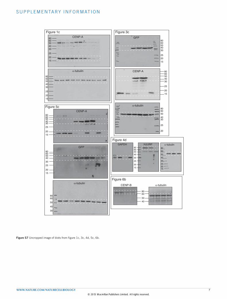
S U P P L E M E N TA RY I N F O R M AT I O N
WWW.NATURE.COM/NATURECELLBIOLOGY 7
Figure 1c
152025
3040506080
Figure 3c
20
25
3040506080
Supplementary figure S7
152025
3040506080 CENP-A
α-tubulin
GFP
CENP-A
α-tubulin
152025
3040506080
152025
3040506080
1520253040506080
Figure 4d
Figure 5c
40506080
30
1520
253040506080
1520
253040506080
CENP-A
GFPGAPDH HJURP α-tubulin
40506080
CENP-B
Figure 6bα-tubulinα-tubulin
1520253040506080
Figure S7 Uncropped image of blots from figure 1c, 3c, 4d, 5c, 6b.
© 2013 Macmillan Publishers Limited. All rights reserved.

S U P P L E M E N TA RY I N F O R M AT I O N
8 WWW.NATURE.COM/NATURECELLBIOLOGY
Supplementary Table Legends
Supplementary Table 1 List of strains used in this study. Fission yeast strains used in figure 7 are listed. The source column indicates where the original strain comes from. The genotype of each strain is also indicated. [68Pidoux, A.L., Richardson, W. & Allshire, R.C. Sim4: a novel fission yeast kinetochore protein required for centromeric silencing and chromosome segregation. J Cell Biol 161, 295-307 (2003)].
Supplementary Table 2 Statistic source data (n < 5).
Supplementary Video Legends
Supplementary Video S1 A method to eliminate Cre-escaper cells. Movie of CENP-A-/F cells expressing the LoxPRFP-STOPLoxP-GFP cassette one hour after infection with Ad-Cre. One frame captured every 5 minutes. Time in top left shows to the minutes after start of filming.
Supplementary Video S2 Lowered levels of CENP-A corresponded with increased rates of chromosome segregation defects. Movie of CENP-A-/F cells expressing Histone H2B-mRFP five days after infection with Ad-Cre. One frame captured every 3 minutes.
© 2013 Macmillan Publishers Limited. All rights reserved.