acehsc - 1. most organisms are active in a limited ... · web viewmaintaining a balance 1. most...
TRANSCRIPT

MAINTAINING A BALANCE1. MOST ORGANISMS ARE ACTIVE IN A LIMITED TEMPERATURE RANGE
IDENTIFY THE ROLE OF ENZYMES IN METABOLISM, DESCRIBE THEIR CHEMICAL COMPOSITION AND USE A SIMPLE MODEL TO DESCRIBE THEIR SPECIFICITY ON SUBSTRATES
◊ Metabolism: the sum of all chemical processes or reactions occurring within an organism.
◊ The rate of reaction is maintained and controlled by large proteins called enzymes
◊ Role in metabolism:
o Enzymes are biological catalysts that support the function of enzymes
o It is a substance that increases the rate of reaction without being changed itself
o Enzyme present -> less energy required to catalyse a chemical reaction
o Enzyme absent -> more energy required and vice versa
◊ Chemical Composition:
o Most enzymes are proteins (a protein is long chain of amino acids joined together by polypeptide bonds)
o A reactant in an enzyme-catalysed reaction is called a substrate
o The area of the enzyme that binds the substrate is called the active site – where the action takes place
◊ Specificity:
o Enzymes are highly specific -> one enzyme catalyses one particular substrate and only that substrate
o This is due to the shape of the active site of the enzyme matches the shape of the substrate material
o When products are formed, they leave the enzyme surface.
◊ Models to explain Specificity:
o Lock and key: suggests that the substrate sits exactly into the active site of the enzyme like a key fit into a lock.
It assumes that the enzyme has an unchanging shape
o Induced fit: suggests that the binding of the substrate induces a temporary change in the shape of the enzyme.
The new shape better accommodates he shape of the substrate.

IDENTIFY THE PH AS A WAY OF DESCRIBING THE ACIDITY OF A SUBSTANCE
◊ pH is the measure of the acidity or the alkalinity of a substance. It is a measure of the concentration of hydrogen ions
◊ pH scale ranges from 1 – 14:
o below 7 -> acidic
o above 7 -> alkaline
o 7 -> neutral
EXPLAIN THE MAINTENANCE OF A CONSTANT INTERNAL ENVIRONMENT IS IMPORTANT FOR OPTIMAL METABOLIC EFFICIENCY
◊ Enzymes facilitate the chemical reactions that are vital to life
◊ However, enzymes are more efficient within a limited range of environmental conditions: perfect temperatures, pH
and substrate concentrations.
◊ If any of these factors are compromised, enzymes will denature, enzyme activity will decrease and so will metabolic
efficiency as a result
◊ Hence, a constant internal environment is needed so that enzymes will always be working at an optimum rate, and thus
metabolism will be at an optimum efficiency.
DESCRIBE HOMEOSTASIS AS A PROCESS BY WHICH ORGANISMS MAINTAIN A RELATIVELY STABLE INTERNAL ENVIRONMENT
◊ Homeostasis: the process by which an organism maintains a relatively stable internal environment, despite changing
external environmental conditions
◊ This is important for enzymes, in which by keeping the body stable at its optimum conditions, we allow the optimal
conditions of enzymes to be met. This in turn allows the body to carry out its reactions efficiently
◊ Homeostasis allows the enzyme’s optimal conditions to be met and the body to work as efficiently as possible
◊ Conditions controlled by homeostasis include: body temperature, pH, water and salt concentrations, sugar levels and
levels of dissolved gases.
EXPLAIN THAT HOMEOSTASIS CONSISTS OF TWO STAGES:
◊ Detecting changes from the stable state: (stage 1)
o These changes are detected by receptors. Receptors can range from a patch of sensitive cells to complex
organs like eyes and ears.
o These changes are caused by the internal and external environment
o Any change or information that catalyses a response is called a stimulus
o Sends messages to the Central nervous system (brain + spinal cord)
o Receptors detect stimuli -> organisms then react to change
o Example : If our body temperature rises, the temperature rise in the blood stimulates the brain’s frontal
hypothalamus (and inhibits the posterior hypothalamus). Alternatively, when a mammal is exposed to cold,
skin receptors increase their activity, sending more nerve impulses to the posterior hypothalamus. Plants
detect gravity, light intensity and direction, and the length of the period of darkness.
◊ Counteracting changes from the stable state: (stage 2)

o After receptors detect changes, organisms can then react to the change.
o This type of response will counteract the change to ensure the stable state is maintained
o Effectors bring about change by movement. Effectors can either be muscles or glands
o Example : After detecting the rise in temperature, the hypothalamus then stimulates heat loss by increasing
blood circulation through the skin, increasing sweating and decreasing metabolic activity and muscular activity.
Thus, the body’s temperature is lowered. After detecting the drop-in temperature, activity in the posterior
hypothalamus stimulates the sympathetic nervous system to activate mechanisms that conserve heat. Plants
may, after detecting increase in concentrations of fluids, release abscisic acid from chloroplasts so there is a
closing of stomates and an increase in the production of watertight resins.
OUTLINE THE ROLE OF THE NERVOUS SYSTEM IN DETECTING AND RESPONDING TO ENVIRONMENTAL CHANGES
◊ The Nervous system provides rapid coordination of internal organ systems and detects and responds to environmental
changes. The Nervous system consists of:
o Central Nervous System (CNS) – consists of the brain and spinal cord; receives information, interprets
information and initiates a response
o Peripheral Nervous System (PNS) – nerves branching throughout body; A means of communication throughout
the body; passes messages rapidly to the CNS and back to the body – allowing the body to respond to changes
it perceives.
◊ Response to changes:
o Counteract changes via the stimulus-response pathway (i.e. stimulus, receptor, control centre, effector,
response)
o For example, the stimulus of high blood pressure reaches he receptor the hypothalamus, which also acts as the
control centre. Effectors like blood vessels stimulate responses such as vasodilation, allowing for a decrease in
blood temperature. The response is generally an example of a negative feedback system; producing a response
that is opposite to the initial situation, then cancelling itself out once the optimal conditions are re-achieved
Table 1: Responding to body temperature increases in mammals
effector Response
Hairs on the body - raise Goosebumps are an attempt to trap a layer of warm air around the body to reduce the amount of heat lost by radiation, convection and conduction.
Arterioles in the skin narrow
Vasoconstriction - muscular walls of the small blood vessels known as arterioles constrict so that most blood flow is redirected to the core of the body, preventing heat loss from the cooler body surface (heat is carried throughout the body in the bloodstream)
Muscles Shivering brought about by rapid small muscle contractions generate heat in the body
Thyroid gland Heat gain centre stimulates the activity of the thyroid gland, causing it to speed up/increases metabolism
Table 2: Responding to body temperature decreases in mammals
effector Response
Arterioles in the skin expand/dilate
Vasodilation - blood carrying heat is directed towards the surface of the body so that heat can be lost by conduction, convection and radiation to the surroundings
Sweat glands Liquid sweat is secreted through the sweat pores onto the surface of the skin and heat is removed from the body to evaporate

the liquid
Thyroid gland Decreased metabolism - heat loss centre causes thyroid gland to lower the rate of metabolism, generating less heat
IDENTIFY THE BROAD RANGE OF TEMPERATURES OVER WHICH LIFE IS FOUND COMPARED WITH THE NARROW LIMITS FOR INDIVIDUAL SPECIES
◊ Ambient Temperature: Temperature of the environment
◊ Room Temperature: Temperature inside a temperature-controlled environment
◊ Most life is found in a temperature ranging of around 0 – 45’C
o Life can still be found in areas with temperature as low as -70’C and of more than 200’C in the deep ocean
o Despite this broad range, most individual animals only tolerate a narrow temperature range
As land temperature varies more than aquatic temperature, land animals naturally need to be better
adapted to accommodate changes in ambient temperature
◊ Examples:
o Water-holding frog: 3’C –> 39’C
o Platypus: -8’C –> 34’C
o Sydney Blue Gum: -1’C –> 34’C
COMPARE RESPONSES OF NAMED AUSTRALIAN ECTOTHERMIC AND ENDOTHERMIC ORGANISMS TO CHANGES IN THE AMBIENT TEMPERATURE AND EXPLAIN HOW THESE REPONSES ASSIST TEMPERATURE REGULATION
DESCRIBE ADAPTATIONS AND RESPONSES THAT HAVE OCCURRED IN AUSTRALIAN ORGANISMS TO ASSIST TEMPERATURE REGULATION
◊ Endotherms: organisms that use internal processes such as metabolism to maintain a constant internal temperature
E.g. mammals and birds
◊ Ectotherms: dependant on energy of the ambient environment to regulate body temperature e.g. invertebrates, fish,
reptiles, amphibians.
◊ Behavioural adaptations: the ways an organism behaves that help it survive in its natural environment
◊ Structural adaptations: have a connection with the physical features of an organism
◊ Physiological adaptations: a feature that helps to regulate a function within an organism
Table 3: Red Kangaroo (Endotherm)
Ambient Env. Physiological Structural Behavioural
Cold conditions (winter months)
Increased metabolic rate to create more heat within the body Vasoconstriction
Layer of fur creates and layer of insulation between the skin and the hair and allows the kangaroo to stay warm
Basking in the sun
Warmconditions
(Summer months)
Decrease in metabolic rate which reduces the amount of heat within the body
- Panting to release heat - Exposed areas of skin on the forelegs to increase evaporative cooling of the blood from this area- vasodilation. - Shunting blood from the tail to the exposed areas of the skin on the forelegs to increase heat loss.
- Nocturnal - Licking forelegs to increase evaporation from the skin - Sitting in the shade
◊ Red kangaroo is the largest marsupial in Australia. Lives in grasslands of dry and arid central part of the country. In this
habitat, temperatures vary from 5’C to 38’C.
◊ For these reasons, the Red kangaroo needs many adaptations to accommodate these drastic changes in temperature
and retain its optimum temperature range at 37.5’C

Table 4: Diamond Python (Ectotherm)
Climate Physiological Structural Behavioural
Cold conditions (winter months)
Lies on eggs and shivers to increase the temperature of incubation
Dark in colour to absorb heat and can therefore tolerate colder temperatures than most snakes
» Basks in the sun to raise body temperature » Hibernation » Migration to warmer areas
Warm conditions (summer months)
» Is nocturnal, so hunts at night to escape the heat during the day » Burrowing during the day
◊ The Diamond Python lives in a variety of habitats (rainforests, temperate forests, grassland, caves and hollow trees).
◊ Eats small mammals, bats, birds and lizards.
◊ Its optimum temperature around 20’C however this depends on the ambient temperature of the python’s surroundings
Comparing the Responses of Endotherms and Ectotherms:
◊ Endotherms need to have a high metabolic rate to maintain this optimum temperature rate in cold conditions and thus,
need to eat large amounts.
◊ Ectotherms do not need to do this however they have greater restrictions placed on their activity as a result.
◊ In hot conditions endotherms, must have specific adaptations to these environmental changes to regulate heat gain so
not to raise their temperature above their optimum temperature level as this can cause severe damage.
◊ This is the same for both endotherms and ectotherms in relation to cold climates.
◊ Ectotherms are not found in extremely cold climates.
IDENTIFY SOME RESPONSES OF PLANTS TO TEMPERATURE RANGE
◊ Plants are ectotherms and cannot maintain a constant temperature. They have various adaptations that enable them to
survive a range of temperatures:
Adaptation Effect
Leaf Fall By dropping leaves, there is less SA exposed to hot conditions and reduces the amount of water lost through transpiration
Radiation Some plants have shiny leaves to reflect solar radiation
Heat Shock Proteins Proteins produced to stop the denaturing of cells
Transpiration Evaporation of water through stomates of the leaves cools the plant. Water travelling up the stem also cools plant
Die-back Shoots and leaves of the plants may die, the bulbs, roots and rhizoids (root hairs) will be left in soil to grow back when conditions improve
Orientation of leaves Leaves may have vertical orientation (hanging down) so that sunlight exposure is reduced
Seed Dispersal Some plants require extreme heat to open their coats. Only when they germinate
Vernalisation Some plants must be exposed to cold conditions for the plant to produce flowers and reproduce

DEVELOP A MODEL OF A FEEDBACK MECHANISM
2. PLANTS AND ANIMALS TRANSPORT DISSOLVED NUTRIENTS AND GASES IN A FLUID MEDIUM
IDENTIFY THE FORM(S) IN WHICH EACH OF THE FOLLOWING IS CARRIED IN MAMMALIAN BLOOD:
Table 5: Forms in which Material is carried in Mammalian blood
Materials Form carried in mammalian blood
Carbon Dioxide Leaves cells and dissolves in plasma to form bicarbonate ions (HCO3-). Oxygen This dissolves into the plasma, and combined with haemoglobin to form oxyhaemoglobin
Water Produced by body cells from respiration, and travels through the body as water molecules in the plasma
Salts Carried in the plasma in the form of ions. E.g. Sodium ions (Na+), calcium ion (Ca2+) etc.
Lipids Carried as lipid droplets but digested lipids are carried in the form of fatty acids and glycerol in plasma
Nitrogenous waste Carried in the plasma in the form urea (NH2CONH2). Formed through the breakdown of proteins
Other products of digestion Sugars are carried in the form of glucose. Proteins are carried in the plasma in the form of amino-acids.
EXPLAIN THE ADAPTIVE ADVANTAGE OF HAEMOGLOBIN
Table 6: Advantages of Haemoglobin
Adaptive advantage Explanation

Each haemoglobin molecule contains four haem units
◊ Increases the oxygen-carrying capacity of blood◊ One haemoglobin molecule can bond with four oxygen molecules◊ More oxygen can be carried in blood cells by haemoglobin than could be
carried dissolved in plasma◊ Can carry far more oxygen than if dissolved in plasma
Oxygen affinity (ability to bind oxygen) is increased once the first O2 molecules binds with haemoglobin
◊ Ability to bind oxygen increases once the first oxygen molecule binds to it ◊ Increases rate and efficiency of oxygen uptake◊ Result: very small increase in oxygen concentration in the lungs can result in
a large increase in the oxygen saturation of blood
Capacity to disassociate increases at lower pH levels -> Bohr effect
◊ Capacity to release oxygen increases when CO2 is present ◊ Metabolising cells release carbon dioxide, which combines with water to
form carbonic acid and this lowers pH◊ Haemoglobin has a reduced affinity for oxygen at lower pH so it releases
oxygen in tissues where it is needed
Capable of binding with carbon dioxide but has higher attraction for oxygen
◊ Haemoglobin has an increased ability to pick up carbon dioxide once it has released oxygen
COMPARE THE STRUCTURE OF ARTERIES, CAPILLARIES AND VEINS IN RELATION TO THEIR FUNCTION
◊ Arteries: o Carry blood away from the hearto Thick muscular wallo Carry oxygenated blood (except for pulmonary artery)o Blood in arteries are under high pressure
◊ Veins :o Thin-walledo Valves are present to prevent backflow of blood o Carries blood back to the hearto Carry deoxygenated blood (except for pulmonary vein)o Blood is under low pressure; movement is assisted by body muscles
◊ Capillaries: o Thin-walled, 1cm thicko Carry blood between veins and arteries
DESCRIBE THE MAIN CHANGES IN THE CHEMICAL COMPOSITION OF THE BLOOD AS IT MOVES AROUND THE BODY AND IDENTIFY TISSUES IN WHICH THESE CHANGES OCCUR
◊ Pulmonary circuit:
o Blood flows from the heart to the lungs and back to the heart
o Blood under low pressure
o Rate of blood is faster
o Very little body fluid is formed
o Blood returning to the body contains high CO2
levels and high O2 levels
◊ Systematic circuit:
o Blood flows from heart to body
o Blood under high pressure due to contraction of
left ventricle of heart, but gradually lessens
o Blood pressure forces some fluid out of the blood
to become body fluid
Tissue Main Changes in Blood
Lung » Increase in oxygen » Decrease in carbon dioxide
Villi of small intestine
» Increase in glucose and other products of digestion (amino acids, lipids, vitamins, minerals, water)
Kidneys » Decrease in nitrogenous wastes (salts and water to form urea)
Other body tissues
» Decrease in oxygen » Decrease in glucose » Increase in carbon dioxide

o In tissues, blood gives up oxygen and other ions and minerals;
produces waste products e.g. Urea, CO2
OUTLINE THE NEED FOR OXYGEN IN LIVING CELLS AND EXPLAIN WHY REMOVAL OF CARBON DIOXIDE FROM CELLS IS ESSENTIAL
◊ Cells require oxygen for respiration – a process vital for making energy
(which we call ‘ATP’)
o Glucose + oxygen + water + ATP
o CO2 is a by-product of this reaction and must be removed to
maintain optimum pH balance in the blood
→ Removing excess CO2 prevents a build-up of pH-lowering carbonic acid that forms when CO2
dissolves in water, and affects enzyme activity
DESCRIBE CURRENT THEORIES ABOUT PROCESSES RESPONSIBLE FOR THE MOVEMENT OF MATERIALS THROUGH PLANTS IN XYLEM AND PHLOEM
◊ Phloem :
o Movement of materials through the phloem is called Translocation. This is an active process that requires energy.
Transports materials up and down
o It is referred to as the source-sink pathway driven by a pressure gradient generated osmotically.
o Loading at the source can either be:
Symplastic loading : where sugars and nutrients move in the cytoplasm from the mesophyll cells to
the sieve elements through the plasmodesmata or,
Apoplastic loading : where movement occurs along a pathway through the cell walls until they reach
the sieve element, then across the cell membrane to enter the phloem tube
o Once loaded at the source, the materials flow towards a sink.
o Sinks are regions of the plant where the nutrients are actively
removed from the phloem, such as roots, stems and flowers.
The pressure difference between the source and the sink
regions drives the phloem sap.
◊ Xylem :
o The Transpiration stream theory persists that due to the
physical forces of water and ions being removed from the plant
stomates by passive transport (i.e. Transpiration), causes a
column of water to be sucked up the stem by the evaporative
pull, also the low concentration of water at the roots allows
more water to diffuse
o Once water has been absorbed into the root plants (by
osmosis), along with mineral ions (by diffusion and active transport), these substances move across the root into
the xylem. A small amount of root pressure results from the continual influx of more water and ions, hence
forcing the solution already present upwards (due to pressure build-up), however this is usually not enough.

o The constant loss of water, leads to a transpiration stream (which is the constant upward flow of water through a
plant), this is because of water’s 2 properties, which are adhesive forces (ability of molecules to attach to walls),
and cohesive forces (the attraction of molecules to each other), hence leading to the capillary (water rising
through bore of tissue) and hence the stream
DEMONSTRATE THE EFFECT OF DISSOLVED CARBON DIOXIDE ON THE PH OF WATER
◊ Aim : to model the effect of carbon dioxide on the pH of water
◊ Equipment:
o 25mL of distilled water
o Universal indicator
o pH probe attached to data logger
o 1 x straw
◊ Risk Assessment:
o Gloves must be worn in case of glass breaking
o The water can be corrosive due to increasing pH, it should not be ingested after use, dispose in an organic
waste container
o Straws should not be used by more than one student, to minimise contracting diseases
◊ Method:
1. In a beaker, pour water
till 25mL grade mark,
then put 3 drops of
universal indicator, this
should now change into
greenish colour
2. Then put pH probe, and
check that pH is about 7
3. Exhale air into the straw
that is dipped into the solution for about 3 minutes
◊ Result:
o After about 30 seconds, the colour of the solution began to change into pale yellow, and the pH on the data
logger started decreasing
o This is because carbon dioxide forms a weak acid, carbonic acid (H2CO3), so the water becomes more acidic.
Carbonic acid in water disassociates to form hydrogen carbonate ions (HCO3-), and some carbonate ions
(CO3^2+)
PERFORM A FIRST-HAND INVESTIGATION USING THE LIGHT MICROSCOPE AND PREPARED SLIDES TO GATHER INFORMATION TO ESTIMATE THE SIZE OF RED AND BLOOD CELLS AND DRAW SCALED DIAGRAMS OF EACH
◊ Aim : To estimate the size of red blood cells and white blood cells seen with the light microscope
◊ Equipment :
o Light Microscope

o Prepared slides of human blood
o 1mm sized mini-grid plastic paper
o Pencil and drawing paper
◊ Risk Assessment :
o Slides can be sharp or unseen flint pieces of glass, gloves and glasses should be worn
o Use commercially prepared microscope slides of blood and not fresh blood, to eliminate the risk of contracting
blood-borne disease.
◊ Method :
1. The microscope on normal view, i.e. 1X, has a limited field of view of 16mm
2. Hence, set your microscope with the millimetre-square graph paper first. Then click the 1X objective lense, this
will show you what the normal eye of 16mm can see.
3. Then click the 10X objective, this will magnify the 10mm by a factor of 10. Hence you’ll see a maximum field
view of 1.6mm, and its sub-ten components (note, 1mm = 1000μm)
4. Now using the 40X objective, this now makes the initial 16mm diameter 4 times less than the 10X, so the
diameter is approximately 0.4mm. Hence the diameter is 0.4mm, or 400μm
5. Now, putting a slide of a prepared blood, 50 red blood cells exist, hence the size of 1 RBC is 400μm divided by
50 is 8μm.
6. Now for white blood cells since there are so few, it is not possible to count the number of white cells across
the diameter, even much more difficult to estimate how many would fit across the diameter. Hence, the size is
estimated by proportion in comparison to that of RBC.
◊ Result :
o Erythrocytes (Red Blood Cells)
Size: 6 - 9μm
Shape: Bi-concave (concave on both sides
Function: to transport oxygen
Have no nuclei, 5 – 6 million in every millilitre of blood
Produced in bone marrow
o Leucocytes (White Blood Cells)
→ Size: 12 - 15μm
→ Shape: irregular, can change shape
→ Function: to defend against disease
→ Have nuclei, produced in lymph glands

IDENTIFY CURRENT TECHNOLOGIES THAT ALLOW MEASUREMENT OF OXYGEN SATURATION AND CARBON DIOXIDE CONCENTRATIONS IN BLOOD AND DESCRIBE AND EXPLAIN THE CONDITIONS UNDER WHIH THESE TECHNOLOGIES ARE USED
IDENTIFY PRODUCTS EXTRACTED FROM DONATED BLOOD AND DISCUSS THEIR USES OF THESE PRODUCTS
◊ Red Blood Cells:
o Used to increase amount of oxygen that can be carried to the body’s tissues
o Given to anaemic patients (whose bone marrow does not make enough red blood cells) or who has lost a lot
of blood
Technique Arterial blood gas analysis
Description of how the technology works
Electrochemical: uses a sensor that translates chemical properties into an electrical signal that can be measured.Involves removing blood from an artery and performing a blood test using computer-based technology to analyse the chemical components in the blood
Placement Invasive - small sample of arterial blood must be withdrawn from the patient or an arterial probe may be inserted into an artery (usually in the arm)
Information re blood gas concentration collected
Oxygen and carbon dioxide levels and pressurepH of bloodLevel of bicarbonate ions
Levels of chemicals in the blood
Conditions where it is used
Only carried out if abnormalities show up in the pulse oximeter readings or in severe cases of breathing disturbance
Study of lung disease and monitor conditions of poor gaseous exchange (respiratory disease e.g. patients with asthma, cystic fibrosis)pH and electrolyte ion levels measured gives important information about how well the kidneys are functioningGeneral: if blood oxygen content seems low or there is a risk of carbon dioxide levels being high or blood pH being too acidic
Benefits Gives more information e.g. CO2, pH, bicarbonate ions - (pressure as well as saturation readings)
Limitations Invasive - bleeding or bruising at puncture siteOnly provides information at one particular time - not continuousFeeling faintBlood accumulating under the skinInfection at the puncture site
Current and future research
Some advanced blood gas analysers can also measure levels of glucose, haemoglobin and electrolytes (salts) in the blood. Research has improved the speed of the machines and in some machines results can be obtained within a few minutes
Technique Pulse oximeter
Description of how the technology works
Two light-emitting diodes, one producing red and one producing infrared light are shone through the finger. The amount of light absorbed by arterial blood determines the level of oxygenation of haemoglobin the blood and thus oxygen saturation is calculated and displayed on a screen.
Placement Non-invasive - consists of a prove attached to the patient's finger or earlobe (anywhere where there is an extremity of blood vessels near the surface)
Information re blood gas concentration collected
Oxygen saturation Pulse rate
Conditions where it is used
General: to determine if blood oxygen is within the normal range or notSpecific: To monitor on an ongoing basis oxygen saturation and pulse rate - indication breathing and circulation are normal:
Prior to anaesthetics Whist under anaesthetics or sedation During recovery after surgery While on oxygen therapy or ventilation Who appear to have breathing difficulty or whose
circulation is abnormal Who are suffering from sleep apnoea Such as premature or newborn babies, who need
ongoing checks
Benefits Non-invasive - proceeds on a continuous basis without the need for a blood sample to be taken
Quick reading Ongoing
Limitations Cannot operate reliably with a poor signal May appear to have a good signal and be displaying a
saturation figure, but either the figure is inaccurate or gives a false sense of security
Delay Nail varnish interference Most do not measure carbon dioxide levels
Current and future research
The latest generation of oximeters (produced from 2005 onwards) have a carbon dioxide sensors and have digital signal processing. They are also designed to allow patients to move about and can be used on non-translucent body parts - particularly effective in newbornsMobile phones are being used to transmit signals

◊ Platelets:
o Essential for blood clotting
o Used for people who have cancer of the blood or lymph such as leukaemia or lymphoma. Patients undergoing
chemotherapy do not have enough blood platelets and are also given platelets extracted from donated blood
◊ Plasma:
o Liquid portion of blood, which contains blood-clotting factors (and immunoglobins).
o Used to treat people with clotting disorders such as haemophilia. Also, used to adjust the osmotic pressure of
blood and pull fluids out of tissues.
o Some products are derived from plasma.
◊ White Blood Cells:
o Another infection-fighting component of the blood.
o Only used occasionally to treat life-threatening infections when the cell count is very low or the white blood
cells are not working properly.
o Most of the time, antibiotics are used in these cases rather than giving white blood cells
◊ Plasma derivatives:
o Separate functions of clotting factors found in whole plasma:
→ Immunoglobins : are infection fighting parts of the blood plasma. Used to treat people who have
difficulty fighting infections and whose immune systems are not working properly because of diseases
such as AIDS.
→ Cryoprecipitate and concentrates of Factors VIII and IX are for patients who suffer a variety of
bleeding disorders.
REPORT ON PROGRESS IN THE PRODUCTION OF ARTIFICIAL BLOOD AND USE AVAILABLE EVIDENCE TO PROPOSE REASONS WHY SUCH RESEARCH IS NEEDED
◊ Artificial blood is only currently used for:
o Increased blood volume – used in cases of severe burns
o Carry oxygen
◊ No substitutes have been developed yet that can replace other functions: immune defence and coagulation
◊ Benefits:
o Free of infection agents and allergens, making them non-toxic and disease-free
o Do not need refrigeration and storage; kept for longer period, 2 – 3 years
o Universal acceptance by all blood groups, allowing transfusion to a person without tests
o Readily available in large supplies, solving the worldwide problem of blood donors
◊ Proposed replacements for blood (oxygen-carrying artificial blood):
o Perfluorochemicals: synthetic materials that can dissolve about 50 times more oxygen than plasma. Cheap to
produce and, because they are synthetic, there is no risk of the material being infected by disease.
o More research is needed because Perfluorochemicals must combine with other substances in order to mix in
the blood stream.
o Haemoglobin-based oxygen carriers (HBOCs): made from haemoglobin extracted from red blood cells. Not
contained in a membrane, therefore do not require blood typing and cross-matching of blood.

o More research is needed because haemoglobin must be modified before it can be used. Current blood
substitutes do not have the enzymes that prevent haemoglobin from oxidising.
PERFORM A FIRST-HAND INVESTIGATION TO GATHER FIRST-HAND DATA TO DRAW TRANSVERSE AND LONGITUDINAL SECTIONS OF PHLOEM AND XYLEM TISSUE
◊ Aim : to draw transverse and longitudinal sections of xylem and phloem tissue
◊ Materials :
o light microscope o watch glass o scalpel
o coverslip o celery stem o dropper
o beaker of water o eosin dye solution o prepared sides of plant stem
both transversal and
longitudinal sections
◊ Risk Assessment:
o When using microscope, if care is not taken, the slide and the objective lens can break into pieces and may
cause lacerations. Only lower objective lenses while looking from the side of the microscope. While looking
through the eyepiece, the lenses must only move upwards to prevent them from crashing onto the slide and
breaking the lense and slide
o Scalpel is very sharp and can cause lacerations. While passing the scalpel, it must be done carefully and must
be pointed downwards, away from the body.
◊ Method:
o A light microscope is set up
o A celery stem that had been left in eosin dye + water solution in beaker for 24 hours was obtained. Excess dye
was washed off with water
o The thinnest section possible is cut from stem using a scalpel. Both longitudinal and transverse sections are cut
out
o The cut sections are placed under the microscope, firstly covered with coverslips.
o The longitudinal and transverse sections were observed under the microscope, firstly under low power than
high power
o The Xylem and Phloem cells were identified in longitudinal
and transverse sections
o Observed the prepared slides of the plant stems that had been
stained to show different tissues.
o Large labelled diagrams were made displaying the
Longitudinal section and transverse section of both
xylem and phloem and a diagram of the transverse
section of the stem was made.

◊ Results:
◊ Conclusion :
o The structure of Xylem is different to the structure of Phloem.
o Xylem is composed of large vessels, whose walls are thickened by lignin. Xylem also contains hard fibre cells,
which adds support to the tissue. Xylem is composed to dead tissue and transports water.
o Phloem is comprised of supporting fibre cells ad two special cell types: sieve tubes and companion cells. Unlike
Xylem, Phloem tissue is alive and uses a process of translocation to transport materials.

3. PLANTS AND ANIMALS REGULATE THE CONCENTRATION OF GASES, WATER AND WASTE PRODUCTS OF METABOLISM IN CELLS AND INTERSTITIAL FLUID
EXPLAIN WHY THE CONCENTRATION OF WATER IN CELLS SHOULD BE MAINTAINED WITHIN A NARROW RANGE FOR OPTIMUM FUNCTION
◊ Osmoregulation: regulation of water concentration to maintain homeostasis
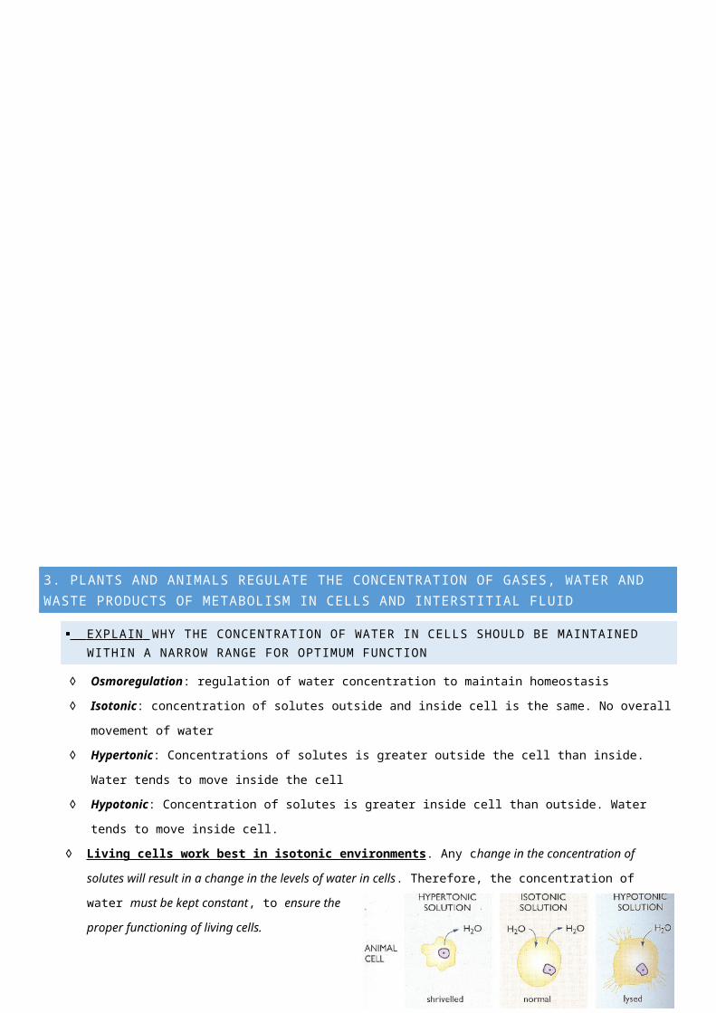
◊ Isotonic: concentration of solutes outside and inside cell is the same. No overall movement of water
◊ Hypertonic: Concentrations of solutes is greater outside the cell than inside. Water tends to move inside the cell
◊ Hypotonic: Concentration of solutes is greater inside cell than outside. Water tends to move inside cell.
◊ Living cells work best in isotonic environments . Any change in the concentration of solutes will result in a change in the
levels of water in cells. Therefore, the concentration of water must be kept constant, to ensure the proper functioning of
living cells.
◊ Concentrations of water must be within a narrow
range because:
o It is vital for metabolic efficiency – it allows
correct concentration of substances to
diffuse across and between cells
o Cells can maintain constant osmotic pressure so water loss through processes such as urination is
compensated for
o It determines the concentrations of various substances in blood
o If not isotonic, cells are vulnerable to losing or gaining too much water – it will move by osmosis
EXPLAIN WHY THE REMOVAL OF WASTES IS ESSENTIAL FOR CONTINUED METABOLIC ACTIVITY
◊ Metabolic waste (CO2, urea, excess salt) need to be removed from the body, otherwise they will disrupt enzyme
activity, or have toxic effects (urea kills cells, excess water lowers cell metabolism by disrupting their osmotic state).

◊ Excretion: removal of metabolic wastes
IDENTIFY THE ROLE OF THE KIDNEY IN THE EXCRETORY SYSTEM OF FISH AND MAMMALS
◊ In mammals and fish, the kidney has a dual role of excreting nitrogenous wastes and maintaining water and salt
concentrations (osmoregulation)
◊ Fresh Water fish:
o Live in rivers and lakes, where the water potential is very high (losing water by osmosis, when concentrations
are high) – these habitats contain few dissolved salts and hence water is freely available.
o Freshwater fish tend to urinate frequently, as water tends to accumulate in their tissues because of passive
movement by osmosis. Their kidneys excrete excess water, as well as nitrogenous wastes.
o Their kidneys are structurally suited to this as they have large glomeruli for the filtration of blood in large
volumes. Their kidneys are not involved in salt balance, as there is no salt accumulation in freshwater fish.
◊ Marine Fish:
o Marine fish urinate less. They tend to lose body water (by osmosis), across the body surface and gills, into their
salty surroundings.
o Excess salt tends to accumulate in their bodies, moving in by diffusion from the surrounding sea water
therefore, to remove excess salt. Marine fish drink the sea water, extract the salt, use the water for
metabolism, then excrete the extracted salt to keep their bodies salt levels at a minimum.
o The kidneys also tend to conserve water rather. The kidney is also responsible for removing nitrogenous
wastes.
◊ Mammals:
o Lose water and solutes because of evaporation from the lung surface during respiration.
o The kidneys of mammals excrete urine, which is composed mainly of water and nitrogenous wastes as well as
some excess salts.

o Mammals have a complex control mechanism to ensure that a balance is maintained between the amounts of
sweat and urine excreted.
o For example, in hot weather, more water is excreted as sweat (since sweat is evaporative cooling, lowering the
bodies temperature) and thus less urine is produced. In colder weather, more water is lost in urine and very
little as sweat.
o A relatively large quantity of salts is also lost during sweating and needs to be replaced to maintain a stable
osmotic pressure. Any adjustment to the water or salt concentration in body fluids is brought about by the
hormone ADH and aldosterone. Urine may be dilute or concentrated depending on the needs of the body.
EXPLAIN WHY THE PROCESSES OF DIFFUSION AND OSMOSIS ARE INADEQUATE IN REMOVING DISSOLVED NITROGENOUS WASTES IN SOME ORGANISMS
◊ Diffusion – the movement of particles from an area of high concentration of particles to an area of low
concentration of particles.
◊ Osmosis – the movement of water particles through a semipermeable membrane into a solution of higher solute
concentration that tends to equalize the concentrations of solute on the two sides of the membrane.
◊ Diffusion and Osmosis are passive forms of transport across cell membranes. They do not require the expenditure
of energy – only active transport requires energy. Diffusion and osmosis are not selective processes; they result in
the movement of any substance small enough to cross the cell membrane where there is a concentration gradient.
◊ These processes are inadequate to remove dissolved nitrogenous wastes in some organisms as they do not occur
fast enough to maintain the requires solute concentrations in cells. They also result in the loss of substances needed
by cells. Consequently, many organisms use active transport to either remove waste substances or to return
requires substances to the cells.
DISTINGUISH BETWEEN ACTIVE AND PASSIVE TRANSPORT AND RELATE THESE TO PROCESSES OCCURRING IN THE MAMMALIAN KIDNEY
◊ Passive Transport: the movement of substances without energy expenditure.
◊ Active transport: the movement of substances across a membrane without energy expenditure.
◊ The kidney is made of millions of Nephrons. Nephrons are regulatory units of the kidney that assist in the processes of
filtration, reabsorption and secretion.
◊ The Nephron is comprised of a Bowmans Capsule, which is connected to the Proximal Tubule, leading to the Loop of
Henle, which then connects to the Distal Tubule. This all connects to the Collecting Duct which leads to the bladder.
◊ In the kidney, both types of transport occur in the nephrons:
o Passive Transport : our body wants to maintain a nice and good concentration of water, so it passively
reabsorbs and secretes water when the glomerular filtrate reaches the descending part of the Loop of Henle
after passing the proximal tubule, until our bodies are at a balance.
o Active Transport : Our bodies need to maintain glucose and other materials such as amino acids, so it actively
reabsorbs them through the filtration process.
EXPLAIN HOW THE PROCESSES OF FILTRATION AND REABSORPTION IN THE MAMMALIAN NEPHRON REGULATE BODY FLUID COMPOSITION
◊ The nephron is a regulatory unit; it selectively reabsorbs materials required to maintain homeostasis. The
readjustments occur as substances are removed from either direction – reabsorption back into the blood or secretion
back into the nephron. This regulation helps to maintain the constant composition of the blood and interstitial fluid.
◊ Filtration occurs in the glomerulus where the blood pressure forces of movement of small molecules from the blood
stream into the Bowman’s capsule (Urea, glucose, amino acids, salts and water). These then form the glomerular
filtrate
◊ Reabsorption occurs in the tubules (proximal, distal and Loop of Henle). It involves both passive and active transport –
whereby solutes required by the body are moved back into the blood stream via the capillary network (ALL glucose and
amino acids, SOME ions and water molecules)
OUTLINE THE ROLE OF HORMONES, ALDOSTERONE AND ADH (ANTI-DIURETIC HORMONE) IN THE REGULATION OF WATER AND SALT LEVELS IN BLOOD
◊ ADH :
o Also, known as Vasopressin is produced by the Hypothalamus and stored in the posterior pituitary gland.
o When water levels are too low, the Osmoreceptors in the hypothalamus monitoring the salt and water levels till
detect this change and ADH is released.
o ADH changes the water permeability of collecting ducts so that more water is reabsorbed into the blood stream
from the ducts.
o This also increases the permeability of the duct walls to urea, which diffuse in from the blood stream. These two
effects cause the urine concentration to increase.
◊ Aldosterone :
o A hormone produced by the adrenal cortex in the
adrenal gland when a decrease in the
concentration of salt is detected.
o This hormone increases the permeability of the
nephron to salt, causing more reabsorption of
sodium ions, and consequently increased
reabsorption of chloride and water (which
follows by osmosis) as they follow the sodium
ions)
o Thus, there is more salt conservation within the
body causing in the rise of blood volume and
pressure.
◊ Both ADH and Aldosterone carry out its homeostatic
functions of Osmoregulatory.
◊ In sum, Aldosterone brings about conservation of
salts (and consequently water) in the body and ADH
brings about water conservation in the body.
DEFINE ENANTIOSTASIS AS THE MAINTENANCE OF METABOLIC AND PHYSIOLOGICAL FUNCTIONS IN RESPONSE TO VARIATIONS IN THE ENVIRONMENT AND DISCUSS ITS IMPORTANCE TO ESTUARINE ORGANISMS IN MAINTAINING APPROPRIATE SALT CONCENTRATIONS
◊ Enantiostasis: the maintenance of body functions in response to variations in the environment.
o It can be difficult to understand how it is different from homeostasis.
o Homeostasis is copying by keeping the internal environment constant, whilst Enantiostasis is coping by
changing the internal environment.

◊ Estuaries: when a river meets a see or when fresh water meets salt water
o Have a salinity gradient – high salinity at ocean and low salinity at river
Have major periodic changes in salinity due to tides, droughts etc.
Organisms with narrow salinity tolerance and that are incapable of Enantiostasis can be killed by
these changes. Some organisms are adapted to be able to avoid this issue.
o Halophytes: plants adapted to living in salt conditions. They can either tolerate higher levels or have special
mechanisms to control their levels of salt.
◊ Example: Blue Crabs:
o They suffer a decrease in internal salt composition when they leave the ocean and enter a lower salinity
environment (an estuary)
o Reduced salt-ion levels inhibit oxygen binding by haemocyanin, a large biomolecule which transports oxygen in
the crabs’ circulatory fluid (like haemoglobin)
o Blue crabs do not suffer from this they make their internal fluids more alkaline by increasing production of
ammonia increases oxygen binding by haemocyanin offsets the ion effects
DESCRIBE ADAPTATIONS OF A RANGE OF TERRESTRIAL AUSTRALIAN PLANTS THAT ASSIST IN MINIMISING WATER LOSS
Table 7: Adaptations to minimise Water loss
Adaptation Plant How?
Phyllodes Acacia group Replaced leaves with a modified leaf steams called phyllodes. They are green and able to photosynthesise life a leaf but contain fewer stomata per square centimetre than normal leaves. Therefore, reduces transpiration and water loss for the plant.
Reduce size of leaves Casuarina equisetifolia
Reduces the amount of stomata present on the leaf’s surface and therefore reduces transpiration stream.
Sunken stomates Wollemi Pine Leaves have stomates that are set into or ‘sunken’ into the leaf. The stomates have no direct contact with the sunlight so water evaporation is reduced.
Hairy Leaves Paper Daisy Leaves and sometimes stems are covered in hairs to reduce water loss. The hairs trap water that has evaporated from the plant, increasing the humidity around this area. This humidity decreases the transpiration rate.
Leaf curl Flax Lilies Will curl their leaves when temperatures get too high. Most of their stomates are located on the upper side of their leaves so when the leaves roll up, the stomates are on the inside protected from heat and evaporation.
Leaf shape Native Pig Face Grows on sand dunes so exposed to sunlight practically all day. Leaves are triangular to reduce the surface area exposed to sunlight and decreasing water loss.
PERFORM A FIRST-HAND INVESTIGATION OF THE STRUCTURE OF THE MAMMALIAN KIDNEY BY DISSECTION, USE A MODEL OR VISUAL RESOURCE AND IDENTIFY REGIONS INVOLVED IN THE EXCRETION OF WASTE PRODUCTS
◊ Aim : To identify the regions of the mammalian kidney, notably the ones involved in the excretion of waste products
◊ Equipment :
o 1 x commercially packed cow kidney
o 1 x sharp scalpel o 1 x dissecting tray o 1 x Clean laying paper
o 1 x pair of latex gloves
o 1 x tissue forceps (it helps pull skin)
o Disinfectant agent
◊ Risk Assessment:

o Gloves and protective face masks are
crucial, in case of a diseased kidney, or
the possible contracting of a disease
when touching
o Gloves are also crucial when handling
sharp objects, in case of accidental
damage
o Disinfectant agent was used on the tray,
and the paper in case of airborne
particles that may be ingested and cause
allergies or disease
o Material are carefully disposed of, to inhibit the build-up of bacteria and other organisms
◊ Method:
1. Carefully remove fat around the kidney
2. Carefully layer the tray with disinfectant agent and paper.
3. Identify and separate the three tubes entering the kidney and leaving the kidney: renal artery, renal vein and
ureter
4. Place the kidney facing sideways, and carefully cut lengthwise, using scalpel and use forceps to separate cut,
leaving the three tubes intact in one side of the dissection.
5. Observe the internal appearance to identify the cortex, medulla and pelvis. Record observations.
◊ Results : The kidney is made up of three sections: the pelvis, the medulla and the cortex where located to observe
where the processes (Filtration Reabsorption Secretion) occur
o Cortex: contains the glomeruli. It is dark red due to the capillaries. It is involved in the filtration of blood.
o Medulla: Contains nephron tubules, as can be observed by the striped appearance of the medulla. It is
involved in the reabsorption and secretion of substances
o Pelvis: it is where all the collecting ducts connect to, i.e. the collecting of urine
COMPARE THE PROCESS OF RENAL DIALYSIS WITH THE FUNCTION OF THE KIDNEY
◊ Renal dialysis : an artificial process in which the waste in the blood is removed by diffusion across a partially permeable
membrane of solution known as dialysis fluid. It is a solution of salts, glucose, dissolved gases and other substances,
wastes diffuse out of the blood into the dialysis fluid. The clean blood is
then returned.
◊ Process :
o Blood is extracted from the body from an artery and passed into a
dialyser, which is a bundle of hollow fibres made of partially
permeable membrane to help units suffering
from artery failure
o The dialyser is in a solution of dialysing fluid,
which has similar concentrations as blood. The
dialyser only allows wastes to pass through,
and not the blood cells and proteins.

o The wastes diffuse into the solution and is constantly replaced. The anti-clotting agent, heparin, is also added
to prevent clotting.
o The blood is then returned to the blood via a vein.
Feature Kidney Renal Dialysis
Structure About 1 million nephrons filtering the blood An A-V fistula in patient’s veins circulates through
semi-permeable membranes that filter toxins from
the blood. Dialysers have a compartment for blood, a
compartment for dialysate, and a semi-permeable
membrane separating the two
Function Removes urea from blood – continuous process Removes urea from blood – slow process
Other functions Maintains salt and water concentrations, and
releases hormones that regulate vital functions
(i.e. blood pressure) into the blood stream
Strictly regulates concentration of desired solutes by
altering the composition of the dialysis fluid
Amount 1.5 – 2.5L urine excreted a day 3 – 4 hour sessions in hospital, a few times a week
Filtration? yes Yes
Reabsorption? yes no
OUTLINE THE GENERAL USE OF HORMONE REPLACEMENT THERAPY IN PEOPLE WHO CANNOT SECRETE ALDOSTERONE
◊ HRT: treatment given when a gland is not producing enough of a hormone.
◊ HRT is used in Addison’s disease. People who are diagnosed with this often results in aldosterone deficiency. This
results in low blood volume, low blood pressure and low sodium content of blood.
◊ This disease is treated with an artificial replacement hormone called Fludrocortisone, which decreases the amount of
salt the body excretes. This however, can lead to side effects such as fluid retention and high blood pressure.
COMPARE AND EXPLAIN THE DIFFERENCES IN URINE CONCENTRATION OF TERRESTIAL ANIMALS, MARINE FISH AND FRESH WATER FISH
General organism Freshwater fish Marine fish Mammal
Specific example/s Carp brim, snapper human
Type of environment
hyposaline (water surrounding fish has lower concentration of salts than its cells) –high water potential
hypersaline (water surrounding fish has higher concentration of salts than its cells) –low water potential
terrestrial – water scare
Osmoregulatory problem
tends to take on water and lose valuable salts to the environment - must be able to eliminate excess water and retain salts
tend to lose water through their membranes - must conserve water and to get rid of excess salts
Type of nitrogen waste produced
creatinine, creatine, ammonia, creatinine, creatine, urea (mainly), some ammonia
urea
Characteristic of excretory organ
- many, large glomeruli- small proximal convoluted tubule- many nephrons
- large proximal convoluted tubule - small glomeruli (or absent)- few nephrons
- variable according to environmental conditions
Urine characteristics (volume, concentration, etc.)
high volume, dilute (low concentration) small volume, highly concentrated depends on water consumption

Drinks? no Constantly Moderately
Role of kidneys Primarily Osmoregulation and excretion Primarily Osmoregulation and excretion Osmoregulation and excretion
Role of gills - uptake of salts and excretion of wastes- gas exchange
- excretion of salts and wastes- gas exchange
N/A
EXPLAIN THE RELATIONSHIP BETWEEN THE CONSERVATION OF WATER AND THE PRODUCTION AND EXCRETION OF CONCENTRATED NITROGENOUS WASTES IN A RANGE OF AUSTRALIAN INSECTS AND TERRESTRIAL MAMMALS
◊ The dryer the environment, the greater the need to conserve water animals in dry environments will produce lower
volumes of concentrated urine.
◊ As water becomes scarcer, the nitrogenous waste changes from ammonia to urea and then to uric acid
Australian insects Australian terrestrial mammals
Examples blowfly, grasshopper human, dingo Australian hopping mouse (desert)
Availability of water in the environment
low low low
Details of excretory organs Malpighian tubules –open directly into end of digestive tract
kidneys kidneys with long Loop of Henle
Nitrogenous wasteToxicity
uric acidlow
ureamore toxic than uric acid
ureamore toxic than uric acid
Energy required for production large amount more energy required than ammonia but less than uric acid
some energy (less than uric acid)
Amount of water lost through excretion
Small (least) small small
Dilute/concentrated urine and explanation why
Concentrated uric acid –low toxicity and very little water lost.
Concentration varies within limited range, depending on water intake and the loss of water through sweating
Very concentrated urea –less water lost in desert conditions
DISCUSS PROCESSES USED BY DIFFERENT PLANTS FOR SALT REGULATION IN SALINE ENVIRONMENTS
◊ Halophyte – a plant that has successfully inhabited areas of high salinity such as Estuaries.
◊ Example : Grey Mangroves (Avicennia Marina)
◊ Leaf adaptations:
o have leaves with glands that excrete salt
o can also tolerate the storage of large amounts of salt in their leaves – which are discarded when the salt load is
too high.
o can also restrict the opening of their stomata (these are small pores through which carbon dioxide and water
vapour are exchanged during photosynthesis). This allows the mangrove to conserve its fresh water, an ability
vital to its survival in a saline environment.
o are able to turn their leaves to reduce the surface area of the leaf exposed to the hot sun. This enables them to
reduce water loss through evaporation.
◊ Root adaptations:

o Roots come in many different shapes and sizes, they all perform an important function – structural support in
the soft soils.
o have pneumatophores, which are above-ground roots. These are filled with spongy tissue and peppered with
small holes that offer structural support and allow oxygen to be transferred to the roots trapped below ground
in the anaerobic (low oxygen) soils.
o adapted to stop the intake of a lot of the salt from the water before it reaches the plant.
◊ Reproductive adaptations:
o evolved to produce seeds that float. The tide acts as the method of dispersal to avoid crowding of young
plants.
o They retain their seeds until after it has germinated and a
long, cylindrical propagule has formed. When it has
matured to this stage, the parent tree drops it into the
water, where it remains dormant until it finds the soil and
can put out roots.
PERFORM A FIRST-HAND INVESTIGATION TO GATHER INFORMATION ABOUT STRUCTURES IN PLANTS THAT ASSIST IN THE CONSERVATION OF WATER
Table 8: Adaptations of various Australian plants that assist in minimising water loss
Name of Australian Plant
Description of plants adaptations How adaptation helps minimise water loss
Spinifex grass Long, thin spiky leaves Reduces surface area of exposure to sunLeaves curved around underside into a needle; stomata is enclosed in tight area
Reducing the amount of water vapour to escape; also, less stomata exposure to drying atmosphere
Underside of leaves have tiny hairs that trap the air
Prevent convection currents and provides another mechanism to prevent water loss
Upper epidermis is covered in waxy cuticles
Reducing the ability of water to diffuse out; also, provides a reflective surface to reflect the sun’s rays
Eucalyptus Nicholii Leaves with waxy cuticles and rough surface
Reduces evaporation from leaves
Leaves hang vertically Reduces surface area exposure to sun and reducing transpiration by avoiding high radiation
Stomata open in early morning and close late morning on dry days
Less heat during the early morning, therefore less water loss
Bottle brush Petal-less flowers No more water lost through petals of plant, therefore conserving water
Waxy cuticles Reducing transpiration and provides a reflective barrier shielding against sun’s rays
Some leaves have tiny hairs all over Decrease air movement close to the surface of the plant, this reduces water loss by evaporation. These hairs also trap water molecules close to the leaf further reducing evaporation
Grevillea Dimorpha Hairy branches Traps air and prevents it from convection currentsCurved leaves Reduces the loss of water vapour therefore conserving
waterSmall leaves Less surface area exposed to sun’s rays thus minimising
transpirationWaxy cuticles Reflecting sun’s heat and therefore reduces evaporation
through the leaves
