achilles lengthening
TRANSCRIPT
Master’s Surgical Technique
Copyright @ 2021 JPOSNA www.jposna.org
Achilles Lengthening
Sean A. Tabaie, MD1 and Anthony J. Videckis, BS2
1Children’s National Hospital, George Washington University School of Medicine, Washington, DC; 2Georgetown University School of Medicine, Washington, DC
Introduction An equinus deformity can be either congenital or ac-quired and can be dynamic or fixed. Unlike a dynamic deformity, a fixed deformity does not correct with pas-sive manipulation.1 The shortening of the Achilles ten-don and/or gastrocsoleus complex arises from an equi-nus or plantarflexed positioning of the calcaneus relative to the tibia.2 Clinical conditions that often necessitate the need for Achilles lengthening include pain with weight-bearing, toe walking, plantar forefoot callosities, inabil-ity to properly fit into orthoses, and/or midfoot pain.
Operative Achilles lengthening is to address fixed ankle equinus that exists with the knee flexed as well as ex-tended which also interferes with normal gait. It is im-portant to understand that surgical management of fixed ankle equinus in only knee extension that resolves with knee flexion (positive Silfverskiold test) should consist of surgery to the gastrocnemius fascia alone.3 The ulti-mate goal is to improve ankle dorsiflexion, ideally to 10 degrees of ankle dorsiflexion past neutral with the knee flexed and 5 degrees with the knee fully extended.4
Abstract: An equinus or plantarflexed positioning of the calcaneus relative to the tibia often results in shortening of the Achilles tendon, gastrocsoleus complex, or both. This may result in a number of patient symptoms including abnormal gait, pain with weight-bearing, toe walking, plantar forefoot callosities, and inability to properly fit into orthoses. When properly indicated, operative Achilles lengthening corrects fixed ankle equinus that exists with the knee flexed as well as extended. The ultimate goal is to improve ankle dorsiflexion, ideally to 10 degrees of ankle dorsiflexion past neutral with the knee flexed and 5 degrees with the knee fully extended. In this article, we discuss the clinical decision-mak-ing, various surgical techniques, and postoperative protocol of Achilles lengthening.
Key Concepts: • Before proceeding with an Achilles lengthening, determine whether fixed ankle equinus exists with both the knee
flexed as well as extended (negative Silfverskiold test).
• All associated joint contractures should be addressed in conjunction with an Achilles lengthening to achieve opti-mal results.
• It is important to prevent overlengthening of the Achilles tendon by repairing the tendon in adequate tension orwith the ankle in neutral dorsiflexion.
• To prevent wound healing complications, the paratenon should not be dissected free from the overlying subcuta-neous tissues posteriorly.
1

JPOSNA Volume 3, Number 3, August 2021
Copyright @ 2021 JPOSNA www.jposna.org
Description of the Method
Indications
Achilles lengthening is indicated in children with an an-kle equinus deformity that is present on physical exami-nation both statically and dynamically. The contracture should be recalcitrant to nonoperative management such as night splints, physical therapy, chemodenervation in certain neuromuscular populations, and serial stretch casting.5 In most younger children with equinus posi-tioning during gait, an Achilles lengthening is often not needed as the soleus is not contracted and surgical inter-vention with a gastrocnemius recession is more appro-priate. The Silfverskiold test is used to adequately assess whether the ankle contracture is the result of a tight Achilles tendon (negative test) or gastrocnemius tight-ness (positive test). The test is performed with the pa-tient supine and by dorsiflexing the foot in a manner that prevents midfoot breech which can minimize the degree of triceps surae contracture. The physician can either su-pinate the forefoot or use one hand to stabilize the talo-navicular joint and forefoot while the other hand neutral-izes the subtalar joint (thus minimizing forefoot break). With the subtalar joint in neutral and the knee fully ex-tended, the foot is dorsiflexed and forefoot is supinated. The dorsiflexion is recorded, and the test is repeated with the knee flexed at 90 degrees. A negative Silfver-skiold test occurs when the ankle cannot be dorsiflexed to neutral with the knee flexed and the hindfoot in-verted.6 This indicates an Achilles tendon contracture ra-ther than gastrocnemius tightness.
Contraindications
A true surgical contraindication to performing an Achil-les lengthening is in the presence of a Silfverskiold test demonstrating the ability to dorsiflex the ankle past neu-tral with the knee flexed.6 In that setting an Achilles lengthening will result is overlengthening of the tendon and can be detrimental to the patient’s gait and function as a result of poor power in toe off and risk of crouching in midstance.
Preoperative Preparation
In preparation to performing an Achilles lengthening for an equinus deformity, a thorough history and physical examination is very important. On a patient history, it is important to inquire about birth history to look for fac-tors that could have contributed to a brain injury leading to cerebral palsy. Family history should also be consid-ered to reveal any correlation of the equinus deformity with a heritable neuromuscular disease or idiopathic toe walking. Additionally, inquiries should be made about post-traumatic development of the equinus deformity following injury.
The physical examination should include a complete in-spection of the entire lower extremities to assess for as-sociated deformities of the hip, knee, and foot. It is rec-ommended to perform the examination both supine and prone on a firm surface in order to obtain an accurate measurement of the ankle range of motion. Testing the alignment and passive range of motion of the proximal joints should be performed as equinus may be a func-tional compensation for coexistent contractures. As men-tioned earlier, the Silfverskiold test is the key diagnostic test to determine if the ankle contracture is due to a fixed Achilles versus gastrocnemius contracture.6 This test should be performed during the initial physical examina-tion and preoperatively. A neurologic exam is also nec-essary to evaluate for spasticity and tone leading to con-tracture. If not properly addressed, these factors would ultimately affect the long-term surgical outcomes. In am-bulatory patients, the final component of the physical exam should be an observational gait analysis. It is im-portant to evaluate for sagittal plane abnormalities such as crouched gait or a stiff knee gait.
Prior to performing an Achilles lengthening, radio-graphic images can be useful for patients who have a history of skeletal dysplasia, prior trauma, unknown syn-drome, prior clubfoot surgery, or could have bony causes to restrict ankle dorsiflexion. An achilles lengthening will be ineffective in improving dorsiflexion if the talus cannot rotate in the mortice as a result of being flat or
2
JPOSNA Volume 3, Number 3, August 2021
Copyright @ 2021 JPOSNA www.jposna.org
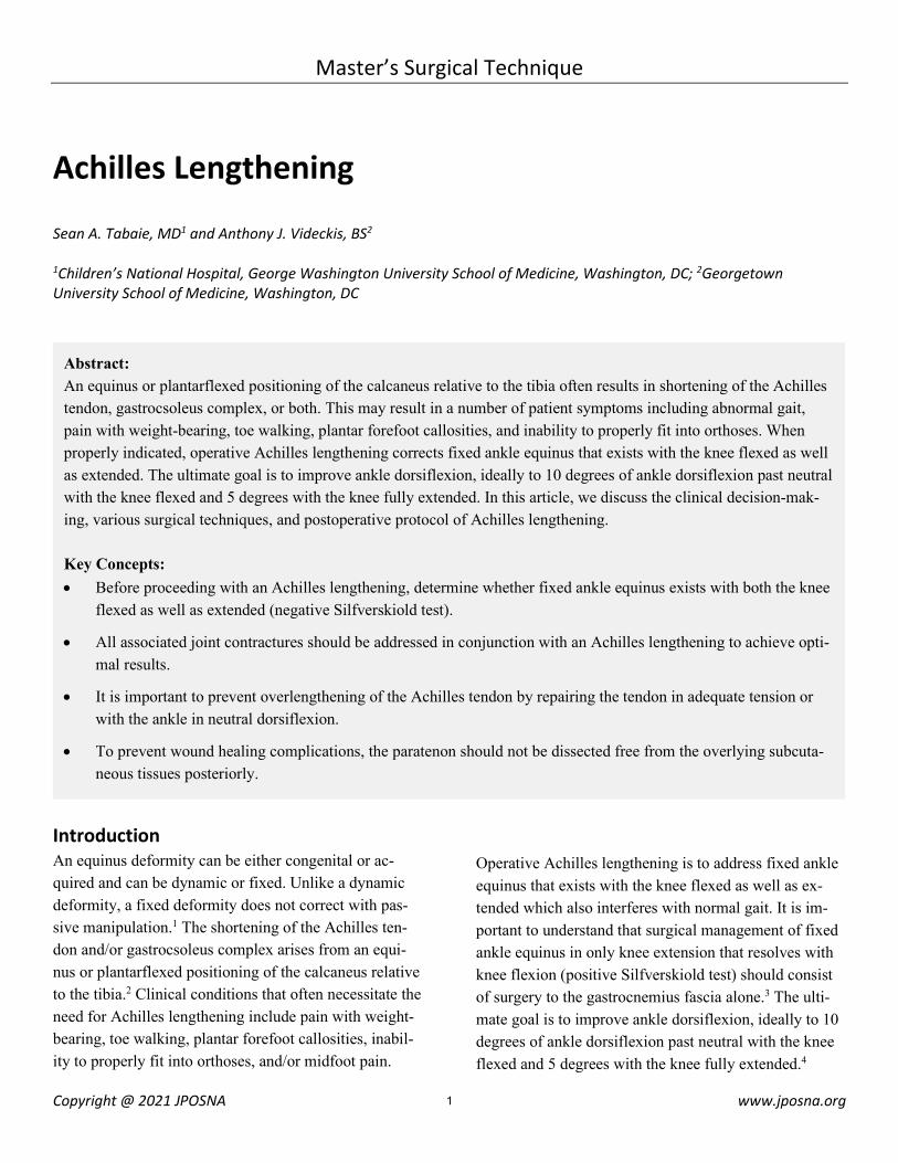
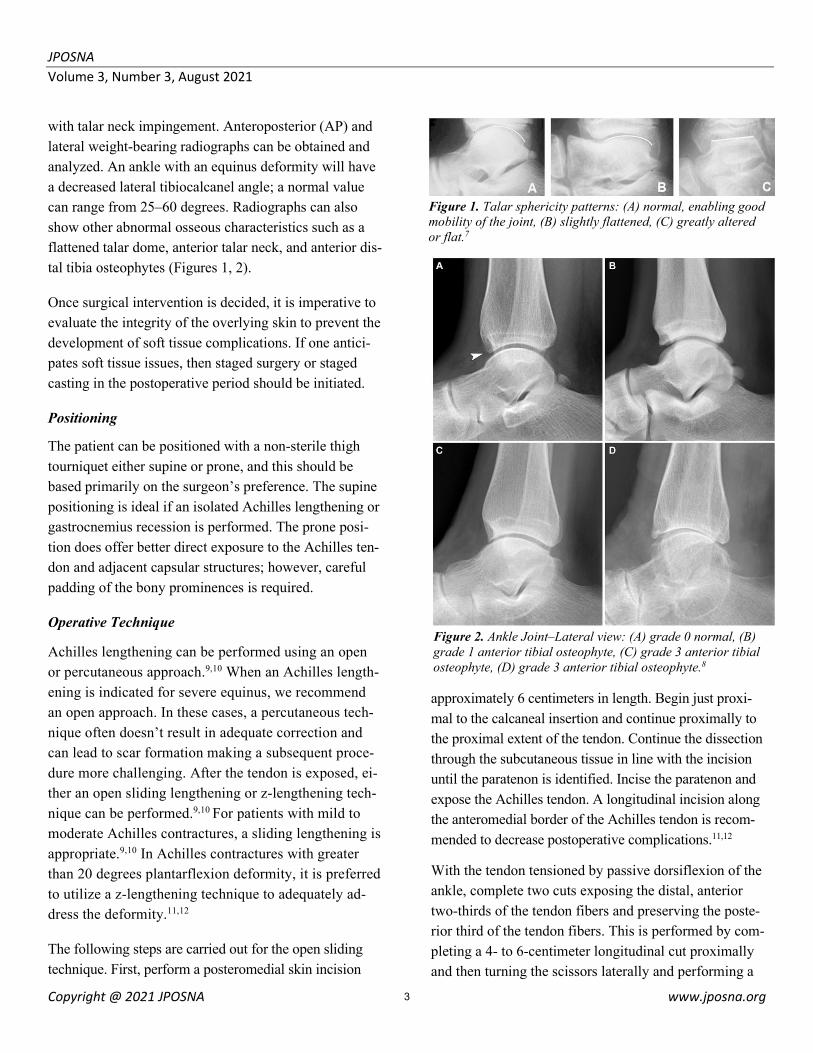
with talar neck impingement. Anteroposterior (AP) and lateral weight-bearing radiographs can be obtained and analyzed. An ankle with an equinus deformity will have a decreased lateral tibiocalcanel angle; a normal value can range from 25–60 degrees. Radiographs can also show other abnormal osseous characteristics such as a flattened talar dome, anterior talar neck, and anterior dis-tal tibia osteophytes (Figures 1, 2).
Once surgical intervention is decided, it is imperative to evaluate the integrity of the overlying skin to prevent the development of soft tissue complications. If one antici-pates soft tissue issues, then staged surgery or staged casting in the postoperative period should be initiated.
Positioning
The patient can be positioned with a non-sterile thigh tourniquet either supine or prone, and this should be based primarily on the surgeon’s preference. The supine positioning is ideal if an isolated Achilles lengthening or gastrocnemius recession is performed. The prone posi-tion does offer better direct exposure to the Achilles ten-don and adjacent capsular structures; however, careful padding of the bony prominences is required.
Operative Technique
Achilles lengthening can be performed using an open or percutaneous approach.9,10 When an Achilles length-ening is indicated for severe equinus, we recommend an open approach. In these cases, a percutaneous tech-nique often doesn’t result in adequate correction and can lead to scar formation making a subsequent proce-dure more challenging. After the tendon is exposed, ei-ther an open sliding lengthening or z-lengthening tech-nique can be performed.9,10 For patients with mild to moderate Achilles contractures, a sliding lengthening is appropriate.9,10 In Achilles contractures with greater than 20 degrees plantarflexion deformity, it is preferred to utilize a z-lengthening technique to adequately ad-dress the deformity.11,12
The following steps are carried out for the open sliding technique. First, perform a posteromedial skin incision
approximately 6 centimeters in length. Begin just proxi-mal to the calcaneal insertion and continue proximally to the proximal extent of the tendon. Continue the dissection through the subcutaneous tissue in line with the incision until the paratenon is identified. Incise the paratenon and expose the Achilles tendon. A longitudinal incision along the anteromedial border of the Achilles tendon is recom-mended to decrease postoperative complications.11,12
With the tendon tensioned by passive dorsiflexion of the ankle, complete two cuts exposing the distal, anterior two-thirds of the tendon fibers and preserving the poste-rior third of the tendon fibers. This is performed by com-pleting a 4- to 6-centimeter longitudinal cut proximally and then turning the scissors laterally and performing a
Figure 1. Talar sphericity patterns: (A) normal, enabling good mobility of the joint, (B) slightly flattened, (C) greatly altered or flat.7
Figure 2. Ankle Joint–Lateral view: (A) grade 0 normal, (B) grade 1 anterior tibial osteophyte, (C) grade 3 anterior tibial osteophyte, (D) grade 3 anterior tibial osteophyte.8
3
JPOSNA Volume 3, Number 3, August 2021
Copyright @ 2021 JPOSNA www.jposna.org
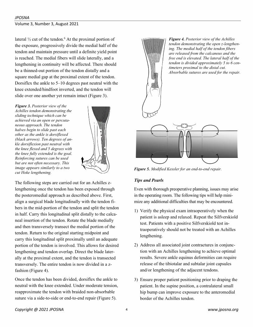
lateral ½ cut of the tendon.6 At the proximal portion of the exposure, progressively divide the medial half of the tendon and maintain pressure until a definite yield point is reached. The medial fibers will slide laterally, and a lengthening in continuity will be affected. There should be a thinned-out portion of the tendon distally and a square medial gap at the proximal extent of the tendon. Dorsiflex the ankle to 5–10 degrees past neutral with the knee extended/hindfoot inverted, and the tendon will slide over one another yet remain intact (Figure 3).
The following steps are carried out for an Achilles z-lengthening once the tendon has been exposed through the posteromedial approach as described above. First, align a surgical blade longitudinally with the tendon fi-bers in the mid-portion of the tendon and split the tendon in half. Carry this longitudinal split distally to the calca-neal insertion of the tendon. Rotate the blade medially and then transversely transect the medial portion of the tendon. Return to the original starting midpoint and carry this longitudinal split proximally until an adequate portion of the tendon is involved. This allows for desired lengthening and tendon overlap. Direct the blade later-ally at the proximal extent, and the tendon is transected transversely. The entire tendon is now divided in a z-fashion (Figure 4).
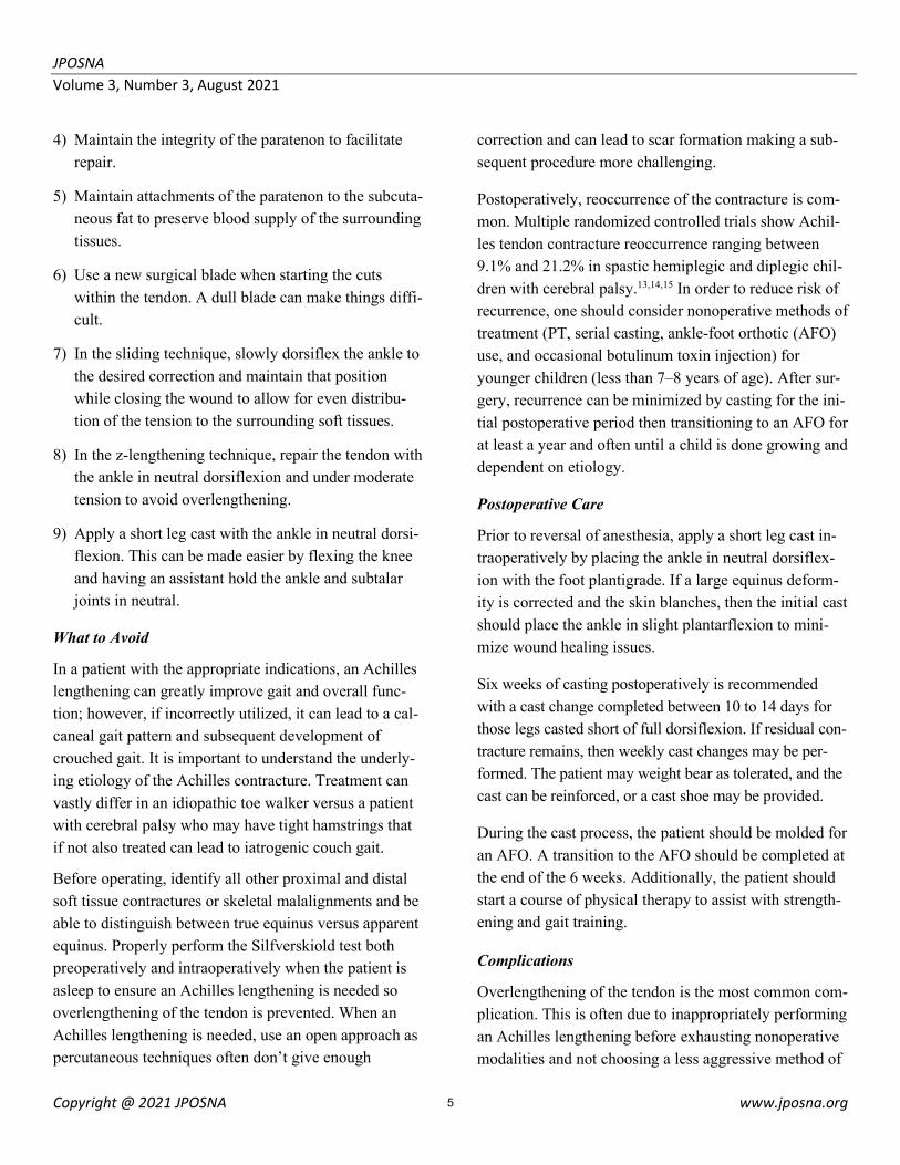
Once the tendon has been divided, dorsiflex the ankle to neutral with the knee extended. Under moderate tension, reapproximate the tendon with braided non-absorbable suture via a side-to-side or end-to-end repair (Figure 5).
Tips and Pearls
Even with thorough preoperative planning, issues may arise in the operating room. The following tips will help mini-mize any additional difficulties that may be encountered.
1) Verify the physical exam intraoperatively when the patient is asleep and relaxed. Repeat the Silfverskiold test. Patients with a positive Silfverskiold test in-traoperatively should not be treated with an Achilles lengthening.
2) Address all associated joint contractures in conjunc-tion with an Achilles lengthening to achieve optimal results. Severe ankle equinus deformities can require release of the tibiotalar and subtalar joint capsules and/or lengthening of the adjacent tendons.
3) Ensure proper patient positioning prior to draping the patient. In the supine position, a contralateral small hip bump can improve exposure to the anteromedial border of the Achilles tendon.
Figure 3. Posterior view of the Achilles tendon demonstrating the sliding technique which can be achieved via an open or percuta-neous approach. The tendon halves begin to slide past each other as the ankle is dorsiflexed (black arrows). Ten degrees of an-kle dorsiflexion past neutral with the knee flexed and 5 degrees with the knee fully extended is the goal. Reinforcing sutures can be used but are not often necessary. This image appears similarly to a two cut Hoke lengthening.
Figure 4. Posterior view of the Achilles tendon demonstrating the open z-lengthen-ing. The medial half of the tendon fibers are released from the calcaneus and the free end is elevated. The lateral half of the tendon is divided approximately 5 to 6 cen-timeters proximal to the distal cut. Absorbable sutures are used for the repair.
Figure 5. Modified Kessler for an end-to-end repair.
4
JPOSNA Volume 3, Number 3, August 2021
Copyright @ 2021 JPOSNA www.jposna.org
4) Maintain the integrity of the paratenon to facilitate repair.
5) Maintain attachments of the paratenon to the subcuta-neous fat to preserve blood supply of the surrounding tissues.
6) Use a new surgical blade when starting the cuts within the tendon. A dull blade can make things diffi-cult.
7) In the sliding technique, slowly dorsiflex the ankle to the desired correction and maintain that position while closing the wound to allow for even distribu-tion of the tension to the surrounding soft tissues.
8) In the z-lengthening technique, repair the tendon with the ankle in neutral dorsiflexion and under moderate tension to avoid overlengthening.
9) Apply a short leg cast with the ankle in neutral dorsi-flexion. This can be made easier by flexing the knee and having an assistant hold the ankle and subtalar joints in neutral.
What to Avoid
In a patient with the appropriate indications, an Achilles lengthening can greatly improve gait and overall func-tion; however, if incorrectly utilized, it can lead to a cal-caneal gait pattern and subsequent development of crouched gait. It is important to understand the underly-ing etiology of the Achilles contracture. Treatment can vastly differ in an idiopathic toe walker versus a patient with cerebral palsy who may have tight hamstrings that if not also treated can lead to iatrogenic couch gait.
Before operating, identify all other proximal and distal soft tissue contractures or skeletal malalignments and be able to distinguish between true equinus versus apparent equinus. Properly perform the Silfverskiold test both preoperatively and intraoperatively when the patient is asleep to ensure an Achilles lengthening is needed so overlengthening of the tendon is prevented. When an Achilles lengthening is needed, use an open approach as percutaneous techniques often don’t give enough
correction and can lead to scar formation making a sub-sequent procedure more challenging.
Postoperatively, reoccurrence of the contracture is com-mon. Multiple randomized controlled trials show Achil-les tendon contracture reoccurrence ranging between 9.1% and 21.2% in spastic hemiplegic and diplegic chil-dren with cerebral palsy.13,14,15 In order to reduce risk of recurrence, one should consider nonoperative methods of treatment (PT, serial casting, ankle-foot orthotic (AFO) use, and occasional botulinum toxin injection) for younger children (less than 7–8 years of age). After sur-gery, recurrence can be minimized by casting for the ini-tial postoperative period then transitioning to an AFO for at least a year and often until a child is done growing and dependent on etiology.
Postoperative Care
Prior to reversal of anesthesia, apply a short leg cast in-traoperatively by placing the ankle in neutral dorsiflex-ion with the foot plantigrade. If a large equinus deform-ity is corrected and the skin blanches, then the initial cast should place the ankle in slight plantarflexion to mini-mize wound healing issues.
Six weeks of casting postoperatively is recommended with a cast change completed between 10 to 14 days for those legs casted short of full dorsiflexion. If residual con-tracture remains, then weekly cast changes may be per-formed. The patient may weight bear as tolerated, and the cast can be reinforced, or a cast shoe may be provided.
During the cast process, the patient should be molded for an AFO. A transition to the AFO should be completed at the end of the 6 weeks. Additionally, the patient should start a course of physical therapy to assist with strength-ening and gait training.
Complications
Overlengthening of the tendon is the most common com-plication. This is often due to inappropriately performing an Achilles lengthening before exhausting nonoperative modalities and not choosing a less aggressive method of
5
JPOSNA Volume 3, Number 3, August 2021
Copyright @ 2021 JPOSNA www.jposna.org
addressing the contracture, such as with a gastrocnemius recession.3 Worsening of the gait can occur, specifically the development of crouched gait. This is due to the lack of understanding between true versus apparent equinus and failure to identify adjacent soft tissue contractures and/or skeletal malalignment.11,12 Reoccurrence of the contracture or soft tissue pressure wounds may occur due to the lack of adequate postoperative casting and or-thotic wear. To prevent the risk of soft tissue pressure wounds, it may be needed to perform cast changes in the postoperative period.
Summary When properly indicated, operative Achilles lengthening corrects fixed ankle equinus and improves ankle dorsi-flexion, ideally to 10 degrees of ankle dorsiflexion past neutral with the knee flexed and 5 degrees with the knee fully extended. Before operating, it is imperative to distin-guish between true equinus versus apparent equinus. Properly perform the Silfverskiold test both preopera-tively and intraoperatively when the patient is asleep to ensure an Achilles lengthening is needed so overlengthen-ing of the tendon is prevented. When an Achilles length-ening is indicated for severe deformity, use an open ap-proach as percutaneous techniques often don’t give enough correction. In the open approach, either an open sliding lengthening or z-lengthening technique can be per-formed once the tendon is exposed. In Achilles contrac-tures with greater than a 20 degrees plantarflexion de-formity, it is preferred to utilize a z-lengthening technique to adequately address the deformity. Postoperatively, re-occurrence of the contracture is common. This can be minimized by casting for the initial postoperative period then transitioning to an AFO.
Additional Links • AAOS Percutaneous Achilles Tendon Repair:
https://www.aaos.org/videos/video-detail-page/20275__Videos
• AAOS Minimally-Invasive Surgical Repair for Achilles Tendon Rupture: https://www.aaos.org/videos/video-detail-page/18309__Videos
References 1. O’Brien M: The anatomy of the Achilles tendon. Foot Ankle Clin 2005;10(2):225-238. 2. Pinney SJ, Hansen ST Jr, Sangeorzan BJ: The effect on ankle dor-siflexion of gastrocnemius recession. Foot Ankle Int 2002;23(1):26-29. 3. Downey MS, Banks AS: Gastrocnemius recession in the treatment of nonspastic ankle equinus: A retrospective study. J Am Podiatr Med Assoc 1989;79(4):159-174. 4. Jahn J, Vasavada AN, McMulkin ML:Calf muscle-tendon lengths before and after Tendo Achilles lengthenings and gastrocnemius lengthenings for equinus in cerebral palsy and idiopathic toe walking. Gait Posture 2009;29(4):612-617. 5. Radford JA, Burns J, Buchbinder R, Landorf KB, Cook C: Does stretching increase ankle dorsiflexion range of motion? A systematic review. Br J Sports Med 2006;40(10):870-875. 6. Silfverskiöld N: Reduction of the uncrossed two-joints muscles of the leg to one-joint muscles in spastic conditions. Acta Chir Scand 1924;65:315-330. 7. Pinto JA, Hernandes AC, Buchaim TP, Blumetti FC, Chertman C, Yamane PC, Rocha Correa Fernandes, A. Radiographic Abnormali-ties of the Talus in Patients with Clubfoot after Surgical Release Us-ing the McKay Technique. Rev Bras Ortop. 2015;46(3):293-298. 8. Kraus, V. B., Kilfoil, T. M., Hash, T. W., 2nd, McDaniel, G., Ren-ner, J. B., Carrino, J. A., & Adams, S. (2015). Atlas of radiographic features of osteoarthritis of the ankle and hindfoot. Osteoarthritis and cartilage, 23(12): 2059–2085. 9. Salamon ML, Pinney SJ, Van Bergeyk A, Hazelwood S: Surgical anatomy and accuracy of percutaneous achilles tendon lengthening. Foot Ankle Int 2006;27(6):411-413. 10. Hoefnagels EM, Waites MD, Belkoff SM, Swierstra BA: Percu-taneous Achilles tendon lengthening: A cadaver-based study of fail-ure of the triple hemisection technique. Acta Orthop 2007;78(6):808-812. 11. McMulkin ML, Baird GO, Caskey PM, Ferguson RL: Compre-hensive outcomes of surgically treated idiopathic toe walkers. J Pedi-atr Orthop 2006;26(5):606-611. 12. Hemo Y, Macdessi SJ, Pierce RA, Aiona MD, Sussman MD: Outcome of patients after Achilles tendon lengthening for treatment of idiopathic toe walking. J Pediatr Orthop 2006;26(3):336-340. 13. Chung CY, Sung KH, Lee KM, Lee SY, Choi IH, Cho TJ, Yoo WJ, Park MS. Recurrence of equinus foot deformity after tendo-achilles lengthening in patients with cerebral palsy. J Pediatr Orthop 2015;35(4):419-25. 14. Ahmed G, Muhammad A, Syed, Tago, Imtiaz. Outcome of Z-Plasty Lengthening of Achilles Tendon for Correction of Equinus Deformity of Foot in Children with Cerebral Palsy. (2013). 15. Khatri, K., Akhund, M., Shah, N., Jhatiyal, R., Jokhio, M., & Tunio, Z. (2020). Results of Achilles Tendon Lengthening Using Z-Plasty in Equinus Feet. PJMHS,14(3):1463–1464.
6