action potential gating of calcium channels and
TRANSCRIPT

Bad trick to play
Action Potential Gating of Calcium Channels and Transmitter Release
by
J Darwin King, Jr.
BS, University of Pittsburgh, 1998
MS, University of Pittsburgh, 2001
Submitted to the Graduate Faculty of
Arts and Sciences in partial fulfillment
of the requirements for the degree of
Doctor of Philosophy
University of Pittsburgh
2007
UNIVERSITY OF PITTSBURGH
School of Arts and Sciences
This dissertation was presented
by
J Darwin King, Jr.
It was defended on
October 29, 2007
and approved by
Committee Chair: Guo-Qiang Bi, Ph.D.
Germán Barrionuevo, M.D.
Alison Barth, Ph.D.
Kevin Currie, Ph.D.
Nathaniel N. Urban, Ph.D.
Dissertation Advisor: Stephen D. Meriney, Ph.D.
ii

Action Potential Gating of Calcium Channels and Transmitter Release
J Darwin King, Jr.
University of Pittsburgh, 2007
The regulation of transmitter release at the neuromuscular junction is tightly regulated by the
influx of calcium in the presynaptic nerve terminal. Interestingly, the probability that release
sites at the neuromuscular junction will liberate transmitter during each action potential is very
low. The reasons for this low probability of release are not well understood. To test the
hypothesis that individual N-type calcium channels open with a low probability, single channel
recordings of N-type voltage-gated calcium channels were performed. Using this approach I
determined the conductance of these channels, their probability of gating during an action
potential waveform, and the magnitude of calcium flux during a single channel opening. I
conclude from these studies that N-type voltage-gated calcium channels have a very low
probability of opening (< 5%) during an action potential and the characteristics of calcium entry
during single channel openings can help to explain the low probability of transmitter release at
release sites in the neuromuscular junction. To understand how calcium current is activated
physiologically, the activation and resulting current from N-type calcium channels elicited by
different action potential waveforms were studied. This work was carried out at both room
temperature and 37˚C to provide a physiological context. Using the whole-cell patch clamp
techniques, I studied the activation of current during various action potential shapes and
conditions, and the kinetics of N- and L-type current activation. Using this approach I
determined that N-type channels activate more slowly than L-type. Furthermore, depending on
the action potential shape used and the temperature, action potentials can activate varying
iii

proportions (I/Imax) of N-type calcium current (ranging from 10-100%). Under physiological
conditions using a frog motoneuron action potential waveform I determined that there was a very
low proportion of calcium current activated by a natural action potential (~32%). Adenosine 5´-
triphosphate (ATP) is co-released with acetylcholine (ACh) at the neuromuscular junction, and
has been found to inhibit transmission. I used the cutaneous pectoris muscle of the Rana pipiens
to study ATP-mediated modulation of ACh release. Intracellular postsynaptic recordings were
used as a measure of ACh release, and agents that perturb the ATP signaling were examined.
iv

TABLE OF CONTENTS
1.0 INTRODUCTION ........................................................................................................ 1
1.1 OVERVIEW ......................................................................................................... 1
1.2 CALCIUM CHANNELS..................................................................................... 2
1.2.1 Calcium channel types..................................................................................... 2
1.2.2 Calcium channel subunits ............................................................................... 3
1.3 CALCIUM TRIGGERED SECRETION .......................................................... 7
1.3.1 Probability of release ....................................................................................... 9
1.3.2 Synaptotagmin ................................................................................................. 9
1.4 G-PROTEIN MODULATION ......................................................................... 11
1.5 ROLE OF ATP IN MODULATION OF RELEASE ..................................... 13
1.6 GOALS OF THESE STUDIES ........................................................................ 17
1.6.1 Single channel recordings of calcium channels reveal a low probability of
opening ........................................................................................................................ 17
1.6.2 Action potentials open a small proportion of calcium current .................. 18
1.6.3 Modulation of calcium entry and transmitter release by ATP ................. 19
2.0 SINGLE CHANNEL RECORDINGS OF CALCIUM CURRENTS REVEAL A
LOW PROBABILITY OF OPENING DURING AN ACTION POTENTIAL STIMULUS
………………………………………………………………………………………...20
v
2.1 SUMMARY ........................................................................................................ 20
2.2 INTRODUCTION ............................................................................................. 21
2.3 METHODS ......................................................................................................... 25
2.3.1 Xenopus Nerve-Muscle Co-culture............................................................... 25
2.3.2 Chick Ciliary Ganglion Cell Culture ........................................................... 25
2.3.3 Cell Attached Patch-Clamp .......................................................................... 26
2.4 RESULTS ........................................................................................................... 27
2.4.1 Single channel recording from Xenopus nerve-muscle co-cultures .......... 27
2.4.2 Calcium Channel Conductance in Chick Ciliary Ganglion ...................... 28
2.4.3 Determination of the Number of Channels within a Patch ....................... 28
2.4.4 Opening of Unitary Conductances During Action Potential Stimuli ....... 29
2.5 DISCUSSION ..................................................................................................... 37
3.0 PROPORTION OF N-TYPE CALCIUM CURRENT ACTIVATED BY
ACTION POTENTIAL STIMULI ............................................................................................ 43
3.1 SUMMARY ........................................................................................................ 43
3.2 INTRODUCTION ............................................................................................. 44
3.3 METHODS ......................................................................................................... 46
3.3.1 Cell culture ..................................................................................................... 46
3.3.2 Electrophysiological recording techniques .................................................. 46
3.3.3 Calcium current activation and analysis ..................................................... 47
3.4 RESULTS ........................................................................................................... 49
3.4.1 Calcium current activation during action potential depolarization ......... 49
3.4.2 Gating characteristics of N- versus L-type current .................................... 51
vi

3.4.3 Effects of temperature on activation of N-type calcium current during
action potential depolarization ................................................................................. 51
3.5 DISCUSSION ..................................................................................................... 62
3.5.1 Use of chick ciliary ganglion as a model system ......................................... 62
3.5.2 Cellular signaling ........................................................................................... 63
3.5.3 Regulation of transmitter release ................................................................. 65
4.0 MODULATION OF NEUROMUSCULAR TRANSMITTER RELEASE BY
ATP ………………………………………………………………………………………...68
4.1 SUMMARY ........................................................................................................ 68
4.2 INTRODUCTION ............................................................................................. 69
4.3 METHODS ......................................................................................................... 73
4.3.1 Tissue preparation ......................................................................................... 73
4.3.2 Intracellular recordings ................................................................................ 73
4.4 RESULTS ........................................................................................................... 74
4.4.1 Suramin .......................................................................................................... 74
4.4.2 Reactive Blue 2 ............................................................................................... 75
4.4.3 MRS2179 ........................................................................................................ 76
4.4.4 AMP-PNP ....................................................................................................... 77
4.4.5 GDPβs ............................................................................................................. 77
4.4.6 GDPβs plus AMP-PNP .................................................................................. 79
4.5 DISCUSSION ..................................................................................................... 87
5.0 GENERAL DISCUSSION ........................................................................................ 93
vii

5.1 BUILDING A RELIABLE NEUROMUSCULAR JUNCTION FROM
UNRELIABLE ELEMENTS ............................................................................................ 94
5.2 CALCIUM TRIGGERING OF TRANSMITTER SECRETION FROM
THE MOTOR NERVE TERMINAL ............................................................................... 99
5.3 ENDOGENOUS CALCIUM CHANNEL MODULATION ALTERS
CALCIUM ENTRY .......................................................................................................... 102
BIBLIOGRAPHY ..................................................................................................................... 105
viii

LIST OF TABLES
Table 1.1 Calcium channel molecular organization, subtype, and function. .................................. 4
Table 5.1 Comparison of the neuromuscular junction and the calyx of Held transmitter release
characteristics. ............................................................................................................................... 98
ix

LIST OF FIGURES
Figure 1.1: Calcium channel structure ............................................................................................ 6
Figure 1.2: Schematic of ATP signaling at the neuromuscular junction ...................................... 16
Figure 2.1 Presynaptic Xenopus varicosity cell-attached recordings. ........................................... 31
Figure 2.2 Representative single channel openings and conductance plots. ................................ 32
Figure 2.3 Representative ramp data ............................................................................................ 33
Figure 2.4 Representative calcium channel openings triggered by an action potential train. ...... 34
Figure 2.5 Characteristics of unitary calcium channel openings triggered by action potential
stimuli. .......................................................................................................................................... 35
Figure 2.6 MCell prediction of single channel current variability through one opening. ............ 36
Figure 3.1 Effects of action potential broadening on calcium current activation in chick ciliary
ganglion neurons. .......................................................................................................................... 55
Figure 3.2 Effects of action potential broadening on calcium current activation in chick ciliary
ganglion neurons using a recorded ciliary ganglion neuron action potential. .............................. 56
Figure 3.3 Calcium current activation during step depolarizations. ............................................. 57
Figure 3.4 Effects of motor nerve terminal action potential broadening at 37°C on calcium
current activation in chick ciliary ganglion neurons. .................................................................... 58
x

xi
Figure 3.5 Effects of action potential broadening on calcium current activation recorded at 37°C
in chick ciliary ganglion neurons using a recorded ciliary ganglion neuron action potential as a
voltage command. ......................................................................................................................... 59
Figure 3.6 Evaluation of calcium current activation using various combinations of action
potential waveforms and recording conditions. ............................................................................ 60
Figure 3.7 Whole-cell recordings from frog varicosities .............................................................. 61
Figure 4.1 Intracellular recordings of EPPs before and after suramin treatment. ......................... 80
Figure 4.2 Representative EPP traces from intracellular recordings before and after RB2
treatment. ...................................................................................................................................... 81
Figure 4.3 Effects of MRS2179 on EPP amplitude. ..................................................................... 82
Figure 4.4 Effects of AMP-PNP on EPP amplitude and paired-pulse facilitation. ...................... 83
Figure 4.5 Effects of GDPβs on EPP amplitude and synaptic strength ....................................... 84
Figure 4.6 Representative EPPs showing the effects of AMP-PNP after pretreatment with
GDPβs. .......................................................................................................................................... 85
Figure 4.7 Summary figure of the effects of agonists and antagonists of the purinergic receptors
on the amplitude of EPPs. ............................................................................................................. 86
Figure 5.1 Comparison of the rate of depression at the neuromuscular junction and at the Calyx
of Held .......................................................................................................................................... 97
Figure 5.2 Calcium release relationship when calcium entry sites are partially blocked ........... 100

1.0 INTRODUCTION
1.1 OVERVIEW
Voltage-gated calcium channels provide an avenue through which calcium ions may enter a cell.
The influx of calcium can trigger many intracellular events such as gene transcription, second
messenger cascades, the secretion of hormones, muscle contraction, or the release of
neurotransmitter. Action potentials propagating down the nerve’s axon depolarize the terminal
opening channels causing an influx of calcium down a large concentration and electrostatic
gradient. This influx of calcium leads to the release of neurotransmitter from synaptic vesicles
docked at active zones in the nerve terminal. In this way, voltage-gated calcium channels serve
as the link between cellular depolarization and the signal transduction involved in triggering
vesicle fusion. Therefore, the study of voltage-gated calcium channel function and regulation
remains an important issue. The work described in this thesis focuses on the likelihood that these
high voltage-gated calcium channels open during an action potential stimulus, addresses some
mechanisms that might underlie this phenomenon, and characterizes the variability in the
magnitude of calcium entry through a single channel opening. To study this issue, cultured
motoneurons (Xenopus laevis) and the adult neuromuscular junction (Rana pipiens) were used as
model experimental systems.
1

1.2 CALCIUM CHANNELS
1.2.1 Calcium channel types
The first electrophysiological studies of calcium channels were performed in cardiac myocytes
(Reuter 1967; 1979). Since that time, many types of calcium current have been defined
physiologically, pharmacologically, and molecularly. Two main categories of calcium channels
exist: low voltage activated (requiring little depolarization from resting potential for activation)
and high voltage activated (requiring large depolarizations above resting membrane potential for
activation). These categories are further divided by pharmacologic sensitivity. L-type channels
are found in cardiac, smooth, skeletal muscle, neurons, and other cell types. L-type are a high
voltage activated channel with large single channel conductance, slow voltage-dependent
inactivation, and are selectively inhibited by dihydropyridines (Reuter 1983). T-type channels
are found in cardiac and skeletal muscle as well as neurons. However, T-type channels activate
near resting membrane potentials, quickly inactivate, slowly deactivate, and have a small single
channel conductance (Carbone and Lux 1984; Fedulova et al. 1985; Nowycky et al. 1985;
Swandulla and Armstrong 1988). T-type channels can be blocked selectively by kurtoxin
(Chuang et al. 1998). N-type channels have a more negative voltage dependence, and gate faster,
than L-type (Nowycky et al. 1985). N-type channels are blocked selectively by the cone snail
peptide ω-conotoxin GVIA (McCleskey et al. 1987; Tsien et al. 1988). P-type channels are
sensitive to the spider toxin ω-agatoxin IVA (Llinas et al. 1989; Mintz et al. 1992). Q-type
discovered in cerebellar granule neurons (Randall and Tsien 1995) are blocked to a lesser degree
by ω-agatoxin IVA, but probably represent a related subtype (same gene) and are often grouped
2
into a “P/Q” type. There is also an R-type that was identified and can be blocked by SNX-482
(Newcomb et al. 1998).
1.2.2 Calcium channel subunits
Despite the continued use of the “lettered” classification system originally proposed by
pharmacologists, calcium channels are now classified by a newer system based on the 10 genes
that have been cloned (Catterall 2000; Ertel et al. 2000). The L-type or Cav1 group includes α1s
(skeletal muscle) and α1c (cardiac) channels that are sensitive to dihydropyridine block. The
second group of calcium channels (Cav2) is often coupled to the release of neurotransmitters.
This Cav2 group includes the N-, P/Q-, and R-types described earlier. Most of the work described
here focuses on the Cav2.2 (N-type) calcium channel. The third group (Cav3) includes the low
voltage activated or T-type channels (see Table 1.1).
3
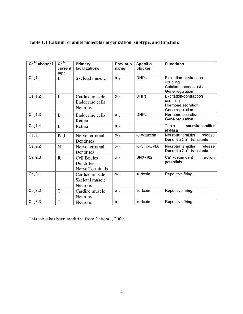
Table 1.1 Calcium channel molecular organization, subtype, and function.
Ca2+ channel Ca2+ current type
Primary localizations
Previous name
Specific blocker
Functions
CaV1.1 L Skeletal muscle α1S DHPs Excitation-contraction coupling Calcium homeostasis Gene regulation
CaV1.2 L Cardiac muscle Endocrine cells Neurons
α1C DHPs Excitation-contraction coupling Hormone secretion Gene regulation
CaV1.3 L Endocrine cells Retina
α1D DHPs Hormone secretion Gene regulation
CaV1.4 L Retina α1F Tonic neurotransmitter release
CaV2.1 P/Q Nerve terminal Dendrites
α1A ω-Agatoxin Neurotransmittler release Dendritic Ca2+ transients
CaV2.2 N Nerve terminal Dendrites
α1B ω-CTx-GVIA Neurotransmittler release Dendritic Ca2+ transients
CaV2.3 R Cell Bodies Dendrites Nerve Terminals
α1E SNX-482 Ca2+-dependent action potentials
CaV3.1 T Cardiac muscle Skeletal muscle Neurons
α1G kurtoxin Repetitive firing
CaV3.2 T Cardiac muscle Neurons
α1H kurtoxin Repetitive firing
CaV3.3 T Neurons α1I kurtoxin Repetitive firing
This table has been modified from Catterall, 2000.
4

Calcium channels were first purified from the transverse tubule membrane of skeletal
muscle (Curtis and Catterall 1984). The α1 subunit is about 2000 amino acids in length and has
four domains (I thru IV) each of which has six transmembrane segments (S1 thru S6). The S4
segment is unique in that it contains both positively charged and hydrophobic amino acids (De
Waard et al. 1996). This segment serves as the ‘voltage-sensor’ within the channels, reacting to
changes in membrane depolarization and inducing conformational changes in the protein that
subsequently opens the channel pore. Point mutations of the amino acid sequence in the S4
region have led to changes in channel gating (Guy and Conti 1990). A pair of glutamate residues
within each domain leads to calcium selectivity (Heinemann et al. 1992). The P loop between the
S5 and S6 segment lines the pore and is the site for drugs that utilize pore blocking as their
method of action (L-type antagonists, dihydropyridines). Each of these subtypes may be
alternatively spliced leading to further diversity (Lipscombe et al. 1989; Hofmann et al. 1994;
Perez-Reyes and Schneider 1995). Calcium channels are heteromultimeric with α1, β, and α2/δ
subunits (McEnery et al. 1991; Witcher et al. 1993; Martin-Moutot et al. 1995; Liu et al. 1996;
Martin-Moutot et al. 1996). Each member of the heteromultimer has a unique function.
Expression of the α1 subunit alone is sufficient to produce a calcium current influx although the
kinetics and voltage dependence are altered (Perez-Reyes et al. 1989). Co-expression with the β
and α2/δ subunits returns kinetics and voltage dependence back to physiological levels (Lacerda
et al. 1991; Singer et al. 1991). Channel kinetics, but not voltage-dependence, have also been
shown to be altered in splice variants of these channels (Lin et al. 2004).
5

Figure 1.1: Calcium channel structure
(modified from Catterall, 2000)
6

The β subunit is completely intracellular and lacks any transmembrane segments. There
are four known β genes named β1-4 and many splice variants of each. Expression of different β
subunits with a particular α1 subunit yields different channel kinetics and voltage dependence of
activation (Dolphin 2003). In this manner, peak current amplitude and channel inactivation
kinetics can be altered yielding diverse voltage-gated calcium channels. The role of the β subunit
was downplayed in studies for many years. However, it is now known that the β subunit is
critical for calcium channel translocation to the membrane and G-protein modulation (Dolphin
2003).
The two components of the α2/δ subunit originate from a single gene product and are
processed to yield two final proteins bound together by a disulfide bond. The α2 subunit is
located extracellularly and only five amino acids of the δ subunit are located intracellularly
(Randall and Benham 1999). Four α2/δ subunit gene splices have been identified (Klugbauer et
al. 1999). Alternative expression of these variants has a lesser effect on calcium channel
properties as compared to the β subunits and generally alters calcium current amplitude by
increasing the number of channels in the membrane (Douglas et al. 2006) and inactivation
kinetics (De Waard et al. 1995).
1.3 CALCIUM TRIGGERED SECRETION
The synapse is a specialized site which translates an electrical signal (action potential) into a
chemical signal (release of transmitter). This conversion occurs at active zones, a term first used
in 1970 by Couteaux and Pecot-Dechavassine (les zones actives). The active zone, regardless of
7

species, has hallmark identifying traits: the plasma membrane is electron dense, synaptic vesicles
cluster, tether, and fuse there, and it is opposed to the postsynaptic density in excitatory
synapses. The active zone can convert the action potential activity into chemical transmitter
release because it clusters voltage-gated calcium channels with release machinery, and uses
calcium ions to trigger the fusion of synaptic vesicles. For this to be a fast process, these
components must be in close proximity to one another. Strikingly, the time from calcium entry to
vesicle fusion is about 0.2 msec (Stanley 1997). Theoretical analysis, predicts that doubling the
distance between the calcium channel and synaptic vesicle will lead to a three-fold decrease in
the probability of release of a single vesicle (Cohen et al. 1991; Bennett et al. 2000).
Immunohistochemistry has shown the close proximity of calcium channels to the active zone
(Robitaille et al. 1990; Cohen et al. 1991).
A freeze fracture electron microscopy study has demonstrated a highly ordered
organization of the active zone (Heuser et al. 1974). Freeze fracture electron micrographs of the
frog neuromuscular junction active zone demonstrate long linear arrays spaced at 1µM
increments, each with ~250 intermembranous particles running the width of the nerve terminal
(Heuser et al. 1979; Heuser and Reese 1981; Pumplin et al. 1981; Meriney et al. 1996; Stanley
1997; Pawson et al. 1998; 1998). These active zones are closely associated with 20-30 vesicles
that contain transmitter as well as other proteins involved in vesicle trafficking and fusion. The
voltage-gated calcium channels that regulate calcium influx into the active zone region of the
nerve terminal are thought to compose a subset of the 250 intermembranous particles.
8

1.3.1 Probability of release
Action potentials travelling down the axon depolarize the nerve terminal and open voltage-gated
calcium channels. This leads to a local rise (Llinas et al. 1995) in intracellular calcium
concentration that triggers exocytosis of transmitter from active zone vesicles. Although,
nonphysiological stimuli such as hypertonic sucrose can cause all vesicles in the ready releasable
pool to undergoes exocytosis (Rosenmund and Stevens 1996), usually a single action potential
leads to the release of a single vesicle from an active zone. The entire frog neuromuscular
junction releases ~175 vesicles with each action potential stimulus. However, the probability of a
single vesicle release from each active zone is about 0.5 (Cho and Meriney, unpublished
observations; Dittrich et al. 2007). Because there are about 20-30 vesicles docked at each active
zone, a particular vesicle at a release site has a very low probability of release (~0.02) (D'Alonzo
and Grinnell 1985; Meriney et al. 1996; Macleod et al. 1999). A low probability for release of a
single vesicle has been reported at central nervous system synapses, and may be important for
the proper functioning of the synapse (Goda and Sudhof 1997).
1.3.2 Synaptotagmin
Synaptotagmin-1/2 is believed to be one of the calcium sensors for the fast exocytosis of
neurotransmitter. There are 13 vertebrate synaptotagmins that are placed into 6 classes (Sudhof
2002). The structure of synaptotagmin includes an N-terminal transmembrane region, a central
linker, and two C-terminal C2 domains (designated C2A and C2B). The term C2 comes from the
fact that these two areas are homologous with previously identified calcium binding domains for
PKC (Coussens et al. 1986). An individual C2 domain is 130-140 residues in length and can bind
9

calcium ions (Rizo and Sudhof 1998). C2A and C2B domains are constructed of a β sandwich
with eight β strands with loops on the top and bottom. Calcium binds to the top loops of the C2
domain. C2A domains can bind 3 calcium ions and C2B binds only 2 calcium ions (Ubach et al.
1998).
Synaptotagmin class 1 includes synaptotagmins 1 and 2. Both of these are localized to
synaptic vesicles and secretory granules placing them in position for fast calcium triggered
exocytosis. A point mutation in the gene coding for synaptotagmin 1 in the mouse cut calcium
affinity in half implicating synaptotagmin’s role (Fernandez-Chacon et al. 2001).
Synaptotagmins are transmembrane proteins within the synaptic vesicle. The vesicular
synaptotagmins exhibit a relatively low calcium affinity, with each of the 5 calcium binding sites
having a different affinity.
The function of synaptotagmin has been discovered through point mutations and knock
out studies. If wild-type synaptotagmin 1 DNA is introduced to cultured hippocampal neurons of
synaptotagmin 1 knock-out mice, then calcium-dependent synchronous transmitter release is
mostly restored. The mutation of either the second or third Asp residue of the C2B domain
inhibits the ability of synaptotagmin 1 to rescue synchronous release. Synaptotagmin 1 with
mutations in the first or fourth Asp residues of the C2B domain partially rescues synchronous
release. Therefore, the C2B domain of synaptotagmin 1 regulates neurotransmitter release and
synchronous release absolutely requires binding of calcium to the second and third Asp residues
in this domain (Nishiki and Augustine 2004).
10

1.4 G-PROTEIN MODULATION
G-protein modulation of calcium influx was first described in chick dorsal root ganglion neurons
(Dunlap and Fischbach 1978; 1981). In these studies, action potential duration in the dorsal root
ganglion neurons was shortened when GABAB, serotonin, or adrenergic receptors were
activated. Later, many other types of G-protein coupled receptors were found, and including the
odorant receptors, there may be thousands of subtypes. Cav2.1 (P/Q-type) and Cav2.2 (N-type)
are commonly regulated by G-protein pathways. Since these calcium channel types trigger the
release of neurotransmitter, G-protein receptor modulation is a common form neurotransmission
regulation (Hille 1994; Jones and Elmslie 1997; Ikeda and Dunlap 1999).
The effect of G-protein mediated inhibition on voltage-gated calcium channels has been
well studied with electrophysiological methods. The peak current amplitudes are often reduced
during whole-cell recordings. The inhibition is greater at more hyperpolarized (negative)
membrane voltages. Therefore, a shift of the activation curve toward more depolarized potentials
is observed. Also, current activation and inactivation are slowed by G-protein modulation (Bean
1989; Kasai and Aosaki 1989; Lipscombe et al. 1989). A “prepulse” to +100mV can relieve G-
protein inhibition, and although such depolarizations do not occur physiologically, trains of
stimuli can also relieve inhibition (Brody et al. 1997).
Single channel recordings of N-type calcium channels have been used to characterize
kinetic changes associated with voltage-dependent G-protein mediated inhibition (Carabelli et al.
1996; Patil et al. 1996). The G-protein inhibition increases latency to first channel opening
causing activation kinetics to slow. The mechanism of this increase in latency has been shown to
be caused by stabilization of the closed state of the channel by Gβγ binding (reluctant state).
When the Gβγ subunits are free, the channel is said to be in a willing state (Bean 1989). The
11

dissociation of the Gβγ complex from the channel allows a reluctant channel to transition to a
willing state. The N-type channel can indeed open even in the reluctant state, however, this has a
very low probability and mean open time (Colecraft et al. 2001).
G-protein modulation of N-type calcium channels may also occur in a voltage-
independent manner. The activation of a G-protein receptor frees both Gα and Gβγ. These
diffuse within the membrane and act on cytoplasmic second messenger systems such as
phospholipase C, phospholipase A2, and adenylyl cyclase. For example, protein kinase C (PKC)
can phosphorylate calcium channels leading to an increase in calcium current. PKC can also
reverse the effects of G-protein mediated inhibition of N-type channels. This PKC
phosphorylation stops G-protein inhibition if the Gβ1 subunit is present. PKC acts on the α1
subunit at the threonine reside (422) located on the I-II linker and blocks the binding of Gβγ
(Zamponi et al. 1997).
With extended activation, G-protein channel signaling undergoes desensitization which is
defined as a loss of effectiveness despite continued exposure to the agonist. This desensitization
can occur through many mechanisms (Ferguson 2001). Rapid desensitization (on the order of
seconds to minutes) occurs due to phosphorylation by G-protein coupled receptor kinases
(GRKs) or second messenger kinases. This phosphorylation uncouples the G-protein from the
receptor. Desensitization may also occur via receptor internalization (Beaumont et al. 1998).
Activated receptors undergo endocytosis via clatherin-mediated mechanisms. Also, G-protein
modulation may be terminated by regulators of G-protein signaling (RGS). These act by
increasing the rate of the hydrolysis of the Gα – bound GTP.
12

1.5 ROLE OF ATP IN MODULATION OF RELEASE
Adenosine 5´-triphosphate (ATP) is co-released with acetylcholine (ACh) at the neuromuscular
junction (Silinsky 1975; Silinsky and Redman 1996). Early work suggests that exogenously
applied ATP can depress synaptic transmission (Ribeiro and Walker 1975). Adenosine and ATP
are members of a larger class of transmitters called purines. Metabotropic purinergic receptors
have been proposed as contributors to synaptic depression (Burnstock and Kennedy 1985). This
study will focus on the role of ATP in mediating a tonic inhibition of transmitter release from the
neuromuscular junction as a possible contributor to the very low probability of opening of
voltage-gated calcium channels.
Both potentiation and depression of transmitter release can occur as a result of purinergic
signaling (Correia-de-Sa et al. 1996). In frogs, release can be either increased or decreased
depending on the developmental stage of the animal being studied. For example, in the Xenopus
tadpole neuromuscular junction, purines increase transmitter release (Fu and Poo 1991).
However, the opposite is true in the adult frog neuromuscular junction where purines depress
release (Giniatullin and Sokolova 1998). The frequency and pattern of stimulation also influence
presynaptic modulation (Correia-de-Sa et al. 1996). These complicated effects also depend on
the type of purine (ATP or adenosine) present, and the receptors that mediate these effects. The
A1 adenosine receptor dominates during the low frequency firing when low levels of adenosine
are present, leading to a depressing effect. The A2a receptors respond only to higher levels of
adenosine present with higher frequency stimulation, and these receptors mediate potentiation of
release. Higher levels of adenosine are present when a larger amount of ATP, which is co-
released with ACh, is cleaved by ectonucleotidases. This raises an important question: if more
release leads to an inhibition then how does the neuromuscular junction continue to function
13

during prolonged periods of activity? It seems that the ectonucleotidases can be inhibited by the
high frequency release of ATP itself, thus the adenosine production and the depression of release
is reduced (Cunha 2001). In addition, the accumulation of adenosine from all of the ATP
released, causes activation of the A2a receptors leading to potentiation (Correia-de-Sa et al.
1996). These two mechanisms serve to keep the neuromuscular junction functioning during
prolonged periods of release which occurs during intense muscle activity.
The hydrolysis of ATP to yield adenosine can activate adenosine receptors which may
also affect calcium channels (Hamilton and Smith 1991). Adenosine A1 receptors have been
shown to inhibit the P/Q-type channel in mammals where P/Q channels are responsible for
calcium entry necessary for transmitter release (Silinsky 2004).
Modulation can also occur directly through ATP receptors. Again the receptor determines
whether the effect is depressing or potentiating. Activation of metabotropic P2 receptors have an
inhibitory effect in the frog neuromuscular junction (Giniatullin and Sokolova 1998). The work
in this study focused on the inhibition of the adult frog NMJ by the release of ATP. Originally it
was thought that the depression of the synapse mediated by purinergic receptors did not alter
presynaptic calcium flux (Redman and Silinsky 1994; Robitaille et al. 1999; Huang et al. 2002).
However, a later study suggested that the activation of the P2Y receptor can lead to a decrease in
presynaptic calcium current in the frog NMJ (Grishin et al. 2005). It is thought that this
depression occurs through the arachidonic acid second messenger pathway (Grishin et al. 2005).
Purinergic signaling at the neuromuscular junction is not limited to the nerve terminal.
The peripheral nervous system has three types of glia: myelinating and nonmyelinating Schwann
cells that are associated with axons, and perisynaptic Schwann cells (PSCs). PSCs are of the
most interest in this context as they are present at the neuromuscular junction and could be a
14
possible influence by releasing ATP and causing inhibition of the synapse. Furthermore, PSCs
also contain adenosine and ATP (both P2X ionotropic and P2Y metabotropic) receptors
(Robitaille 1995). Suramin, a P2 receptor antagonist, has been shown to reduce nerve
stimulation-evoked PSC calcium responses by ~50%. Thus purinergic signaling has a strong
influence on calcium flux in glia, and this can also alter nerve terminal transmitter release.
In summary, adenosine and ATP can alter calcium influx, transmitter release, and
activate intracellular cascades by many potential mechanisms. Because purinergic signaling can
both depress and potentiate vesicular release at the neuromuscular junction by multiple methods,
much work needs to be completed in this area before this complex signaling system can be fully
understood.
15
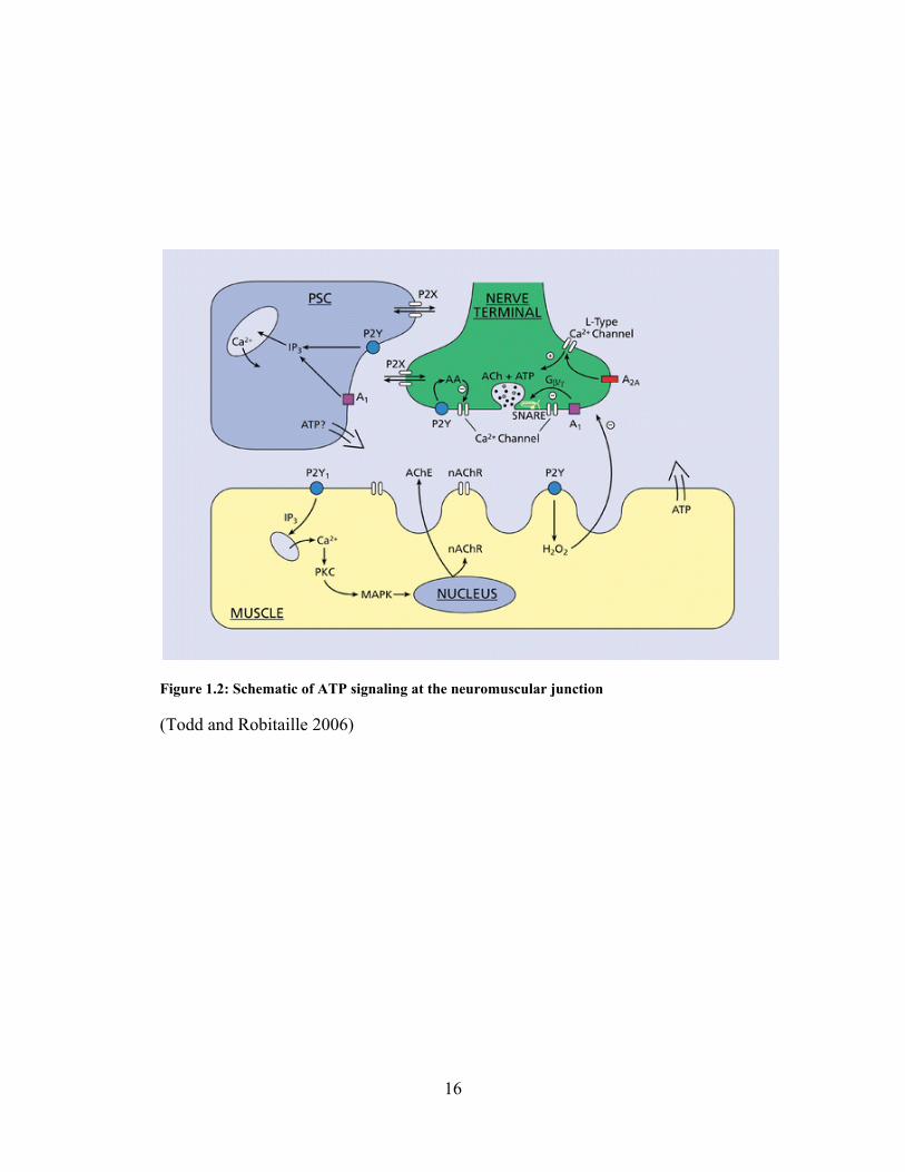
Figure 1.2: Schematic of ATP signaling at the neuromuscular junction
(Todd and Robitaille 2006)
16
1.6 GOALS OF THESE STUDIES
The regulation of transmitter release at the neuromuscular junction is tightly regulated by the
influx of calcium in the presynaptic nerve terminal. Interestingly, the probability that release
sites at the neuromuscular junction will liberate transmitter during each action potential is very
low. The reasons for this low probability of release are not well understood. One hypothesis is
that presynaptic calcium channels open with a very low probability at release sites. I have tested
this hypothesis by studying the gating of the type of calcium channel (N-type) present in the frog
presynaptic motor nerve terminal. Furthermore, I have examined a modulatory influence that
exists at the neuromuscular junction (ATP) and studied its effect on transmitter release. I
hypothesize that the corelease of ATP with acetylcholine, mediated by metabotropic purinergic
receptors, helps to maintain a low probability of release at the neuromuscular junction that is
expected to be critical to the continued release of transmitter during high frequency, repeated
firing at this synapse. My work examining these issues is divided into three chapters.
1.6.1 Single channel recordings of calcium channels reveal a low probability of opening
To test the hypothesis that individual N-type calcium channels open with a low probability,
single channel recordings of N-type voltage-gated calcium channels were performed. As this
investigation is aimed at understanding the calcium control of release from the neuromuscular
junction, some preliminary data were collected at the cultured frog neuromuscular junction nerve
terminal. Since recording from the release face of these natural nerve terminals required the
physical removal of the muscle from the varicosity, these experiments were technically
challenging. Thus, for the majority of the data acquisition used to test my hypotheses, a model
17

system (chick ciliary ganglion neurons) was used for cell-attached patch recordings. Using this
approach I determined the conductance of these channels, their probability of gating during an
action potential waveform, and the magnitude of calcium flux during a single channel opening. I
conclude from these studies that N-type voltage-gated calcium channels have a very low
probability of opening during an action potential and the characteristics of calcium entry during
single channel openings can help to explain the low probability of transmitter release at release
sites in the neuromuscular junction.
1.6.2 Action potentials open a small proportion of calcium current
To understand how calcium current is activated physiologically, the activation and resulting
current from N-type calcium channels elicited by different action potential waveforms were
studied. This work was carried out at both room temperature and 37˚C to provide a physiological
context. Using the whole-cell patch clamp techniques, I studied the activation of current during
various action potential shapes and conditions, and the kinetics of N- and L-type current
activation. Using this approach I determined that N-type channels activate more slowly than L-
type. Furthermore, depending on the action potential shape used and the temperature, action
potentials can activate varying proportions (I/Imax) of N-type calcium current (ranging from 10-
100%). Under physiological conditions using a frog motoneuron action potential waveform I
determined that there was a very low proportion of calcium current activated by a natural action
potential (~32%).
18

1.6.3 Modulation of calcium entry and transmitter release by ATP
In addition to the intrinsic properties of the presynaptic N-type calcium channel (studied in
chapters 2 and 3), there are likely to be many local influences that maintain a very low
probability of transmitter release at the neuromuscular junction. This last chapter focuses on one
possible mechanism that could provide a tonic inhibition of release at the neuromuscular
junction. Adenosine 5´-triphosphate (ATP) is co-released with acetylcholine (ACh) at the
neuromuscular junction (Silinsky 1975; Silinsky and Redman 1996), and has been found to
inhibit transmission (Ribeiro and Walker 1975). I used the cutaneous pectoris muscle of the
Rana pipiens as a model synapse to study ATP-mediated modulation of ACh release.
Intracellular postsynaptic recordings were used as a measure of ACh release, and agents that
perturb the ATP signaling were examined. The data presented in this chapter support the
hypothesis that ATP mediates a tonic inhibition of transmitter release from the neuromuscular
junction, and this inhibition may serve to control the very low probability of opening of voltage-
gated calcium channels and release of ACh-containing vesicles.
19

2.0 SINGLE CHANNEL RECORDINGS OF CALCIUM CURRENTS REVEAL A
LOW PROBABILITY OF OPENING DURING AN ACTION POTENTIAL STIMULUS
2.1 SUMMARY
Although it is known that presynaptic calcium entry triggers transmitter release, there is some
debate as to how many calcium channels open in an active zone to cause each vesicle fusion
event. Xenopus nerve-muscle co-cultures and chick ciliary ganglion neurons were used to study
the activation of unitary N-type calcium currents during a motor nerve terminal action potential
depolarization. An action potential that was recorded from a cultured frog motor nerve terminal
was used as a voltage command template. In both presynaptic varicose structures from Xenopus
nerve-muscle co-cultures and cultured chick ciliary ganglion neurons, the native frog motor
neuron action potential revealed a very low probability of opening of the N-type calcium channel
(3.59 ± 0.45%). These results suggest that under physiological conditions, an action potential is
not very effective at gating N-type calcium channels. This finding is consistent with the
hypothesis that few (possibly even one) N-type calcium channels open during an action potential
in the frog motor nerve terminal to trigger transmitter release.
20

2.2 INTRODUCTION
The neuromuscular junction is a very reliable synapse such that depolarization of the presynaptic
motoneuron nearly always leads to contraction of the postsynaptic muscle fiber. A single action
potential can lead to the release of hundreds of synaptic vesicles which contain thousands of
molecules of transmitter. In addition, the postsynaptic membrane has thousands of receptors that
are sensitive for transmitter.
For synaptic transmission to be fast, the presynaptic active zone must be specialized. This
includes the localization of proteins involved in release. These proteins are involved in the
conversion of the electrical signal of the action potential depolarization into the chemical signal
of the release of neurotransmitter. The structure of the active zone has been studied at the frog
neuromuscular junction using the freeze fracture electron microscope technique. The release
face, the presynaptic membrane directly in contact with the postsynaptic muscle cell, has a series
of well organized double rows of intramembranous particles that are 1 µm long and spaced 1 µm
apart (Heuser et al. 1974; Pumplin et al. 1981). Some of these particles are voltage-gated
calcium channels that appear to be tightly co-localized with the release of transmitter from the
active zone (Robitaille et al. 1990; Cohen et al. 1991; Llinas et al. 1992; Stanley 1997).
Voltage-gated calcium channels perform many functions in neurons. Different types of
calcium channels have been identified with varying properties that are well suited for specific
roles in different compartments of the cell (De Waard et al. 1996). In particular, some types of
calcium channels are clustered at nerve terminals for the calcium-triggered vesicle fusion that
underlies neurotransmitter release (Stanley 1997). These types are generally activated by
relatively large depolarizations from rest as is experienced during action potential invasion of the
nerve terminal. The active zone is the specialized region (Heuser et al. 1974) of the presynaptic
21

nerve terminal from which transmitter is released (Katz, 1969). This specialized transmitter
release region of the nerve terminal (active zone) is characterized by the presence of large
numbers of voltage-gated calcium channels and associated transmitter-containing synaptic
vesicles. Interestingly, despite the availability of many release-ready vesicles at a typical active
zone, only one is usually released following single action potential invasion. Vesicle fusion
occurs with low probability despite a colocalization of many presynaptic calcium channels with
release-ready vesicles (Rosenmund and Stevens 1996; Goda and Sudhof 1997). This thesis is
focused on the contribution of calcium channel gating to the low probability of transmitter
release from each active zone. The depolarizing phase of the action potential opens calcium
channels, but the driving force for calcium is low at the peak of the action potential. Driving
force for calcium increases with action potential repolarization, and this generates a "tail" current
of calcium influx (Llinas et al. 1981; Toth and Miller 1995; Sabatini and Regehr 1996; Borst and
Sakmann 1998). At the nerve terminal, this brief influx of calcium creates a local "nanodomain"
(Chad and Eckert 1984; Llinas et al. 1995) of elevated intracellular calcium around the mouth of
each open channel that triggers vesicle fusion and transmitter release.
Currently, there is some debate regarding the number of calcium channels that must be
activated in an active zone to cause sufficient levels of intracellular calcium to trigger vesicle
fusion. Some have argued that the nanodomain of calcium that results from a single calcium
channel opening is sufficient to trigger the fusion of a nearby docked vesicle. One study
measured dose-response relationships between various calcium channel blockers and transmitter
release at the frog neuromuscular junction, and performed computational analyses that led them
to hypothesize that the activity of a single calcium channel mediates vesicle fusion at an
individual transmitter release site (Yoshikami et al. 1989). Another group reached a similar
22

conclusion by using a combination of patch clamp recordings of single calcium channels and a
chemi-luminescent method to detect transmitter release in the calyciform presynaptic terminal of
the chick ciliary ganglion (Stanley 1993). Further support for single channel triggering of vesicle
fusion was provided in a computational study (Bertram et al. 1996). A study in the Meriney lab
(Poage and Meriney 2002; Wachman et al. 2004) used a fast imaging approach at the adult frog
neuromuscular junction to demonstrate that the spatial distribution of calcium entry at release
sites was altered by calcium channel blockers in a manner consistent with very few, perhaps only
one calcium channel opening underlying each calcium entry site. These studies suggest that, in
these preparations, the flux of calcium through a single calcium channel opening normally
triggers each transmitter release event.
At other synapses, studies conclude that multiple calcium channel openings are necessary
for transmitter release. Data from the calyx of Held in the rat medial nucleus of the trapezoid
suggest that more than 60 calcium channel openings occur with each vesicle fusion event (Borst
and Sakmann 1996). Furthermore, because the slow calcium buffer EGTA was able to reduce
transmitter release even at concentrations as low as 1 mM, the authors conclude that calcium ions
must be acting to trigger transmitter release over significant distances within the nerve terminal
and that calcium entry through multiple channels normally triggers each vesicle fusion event.
Data from hippocampal commissural synapses onto CA1 pyramidal neurons are most consistent
with a large proportion of available calcium channels opening with each action potential stimulus
(Dunlap et al. 1995). At these synapses, synaptic transmission is completely blocked only when
a combination of N- and P/Q-type channel blockers are used, with each blocker in isolation
blocking only a portion of release (Wheeler et al. 1994). These data suggest that not only are
there more than one calcium channel type present at these synapses, but that they need to act in
23

concert to trigger vesicle fusion. In order for this to occur, one would predict that a large
percentage of available channels would need to be activated by each action potential. In fact,
Borst and Sakmann (1998) have shown that an action potential is very effective at activating
presynaptic calcium current at the rat calyx of Held, with about 70% of maximal current
activated by a native action potential. These data support the hypothesis that a majority of
presynaptic calcium channels open with each action potential and are consistent with the idea
that the flux through many channels normally triggers each vesicle fusion event at these types of
synapses.
In this chapter most experiments used the chick ciliary ganglion as a model system to
study calcium channel gating. The chick ciliary ganglion acute cell culture system was used in
the Meriney lab for other studies, but is also useful for the study of calcium channel gating.
These cells are spherical and can be easily controlled in a voltage clamp experiment. The cell
membranes are patched with relative ease, and a wealth of literature has been published on this
system. The chick ciliary ganglion has two populations of parasympathetic motoneurons. The
ciliary neurons innervate the striated muscle of the ciliary body and iris of the eye. The choroid
neurons innervate the smooth muscle of the choroid coat in the eye (Marwitt et al. 1971). Chick
ciliary ganglion neurons express approximately 74% N-type and about 24% L-type calcium
current at stage 40 (White et al. 1997) making them ideal to study N-type current when
nifedipine is present. As described earlier in this introduction, there is some debate as to how
many calcium channels open in an active zone to cause each vesicle fusion event, and the answer
to this issue may be preparation dependent. Chick ciliary ganglion neurons were used for most
experiments in this chapter to study the gating of single N-type calcium channels during action
potential depolarization. In addition to characterizing these channel gating events, I have
24

examined their activation during action potentials, including variability in the flux of ions. It is
anticipated that these issues will be important in the calcium triggering of transmitter release at
synapses.
2.3 METHODS
2.3.1 Xenopus Nerve-Muscle Co-culture
Nerve muscle co-cultures were prepared as previously described (Yazejian et al. 1997). After 1-2
days in culture, the presynaptic contact occasionally takes the form of a varicosity sufficiently
large enough to be studied using patch clamp recording techniques. For cell-attached patch
recordings at the release face of the neurons, the postsynaptic muscle was physically removed by
mechanical means to expose the membrane formerly in contact with the muscle.
2.3.2 Chick Ciliary Ganglion Cell Culture
Cell attached patch recordings were obtained using ciliary ganglia from White Leghorn chicken
embryos at stage 40 (Hamburger & Hamilton 1951). Embryos were removed from the shell and
rapidly decapitated in accordance with the University of Pittsburgh’s Institutional Animal Care
and Use Committee. Cells were dissected in an oxygenated Tyrode solution containing (in mM)
134 NaCl, 3 KCl, 3 CaCl2, 1 MgCl2, 12 glucose, and 20 NaH2CO3 with a pH of 7.3. The ganglia
were incubated in 0.08% trypsin at 37˚C in calcium and magnesium free tyrode for 20 minutes to
digest the ganglion sheath. After three washes to inactivate the enzyme, tissue was then gently
25

triturated and placed in minimum essential medium (MEM) with 10% heat-inactivated horse
serum, centrifuged for 5 minutes at 100 x g, resuspended in MEM plus 10% chick embryo
extract and 10% heat-inactivated horse serum, and plated in poly-D-lysine coated 35 mm plastic
culture dishes. Cells were then incubated in 5% CO2 at 37˚C until use 1 to 4 hours later.
2.3.3 Cell Attached Patch-Clamp
To minimize noise, electrodes were manufactured of quartz glass (OD 1.5mm, ID 0.5mm) pulled
on a P-2000 laser micropipette puller (Sutter Instruments) and coated with Sylgard (Dow
Corning). Bath recording solution for frog contained (in mM) 120 K-Asp, 10 HEPES, 5 EGTA,
and 5 MgCl2. The pipette solution contained (in mM) 100 BaCl2, 15 TEA-Cl, 10 HEPES, 2 µM
TTX, and 1 µM nifedipine. To compensate for osmolarity differences, the chick ciliary ganglion
bath solution contained (in mM) 140 K-Asp, 10 HEPES, 5 EGTA, and 5 MgCl2. The chick
pipette solution contained (in mM) 110 BaCl2, 30 TEA-Cl, 10 HEPES, 2 µM TTX, and 1 µM
nifedipine. The action potential waveform command was recorded from a frog motoneuron
varicosity (nerve terminal). Data were acquired using pClamp6 software (Molecular Devices),
filtered at 2 kHz (8 pole Bessell filter), and digitized at 100 kHz. Liquid junction potential was
corrected for before all recordings. Clampfit 9.2 was used to leak subtract a control waveform
command of equal, but opposite amplitude such that the area of each event could be integrated.
The amplitude and width at half amplitude of the single channel events were measured using
Clampfit 9.2. Then the integral under each single channel event was found using the integrate
function of Clampfit 9.2. Events with an open time at half amplitude of less than 0.5 msec were
discarded since the sampling frequency used could have truncated the true amplitude of the
event.
26
2.4 RESULTS
2.4.1 Single channel recording from Xenopus nerve-muscle co-cultures
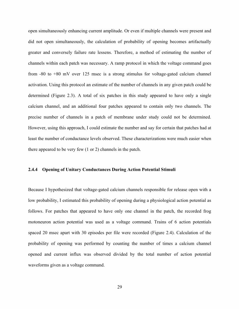
Initially, I performed single channel recordings at the release face of the synaptic boutons
in Xenopus nerve-muscle co-cultures. These experiments were accomplished by physically
removing the postsynaptic muscle such that a patch electrode could be introduced to the
presynaptic membrane (Figure 2.1 A). I hypothesized that action potential stimuli would not be
very effective at activating N-type calcium channels in the active zone. I collected single channel
data using this approach (Figure 2.1 B & C), but obstacles stood in the way of continuing to use
this approach to test my hypothesis. Primarily, this technique was only successful a small
percentage of the time, and when patches were obtained, they did not last long due to the fragile
nature of synaptic varicosities. Therefore, I turned to using a somal model to test my hypothesis.
27

2.4.2 Calcium Channel Conductance in Chick Ciliary Ganglion
Chick ciliary ganglion neurons were used for the remainder of work reported in this chapter. To
characterize the conductance of N-type channels in these neurons, voltage steps of 100 msec in
duration were applied from a resting potential of -80 mV to a voltage of -30, -20, -10, 0, +10,
+20, or +30 mV. Pooled data from multiple patches reveals an average conductance of 1.38 ±
0.07 pA for steps to -30 pA when driving force for calcium is very high. A step to +30 revealed
an average current amplitude of 0.49 ± 0.03 pA due to a low driving force for calcium. By
Ohm’s Law, resistance is defined as voltage divided by current (V = IR or R = V/I).
Conductance (G) is the inverse of resistance (G = 1/R) and can be determined as the slope of the
I-V curve (G = I/V). By plotting current amplitude at different potentials for many patches
(n=13) and fitting a line to this plot, a conductance was calculated from the slope of the line.
Figure 2.2 shows representative currents evoked by step depolarizations that were leak
subtracted. In the same figure, conductance plots resulting from the pooling of all data at each
potential (Figure 2.2B), and the individual data points from each patch (Figure 2C) are shown.
Using this approach, the conductance of the channel under investigation was determined to be
13.05 pS. This is consistent with what has been reported as the conductance (13-25 pS) for N-
type channels (Fox et al. 1987; Elmslie 1997).
2.4.3 Determination of the Number of Channels within a Patch
For some aspects of the analysis, it was critical to determine the number of channels within a
patch of membrane. The goal of the study was to determine the probability of opening of a given
calcium channel upon physiological depolarization. If multiple channels were present, they could
28
open simultaneously enhancing current amplitude. Or even if multiple channels were present and
did not open simultaneously, the calculation of probability of opening becomes artifactually
greater and conversely failure rate lessens. Therefore, a method of estimating the number of
channels within each patch was necessary. A ramp protocol in which the voltage command goes
from -80 to +80 mV over 125 msec is a strong stimulus for voltage-gated calcium channel
activation. Using this protocol an estimate of the number of channels in any given patch could be
determined (Figure 2.3). A total of six patches in this study appeared to have only a single
calcium channel, and an additional four patches appeared to contain only two channels. The
precise number of channels in a patch of membrane under study could not be determined.
However, using this approach, I could estimate the number and say for certain that patches had at
least the number of conductance levels observed. These characterizations were much easier when
there appeared to be very few (1 or 2) channels in the patch.
2.4.4 Opening of Unitary Conductances During Action Potential Stimuli
Because I hypothesized that voltage-gated calcium channels responsible for release open with a
low probability, I estimated this probability of opening during a physiological action potential as
follows. For patches that appeared to have only one channel in the patch, the recorded frog
motoneuron action potential was used as a voltage command. Trains of 6 action potentials
spaced 20 msec apart with 30 episodes per file were recorded (Figure 2.4). Calculation of the
probability of opening was performed by counting the number of times a calcium channel
opened and current influx was observed divided by the total number of action potential
waveforms given as a voltage command.
29

The number of channels estimated to be in a patch certainly had an effect on the
probability of opening. If a patch of membrane contained multiple channels, it follows that the
likelihood of observing a calcium channel opening would increase. Recordings with a single
calcium channel present were the most useful in determining the probability of opening.
However, a plot of probability of opening versus estimated number of channel was performed to
demonstrate this relationship. For the six patches with only one calcium channel present the
probabilities of opening were 2.33, 2.59, 3.15, 4.44, 4.88, and 4.91%. These data result in a mean
probability of opening of 3.59 ± 0.45% (n = 6). As the number of channels within a given patch
rise, as estimated by the ramp protocol, the probability of opening also rises.
Another important variable in calcium triggered secretion is the flux of calcium through
an open channel. To determine the amount of calcium entering the cell through a single channel
upon action potential stimulation, a histogram of current integral was generated. Histograms
including only events from the patches with a single channel present, and patches with 1 or 2
channels present were made (Figure 2.5 A and B, respectively). The area under the current trace
for each event was determined as described in the methods.
These cell attached recordings revealed many characteristics of the N-type current in
chick ciliary ganglia. Recordings in which there was a single channel in the patch demonstrated a
very low probability of opening. These data suggest that a particular voltage-gated calcium
channel is very unlikely to open with a physiological stimulus. In those cases when only a single
channel opening is triggered by an action potential stimulus, it is important to determine the
variability in the total calcium flux as this probably is critical in the determination of whether this
unitary flux triggers local vesicle fusion.
30
Figure 2.1 Presynaptic Xenopus varicosity cell-attached recordings. A) Picture demonstrating the technique of physically removing the postsynaptic muscle fiber such that the release face of the presynaptic varicosity can be exposed for patch-clamp experiments. B) Ramp protocol of single channel recordings from a frog varicosity. C)Representative single channel currents evoked by step depolarizations.
31

Figure 2.2 Representative single channel openings and conductance plots. A) Sample single channel openings through calcium channels in chick ciliary ganglia in response to a square step depolarization. B&C) Amplitude of current from unitary N-type calcium channel openings is plotted versus membrane voltage. Data were obtained by stepping chick ciliary ganglion neurons from -80 mV to voltages plotted on the x-axis. B) Data from multiple patches, but specific voltage steps are pooled together (conductance = 13.05 pS). C) Data from individual patches are displayed separately (conductance = 12.86 pS).
32

Figure 2.3 Representative ramp data The chick ciliary ganglion cell is ramped from -80 to +80 mV over 125 milliseconds. Voltage-dependent calcium channels open in response to this strong stimulus. This method is used to estimate the number of channels within the patch of membrane under investigation. A)Example data from a patch that appeared to have only a single channel. B) Example data from a patch that appeared to have multiple channels.
33

Figure 2.4 Representative calcium channel openings triggered by an action potential train. Top trace: Action potential voltage command used to open channels in the patch. Bottom trace: N-type calcium channel currents recorded in response to the action potential train. The inset shows an expanded time-base for one representative single channel opening. Interstimulus interval for action potential train is 20 msec.
34

Figure 2.5 Characteristics of unitary calcium channel openings triggered by action potential stimuli. A) Distribution of single channel integrals when only one channel is estimated to be in the patch (n=6). B) Distribution of single channel integrals when one or two channels are thought to be in the patch. Since Po is low integrals in this plot are also predicted to still be from single channel openings.
35

Figure 2.6 MCell prediction of single channel current variability through one opening. Using a realistic 3-Dimensional computer model of a cell with only a single calcium channel present, action potential waveforms were added to the model to determine the variability in calcium flux through stochastically gated single channel openings at different phases of the action potential waveform.
36
2.5 DISCUSSION
N-type calcium channels expressed in chick ciliary ganglia cultured motoneurons have been used
as a model system to study N-type channel activation at the frog neuromuscular junction. Direct
patch-clamp recording from the frog motor nerve terminal although not impossible, had many
technical challenges. The frog motoneuron forms a very small varicose structure on the
postsynaptic muscle. This varicose structure is small in size making cell-attached recording
difficult. Often times, the varicosity was similar in size as compared to the recording pipette.
Furthermore, these embryonic presynaptic structures were fragile and had little cytoskeletal
rigidity. Therefore, the presynaptic varicosity could be accidentally aspirated into the patch
electrode. In addition, there is the issue of where on the membrane the calcium channels
involved in the release of transmitter are located. If voltage-gated calcium channels are
colocalized with presynaptic release machinery, then it follows that the calcium channels would
be on the presynaptic membrane directly opposite the postsynaptic muscle fiber. Therefore, cell-
attached patch clamp needs to be performed on the membrane that is directly opposite the muscle
cell which is inaccessible. Attempts to record from this “release face” of the presynaptic terminal
were completed, but proved to be quite difficult. Both the small size of the terminal and the fact
that often times the synaptic terminal floated freely in the culture dish after being physically
removed from the muscle cell made electrophysiological studies difficult. For these reasons, I
turned to the chick ciliary neurons as models for study. They contain a large proportion of N-
type calcium current (~75%) that can be pharmacologically isolated. These cells are round and
easy to patch clamp. Also, chick ciliary ganglion neurons are structurally robust lending
themselves quite well to the patch clamp technique.
37

The measured frequency of opening of N-type calcium channels reported in this chapter
lead me to hypothesize that very few of the calcium channels in an active zone will open during a
physiological action potential stimulus. These conclusions are in contrast with some previous
reports, and these data contribute to the debate regarding the manner in which calcium channel
openings trigger vesicle fusion. Does a large proportion of the calcium channels in each active
zone open with each action potential and create a cloud of calcium that sums to trigger vesicle
fusion (Dunlap et al. 1995; Borst and Sakmann 1996; 1998)? Alternatively, do a few calcium
channels open in each active zone and the flux of calcium through a single channel triggers
vesicle fusion (Stanley 1993; Bertram et al. 1996; Wachman et al. 2004)? The different
conclusions from these two sets of studies could be due to differences in the populations of
calcium channel types found in the model preparations studied. One can group the two different
kinds of results into those from preparations that express predominately P/Q-type channels
versus those that express predominately N-type channels. P/Q-type channels have been shown to
represent 96% of the calcium channel population in the calyx of Held (Iwasaki et al. 2000), and
P/Q-type blockers in isolation block the majority of transmitter release in hippocampal
commisural synapses (Wheeler et al. 1994). On the other hand, ciliary ganglion neurons and the
frog neuromuscular junction contain predominately N-type channels (Yoshikami et al. 1989;
Yawo and Momiyama 1993; Dryer 1994; White et al. 1997; Yazejian et al. 1997). If N- versus
P/Q-type channels show differences in their activation kinetics, this may explain some of the
observed differences among preparations. Consistent with this possibility, previous work
comparing P/Q-type channels with N- and L-type channels provides data to suggest that P/Q-
type channels may activate with faster kinetics than N-type channels (Mintz et al. 1992; Sather et
al. 1993). However, recent work from more mature synapses suggests that P/Q-type channels at
38
the mature Calyx of Held can exhibit a low probability of opening and that few channels lead to
a fusion event (Fedchyshyn and Wang 2005).
The close spatial association between voltage-gated calcium channels and the calcium
sensor that triggers transmitter release has fueled speculation about the stoichiometry of calcium
influx sites and the calcium sensor. Competing hypotheses suggest that (1) the calcium signal
that triggers transmitter release is produced by influx through a single calcium channel or (2)
that the activity of multiple calcium channels expose the calcium sensor to overlapping calcium
domains and that this overlap is required to trigger release under normal conditions. Theoretical
and experimental results predict a local “nanodomain” of high calcium concentration around the
mouth of an open calcium channel (Chad and Eckert 1984; Fogelson and Zucker 1985; Simon
and Llinas 1985). During an action potential in a presynaptic terminal, these transient domains
of calcium are predicted to reach values of ~100 μM within 20 nm of an open channel (Neher
1998). The location of the calcium sensor for release relative to these domains of high calcium
has important implications for the calcium-dependence of transmitter release.
My data demonstrate that a minority of available N-type calcium channels open with
each action potential at room temperature at the frog neuromuscular junction (physiological
conditions for frogs). These data are most consistent with a single channel opening providing the
trigger for each release event. In synapses that express only N-type channels in the active zone,
each vesicle fusion event is hypothesized to be associated with a single local calcium channel
opening (Yoshikami et al. 1989; Bertram et al. 1996). For synapses than contain both N- and
P/Q-type channels, there may be a more complex relationship between channel gating and
vesicle fusion. If P/Q channels open with higher probability in a given active zone, an action
potential would produce a cloud of calcium (nanodomain) that could trigger the release of
39

several neighboring vesicles. Within these synapses, N-type channels may only add to that large
cloud, but depending on the stoichiometry of the different channel types associated with each
release site, there may be different relationships between specific calcium channels and the
triggering of vesicle fusion.
There are also implications for G-protein modulation of presynaptic calcium channels. At
the single channel level, G-protein modulation causes calcium channels to open with a delayed
latency, and with a lower probability of opening in response to a prolonged depolarizing stimulus
(Carabelli et al. 1996; Patil et al. 1996). In terms of physiological importance during action
potential stimulation, however, it appears that modulated calcium channels open so slowly that
they do not contribute significantly to action potential-evoked calcium current (Artim and
Meriney 2000). In this sense, inhibitory modulation essentially eliminates calcium channels
from opening during action potential stimulation. In the context of my studies of N-type calcium
channel activation at the motor synapse, one would predict that G-protein-coupled receptor
inhibition of N-type calcium channels would decrease the number of available calcium channels.
If single channel openings are sufficient to trigger vesicle fusion, then G-protein-coupled
receptor modulation, rather than decreasing the probability of release evenly across all
modulated release sites, would decrease the number of available release sites.
The data collected in this chapter aid in our understanding of presynaptic influx of
calcium during an action potential and allow me to hypothesize how this flux leads to the release
of transmitter. It should be noted that are two caveats to the collected data. First there could be
inactivation occurring during the trains of action potential stimuli. Second, calcium channel
under observation may be shifting into different gating modes. Both of these variables could
reduce my observed probability of opening. The action potential are spaced 20 msec apart, but
40

the train is not a true 50 Hz stumulis. Each train consisting of 6 bursts has a very slight pause in
between due to software acquisition. Also, after each 30 trains of 6 action potentials (180 action
potentials total), there is an even longer pause to reload the acquisition file in the software.
Therefore, this 50 Hz trains is paused and the stimulation rate is slower. This could lower
activation of the voltage-gated calcium channels. The data suggest that during a physiological
stimulus, such as the frog motoneuron action potential, a given voltage-gated N-type calcium
channel has a very low probability of opening. And yet despite this very low probability of
opening, the neuromuscular synapse is highly effective at synaptic transmission. This strength
arises from the fact that although a given calcium channel has a low probability of opening, the
entire active zone has hundreds of release sites leading to an average consistent release of
transmitter that is necessary for reliable muscle contraction.
If single channel openings are providing the calcium that triggers each vesicle fusion
event, one question I addressed is how does calcium influx vary during each gating event? This
is important because we do not know how much calcium influx is necessary for the release of a
synaptic vesicle. To compare with my data, and provide a higher resolution evaluation of this
issue, the calcium current influx through a single calcium channel has been modeled by Markus
Dittrich and Joel Stiles in collaboration with the Meriney lab (Figure 2.6). In this model, a
realistic channel gating scheme, the frog nerve terminal action potential waveform, a modeled
cell volume, and the known flux rates of calcium ions through stochastically generated single
channel openings were combined to reproduce the expected variability in flux through single
channel openings. Therefore, this modeling work takes into account changes in driving force
during an action potential waveform and variability in the mean open time of actual channels.
The kinetics of gating were modeled using a scheme with three closed states and one open state,
41

and kinetic parameters were scaled to provide a probability of opening of about 0.2 to match
experimental data. Interestingly, the distribution of these modeling data have a similar pattern as
my data of recorded variability in single channel integrals (Figure 2.6). I hypothesize that not all
of these fluxes would provide enough calcium to trigger fusion at synapses where each fusion
event is governed by a single channel opening. Further work would be required to determine a
threshold calcium entry through a single channel that could trigger vesicle fusion. Calcium
channel openings that result in flux below that threshold would further decrease the probability
of release.
42
3.0 PROPORTION OF N-TYPE CALCIUM CURRENT ACTIVATED BY ACTION
POTENTIAL STIMULI
3.1 SUMMARY
N-type calcium currents are important in many neuronal functions, including cellular signaling,
regulation of gene expression, and triggering of neurotransmitter release. Often the control of
these diverse cellular functions is governed by the spatial and temporal patterns of calcium entry
in subcellular compartments. Underlying this issue is the effectiveness of action potentials at
triggering calcium channel opening. Chick ciliary ganglion neurons were used as model cells to
study the activation of N-type calcium current during action potential depolarization. Several
different action potential shapes were recorded, used as voltage command templates, and altered
such that control action potential–evoked currents could be compared with those elicited by
broadened action potential commands. Depending on the action potential shape used to activate
calcium currents in chick ciliary ganglion neurons, and the temperature at which recordings were
performed, varying proportions (I/Imax) of N-type calcium current could be activated. The largest
proportion measured occurred using a broad ciliary ganglion cell soma action potential to activate
calcium current at 37°C (100%). The smallest proportion measured occurred using a fast, high-
temperature–adjusted frog motoneuron nerve terminal action potential to activate calcium current
43

at room temperature (10%). These data are discussed with respect to the impact on cellular
signaling and the regulation of transmitter release.
3.2 INTRODUCTION
Different types of voltage-gated calcium channels have been identified with varying properties
that are well suited for specific roles in different compartments of the cell (De Waard et al. 1996;
Tully and Treistman 2004). In particular, N-type calcium channels have diverse functions that
include cellular signaling (Saffell et al. 1992; Usachev and Thayer 1997; Sandler and Barbara
1999; Shah and Haylett 2000; Balkowiec and Katz 2002), regulation of gene expression
(Brosenitsch and Katz 2001), and the calcium-triggered vesicle fusion that underlies
neurotransmitter release (Stanley 1997). The specific role that N-type calcium channels play in a
neuron depends on the subcellular distribution of these ion channels, the macromolecular
complex of other proteins with which they are tightly associated, and the patterns of calcium
entry through these channels. The specific spatial and temporal patterns of calcium entry through
N-type channels are governed by neuronal activity patterns and the response characteristics of N-
type calcium channels. Some cellular events may be triggered by the combined flux through
many local open channels, whereas others may be triggered by the flux through a single open
channel.
One critical characteristic that contributes to the pattern of calcium entry is the probability
that a channel will open in response to a single action potential stimulus. This characteristic is
controlled by the activation kinetics of the channel, and whether the channel is in an inactivated
state or under modulatory control by G-proteins or some other cellular modulator. Different
44

calcium channel types have characteristic kinetics of activation that depend on specific channel
type, splice variant, and auxiliary subunits (Jones 1998; Hering et al. 2000; Magistretti et al.
2003; Lin et al. 2004). G-protein modulation often makes calcium channels reluctant to open,
slowing activation kinetics of whole cell current (Jones 1998), and essentially eliminating
modulated channels from contributing significantly to the current activated by a single action
potential stimulus (Artim and Meriney 2000).
I have studied the activation of N-type calcium channels by various action potential
shapes (at room temperature and at 37°C) using parasympathetic chick ciliary ganglion neurons
as model cells. Acute cultures of chick ciliary ganglion neurons provide a homogeneous
population of neurons that, when grown on the appropriate substrate, do not elaborate neurites.
As such, they provide the opportunity to study calcium current under excellent voltage control.
At embryonic stage 40, chick ciliary ganglion neurons express about 74% N-type, about 24% L-
type, with only about 2% resistant calcium current (White et al. 1997). These cells have little or
no tonic G-protein modulation (DE Artim and SD Meriney, unpublished observations) and allow
me to evaluate the proportion of current that can be activated by action potentials. Therefore I
used this model system to study N-type calcium current activation during action potential stimuli
of various shapes. I find that, depending on the action potential shape used and the temperature,
action potentials can activate varying proportions (I/Imax) of N-type calcium current (ranging
from 10 to 100%). Implications of these findings for various neuronal functions are considered.
45

3.3 METHODS
3.3.1 Cell culture
Ciliary ganglia were dissected from stage 40 White Leghorn chick embryos after rapid
decapitation, in accordance with the University of Pittsburgh’s Institutional Animal Care and
Use Committee, in Tyrode solution containing (in mM): 134 NaCl, 3 KCl, 3 CaCl2, 1 MgCl2, 12
glucose, and 20 NaHCO3; pH 7.3; Hamburger and Hamilton 1951). Dissected ganglia were
incubated for 20 min in 0.08% trypsin in Ca2+-and Mg2+-free Tyrode solution at 37°C. Removal
and inhibition of the trypsin was accomplished by washing three times in minimal essential
medium (MEM) plus 10% heat-inactivated horse serum. Neurons were mechanically dissociated
from ganglia by trituration through a polished Pasteur pipette. The suspension of cells was
centrifuged at 100 x g for 6–8 min and resuspended in MEM plus 10% heat-inactivated horse
serum. The cells were plated onto poly-D-lysine coated 35 mm plastic dishes, incubated at 37°C
in 5% CO2, and used for experimentation after 1–4 hours of incubation.
3.3.2 Electrophysiological recording techniques
Recordings of calcium current from chick ciliary ganglion neurons were performed using the
whole cell patch-clamp technique (Hamill et al. 1981). External bath solution was as follows (in
mM): 100 NaCl, 50 TEA-Cl, 10 HEPES, 5 glucose, 5 KCl, 5 CaCl2, 2 MgCl2, and 1 µM
tetrodotoxin (pH 7.3). The use of 5 mM external calcium (to increase current amplitude), instead
of the more physiologic 2 mM calcium, results in a slight shift in calcium current activation
(attributed to increased surface charge) to more positive voltages (predicted to be <5 mV) (Smith
46
et al. 1993). Pharmacological agents ω-conotoxin (ω-CgTx) from the venom of the cone shell
Conus geographus (GVIA, 500 nM) and nitrendipine (1 µM in 0.01% DMSO) were added to
isolate L- or N-type calcium currents, respectively. A combination of ω-CgTx GVIA and
nitrendipine blocks about 98% of calcium current in these neurons (White et al. 1997).
Borosilicate glass pipettes were pulled on a Flaming/Brown micropipette puller (Sutter
Instruments, Model P-97), coated with Sylgard (Dow Corning) and fire polished to a diameter
with a measured electrical resistance of 0.5–2 MΩ. Internal solution used in the patch pipettes
included (in mM): 120 CsCl, 10 HEPES, 11 EGTA, 5 TEA-Cl, 1 CaCl2, and 4 MgCl2. To retard
calcium current rundown, the following were added to the internal solution fresh daily (in mM):
4 Mg-ATP, 0.3 Na-GTP, and 0.1 leupeptin. Correction was made for a –6 mV liquid junction
potential before all recordings. Series resistance averaged 6.0 ± 0.4 MΩ (means ± SE; n = 26)
and was compensated by 85%. Cell capacitance averaged 12.0 ± 0.9 pF. Data were leak
subtracted using a P/4 protocol and collected through the use of the Axopatch 200A amplifier
(Axon Instruments) and the pClamp 6.0 software package (Axon Instruments) running on a
Pentium processor–based computer.
3.3.3 Calcium current activation and analysis
Calcium current was activated by square voltage steps and action potential waveforms.
Representative action potential waveforms, to be used as voltage commands, were recorded
using the fast current-clamp mode of an Axopatch 200B amplifier from two model preparations:
cultured parasympathetic chick ciliary ganglion neuron somata and cultured Xenopus frog
motoneuron nerve terminal varicosity (Pattillo et al. 1999). These action potentials were digitized
and the resulting waveforms were used as voltage commands. At room temperature (20–22°C),
47

the ciliary ganglion action potential had a resting potential of –60 mV, a peak amplitude of +25
mV, and a duration at half-amplitude of 2.1 msec. At 37°C, duration at half-amplitude shortened
to 0.65 msec. The motor nerve terminal action potential waveform had a resting potential of –60
mV, a peak voltage of +30 mV, and a duration at half-amplitude of 0.85 msec. At 37°C, duration
at half-amplitude was shortened to 0.24 msec. To assess the proportion of calcium current
activated, the action potential waveforms were altered such that the duration of the peak (most
depolarized) voltage was 0.02, 0.06, 0.26, 0.46, 0.66, 0.86, 1.06, 1.26, 1.46, 2.46, 3.46, or 4.46
msec. In the control nerve action potentials, the duration at the peak was 0.06 msec.
For analysis, each current amplitude was normalized to the peak tail current amplitude
recorded with the 3.46-ms peak duration action potential. Rundown was adjusted for by using a
double-pulse protocol (64-ms interpulse interval), which allowed comparison of the calcium
current elicited by the test action potentials of varying duration (first pulse) with the 3.46-ms
peak-broadened action potential (second pulse). By comparing the calcium current evoked by
these test action potentials to the calcium current that could be evoked by a standard broad-
duration action potential, I estimated the proportion of current (I/Imax) that could be evoked by an
action potential waveform. Data were plotted relative to the duration of the action potential at
half-amplitude. Even with prolonged strong-voltage depolarizations not all available calcium
channels will be open at any one point in time. I broadened action potential shapes until I/Imax
approached a maximum at or near 1.0 (defined as 100%). For these studies, this is defined as
maximal current activation. Maximal current activation does not represent a condition in which
all available calcium channels are open.
N-type calcium channels have been shown to have multiple gating modes that have a
variable probability of opening (Delcour et al. 1993), with a predicted mean probability of
48

opening around 0.5 estimated from prolonged exposure to voltages reached by action potentials
under physiological conditions (Lee and Elmslie 1999; Colecraft et al. 2001). Consequently the
I/Imax values cited in this report are an overestimate of the actual probability of channel opening
during an action potential. Estimating the actual probability of channel opening is difficult in
whole cell recording conditions, but is dealt with in Chapter 2. Within each condition studied
(action potential shape and temperature), there were no changes in the voltage at which peak
current was measured as action potentials were broadened. As such, within each experimental
condition, there were no changes in driving force for calcium entry.
To evaluate the kinetics of calcium current activation, currents were activated by 5-ms
steps from –80 to 0 mV. Kinetics of calcium current activation were measured by fitting a single
exponential to the current trace beginning at the time that current began to flow inward and
ending at the time of maximal current (Jones and Marks 1989).
3.4 RESULTS
3.4.1 Calcium current activation during action potential depolarization
Before isolating N-type calcium current, I evaluated the activation of all calcium currents
expressed in ciliary ganglion neurons elicited by a motor nerve terminal action potential
waveform. Figure 3.1A shows the proportion (I/Imax) of ciliary ganglion calcium current activated
by a nerve terminal action potential at room temperature (20–22°C). These nerve terminal action
potentials activated only 36.5 ± 3.7% (means ± SE) of maximal calcium current in ciliary
ganglion neurons (arrow in Figure 3.1A). This relatively small proportion of total calcium current
49

activation led me to investigate isolated calcium channel types. I evaluated the activation of L- or
N-type currents selectively by exposure to either 1 µM nitrendipine (to isolate N-type) or 500 nM
ω-CgTX GVIA (to isolate L-type). After isolation of either N-type or L-type current, I estimated
the proportion of current activated by the nerve terminal action potential. A significantly greater
proportion of L-type calcium current (73.2 ± 12.6%) than N-type (32.8 ± 6.3%) was evoked by
the nerve terminal action potential (Figure 3.1B; P < 0.001, two-tailed Student's t-test,
independent groups, n = 12 for L-type and n = 18 for N-type). Representative current examples
are shown in Figure 3.1C.
I also evaluated the activation of calcium currents expressed in ciliary ganglion neurons
during an action potential recorded from a ciliary ganglion neuronal soma. Figure 3.2A shows
the proportion of total current activated by the ciliary ganglion action potential at room
temperature. These action potentials are much broader than those recorded from the motoneuron
nerve terminals and, as a result, are more effective at activating calcium current, with 82.4 ±
2.3% activated using the native ciliary ganglion action potential waveform. In this case,
eliminating the L-type channel contribution (using 1 µM nitrendipine) and examining solely N-
type current had little effect on the proportion of current activated by a native ciliary ganglion
action potential (76.9 ± 3.3%; see Figure 3.2B). Figure 3.2C shows representative currents
activated by the ciliary ganglion action potential as it is broadened. Because these native ciliary
ganglion action potential waveforms are so effective at activating N-type current, and N-type
channels are the focus of my study, I did not examine the activation of isolated L-type channels
using this action potential waveform.
50

3.4.2 GGating charaacteristics oof N- versus L-type currrent
Next I fo
the obser
3.1B). R
for L-typ
calcium
currents
different
Figure 3
the kinet
activated
activation
with such
3.4.3 E
potentia
Although
calcium c
physiolog
and calci
of calciu
peak of
ocused on po
rved differe
Representativ
pe compared
current (fro
averaged 0.
(means ± SE
.3A), consis
tics of activ
d by a moton
n of the L-ty
h a brief acti
Effects of tem
l depolariza
h I found th
current at ro
gical temper
ium imaging
um influx du
the action p
otential diffe
nces in curr
ve traces (se
d with N-ty
om –80 to 0
.65 ± 0.07 a
E; P < 0.001
stent with pr
vation for N
neuron nerve
ype channel
ion potential
mperature o
ation
hat a nerve t
oom temperat
ratures of 36
g in the rat c
uring an acti
potential and
rences betw
rent activatio
e Figure 3.3
ype current d
0 mV). The
and 1.85 ±
1, one-way A
reviously rep
N- and L-typ
terminal act
s would pre
stimulus.
ween N- and L
on during a
3B) demonst
during a ste
e kinetics o
0.11 ms, re
ANOVA, n =
ported work
pe channels
tion potentia
dict a greate
L-type chann
a nerve term
trate the fast
p depolariza
f activation
spectively,
= 10 for L-ty
(Kasai and
may explai
al (Figure 3.
er activation
nel gating th
minal action
ter time cou
ation that ac
( ) for L-t
and these w
ype and n =
Neher 1992
in the propo
1B) because
of available
hat could und
potential (F
urse of activ
ctivates max
type and N
were signific
7 for N-type
2). Differenc
ortion of cu
e faster kinet
e calcium cu
derlie
Figure
vation
ximal
N-type
cantly
e; see
ces in
urrent
tics of
urrent
on activation of N-type calcium cuurrent durinng action
terminal act
ture, I quest
6–38°C. Sab
cerebellum to
ion potentia
d calcium in
tion potentia
ioned wheth
batini and Re
o document
al. At 37°C t
nflux was si
al is not ver
her the same
egehr (1996
the effects
they showed
ignificantly
ry effective
would be tr
) used whol
of temperatu
d that the de
reduced as
at gating N
rue at mamm
le cell record
ure on the ti
elay betwee
a result of
N-type
malian
dings
iming
n the
rapid
51
kinetics of channel gating. First, I measured the kinetics of activation of N-type channels at 37°C.
Consistent with previous observations (van Lunteren et al. 1993), I found that N-type channel
gating was significantly faster at 37°C such that current activated with a τ of 0.31 ± 0.03 ms
(means ± SE; P < 0.001, one-way ANOVA, n = 9; Figure 3.3). This faster activation at higher
temperature would be predicted to yield a greater proportion of current activation during an
action potential.
Because the Xenopus preparation does not tolerate patch-clamp recording at 37°C
(Meriney, unpublished observations), I could not record a native action potential from
motoneuron nerve terminals at this temperature. Instead, I altered the room-temperature–
recorded nerve terminal action potential shape as it would be predicted to be changed at this
higher temperature. I scaled this nerve terminal action potential with respect to the duration at the
base, duration at half-amplitude, rise time, and repolarization time in a manner that was
proportional to that shown by Sabatini and Regehr (1996). This scaled action potential shape is
consistent with the time course of action potentials recorded from other preparations at this
temperature (Sabatini and Regehr 1996; Borst and Sakmann 1998). This fast (rescaled) motor
nerve terminal action potential was then used to investigate calcium current activation at 37°C.
Figure 3.4A shows the rescaled action potential command at 37°C (solid line) compared with the
native nerve terminal action potential recorded at room temperature (dotted line). Using this
rescaled action potential, I found that 60.3 ± 5.7% of available N-type current was activated at
37°C (Figure 3.4B, filled circles). Representative currents are shown in Figure 3.4C. Therefore
unlike at room temperature, the nerve terminal action potential is reasonably effective at
activating N-type channels at 37°C (32.8 ± 6.3 vs. 60.3 ± 5.7%; P < 0.001, Student's t-test). In
52

this condition, it seems as if the faster activation kinetics for N-type calcium current (tact = 0.31
ms) is not completely offset by the faster action potential duration at half-amplitude.
Next I investigated the activation of N-type calcium current using the ciliary ganglion
action potential recorded at 37°C. Using this native action potential, N-type activation was not
significantly changed (76.9 ± 3.3% at room temperature vs. 71.8 ± 8.8% at 37°C; P = 0.55,
Student's t-test; see Figure 3.5A). Apparently, temperature-dependent changes in action potential
shape almost exactly offset those in calcium current kinetics for this relatively broad native
action potential waveform. Representative currents are shown in Figure 3.5B. The most obvious
difference in the currents evoked by the ciliary ganglion action potentials when compared at
room temperature and at 37°C was the steepness of the relationship between the proportion of
current activated and the broadening of the action potential. At 37°C maximal activation was
achieved with only slight broadening (filled circles in Figure 3.5A) compared with that at room
temperature (open circles in Figure 3.5A). This temperature-dependent increase in steepness of
the relationship was observed using both action potential waveforms (motor nerve terminal and
ciliary ganglion; compare Figure 3.4B and Figure 3.5A).
To more thoroughly investigate the effects of temperature and action potential waveform
shape, I mixed conditions to examine effects on N-type calcium current activation. Figure 3.6A
demonstrates that the native ciliary ganglion action potential shape recorded at 37°C (0.65-ms
duration at half-amplitude) activated about 25% (I/Imax = 0.25) of available N-type current
recorded at room temperature (where tact = 1.85 ms). However, when the ciliary ganglion action
potential recorded at room temperature was used at 37°C (Figure 3.6B), this broadest action
potential shape (2.1-ms duration at half-amplitude) activates maximal N-type calcium current
(100%; I/Imax = 1.0). At this temperature, N-type calcium current activates very quickly (tact =
53

0.31 ms). When the motor nerve terminal action potential is adjusted to reflect the fast kinetics
expected at high temperature (0.24-ms duration at half-amplitude), a very small proportion of N-
type current is recorded at room temperature (10%; see Figure 3.6C). As expected, using the
motor nerve terminal action potential recorded at room temperature (0.85-ms duration at half-
amplitude) to record calcium current at 37°C, this native action potential shape activated a very
large proportion (about 90–95%) of N-type calcium current (see Figure 3.6D).
54

Figure 3.1 Effects of action potential broadening on calcium current activation in chick ciliary ganglion neurons. A) plot of calcium current elicited by motor nerve terminal action potentials of varying duration at half-amplitude, normalized to the current elicited by the broadest action potential (I/Imax). Nerve terminal action potential (arrow) activated 36.5 ± 3.7% of maximal calcium current. B) plot of calcium current as described in A after isolation of L- (open circles) or N-type (filled circles) calcium currents through the use of ω-conotoxin (ω-CgTX) from the venom of the cone shell Conus geographus (GVIA) (500 nM) or nitrendipine (1 µM), respectively. Nerve terminal action potential activates 73.2 ± 12.6% of L-type current (arrow, open circle) but only 32.8 ± 6.3% of N-type current (arrow, filled circle). Inset: nerve terminal action potential voltage command and associated calcium current for both L-type (left) and N-type (right) calcium currents. C) representative current traces and voltage commands for both L- and N-type calcium currents. Starting with the nerve terminal action potential, every other trace has been omitted for clarity. In A and B, data are fit to a single exponential function.
55

Figure 3.2 Effects of action potential broadening on calcium current activation in chick ciliary ganglion neurons using a recorded ciliary ganglion neuron action potential. A) plot of calcium current elicited by action potentials of varying durations, normalized to the current elicited by the broadest action potential (I/Imax). Ciliary ganglion neuron action potential (arrow) activated 82.4 ± 2.3% of maximal calcium current. B) plot of calcium current as described in A after isolation of N-type calcium currents through the use of nitrendipine (1 µM). Ciliary ganglion neuron action potential activated 76.9 ± 3.3% of N-type current (arrow). Inset: ciliary ganglion neuron action potential voltage command and associated calcium current for N-type calcium currents. In A and B, data are fit to a single exponential function. C) representative current traces and voltage commands for N-type calcium currents. Starting with the ciliary ganglion neuron action potential, every other trace has been omitted for clarity.
56

Figure 3.3 Calcium current activation during step depolarizations. A) comparison of the kinetics of activation as measured using a single exponential fit of current traces for L- and N-type currents elicited by step depolarizations from –80 to 0 mV. B) representative calcium currents from each experimental condition.
57

Figure 3.4 Effects of motor nerve terminal action potential broadening at 37°C on calcium current activation in chick ciliary ganglion neurons. A) nerve terminal action potential waveforms used at room temperature (dotted line) and 37°C (solid line) are shown for comparison. B) plot of normalized N-type calcium current activation (as shown in Fig. 1) at 37°C (filled circles) in comparison with the room-temperature data from Fig. 1B (open circles). Nerve terminal action potentials (arrows) activated 60.3 ± 5.7% of maximal current at 37°C compared with 32.8 ± 6.3% at room temperature. Data are fit to a single exponential function. Inset: nerve terminal action potential voltage command and associated calcium current recorded at 37°C. C) representative current traces and voltage commands for the data collected at 37°C. Starting with the nerve terminal action potential, every other trace has been omitted for clarity.
58

Figure 3.5 Effects of action potential broadening on calcium current activation recorded at 37°C in chick ciliary ganglion neurons using a recorded ciliary ganglion neuron action potential as a voltage command. A) plot of normalized N-type calcium current activation recorded at 37°C (filled circles) in comparison with the room-temperature data from Fig. 2B (open circles). Ciliary ganglion neuron action potentials (arrows) activated 71.8 ± 8.8% of maximal current at 37°C compared with 76.9 ± 3.3% at room temperature. Data are fit to a single exponential function. Inset) ciliary ganglion neuron action potential voltage command and associated calcium current recorded at 37°C. B) Representative current traces and voltage commands for the data collected at 37°C. Starting with the ciliary ganglion neuron action potential, every other trace has been omitted for clarity.
59

Figure 3.6 Evaluation of calcium current activation using various combinations of action potential waveforms and recording conditions. A) plot of normalized N-type calcium current activation when the ciliary ganglion action potential that was recorded at 37°C (see Fig. 5) was used to activate calcium current recorded at room temperature. This control action potential activated 27.8 ± 4.1% of maximal calcium current. B) plot of normalized N-type calcium current using the ciliary ganglion action potential recorded at room temperature (see Fig. 2) to activate calcium currents at 37°C. This control action potential activated 107.4 ± 2.4% of maximal current (data are fit with a linear regression). C) plot of normalized N-type calcium current activation when the motor nerve terminal action potential that was scaled as it is predicted to change at 37°C (see Fig. 4) was used to activate calcium current recorded at room temperature. This control action potential shape activated 11.7 ± 2.0% of maximal current. D) plot of normalized N-type calcium current using the motor nerve terminal action potential recorded at room temperature (see Fig. 1) to activate calcium currents at 37°C. This control action potential shape activated 94.0 ± 1.9% of maximal current. For plots A, C, and D, data are fit to a single exponential function. In all plots the control action potential duration (arrow) is altered at the peak as described above.
60

Figure 3.7 Whole-cell recordings from frog varicosities A&B) Photo of Xenopus nerve-muscle cocultures before (A) and after introduction of the patch pipette to the varicosity (B). B) Plot of calcium current elicited by motor nerve terminal action potentials of varying duration at half-amplitude, normalized to the current elicited by the broadest action potential (I/Imax). Nerve terminal action potential (arrow) activated ~25% of maximal calcium current.
61

3.5 DISCUSSION
I have shown that action potentials are not always effective at activating calcium current
compared with maximal activation evoked by a broadened waveform. As would be expected,
broad action potentials are more effective at activating calcium current than brief action
potentials. Furthermore, because calcium current activation kinetics can vary, this aspect of
channel gating is also critical in the determination of the proportion of current that will be
activated by an action potential. The proportion of calcium current activated by an action
potential can be important in several respects.
3.5.1 Use of chick ciliary ganglion as a model system
The chick ciliary ganglion was used as a model system to obtain quality whole-cell recordings of
voltage-gated N-type calcium channels. The issue arises of how useful is the chick preparation
for the understanding of the frog neuromuscular junction? Despite sequence homology between
the two species, there are splice variants. CaV2.2 (N-type) exists in both a short and long splice
variant (Lu and Dunlap 1999). The long variant has 211 amino acids found to be involved with
targeting the long variant to the presynaptic terminal (Khanna et al. 2006). Preliminary work for
this study was performed in the Xenopus nerve muscle coculture system by Robert Poage (see
Figure 3.7). This early work demonstrated that the proportion of available calcium current
elicited by a natural action potential is very similar to that found when the same action potential
is used as a command voltage in chick ciliary ganglion neurons. Therefore, for the characteristics
of calcium current activation examined in this thesis, the chick ciliary ganglion appears to be a
good model for calcium current activation at frog neuromuscular synapses.
62

3.5.2 Cellular signaling
Calcium is the intracellular signal for many cellular events. As these events have become more
fully understood, it has become clear that the detailed characteristics of intracellular calcium
elevations are very important. The specific transmembrane source and subcellular localization, as
well as the pattern, time course, and magnitude of calcium entry, can be critical in determining
the cellular events that are triggered (Fields et al. 1997; Balkowiec and Katz 2000; 2002).
Furthermore, at subcellular sites of calcium entry there may be mechanisms to amplify the
calcium signal through calcium-induced calcium release from intracellular stores (Tully and
Treistman 2004). These issues have often been studied in neurons with respect to the regulation
of gene expression and the plasticity of synaptic transmission. In terms of calcium entry through
voltage-gated calcium channels, different calcium channel types appear to be coupled to the
regulation of different genes (Finkbeiner and Greenberg 1998; Brosenitsch and Katz 2001).
Previous work using a high potassium stimulation paradigm may have favored study of the role
of L-type, and overlooked the contribution of N-type calcium channels, because the N-type may
inactivate during prolonged high potassium stimulation (Nowycky et al. 1985). Using
physiological patterns of stimulation in primary sensory neurons, Brosenitsch and Katz (2001)
have shown that the calcium flux through N-type channels appears to trigger the expression of
several immediate early genes and tyrosine hydroxylase. This is likely attributable to a
subcellular co-localization of particular channel types with specific signaling cascades. The
reliability of this local signaling will depend on the probability that channels open during action
potential stimuli.
In terms of synaptic plasticity, long-term changes in synaptic strength are known to be
triggered by calcium entry (Miller and Kennedy 1986; Lisman and Goldring 1988; Lisman 1989;
63

Artola and Singer 1993). Recently, it has been shown that the specific temporal pattern of
calcium entry can determine the type of synaptic plasticity that results (Yang et al. 1999; Zucker
1999; Ismailov et al. 2004). The patterns of calcium entry during activity depend critically on the
probability that voltage-gated calcium channels open during these physiological stimuli. With a
low probability of specific channels opening during an action potential, calcium entry is
predicted to be sporadic at particular entry sites, be restricted to small subcellular compartments,
and generally not accumulate during low-frequency action potential activity or during very short
bursts of higher-frequency activity. On the other hand, if channels have a high probability of
opening during action potential activity, the local "flood" of calcium could create a larger
intracellular cloud that temporarily overcomes local buffering, and this signal would be predicted
to be consistent after each action potential stimulus. In this scenario, calcium signals would be
more reliably generated to accurately report action potential activity in the cell.
Voltage-gated calcium channels are common targets for G-protein modulation. The
proportion of calcium channels that are normally activated by an action potential can have
important implications related to the potential for neuromodulation. In cases where there is a very
high probability for calcium channel opening during an action potential, there is not much room
for modulation that would increase channel open probability, but a large dynamic range over
which inhibitory modulation could act. If calcium channels have a very low probability of
responding during action potential stimuli, the opposite would be true and there would be much
more room for modulation that would serve to increase calcium entry. Furthermore,
neuromodulation often targets potassium channels that indirectly alter calcium entry by changing
action potential shape. Even subtle changes in action potential shape can have significant effects
on calcium entry (Pattillo et al. 1999). Furthermore, based on the steepness of the relationship
64

between action potential duration and I/Imax observed at 37°C (filled symbols of Figure 3.4B and
Figure 3.5A), I predict that slight differences in action potential shape will have more dramatic
effects on action potential–evoked activation of N-type calcium current at 37°C than that at room
temperature.
3.5.3 Regulation of transmitter release
I have shown that a nerve terminal action potential recorded from a cultured frog motor synapse
was not very effective at activating N-type calcium current at room temperature. These data lead
us to hypothesize that very few of the N-type calcium channels at a frog motor nerve terminal
will open with each action potential invasion. Recently, Wachman et al. (2004) used a fast
calcium imaging approach at the adult frog neuromuscular junction to provide evidence that very
few of the calcium channels at this synapse open during an action potential. Furthermore, they
demonstrated that the spatial distribution of calcium entry at release sites was altered by calcium
channel blockers in a manner consistent with very few—perhaps only one—calcium channel
opening underlying each action potential–evoked calcium entry site. Interestingly, when a
binomial analysis was applied to imaged calcium entry domains in this neuromuscular
preparation, the predicted mean probability of calcium channel opening during a single action
potential was about 0.12 (Luo et al. 2005). This is remarkably consistent with the proportion of
N-type calcium current (I/Imax ~0.25) recorded from Xenopus presynaptic varicosities in vitro
when activated by the motor nerve terminal action potential (Poage and Meriney 2002) after
multiplication by the predicted mean opening probability of N-type calcium channels previously
reported in the literature during long, strong depolarizations (Lee and Elmslie 1999; Colecraft et
65

al. 2000). Furthermore, my data from ciliary ganglion cell soma reported here are also consistent
with these observations.
This low open probability further suggests that when vesicles are triggered to fuse by the
calcium flux through N-type calcium channels, each vesicle fusion event might be triggered by
the opening of a single N-type calcium channel. This hypothesis was first proposed by
Yoshikami et al. (1989), who measured dose–response relationships between various calcium
channel blockers and transmitter release at the adult frog neuromuscular junction, and performed
computational analyses that led them to hypothesize that the activity of a single N-type calcium
channel mediates vesicle fusion at an individual transmitter release site. Evidence in favor of this
idea has previously been reported at other synapses controlled by calcium influx through N-type
channels. Stanley (1993) used a combination of patch-clamp recordings of single N-type calcium
channels and a chemiluminescent method to detect transmitter release in the calyciform
presynaptic terminal of the chick ciliary ganglion to show that single N-type calcium channel
openings can trigger vesicle fusion. Further support for this idea was provided in a computational
study (Bertram et al. 1996). These studies suggest that, in these preparations at room
temperature, the flux of calcium through a single N-type calcium channel opening normally
triggers each transmitter release event.
The results at the frog neuromuscular junction and chick ciliary ganglion are in contrast
with what has been reported at the rat calyx of Held and hippocampal mossy fiber synapses
where action potentials appear to be very effective (70–85%) at activating the predominately
P/Q-type channels at these CNS synapses at room temperature (Borst and Sakmann 1998;
Bischofberger et al. 2002). These data have led to the conclusion that a majority of presynaptic
calcium channels open with each action potential, and the flux through many open channels sums
66

to create the calcium trigger for vesicle fusion (Borst and Sakmann 1998; Bischofberger et al.
2002). At the rat calyx of Held, direct patch-clamp studies led to the conclusion that more than
60 calcium channel openings provide the calcium flux to trigger each vesicle fusion event (Borst
and Sakmann 1996).
If different channel types show differences in their activation kinetics, this may explain
some of the observed differences among preparations. Consistent with this possibility, previous
work comparing P/Q-type channels with N- and L-type channels provides data to suggest that
P/Q-type channels may activate with faster kinetics than N-type channels (Mintz et al. 1992;
Sather et al. 1993; Bischofberger et al. 2002). However, measured differences between channel
types in particular preparations may not be universally applicable because activation kinetics may
vary significantly between splice variants of the same channel type and depend on specific
interactions with auxiliary subunits (Lin et al. 1997; Lin et al. 1999; Lin et al. 2004).
67

4.0 MODULATION OF NEUROMUSCULAR TRANSMITTER RELEASE BY ATP
4.1 SUMMARY
The nerve-muscle synapse is one of the most reliable connections in the nervous system. At most
neuromuscular junctions, each action potential in the nerve terminal leads to sufficient
depolarization in the postsynaptic muscle cell to bring that cell to threshold and cause muscle
contraction. To accomplish this, the neuromuscular junction is constructed of hundreds of
individual release sites (active zones) each with tens of synaptic vesicles in a “ready to release”
position. This low probability for vesicle release from each release site reduces the likelihood of
fatigue with repeated use. In addition to intrinsic properties of the calcium-triggered release
process that contributes to this low probability for release, I hypothesize that these individual
release sites are under a tonic inhibition that serves to further reduce the probability for
transmitter release. Transmitter release reliability at the frog neuromuscular junction is vital to
animal locomotion as muscles need to be used repeatedly without fatigue. Despite large numbers
of calcium channels associated with active zone synaptic vesicles at the frog neuromuscular
junction (NMJ), the probability of release of a single vesicle is very low. The following section
aims to study a possible mechanism for the tonic inhibition of the NMJ. Adenosine triphosphate
(ATP) is known to be coreleased along with the neurotransmitter acetylcholine (ACh). This ATP
can act on local ATP receptors, and be degraded in the synaptic cleft into adenosine which can
68

also act on adenosine receptors on the presynaptic membrane. The activation of both types of
purinergic receptors (ATP and adenosine) may lead to a decrease in transmitter release. The
experiments described in this chapter utilize the cutaneous pectoris nerve-muscle preparation
from the Rana pipiens frog as a model to investigate ATP-mediated modulation of the
neuromuscular junction.
4.2 INTRODUCTION
ATP is most commonly known as an intracellular energy source. Acceptance of its role in
signaling between cells has taken considerable time. The first study to describe the effects of
ATP were performed on cardiovascular tissue (Drury and Szent-Gyorgyi 1929). This early study
injected ATP and adenosine, taken from extracts of the heart muscle, were injected intravenously
into the guinea pig. Effects included slowing the heart beat, impaired conduction from the auricle
to the ventricle, and lowering arterial pressure (due to heartbeat slowing and arterial dilation).
Work in the cat demonstrated that intravenous injection of ATP could alter peripheral and central
nervous system transmission (Emmelin and Feldberg 1948). The role of ATP modulation at the
denervated frog neuromuscular junction showed that ACh induced contraction of the skeletal
muscle could be potentiated by the application of ATP (Buchthal and Folkow 1948). And ATP
injection into the cat lateral ventricle caused muscle weakness, ataxia, and lethargy (Feldberg
and Sherwood 1954). The release of ATP in the peripheral nervous system was first
demonstrated in the sensory nerves that innervate the rabbit ear artery leading to vasodilation
(Holton 1959) demonstrating that ATP in addition to being a ubiquitous molecule in the body
could also serve as a messenger released by the nervous system. Purinergic presynaptic
69
modulation was also discovered at the rat neuromuscular junction (Ginsborg and Hirst 1972). In
this study, adenosine was applied to the rat phrenic nerve-diaphragm preparation and reversibly
reduced the quantum content of end-plate potentials and the frequency of miniature end-plate
potentials to about half the control.
Nonadrenergic noncholinergic (NANC) inhibitory neurotransmission was discovered in
the gastrointestinal tract (Abrahamsson 1986). Originally nitric oxide was thought to be the only
transmitter involved in NANC transmission in the gut (Rand 1992; Bauer 1993) and in the feline
airway (Takahashi et al. 1995; Tanaka et al. 1996). However, further studies in rat pyloric
sphincter revealed that both nitric oxide and ATP mediated muscle inhibition when reactive blue
2 (P2Y receptor antagonist) was applied (Soediono and Burnstock 1994). Now, purinergic
signaling is well known to contribute to NANC transmission. The discovery of this type of
signaling by Geoffrey Burnstock, Graeme Campbell, and Max Bennett occurred in guinea pig. In
1962, they were observing mechanical and electrical activity of smooth muscle (taenia-coli) after
blocking responses of two neurotransmitters (acetylcholine and noradrenaline). The researchers
expected muscle contraction and depolarization with stimulation of the smooth muscle.
However, single electrical stimuli caused muscle relaxation and hyperpolarization. The
hyperpolarization was prevented with the use of tetrodotoxin which blocked nerve conduction,
but not muscle response. Thus, a NANC form of transmission was discovered (Burnstock et al.
1963; 1964). In addition to the guinea pig taenia-coli, the guinea pig stomach, rabbit ileum, frog
stomach, and turkey gizzard were found to use this type of NANC transmission as well
(Burnstock et al. 1970).
With respect to ATP and adenosine effects on cells, there are many types and subtypes of
purinergic receptors. The P1 receptors are activated by adenosine and the P2 receptors are
70

activated by ATP. The P2 family is further divided into P2X (ligand-gated ion channels) and
P2Y (G-protein coupled receptors). There are currently seven P2X subtypes and eight P2Y
subtypes known. The P1 (adenosine) receptors have been cloned and include four subtypes; A1,
A2a, A2B, and A3 (Daly 1985; Ralevic and Burnstock 1998; Fredholm et al. 2001; Cobb et al.
2003). All P1 receptors couple to G-proteins, and both agonists and antagonists have been found
for the P1 subtypes (Jacobson and Gao 2006). P2X receptors have seven subtypes (P2X1-7) and
are ligand gated nonselective cation channels. The seven subtypes share a 30-50% sequence
identity. P2X receptors contain three subunits to form a trimer (Nicke et al. 1998; Khakh et al.
2001; Barrera et al. 2005; Mio et al. 2005). Differential expression of subunits leads to variable
receptor properties such as kinetics of activation, inactivation, deactivation, and permeability to
calcium and chloride (Salter and Hicks 1995; Khakh et al. 2001; North 2002; Stojilkovic et al.
2005; Egan et al. 2006). In this manner, diversity of receptors can be achieved.
The metabotropic P2Y receptors are of particular importance to this study. There are
eight subtypes: P2Y1, P2Y2, P2Y4, P2Y6, P2Y11, P2Y12, P2Y13, and P2Y14. It should be noted
that P2Y5,9,10 are currently not thought to be nucleotide receptors and are not involved in second
messenger cascades. The intracellular loops and –COOH termini of the P2Y receptors are
diverse among the variants and dictate which type of G-proteins are bound. P2Y1,2,4,6 bind to
Gq/11. P2Y11 binds to both Gq/11 and Gs. P2Y12,13 bind Gi. P2Y14 binds to Gi/o (Abbracchio et al.
2006). A given cell may express many types of P2Y receptors. Activation of these P2Y receptors
can occur through the binding of different ligands; nucleoside diphosphates (P2Y2,4,6), nucleoside
triphosphates (P2Y2,4), or purine nucleotides (P2Y1,11,12). The transduction mechanism for the
P2Y receptors are varied and act either through cAMP, phospholipase C, or arachidonic acid
pathways (Grishin et al. 2005; Abbracchio et al. 2006). The arachidonic acid pathway is of
71

particular interest to this study since it has been reported to decrease calcium influx through
voltage-gated calcium channels (Todd and Robitaille 2006). P2Y receptor mediated reduction in
N-type calcium current has been observed in chromaffin cells (Currie and Fox 1996),
sympathetic neurons (Filippov et al. 1998; Liu and Rittenhouse 2003), and sensory neurons
(Gerevich et al. 2004). Arachidonate mimicked the depressant action of ATP both on calcium
current and end-plate current in frog sartorius muscle (Grishin et al. 2005) and the action of ATP
was shown to be PLA2 dependent (Sokolova et al. 2003). Therefore, the action of ATP on the
inhibition of calcium current at the neuromuscular junction is likely to be mediated via the
arachidonic acid pathway.
ATP can function as a neurotransmitter because it is concentrated in cholinergic synaptic
vesicles (up to 100 mM) as compared to the cytoplasm (2-5 mM). Furthermore, synaptic vesicles
contain many nucleotides such as AMP, ADP, and GTP, although in lower concentrations as
compared to ATP. The transport of ATP in the vesicle is unique in that it does not use a
nucleotide or proton exchange. There is possibly a “channel-like activity” or a diffusion
facilitator that might move ATP into vesicles (Zalk and Shoshan-Barmatz 2006). Once ATP is
released there are extracellular mechanisms to terminate its actions. ATP can either be broken
down by nonspecific enzymes or ectonucleotidases which can be divided into two families. E-
NTPDases hydrolyze tri- and diphosphates. Ectoenzymes serve to regulate synaptic activity by
controlling ATP and adenosine concentrations in the synaptic cleft (Zimmermann 2006). All of
these hydrolysis pathways are thought to be relatively slow as compared to acetylcholine
hydrolysis by acetylcholinesterase. As such, ATP may linger in the synaptic cleft for a
significant amount of time after vesicle fusion.
72

4.3 METHODS
4.3.1 Tissue preparation
Adult frogs (Rana pipiens) of 3-4 cm in length from Connecticut Valley Biological
(Southampton, MA) were anesthetized in 0.1% tricaine methane sulfonate, decapitated, and
pithed in accordance with the University of Pittsburgh’s Institutional Animal Care and Use
Committee. The cutaneous pectoris muscle was dissected bilaterally and bathed in normal frog
ringer (NFR; containing in mM 116 NaCl, 2 KCl, 1.8 CaCl2, 1 MgCl2, and 5 HEPES; pH 7.4).
This preparation was maintained in NFR at 2-8˚C until recording. To prepare for intracellular
recordings, the muscle was stretched to ~100% of in vivo length and pinned down in a Sylgard
(Dow Corning) coated dish.
4.3.2 Intracellular recordings
Intracellular recordings from the frog neuromuscular junction are performed in the
following manner. The nerve of the cutaneous pectoris muscle was drawn into a suction
electrode and stimulated with a current 5X the threshold to evoke a muscle twitch. Intracellular
recordings are performed with glass pipettes (Warner Instruments, filament glass catalog #64-
0787 O.D. 1mm I.D. 0.058) pulled to a tip with a resistance of ~25MΩ, and filled with 3M
potassium acetate. Muscle fiber penetrations were performed under visual control with a long
working distance water-immersion objective (40x, 3mm working distance). Muscles were
required to have a resting potential of -70mV or more hyperpolarized to be included for
recording and analysis. Muscle penetrations are made near the site of visible presynaptic nerve
73

terminals (within 50 µM). Recordings were made with a Dagan IX2-700 amplifier in NFR with
curare added to prevent muscle contraction. Data was acquired with Clampex 9 software (Axon
Instruments) running on a Pentium based computer. Data analysis was performed with Clampfit
9.2 software.
4.4 RESULTS
To test the hypothesis that ATP might serve as a tonic modulator of transmitter release from the
frog neuromuscular junction, I tested the effects of several P2Y receptor agonists and
antagonists.
4.4.1 Suramin
Suramin is a non-selective P2X and P2Y antagonist. Since suramin is a P2 antagonist for both
the P2X (ionotropic) and P2Y (metabotropic) purinoceptors, this drug serves as a crude tool to
evaluate the potential contribution of ATP signaling at the frog neuromuscular junction. If the
release of ATP leads to a tonic inhibition of the release of neurotransmitter, then suramin
application should block this ATP action. For these recordings, the muscle was bathed in NFR
with 4-7 µM curare. Each unique muscle fiber served as its own control and was recorded from
in both control and suramin treated conditions. Stimulus-evoked EPP amplitude was recorded in
both control conditions and after a 30 minute exposure to 100 µM suramin.
Suramin is hypothesized to block the tonic inhibitory effects of the ATP receptor
activation leading to an increase in transmitter release and EPP amplitude. Along these lines,
74

after suramin exposure, I recorded a massive increase in EPP amplitude under these conditions
(Figure 4.1A and 4.7; 313.2 ± 42.8%). This large increase in release caused the muscle to twitch
even in the presence of relatively high (7 µM) curare. As such, it was difficult to perform these
recordings, and more selective P2 receptor blockers were investigated. In addition, a paired-pulse
protocol was used to determine if suramin’s effect was presynaptic. The rationale for this was
that if suramin blocks the action of the presynaptic receptors that normally cause tonic inhibition
by inhibiting calcium channels, then increased release would be expected to be accompanied by
decreased paired-pulse facilitation. A plot of paired-pulse ratio at 6 intervals for control (closed
circles, Figure 4.1B) and 100 µM suramin (open circles) revealed no significant effect of
suramin on paired-pulse ratio, and thus it appears unlikely that the suramin effect is mediated by
alteration in presynaptic calcium entry. The suramin effects here may be due to its reported
inhibition of tyrosine phosphotases, phospholipase D, or some of its other non-specific effects.
4.4.2 Reactive Blue 2
Reactive blue 2 (RB2) is more selective than suramin. RB2 is a selective P2Y
purinoceptor antagonist. These experiments were performed in a manner similar to those
described above for suramin treatment. Stimulus evoked EPP amplitude was recorded in the
same muscle fibers before (control) and after exposure to 30 µM RB2 (30 min). Each unique
muscle fiber serves as its own control. As I expect ATP receptor activation through P2Y
receptors would lead to a tonic inhibition of transmitter release, I hypothesized that RB2 would
increase EPP amplitude. As predicted, RB2 increased EPP amplitude by 580.1% (Figure 4.2 &
4.7). To confirm that this RB2 treatment completely blocked ATP signaling, a non-hydrolyzable
form of ATP (AMP-PNP) was added to the nerve-muscle preparation in the presence of RB2.
75

The application of AMP-PNP in the presence of RB2 had no significant effect on EPP amplitude
(11 ± 19% increase; p=0.5).
4.4.3 MRS2179
The effects of RB2 lead to the hypothesis that there is tonic P2Y receptor activation at the
frog neuromuscular junction. There are many P2Y receptor subtypes, but only a few P2Y
subtype selective antagonists are available. In an attempt to evaluate one potential subtype of
P2Y receptors further, a subtype specific antagonist was used. MRS2179 is a selective,
competitive P2Y1 purinoceptor antagonist. This drug was used to see if P2Y1 purinoceptors
modulate the effect of ATP at the frog neuromuscular junction. Intracellular recordings of EPP
amplitude were performed as described above and EPP amplitude was compared in both control
and 30 µM MRS2179 treated conditions (30 min). As above, each unique muscle fiber served as
its own control. Since the MRS2179 drug is a selective P2Y1 purinoceptor antagonist, if this
subtype of P2Y receptor was mediating tonic ATP effects, then the EPP amplitude was expected
to be increased by exposure to MRS2179. However, in these studies MRS2179 did not
significantly change EPP amplitude (Figure 4.3 & 4.7; 13.70 ± 4.1% decrease; p = 0.77). The
possibility exists that this subset of purinergic receptors may not be present at the Rana pipiens
neuromuscular junction. Thus, a tonic inhibition of ATP on neurotransmission is likely mediated
by a different subtype of P2 receptors. Alternatively, frog P2Y1 receptors may not be sensitive to
this compound developed for use in mammals.
76
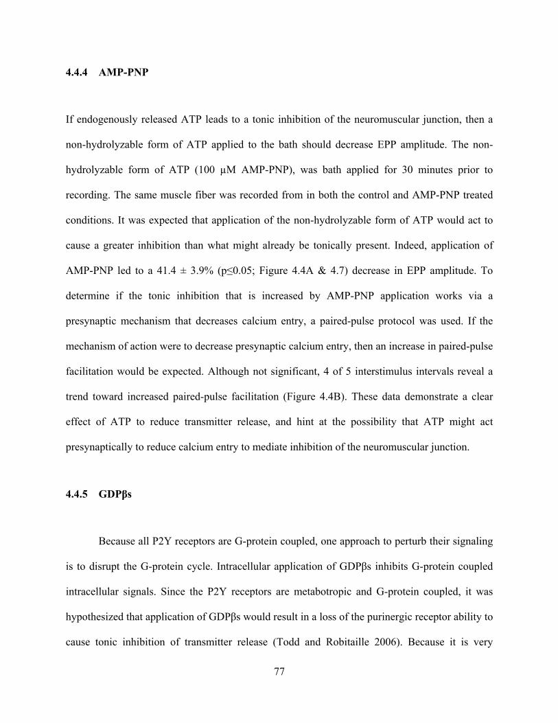
4.4.4 AMP-PNP
If endogenously released ATP leads to a tonic inhibition of the neuromuscular junction, then a
non-hydrolyzable form of ATP applied to the bath should decrease EPP amplitude. The non-
hydrolyzable form of ATP (100 µM AMP-PNP), was bath applied for 30 minutes prior to
recording. The same muscle fiber was recorded from in both the control and AMP-PNP treated
conditions. It was expected that application of the non-hydrolyzable form of ATP would act to
cause a greater inhibition than what might already be tonically present. Indeed, application of
AMP-PNP led to a 41.4 ± 3.9% (p≤0.05; Figure 4.4A & 4.7) decrease in EPP amplitude. To
determine if the tonic inhibition that is increased by AMP-PNP application works via a
presynaptic mechanism that decreases calcium entry, a paired-pulse protocol was used. If the
mechanism of action were to decrease presynaptic calcium entry, then an increase in paired-pulse
facilitation would be expected. Although not significant, 4 of 5 interstimulus intervals reveal a
trend toward increased paired-pulse facilitation (Figure 4.4B). These data demonstrate a clear
effect of ATP to reduce transmitter release, and hint at the possibility that ATP might act
presynaptically to reduce calcium entry to mediate inhibition of the neuromuscular junction.
4.4.5 GDPβs
Because all P2Y receptors are G-protein coupled, one approach to perturb their signaling
is to disrupt the G-protein cycle. Intracellular application of GDPβs inhibits G-protein coupled
intracellular signals. Since the P2Y receptors are metabotropic and G-protein coupled, it was
hypothesized that application of GDPβs would result in a loss of the purinergic receptor ability to
cause tonic inhibition of transmitter release (Todd and Robitaille 2006). Because it is very
77

difficult to inject this compound into the frog motor nerve terminal, the loading of GDPβs
through the cut end of the nerve was attempted. This approach had the further potential
advantage of dissociating the role of ATP on the presynaptic nerve terminal versus surrounding
glia. Because the Meriney lab had previously used this approach to load calcium sensitive dye of
similar molecular weight, the GDPβs was loaded through the cut end of the nerve of the Rana
pipiens cutaneous pectoris muscle for a period of 4 hours. A test loading of a visible dye of a
similar molecular weight (Alexa Fluor 488, molecular weight 570) was used to determine the
distance the GDPβs (30 mM) diffuses down the nerve. Intracellular recordings were made from
the proximal portions of the muscle at positions consistent with the expected diffusion of GDPβs.
Since GDPβs prevents G-protein couple signaling, I expected to observe an increase in EPP
amplitude. However, there was no significant effect (Figure 4.5A & 4.7; 15.2 ± 7.6% decrease; p
= 0.3). Interestingly, there was quite a large variability in these data (range 50% decrease to 40%
increase), and when the effects of GDPβs were plotted against a measure of the strength of the
synapse, a significant correlation was found (Figure 4.5B; R=0.688). In this evaluation, weaker
synapses showed no effect, while strong synapses showed a GDPβs-mediated decrease in EPP
amplitude. This is the opposite of what would be predicted if GDPβs was mediating a block of
tonically active P2Y receptors. It is hard to put much confidence in this correlation as the number
of observations was small, but this could suggest that there is a net change in strong synapses
mediated by the combination of the many types of G-protein receptors that may be found at the
neuromuscular junction.
78
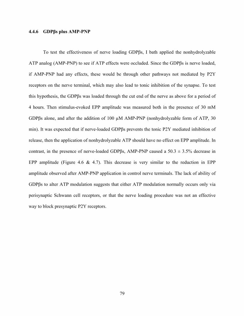
4.4.6 GDPβs plus AMP-PNP
To test the effectiveness of nerve loading GDPβs, I bath applied the nonhydrolyzable
ATP analog (AMP-PNP) to see if ATP effects were occluded. Since the GDPβs is nerve loaded,
if AMP-PNP had any effects, these would be through other pathways not mediated by P2Y
receptors on the nerve terminal, which may also lead to tonic inhibition of the synapse. To test
this hypothesis, the GDPβs was loaded through the cut end of the nerve as above for a period of
4 hours. Then stimulus-evoked EPP amplitude was measured both in the presence of 30 mM
GDPβs alone, and after the addition of 100 µM AMP-PNP (nonhydrolyzable form of ATP, 30
min). It was expected that if nerve-loaded GDPβs prevents the tonic P2Y mediated inhibition of
release, then the application of nonhydrolyzable ATP should have no effect on EPP amplitude. In
contrast, in the presence of nerve-loaded GDPβs, AMP-PNP caused a 50.3 ± 3.5% decrease in
EPP amplitude (Figure 4.6 & 4.7). This decrease is very similar to the reduction in EPP
amplitude observed after AMP-PNP application in control nerve terminals. The lack of ability of
GDPβs to alter ATP modulation suggests that either ATP modulation normally occurs only via
perisynaptic Schwann cell receptors, or that the nerve loading procedure was not an effective
way to block presynaptic P2Y receptors.
79

Figure 4.1 Intracellular recordings of EPPs before and after suramin treatment. A) Representative voltage traces in control (black) and suramin treated (red; 100 µM) conditions. Suramin treatment increases EPP amplitude by 313.2%. B) No significant difference in paired-pulse facilitation was observed following suramin treatment.
80

Figure 4.2 Representative EPP traces from intracellular recordings before and after RB2 treatment. Control (black) and RB2 treatment (red) which increased EPP amplitude by 580.1%
81

Figure 4.3 Effects of MRS2179 on EPP amplitude. MRS2179 (30 µM) had no significant effect on EPP amplitude (13.7 ± 4.1%; p = 0.77).
82

Figure 4.4 Effects of AMP-PNP on EPP amplitude and paired-pulse facilitation. A) Representative EPPs demonstrate a 41.4% decrease in amplitude. B) Although not significant, there was a trend for paired-pulse facilitation to increase following AMP-PNP treatment.
83
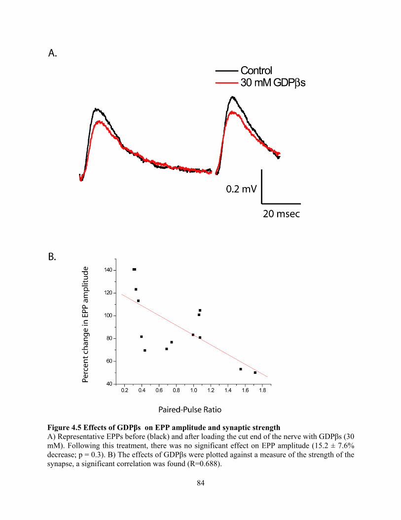
Figure 4.5 Effects of GDPβs on EPP amplitude and synaptic strength A) Representative EPPs before (black) and after loading the cut end of the nerve with GDPβs (30 mM). Following this treatment, there was no significant effect on EPP amplitude (15.2 ± 7.6% decrease; p = 0.3). B) The effects of GDPβs were plotted against a measure of the strength of the synapse, a significant correlation was found (R=0.688).
84

Figure 4.6 Representative EPPs showing the effects of AMP-PNP after pretreatment with GDPβs. Following the addition of AMP-PNP (100 µM) to a muscle preparation pretreated with GDPβs (30 mM), the EPP amplitude showed a 50.3 ± 3.5% decrease.
85

Figure 4.7 Summary figure of the effects of agonists and antagonists of the purinergic receptors on the amplitude of EPPs.
86

4.5 DISCUSSION
The above experiments attempt to explore one possible mechanism at the frog neuromuscular
junction that might contribute to the very low probability of release. ATP has been shown to be
coreleased with ACh at the neuromuscular junction, and ATP is known as a neurotransmitter that
can mediate inhibition through P2Y receptors. However, there is some debate as to which cells
release ATP and where ATP receptors exist. Some data suggests that bidirectional interactions
occur between neurons and perisynaptic Schwann cells (PSCs).
At the amphibian neuromuscular junction, it has been shown that intracellular calcium
rises in PSCs in response to transmitter release (Jahromi et al. 1992; Robitaille 1995). This
demonstrates the glial sensitivity to synaptic function contradicting the previous notion that glia
are simply passive and supportive cells in the nervous system. Thus the glia must possess
receptors that are sensitive to ligands released during synaptic transmission. PSCs have
neurokinin-1, muscarinic, and purinergic receptors (Robitaille 1995; Robitaille et al. 1997;
Bourque and Robitaille 1998). Using these, it has been shown that PSCs can regulate nerve
terminal transmitter release (Bourque and Robitaille 1998; Castonguay and Robitaille 2001).
Exogenously applied ATP has been shown to induce the release of calcium from internal
stores in PSCs at the Rana pipiens NMJ (Robitaille 1995). Using the membrane permeant
calcium indicator fluo-3 a 262% increase in ΔF/F in PSCs was observed following application of
20 µM ATP. Since ecto-ATPases are present in the synaptic cleft that can dephosphorylate ATP
to adenosine, the potential for adenosine effects were also examined (Lai and Wong 1991; Salter
87
et al. 1993). Local exogenous application of 10 µM adenosine also led to an intracellular calcium
increase of 229% (ΔF/F) in the PSCs. In these studies, the response evoked by adenosine
application was not significantly different from that evoked by ATP. Therefore, it is possible that
the intracellular calcium responses evoked by exogenous ATP application could be explained by
the activation of adenosine receptors. This hypothesis was supported by a lack of effect of ATP
after the application of the A1 receptor antagonist 2 µM DPCPX (ΔF/F = 4 ± 1%; Robitaille
1995). In this study, the authors were careful to eliminate the possibility of a nonspecific effect
of the DPCPX by the application of a muscarinic agonist (muscarine 1 µM) which still evoked
intracellular calcium responses at control levels. Furthermore a different A1 antagonist (CPT 10
µM) also reduced the ATP-mediated calcium response. Lastly, local adenosine application could
mediate a strong PSC intracellular calcium signal after the washout of antagonist. It should be
noted that A2 adenosine receptor antagonists (50µM CP-66,713 or 50µM CGS 15943) alone did
not have an effect on the PSC calcium levels.
Since Robitaille’s laboratory found the intracellular release of calcium within PSCs to be
mediated by A1 receptors in the frog neuromuscular junction, he wanted to discern which second
messenger cascade was involved. Pertussis toxin (PTX) catalyzes the ADP-ribosylation of the α
subunits of the heteromultimericic guanine nucleotide regulatory proteins Gi and Go. This
prevents the G-protein heteromultimerics from interacting with receptors which blocks their
coupling and activation. When the Robitaille group used this toxin for 12-14 hours (2 µg/ml
PTX), no adenosine-evoked intracellular calcium release was observed in PSC cells. Again the
application of muscarine could still lead to an intracellular calcium response after PTX
incubation. The presence of active PTX at the neuromuscular junction was confirmed by the fact
88
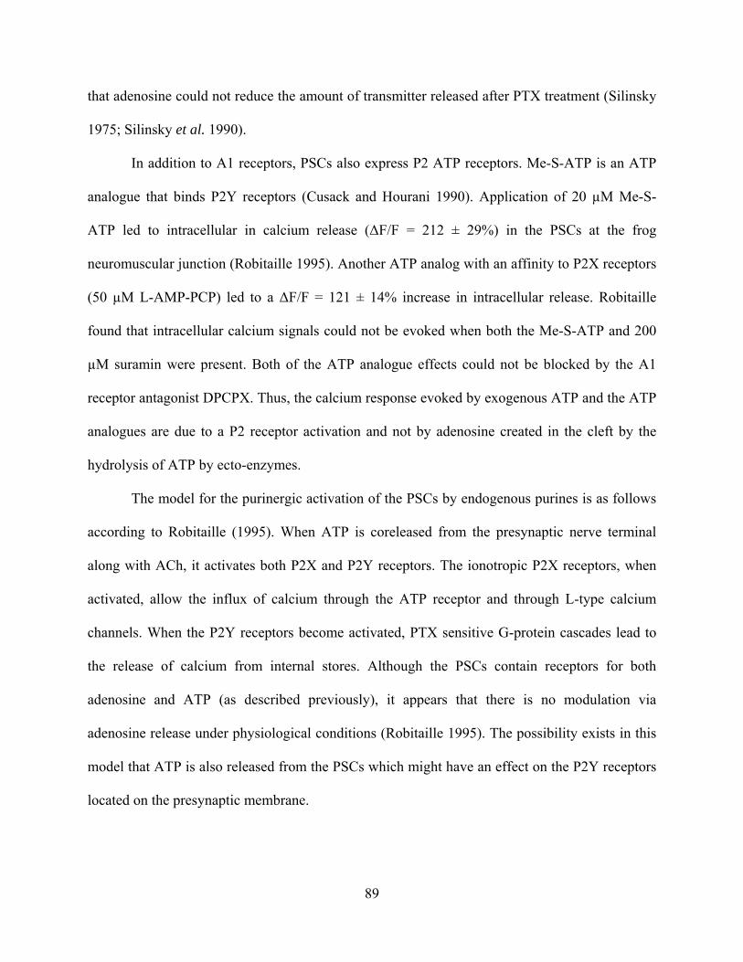
that adenosine could not reduce the amount of transmitter released after PTX treatment (Silinsky
1975; Silinsky et al. 1990).
In addition to A1 receptors, PSCs also express P2 ATP receptors. Me-S-ATP is an ATP
analogue that binds P2Y receptors (Cusack and Hourani 1990). Application of 20 µM Me-S-
ATP led to intracellular in calcium release (ΔF/F = 212 ± 29%) in the PSCs at the frog
neuromuscular junction (Robitaille 1995). Another ATP analog with an affinity to P2X receptors
(50 µM L-AMP-PCP) led to a ΔF/F = 121 ± 14% increase in intracellular release. Robitaille
found that intracellular calcium signals could not be evoked when both the Me-S-ATP and 200
µM suramin were present. Both of the ATP analogue effects could not be blocked by the A1
receptor antagonist DPCPX. Thus, the calcium response evoked by exogenous ATP and the ATP
analogues are due to a P2 receptor activation and not by adenosine created in the cleft by the
hydrolysis of ATP by ecto-enzymes.
The model for the purinergic activation of the PSCs by endogenous purines is as follows
according to Robitaille (1995). When ATP is coreleased from the presynaptic nerve terminal
along with ACh, it activates both P2X and P2Y receptors. The ionotropic P2X receptors, when
activated, allow the influx of calcium through the ATP receptor and through L-type calcium
channels. When the P2Y receptors become activated, PTX sensitive G-protein cascades lead to
the release of calcium from internal stores. Although the PSCs contain receptors for both
adenosine and ATP (as described previously), it appears that there is no modulation via
adenosine release under physiological conditions (Robitaille 1995). The possibility exists in this
model that ATP is also released from the PSCs which might have an effect on the P2Y receptors
located on the presynaptic membrane.
89

The possibility of glia being involved in the tonic inhibition of synaptic transmission was
difficult to address in this study. Experimentally, I added the non-displaceable GDPβs through
the cut end of the nerve. This potentially allows the treatment of only the presynaptic nerve
terminal with GDPβs and does not affect the PSCs. This treatment should block inhibition that is
mediated by G-protein pathways within the loaded nerve fibers. In my experiments, the
application of GDPβs alone had no significant effect on transmitter release, and subsequent
application of AMP-PNP resulted in an effect that was similar to the effects of AMP-PNP
applied to an untreated preparation. Therefore, these data may be difficult to interpret. It is
possible that GDPβs is so unstable that it does not have activity within the nerve terminal after 3-
5 hours (the time necessary for diffusion down the axon to the nerve terminal). Alternatively, it
may not have diffused into the distant release sites from the cut end of the nerve. Further work
will be required to evaluate these hypotheses.
The hypothesis that ATP can modulate neurotransmission is not new. Many studies have
focused on G-protein coupled P2Y ATP receptors as likely candidates for transmitter release
modulation (Burnstock and Kennedy 1985; Chen et al. 1995; Collo et al. 1996). ATP is known
to be coreleased with ACh at the frog neuromuscular junction (Silinsky and Redman 1996) and
has been proposed as a regulator of synaptic transmission (Ribeiro and Walker 1975). However,
early work focused on adenosine as the candidate which is a product of ATP’s degradation
(Ribeiro and Sebastiao 1987; Meriney and Grinnell 1991; Redman and Silinsky 1994). Work in
another frog muscle (sartorius) has revealed evidence for both adenosine and ATP receptors at
the frog neuromuscular junction that can modulate transmitter release (Giniatullin and Sokolova
1998). The sartorius data (Giniatullin and Sokolova 1998) provide evidence that both adenosine
and ATP reduce EPP amplitude to similar degrees at the frog neuromuscular junction. In these
90

studies, spontaneous events (miniature end plate potentials, mEPPs) were not altered in
amplitude, decay time constant, nor the voltage-dependence of decay. This suggests that both
ATP and adenosine modulation of synaptic transmission is presynaptic. In addition, Giniatullin
and Sokolova (1998) report increased paired-pulse facilitation. With reduced quantal release an
increase in paried-pulse facilitation is predicted if these purinergic effects are calcium-dependent
(Zucker 1989).
In further consideration of purinergic candidates for modulation, the degradation of ATP
to adenosine may be too slow to account for significant tonic inhibition of transmitter release. In
fact, Meriney and Grinnell (1991) report a small adenosine effect during repetitive stimuli. It has
been estimated that 50 µM ATP would be hydrolyzed in about 2 minutes (Smith 1991). By
contrast, ACh is broken down by about 600 sites per µm2 yielding a time of 9.5 s-1 (Anglister et
al. 1994). Therefore, the effect of ACh is terminated long before the production of adenosine
from the hydrolysis of ATP. These data would argue that ATP might be in a position to be the
most important modulator.
This work in the cutaneous pectoris muscle of the Rana pipiens adds new information to
the hypothesis that ATP is a neuromodulator at the neuromuscular junction. Although it has long
been known that ATP is coreleased and can indeed exert effects of both PSCs and presynaptic
terminals, there is still much to learn about this process. A non-hydrolyzable form of ATP
(AMP-PNP) can mimic the hypothesized inhibition of transmitter release. The purinergic
receptor antagonist RB2 can relieve the inhibition cause by endogenous P2Y receptor activation.
I propose that tonic ATP inhibition of the frog neuromuscular junction may modulate
presynaptic calcium channels and contribute to the very low probability of opening of voltage-
gated calcium channels involved during an action potential (see Chapter 3). Thus, I hypothesize
91

that the release of ATP at the frog neuromuscular junction serves to provide an endogenous
mechanism to further reduce the opening of voltage-gated calcium channels during an action
potential, and the subsequent release of transmitter.
92

5.0 GENERAL DISCUSSION
It has been known for some time that action potential invasion of the nerve terminal leads to the
opening of voltage-gated calcium channels which, in turn, causes exocytosis of transmitter into
the synaptic cleft. However, much about the role of the voltage-gated calcium channels at the
neuromuscular junction remains to be studied. This body of work focused on the role of voltage-
gated calcium channels in transmitter release at the neuromuscular junction, and how these
channels are gated by action potentials, and may be modulated endogenously. Single channel
recordings both at the frog neuromuscular junction and in chick ciliary ganglia revealed a very
low probability of opening during a physiological depolarization, a motoneuron action potential.
Cell-attached patch clamp recordings of a single N-type calcium channel revealed that channels
open ~5% of the time when a physiological action potential waveform is used as a command
voltage. Also, the macroscopic recording of whole-cell current demonstrates that only ~30% of
available calcium current is evoked using a physiological depolarization. Lastly, the co-release
of ATP with acetylcholine was also explored as a possible mechanism for the tonic inhibition of
voltage-gated calcium channels at the neuromuscular junction.
93

5.1 BUILDING A RELIABLE NEUROMUSCULAR JUNCTION FROM
UNRELIABLE ELEMENTS
The neuromuscular junction must be both a strong and reliable synapse for an animal to survive.
An action potential depolarization in a motoneuron always leads to muscle contraction. The
strength of the neuromuscular junction ironically arises from the fact that it is comprised of
hundreds of unreliable elements (active zones). The large overall structure of the neuromuscular
junction, with a multitude of release sites, leads me to hypothesize that the high reliability comes
from the repetitive placement of many transmitter release elements along the length of the nerve
terminal. The neuromuscular junction has row upon row of active zones, characterized by freeze
fracture as a linear array of hundreds of particles, some of which are likely calcium channels, and
some of which are likely calcium-activated potassium channels and perhaps integrin proteins.
Each active zone contains ten’s of vesicles in close proximity to the linear array of voltage-gated
calcium channels. Despite this large linear array of release machinery, a given action zone
releases a vesicle only every other time that the terminal is depolarized by a physiological action
potential. Thus despite the certainty of muscle contraction, it appears that probability of opening
of a given voltage-gated calcium channel involved in transmitter release is very low. This low
probability is evident in the single channel recordings in this study. Patches believed to contain a
single calcium channel opened on average 4.59% of the time an action potential stimulus was
introduced. To put this in perspective, imagine a nerve terminal with ~350 active zones, and each
active zone has ~30 vesicles of neurotransmitter docked and waiting for calcium entry. Action
potential depolarization would lead to the release of ~175 vesicles. Thus a given depolarization
only releases transmitter from half of the active zones, and thus each of the active zones has
multiple vesicles of transmitter available for future release. Given that muscle contraction results
94

from motoneuron activation in the range of 20-100 Hz in bursts of 3-40 action potentials,
(Hennig and Lomo 1985) motoneurons need to be capable of maintaining reliable release under
these conditions. Therefore, even with trains of action potentials necessary for muscle
contraction, depletion of vesicles is not very prominent (Zengel and Sosa 1994) at the
neuromuscular junction (Figure 5.1A). It appears as if the low probability of opening of the
voltage-gated channel serves as a mechanism to set a low probability of release from
neuromuscular junction active zones, and thus maintain a pool of ready vesicles near release
machinery that is almost never depleted with normal use.
It is interesting to compare how other strong synapses are thought to function in the
nervous system. The functional organization at the neuromuscular junction appears to be in
contrast with that of the calyx of Held (Satzler et al. 2002; Taschenberger et al. 2002). This
synapse is glutamatergic and needs to be strong and fast to allow the localization of sound using
transmission of information through the auditory brainstem. The calyx does not need to be as
reliable at the neuromuscular junction since once an auditory cue is located, the animal requires
no further information. Therefore, both synapses (the neuromuscular junction and the calyx of
Held) are specialized for strong transmission. The calyx of Held is a large structure (5-10 µM in
cat) with more active zones (~500) as compared to the frog nerve terminal (~350). Perhaps most
strikingly, the calyx has 10 times fewer vesicles (3-4) per active zone as compared to the number
of vesicles per active zone at the frog neuromuscular junction (~30). Upon depolarization, the
calyx releases a similar number of vesicles (~175) in total from the synapse. In the young calyx,
it appears that multiple calcium channel openings (possibly 60) contribute to the release of a
single vesicle (Borst and Sakmann 1996). However in the adult calyx the number of calcium
channels required for vesicles fusion reduces to 1-2 (Fedchyshyn and Wang 2005), and thus is
95

similar to the neuromuscular junction in this regard. The probability of release at a given active
zone at the calyx of Held is somewhat lower (0.35) than at the frog neuromuscular junction (0.5).
Another major difference between the two synapses arises when the probability of release of a
docked vesicle is compared. The probability of release of a docked vesicle in the neuromuscular
junction is 0.017, but the calyx it is estimated to be about 0.1. This difference arises because
there is such a difference in size between the two types of active zones. This six-fold increase in
release probability likely leads to a faster depletion of ready vesicles at the calyx of Held during
high frequency stimulation as each active zone does not have as many docked vesicles. While a
neuromuscular synapse can continue to fire at high frequency for a prolonged period without
depression, the calyx exhibits rapid depression (Figure 5.1B). These differences are likely to
reflect the different role that each synapse plays in the animal. To localize sound, the calyx of
Held synapse needs to be fast and strong, but may not need to be used repeatedly at high
frequency. In contrast, the neuromuscular junction must be a strong synapse even during periods
of high frequency activity that are required for a maintained muscle contraction.
96

Figure 5.1 Comparison of the rate of depression at the neuromuscular junction and at the Calyx of Held Note that the neuromuscular junction can sustain EPP amplitude over time while EPCs amplitude diminishes dramatically at the calyx.
97
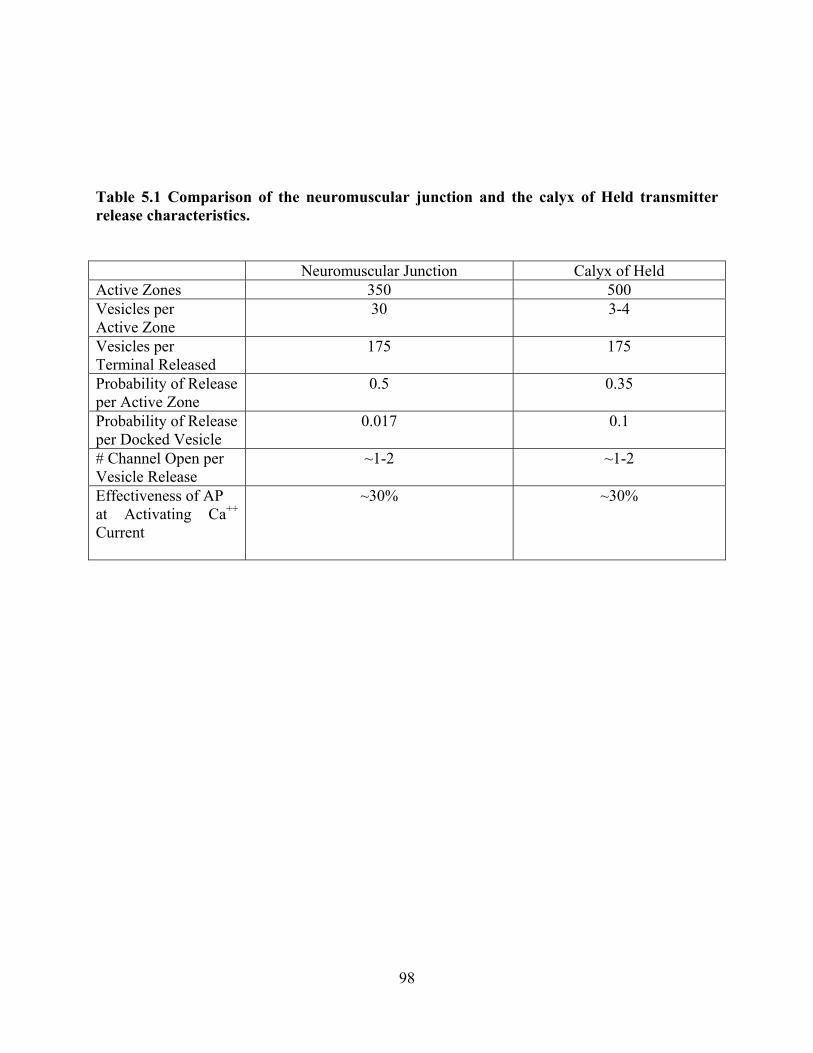
Table 5.1 Comparison of the neuromuscular junction and the calyx of Held transmitter release characteristics.
Neuromuscular Junction Calyx of Held Active Zones 350 500 Vesicles per Active Zone
30 3-4
Vesicles per Terminal Released
175 175
Probability of Release per Active Zone
0.5 0.35
Probability of Release per Docked Vesicle
0.017 0.1
# Channel Open per Vesicle Release
~1-2 ~1-2
Effectiveness of AP at Activating Ca++ Current
~30% ~30%
98

5.2 CALCIUM TRIGGERING OF TRANSMITTER SECRETION FROM THE
MOTOR NERVE TERMINAL
The work completed in this study, focused on the low probability of opening of voltage-gated
calcium channels, impacts upon a larger question: how many calcium channel openings normally
trigger the release of a single vesicle? To address this question I have collected some data in
collaboration with another graduate student in the Meriney lab. I have used intracellular
recordings of EPPs from muscle fibers (as described in Chapter 4) after gradual block of
presynaptic calcium channels at the frog neuromuscular junction (using ω-conotoxin GVIA), and
compared my data with intracellular calcium imaging data (performed by Fujun Luo) after the
same conotoxin treatment.
My recordings were carried out as described in chapter 4 in both control and after a 30
minute exposure to 25, 50, 75, 100 nM ω-conotoxin GVIA. Fujun loaded the cutaneous pectoris
muscle nerve of the Rana pipiens with the calcium sensitive intracellular dye Calcium Green-1
(30 mM) for 8 hours through the cut end of the nerve. He also used alexa 594-α-BTX to stain the
postsynaptic acetylcholine receptors (as an aid to select nerve terminals in the same focal plane).
The imagining technique was as described in Wachman et al. 2004. Using both experimental
approaches, as higher concentrations of ω-CgTX GVIA were used, the number of available
calcium channels in the nerve terminal active zone was reduced. We combined our data to
compare the fractional block of transmitter release (EPP amplitude in my experiments) after the
same series of ω-CgTX GVIA treatments. The hypothesis was that if a single calcium channel
99

Figure 5.2 Calcium release relationship when calcium entry sites are partially blocked using Conotoxin GVIA. Conotoxin GVIA applied at 25, 50, 75, and 100 nM concentrations. The best fit line with a slope of two indicates a single vesicle fusion event is mediated by two calcium channels.
100

leads to the fusion of each vesicle, a slope of one would be observed if fractional block of
transmitter release is plotted versus fractional block of calcium entry as calcium channels and
release sites would be removed together. The observations in figure 5.2 show that after expose to
low concentrations of ω-CgTX GVIA, the data mainly fell along the line with a slope of two,
suggesting that the opening of very few (perhaps between 1-3 calcium channels) is required for
the fusion of a single vesicle. After high concentrations of ω-CgTX GVIA treatment, calcium
entry sites are reduced to such a degree that, the relationship between block of transmitter release
and block of calcium entry trends toward a slope of one. Therefore, when the number of calcium
entry sites becomes very low, it appears likely that a single calcium channel opening is sufficient
to trigger vesicle fusion. These data suggest that between 1-3 calcium channel openings trigger
each vesicle fusion event under normal calcium conditions, but that a single channel opening can
trigger a single vesicle fusion event. These data are consistent with prior work from the Meriney
lab which indicate that discrete imaged calcium entry domains within an active zone are created
by the opening of single calcium channels. In these experiments, when calcium entry domains
are reduced by partial calcium channel blockade, the intensity of the domains that remain
unblocked are unchanged (Poage and Meriney 2002; Wachman et al. 2004). This finding is in
contrast to the known relationship of calcium influx to transmitter release which is known to be
fourth order (Dodge and Rahamimoff 1967). Such a fourth order relationship would be predicted
to be observed in our experiments if extracellular calcium was reduced.
101

5.3 ENDOGENOUS CALCIUM CHANNEL MODULATION ALTERS CALCIUM
ENTRY
This work hypothesized that there was a tonic inhibitory modulation mediated by co-released
ATP, that served as a possible mechanism for the tonic inhibition of calcium channels and
transmitter release at the neuromuscular junction. Decades ago it was discovered that ATP is
coreleased with acetylcholine at the neuromuscular junction (Silinsky 1975). This ATP-mediated
modulation of acetylcholine release was shown to be calcium dependent (Silinsky and Redman
1996). Since this discovery many studies have focused on the possible roles of ATP as a
transmitter. An ATP-mediated tonic inhibition of the frog neuromuscular junction fits very well
into the idea that the neuromuscular junction is a structure of many very low probability release
sites. Under this scenario, the release of ATP with each vesicle fusion event, under conditions in
which the ATP is not degraded quickly, could lead to an inhibitory tone over the presynaptic
calcium channels that would serve to ensure that each active zone functions at very low
probability. This would maintain a relatively large pool of “ready” vesicles and make it hard for
bursts of activity during muscle contraction to deplete transmitter.
The method by which ATP leads to a tonic inhibition is not completely understood. Since
ATP-mediated modulation of transmitter release was shown by some to be calcium dependent,
work has focused on mechanisms that control the levels of calcium in the presynaptic terminal.
When examining the history of literature on this topic, there is some disagreement. Some studies
have suggested that presynaptic calcium in frog is not altered by ATP modulation (Redman and
Silinsky 1994; Robitaille et al. 1999; Huang et al. 2002). Another study suggests that presynaptic
calcium levels are lowered with ATP modulation (Grishin et al. 2005). As part of my work, I
attempted to image ATP-mediated alterations in calcium within the presynaptic nerve terminal of
102

the Rana pipiens cutaneous pectoris muscle preparation. First, the P2Y antagonist RB2 could not
be used because it quenched the Alexa 594 α-bungarotoxin fluorescence necessary to visualize
and focus the nerve terminal prior to laser illumination of the intracellular calcium sensitive dye.
I also attempted to use the nonhydrolyzable ATP analog (AMP-PNP), but there were no
consistent effects observed, perhaps because one would not expect a very large effect on total
calcium entry.
Certainly, there are other possible mechanisms that could lead to tonic inhibition of the
neuromuscular junction when ATP is released. In addition, different species may use different
mechanisms. In mammals the released ATP is broken down to adenosine which acts on
adenosine receptors to cause a decrease in presynaptic calcium levels (Hamilton and Smith 1991;
Silinsky 2004). In amphibians, it is believed that ATP inhibits transmitter release without being
degraded to adenosine (Giniatullin and Sokolova 1998; Sokolova et al. 2003). In frog sartorius
muscle, arachidonic acid has been shown to mimic the effects of P2Y receptor activation
(Grishin et al. 2005). The application of a PLA2 inhibitor, PACOCF3, reduced the effect of ATP
on end plate currents by 84 ± 3%. In addition, the application of sodium arachidonate reduced
EPP to 81 ± 2% of control amplitude. Thus, a plausible intracellular cascade linking the
activation of P2Y receptors with a decrease in release at the frog neuromuscular junction has
been postulated. It is still possible that this arachidonic acid-mediated mechanism acts
downstream of calcium entry and possibly targets the release machinery.
It should be mentioned that in addition to the down-regulation of calcium channels there
are reported mechanisms by which voltage-gated calcium channel current can be up regulated as
well. One of these mechanisms involves the release of norepinephrine (NE) from sympathetic
axons that course through the muscle. NE is known to increase transmitter release from the frog
103

neuromuscular junction in a calcium-dependent manner (Jenkinson et al. 1968; Bergman et al.
1981). This modulator appears to work through a cAMP-dependent cascade to decrease the
release latency distribution at the neuromuscular junction (Bukcharaeva et al. 1999; Bukharaeva
et al. 2001). Recently Fujun Luo in the Meriney laboratory has demonstrated that there is a large
increase in presynaptic calcium entry in frog motor nerve terminals after NE exposure (Luo and
Meriney, unpublished observations). It is interesting to speculate that, during periods of
increased sympathetic activity (fight or flight reponse), NE could act to alter the function of the
active zone. This may occur by increasing the probability of presynaptic calcium channel
opening, the amount of transmitter released, and by shortening the release latency. This
shortened release latency would likely be mediated by the synchronizing action of NE on
transmitter release (Bukharaeva et al. 2001) and might be expected to be the most significant as
motor synapses are normally strong enough to bring muscle fibers to threshold even without NE.
In this manner, NE could increase the speed of motor nerve-evoked muscle contraction in the
fight or flight response by up-modulating the normally unreliable elements (active zones) that
comprise the adult frog neuromuscular junction. This hypothesis will require further
investigation.
104
BIBLIOGRAPHY
Abbracchio, M. P., G. Burnstock, et al. (2006). "International Union of Pharmacology LVIII: update on the P2Y G protein-coupled nucleotide receptors: from molecular mechanisms and pathophysiology to therapy." Pharmacol Rev 58(3): 281-341.
Abrahamsson, H. (1986). "Non-adrenergic non-cholinergic nervous control of gastrointestinal motility patterns." Arch Int Pharmacodyn Ther 280(2 Suppl): 50-61.
Anglister, L., J. R. Stiles, et al. (1994). "Acetylcholinesterase density and turnover number at frog neuromuscular junctions, with modeling of their role in synaptic function." Neuron 12(4): 783-94.
Artim, D. E. and S. D. Meriney (2000). "G-protein-modulated Ca(2+) current with slowed activation does not alter the kinetics of action potential-evoked Ca(2+) current." Journal of Neurophysiology 84(5): 2417-25.
Artola, A. and W. Singer (1993). "Long-term depression of excitatory synaptic transmission and its relationship to long-term potentiation." Trends in Neurosciences 16(11): 480-7.
Balkowiec, A. and D. M. Katz (2000). "Activity-dependent release of endogenous brain-derived neurotrophic factor from primary sensory neurons detected by ELISA in situ." Journal of Neuroscience 20(19): 7417-23.
Balkowiec, A. and D. M. Katz (2002). "Cellular mechanisms regulating activity-dependent release of native brain-derived neurotrophic factor from hippocampal neurons." Journal of Neuroscience 22(23): 10399-407.
Barrera, N. P., S. J. Ormond, et al. (2005). "Atomic force microscopy imaging demonstrates that P2X2 receptors are trimers but that P2X6 receptor subunits do not oligomerize." J Biol Chem 280(11): 10759-65.
Bauer, V. (1993). "NANC transmission in intestines and its pharmacological modulation." Acta Neurobiol Exp (Wars) 53(1): 65-77.
Bean, B. P. (1989). "Neurotransmitter inhibition of neuronal calcium currents by changes in channel voltage dependence." Nature 340(6229): 153-6.
Beaumont, V., M. B. Hepworth, et al. (1998). "Somatostatin receptor desensitization in NG108-15 cells. A consequence of receptor sequestration." Journal of Biological Chemistry 273(50): 33174-83.
Bennett, M. R., L. Farnell, et al. (2000). "The probability of quantal secretion near a single calcium channel of an active zone." Biophysical Journal 78(5): 2201-21.
Bergman, H., S. Glusman, et al. (1981). "Noradrenaline augments tetanic potentiation of transmitter release by a calcium dependent process." Brain Res 214(1): 200-4.
Bertram, R., A. Sherman, et al. (1996). "Single-domain/bound calcium hypothesis of transmitter release and facilitation." Journal of Neurophysiology 75(5): 1919-31.
105
Bischofberger, J., J. R. Geiger, et al. (2002). "Timing and efficacy of Ca2+ channel activation in hippocampal mossy fiber boutons." Journal of Neuroscience 22(24): 10593-602.
Borst, J. G. and B. Sakmann (1996). "Calcium influx and transmitter release in a fast CNS synapse.[see comment]." Nature 383(6599): 431-4.
Borst, J. G. and B. Sakmann (1998). "Calcium current during a single action potential in a large presynaptic terminal of the rat brainstem." Journal of Physiology 506(Pt 1): 143-57.
Bourque, M. J. and R. Robitaille (1998). "Endogenous peptidergic modulation of perisynaptic Schwann cells at the frog neuromuscular junction." J Physiol 512 ( Pt 1): 197-209.
Brody, D. L., P. G. Patil, et al. (1997). "Bursts of action potential waveforms relieve G-protein inhibition of recombinant P/Q-type Ca2+ channels in HEK 293 cells." J Physiol 499 ( Pt 3): 637-44.
Brosenitsch, T. A. and D. M. Katz (2001). "Physiological patterns of electrical stimulation can induce neuronal gene expression by activating N-type calcium channels." Journal of Neuroscience 21(8): 2571-9.
Bukcharaeva, E. A., K. C. Kim, et al. (1999). "Noradrenaline synchronizes evoked quantal release at frog neuromuscular junctions." J Physiol 517 ( Pt 3): 879-88.
Bukharaeva, E. A., D. V. Samigullin, et al. (2001). "The role of intracellular cAMP in mediating the synchronizing action of noradrenaline on the evoked release of quanta of mediator in the frog synapse." Neurosci Behav Physiol 31(5): 473-80.
Burnstock, G., G. Campbell, et al. (1963). "Inhibition of the Smooth Muscle on the Taenia Coli." Nature 200: 581-2.
Burnstock, G., G. Campbell, et al. (1964). "Innervation of the Guinea-Pig Taenia Coli: Are There Intrinsic Inhibitory Nerves Which Are Distinct from Sympathetic Nerves?" Int J Neuropharmacol 3: 163-6.
Burnstock, G., G. Campbell, et al. (1970). "Evidence that adenosine triphosphate or a related nucleotide is the transmitter substance released by non-adrenergic inhibitory nerves in the gut." Br J Pharmacol 40(4): 668-88.
Burnstock, G. and C. Kennedy (1985). "Is there a basis for distinguishing two types of P2-purinoceptor?" Gen Pharmacol 16(5): 433-40.
Carabelli, V., M. Lovallo, et al. (1996). "Voltage-dependent modulation of single N-Type Ca2+ channel kinetics by receptor agonists in IMR32 cells." Biophysical Journal 70(5): 2144-54.
Carbone, E. and H. D. Lux (1984). "A low voltage-activated, fully inactivating Ca channel in vertebrate sensory neurones." Nature 310(5977): 501-2.
Castonguay, A. and R. Robitaille (2001). "Differential regulation of transmitter release by presynaptic and glial Ca2+ internal stores at the neuromuscular synapse." J Neurosci 21(6): 1911-22.
Catterall, W. A. (2000). "Structure and regulation of voltage-gated Ca2+ channels." Annual Review of Cell & Developmental Biology 16: 521-55.
Chad, J. E. and R. Eckert (1984). "Calcium domains associated with individual channels can account for anomalous voltage relations of CA-dependent responses." Biophys J 45(5): 993-9.
Chen, C. C., A. N. Akopian, et al. (1995). "A P2X purinoceptor expressed by a subset of sensory neurons." Nature 377(6548): 428-31.
Chuang, R. S., H. Jaffe, et al. (1998). "Inhibition of T-type voltage-gated calcium channels by a new scorpion toxin." Nat Neurosci 1(8): 668-74.
106

Cobb, B. R., L. Fan, et al. (2003). "Adenosine receptors and phosphodiesterase inhibitors stimulate Cl- secretion in Calu-3 cells." Am J Respir Cell Mol Biol 29(3 Pt 1): 410-8.
Cohen, M. W., O. T. Jones, et al. (1991). "Distribution of Ca2+ channels on frog motor nerve terminals revealed by fluorescent omega-conotoxin." J Neurosci 11(4): 1032-9.
Colecraft, H. M., D. L. Brody, et al. (2001). "G-protein inhibition of N- and P/Q-type calcium channels: distinctive elementary mechanisms and their functional impact.[erratum appears in J Neurosci 2001 Mar 15;21(6):1a]." Journal of Neuroscience 21(4): 1137-47.
Colecraft, H. M., P. G. Patil, et al. (2000). "Differential occurrence of reluctant openings in G-protein-inhibited N- and P/Q-type calcium channels." J Gen Physiol 115(2): 175-92.
Collo, G., R. A. North, et al. (1996). "Cloning OF P2X5 and P2X6 receptors and the distribution and properties of an extended family of ATP-gated ion channels." J Neurosci 16(8): 2495-507.
Correia-de-Sa, P., M. A. Timoteo, et al. (1996). "Presynaptic A1 inhibitory/A2A facilitatory adenosine receptor activation balance depends on motor nerve stimulation paradigm at the rat hemidiaphragm." Journal of Neurophysiology 76(6): 3910-9.
Coussens, L., P. J. Parker, et al. (1986). "Multiple, distinct forms of bovine and human protein kinase C suggest diversity in cellular signaling pathways." Science 233(4766): 859-66.
Cunha, R. A. (2001). "Adenosine as a neuromodulator and as a homeostatic regulator in the nervous system: different roles, different sources and different receptors." Neurochemistry International 38(2): 107-25.
Currie, K. P. and A. P. Fox (1996). "ATP serves as a negative feedback inhibitor of voltage-gated Ca2+ channel currents in cultured bovine adrenal chromaffin cells." Neuron 16(5): 1027-36.
Curtis, B. M. and W. A. Catterall (1984). "Purification of the calcium antagonist receptor of the voltage-sensitive calcium channel from skeletal muscle transverse tubules." Biochemistry 23(10): 2113-8.
Cusack, N. J. and S. M. Hourani (1990). "Subtypes of P2-purinoceptors. Studies using analogues of ATP." Ann N Y Acad Sci 603: 172-81.
D'Alonzo, A. J. and A. D. Grinnell (1985). "Profiles of evoked release along the length of frog motor nerve terminals." Journal of Physiology 359: 235-58.
Daly, J. W. (1985). "Adenosine receptors." Adv Cyclic Nucleotide Protein Phosphorylation Res 19: 29-46.
De Waard, M., C. A. Gurnett, et al. (1996). "Structural and functional diversity of voltage-activated calcium channels." Ion Channels 4: 41-87.
De Waard, M., D. R. Witcher, et al. (1995). "Properties of the alpha 1-beta anchoring site in voltage-dependent Ca2+ channels." J Biol Chem 270(20): 12056-64.
Delcour, A. H., D. Lipscombe, et al. (1993). "Multiple modes of N-type calcium channel activity distinguished by differences in gating kinetics." Journal of Neuroscience 13(1): 181-94.
Dodge, F. A., Jr. and R. Rahamimoff (1967). "On the relationship between calcium concentration and the amplitude of the end-plate potential." J Physiol 189(2): 90P-92P.
Dolphin, A. C. (2003). "Beta subunits of voltage-gated calcium channels." Journal of Bioenergetics & Biomembranes 35(6): 599-620.
Douglas, L., A. Davies, et al. (2006). "Do voltage-gated calcium channel alpha2delta subunits require proteolytic processing into alpha2 and delta to be functional?" Biochem Soc Trans 34(Pt 5): 894-8.
107

Drury, A. N. and A. Szent-Gyorgyi (1929). "The physiological activity of adenine compounds with especial reference to their action upon the mammalian heart." J Physiol 68(3): 213-37.
Dryer, S. E. (1994). "Functional development of the parasympathetic neurons of the avian ciliary ganglion: a classic model system for the study of neuronal differentiation and development." Prog Neurobiol 43(3): 281-322.
Dunlap, K. and G. D. Fischbach (1978). "Neurotransmitters decrease the calcium ocmponent of sensory neurone action potentials." Nature 276(5690): 837-9.
Dunlap, K. and G. D. Fischbach (1981). "Neurotransmitters decrease the calcium conductance activated by depolarization of embryonic chick sensory neurones." Journal of Physiology 317: 519-35.
Dunlap, K., J. I. Luebke, et al. (1995). "Exocytotic Ca2+ channels in mammalian central neurons." Trends Neurosci 18(2): 89-98.
Egan, T. M., D. S. Samways, et al. (2006). "Biophysics of P2X receptors." Pflugers Arch 452(5): 501-12.
Elmslie, K. S. (1997). "Identification of the single channels that underlie the N-type and L-type calcium currents in bullfrog sympathetic neurons." Journal of Neuroscience 17(8): 2658-68.
Ertel, E. A., K. P. Campbell, et al. (2000). "Nomenclature of voltage-gated calcium channels.[comment]." Neuron 25(3): 533-5.
Fedchyshyn, M. J. and L. Y. Wang (2005). "Developmental transformation of the release modality at the calyx of held synapse." Journal of Neuroscience 25(16): 4131-40.
Fedulova, S. A., P. G. Kostyuk, et al. (1985). "Two types of calcium channels in the somatic membrane of new-born rat dorsal root ganglion neurones." Journal of Physiology 359: 431-46.
Feldberg, W. and S. L. Sherwood (1954). "Infections of drugs into the lateral ventricle of the cat." J Physiol 123(1): 148-67.
Ferguson, S. S. (2001). "Evolving concepts in G protein-coupled receptor endocytosis: the role in receptor desensitization and signaling." Pharmacological Reviews 53(1): 1-24.
Fernandez-Chacon, R., A. Konigstorfer, et al. (2001). "Synaptotagmin I functions as a calcium regulator of release probability." Nature 410(6824): 41-9.
Fields, R. D., F. Eshete, et al. (1997). "Action potential-dependent regulation of gene expression: temporal specificity in ca2+, cAMP-responsive element binding proteins, and mitogen-activated protein kinase signaling." Journal of Neuroscience 17(19): 7252-66.
Filippov, A. K., T. E. Webb, et al. (1998). "P2Y2 nucleotide receptors expressed heterologously in sympathetic neurons inhibit both N-type Ca2+ and M-type K+ currents." J Neurosci 18(14): 5170-9.
Finkbeiner, S. and M. E. Greenberg (1998). "Ca2+ channel-regulated neuronal gene expression." Journal of Neurobiology 37(1): 171-89.
Fogelson, A. L. and R. S. Zucker (1985). "Presynaptic calcium diffusion from various arrays of single channels. Implications for transmitter release and synaptic facilitation." Biophys J 48(6): 1003-17.
Fox, A. P., M. C. Nowycky, et al. (1987). "Single-channel recordings of three types of calcium channels in chick sensory neurones." Journal of Physiology 394: 173-200.
Fredholm, B. B., I. J. AP, et al. (2001). "International Union of Pharmacology. XXV. Nomenclature and classification of adenosine receptors." Pharmacol Rev 53(4): 527-52.
108

Fu, W. M. and M. M. Poo (1991). "ATP potentiates spontaneous transmitter release at developing neuromuscular synapses." Neuron 6(5): 837-43.
Gerevich, Z., S. J. Borvendeg, et al. (2004). "Inhibition of N-type voltage-activated calcium channels in rat dorsal root ganglion neurons by P2Y receptors is a possible mechanism of ADP-induced analgesia." J Neurosci 24(4): 797-807.
Giniatullin, R. A. and E. M. Sokolova (1998). "ATP and adenosine inhibit transmitter release at the frog neuromuscular junction through distinct presynaptic receptors." British Journal of Pharmacology 124(4): 839-44.
Ginsborg, B. L. and G. D. Hirst (1972). "The effect of adenosine on the release of the transmitter from the phrenic nerve of the rat." J Physiol 224(3): 629-45.
Goda, Y. and T. C. Sudhof (1997). "Calcium regulation of neurotransmitter release: reliably unreliable?" Current Opinion in Cell Biology 9(4): 513-8.
Grishin, S., A. Shakirzyanova, et al. (2005). "Mechanisms of ATP action on motor nerve terminals at the frog neuromuscular junction." European Journal of Neuroscience 21(5): 1271-9.
Guy, H. R. and F. Conti (1990). "Pursuing the structure and function of voltage-gated channels." Trends Neurosci 13(6): 201-6.
Hamill, O. P., A. Marty, et al. (1981). "Improved patch-clamp techniques for high-resolution current recording from cells and cell-free membrane patches." Pflugers Archiv - European Journal of Physiology 391(2): 85-100.
Hamilton, B. R. and D. O. Smith (1991). "Autoreceptor-mediated purinergic and cholinergic inhibition of motor nerve terminal calcium currents in the rat." Journal of Physiology 432: 327-41.
Heinemann, S. H., H. Terlau, et al. (1992). "Calcium channel characteristics conferred on the sodium channel by single mutations." Nature 356(6368): 441-3.
Hennig, R. and T. Lomo (1985). "Firing patterns of motor units in normal rats." Nature 314(6007): 164-6.
Hering, S., S. Berjukow, et al. (2000). "Molecular determinants of inactivation in voltage-gated Ca2+ channels." Journal of Physiology 528 Pt 2: 237-49.
Heuser, J. E. and T. S. Reese (1981). "Structural changes after transmitter release at the frog neuromuscular junction." Journal of Cell Biology 88(3): 564-80.
Heuser, J. E., T. S. Reese, et al. (1979). "Synaptic vesicle exocytosis captured by quick freezing and correlated with quantal transmitter release." Journal of Cell Biology 81(2): 275-300.
Heuser, J. E., T. S. Reese, et al. (1974). "Functional changes in frog neuromuscular junctions studied with freeze-fracture." Journal of Neurocytology 3(1): 109-31.
Hille, B. (1994). "Modulation of ion-channel function by G-protein-coupled receptors." Trends in Neurosciences 17(12): 531-6.
Hofmann, F., M. Biel, et al. (1994). "Molecular basis for Ca2+ channel diversity." Annual Review of Neuroscience 17: 399-418.
Huang, S. M., A. Kitamura, et al. (2002). "Adenosine depresses a Ca(2+)-independent step in transmitter exocytosis at frog motor nerve terminals." European Journal of Neuroscience 15(8): 1291-8.
Ikeda, S. R. and K. Dunlap (1999). "Voltage-dependent modulation of N-type calcium channels: role of G protein subunits." Advances in Second Messenger & Phosphoprotein Research 33: 131-51.
109

Ismailov, I., D. Kalikulov, et al. (2004). "The kinetic profile of intracellular calcium predicts long-term potentiation and long-term depression." Journal of Neuroscience 24(44): 9847-61.
Iwasaki, S., A. Momiyama, et al. (2000). "Developmental changes in calcium channel types mediating central synaptic transmission." J Neurosci 20(1): 59-65.
Jacobson, K. A. and Z. G. Gao (2006). "Adenosine receptors as therapeutic targets." Nat Rev Drug Discov 5(3): 247-64.
Jahromi, B. S., R. Robitaille, et al. (1992). "Transmitter release increases intracellular calcium in perisynaptic Schwann cells in situ." Neuron 8(6): 1069-77.
Jenkinson, D. H., B. A. Stamenovic, et al. (1968). "The effect of noradrenaline on the end-plate potential in twitch fibres of the frog." J Physiol 195(3): 743-54.
Jones, S. W. (1998). "Overview of voltage-dependent calcium channels." Journal of Bioenergetics & Biomembranes 30(4): 299-312.
Jones, S. W. and K. S. Elmslie (1997). "Transmitter modulation of neuronal calcium channels." Journal of Membrane Biology 155(1): 1-10.
Jones, S. W. and T. N. Marks (1989). "Calcium currents in bullfrog sympathetic neurons. I. Activation kinetics and pharmacology." Journal of General Physiology 94(1): 151-67.
Kasai, H. and T. Aosaki (1989). "Modulation of Ca-channel current by an adenosine analog mediated by a GTP-binding protein in chick sensory neurons." Pflugers Archiv - European Journal of Physiology 414(2): 145-9.
Kasai, H. and E. Neher (1992). "Dihydropyridine-sensitive and omega-conotoxin-sensitive calcium channels in a mammalian neuroblastoma-glioma cell line." Journal of Physiology 448: 161-88.
Khakh, B. S., G. Burnstock, et al. (2001). "International union of pharmacology. XXIV. Current status of the nomenclature and properties of P2X receptors and their subunits." Pharmacol Rev 53(1): 107-18.
Khanna, R., L. Sun, et al. (2006). "Long splice variant N type calcium channels are clustered at presynaptic transmitter release sites without modular adaptor proteins." Neuroscience 138(4): 1115-25.
Klugbauer, N., L. Lacinova, et al. (1999). "Molecular diversity of the calcium channel alpha2delta subunit." Journal of Neuroscience 19(2): 684-91.
Lacerda, A. E., H. S. Kim, et al. (1991). "Normalization of current kinetics by interaction between the alpha 1 and beta subunits of the skeletal muscle dihydropyridine-sensitive Ca2+ channel." Nature 352(6335): 527-30.
Lai, K. M. and P. C. Wong (1991). "Metabolism of extracellular adenine nucleotides by cultured rat brain astrocytes." J Neurochem 57(5): 1510-5.
Lee, H. K. and K. S. Elmslie (1999). "Gating of single N-type calcium channels recorded from bullfrog sympathetic neurons." Journal of General Physiology 113(1): 111-24.
Lin, Y., S. I. McDonough, et al. (2004). "Alternative splicing in the voltage-sensing region of N-Type CaV2.2 channels modulates channel kinetics." Journal of Neurophysiology 92(5): 2820-30.
Lin, Z., S. Haus, et al. (1997). "Identification of functionally distinct isoforms of the N-type Ca2+ channel in rat sympathetic ganglia and brain." Neuron 18(1): 153-66.
Lin, Z., Y. Lin, et al. (1999). "Alternative splicing of a short cassette exon in alpha1B generates functionally distinct N-type calcium channels in central and peripheral neurons." Journal of Neuroscience 19(13): 5322-31.
110

Lipscombe, D., S. Kongsamut, et al. (1989). "Alpha-adrenergic inhibition of sympathetic neurotransmitter release mediated by modulation of N-type calcium-channel gating." Nature 340(6235): 639-42.
Lisman, J. (1989). "A mechanism for the Hebb and the anti-Hebb processes underlying learning and memory." Proceedings of the National Academy of Sciences of the United States of America 86(23): 9574-8.
Lisman, J. E. and M. A. Goldring (1988). "Feasibility of long-term storage of graded information by the Ca2+/calmodulin-dependent protein kinase molecules of the postsynaptic density." Proceedings of the National Academy of Sciences of the United States of America 85(14): 5320-4.
Liu, H., M. De Waard, et al. (1996). "Identification of three subunits of the high affinity omega-conotoxin MVIIC-sensitive Ca2+ channel." Journal of Biological Chemistry 271(23): 13804-10.
Liu, L. and A. R. Rittenhouse (2003). "Arachidonic acid mediates muscarinic inhibition and enhancement of N-type Ca2+ current in sympathetic neurons." Proc Natl Acad Sci U S A 100(1): 295-300.
Llinas, R., I. Z. Steinberg, et al. (1981). "Presynaptic calcium currents in squid giant synapse." Biophys J 33(3): 289-321.
Llinas, R., M. Sugimori, et al. (1992). "Microdomains of high calcium concentration in a presynaptic terminal." Science 256(5057): 677-9.
Llinas, R., M. Sugimori, et al. (1995). "The concept of calcium concentration microdomains in synaptic transmission." Neuropharmacology 34(11): 1443-51.
Llinas, R. R., M. Sugimori, et al. (1989). "Voltage-dependent calcium conductances in mammalian neurons. The P channel." Annals of the New York Academy of Sciences 560: 103-11.
Lu, Q. and K. Dunlap (1999). "Cloning and functional expression of novel N-type Ca(2+) channel variants." J Biol Chem 274(49): 34566-75.
Macleod, G. T., J. Gan, et al. (1999). "Vesicle-associated proteins and quantal release at single active zones of amphibian (Bufo marinus) motor-nerve terminals." Journal of Neurophysiology 82(3): 1133-46.
Magistretti, J., L. Castelli, et al. (2003). "Dual effect of Zn2+ on multiple types of voltage-dependent Ca2+ currents in rat palaeocortical neurons." Neuroscience 117(2): 249-64.
Martin-Moutot, N., N. Charvin, et al. (1996). "Interaction of SNARE complexes with P/Q-type calcium channels in rat cerebellar synaptosomes." Journal of Biological Chemistry 271(12): 6567-70.
Martin-Moutot, N., C. Leveque, et al. (1995). "Properties of omega conotoxin MVIIC receptors associated with alpha 1A calcium channel subunits in rat brain." FEBS Letters 366(1): 21-5.
Marwitt, R., G. Pilar, et al. (1971). "Characterization of two ganglion cell populations in avian ciliary ganglia." Brain Res 25(2): 317-34.
McCleskey, E. W., A. P. Fox, et al. (1987). "Omega-conotoxin: direct and persistent blockade of specific types of calcium channels in neurons but not muscle." Proceedings of the National Academy of Sciences of the United States of America 84(12): 4327-31.
McEnery, M. W., A. M. Snowman, et al. (1991). "Purified omega-conotoxin GVIA receptor of rat brain resembles a dihydropyridine-sensitive L-type calcium channel.[erratum appears
111

in Proc Natl Acad Sci U S A 1992 Apr 1;89(7):3164]." Proceedings of the National Academy of Sciences of the United States of America 88(24): 11095-9.
Meriney, S. D. and A. D. Grinnell (1991). "Endogenous adenosine modulates stimulation-induced depression at the frog neuromuscular junction." J Physiol 443: 441-55.
Meriney, S. D., B. Wolowske, et al. (1996). "Low calcium-induced disruption of active zone structure and function at the frog neuromuscular junction." Synapse 24(1): 1-11.
Miller, S. G. and M. B. Kennedy (1986). "Regulation of brain type II Ca2+/calmodulin-dependent protein kinase by autophosphorylation: a Ca2+-triggered molecular switch." Cell 44(6): 861-70.
Mintz, I. M., M. E. Adams, et al. (1992). "P-type calcium channels in rat central and peripheral neurons." Neuron 9(1): 85-95.
Mio, K., Y. Kubo, et al. (2005). "Visualization of the trimeric P2X2 receptor with a crown-capped extracellular domain." Biochem Biophys Res Commun 337(3): 998-1005.
Neher, E. (1998). "Vesicle pools and Ca2+ microdomains: new tools for understanding their roles in neurotransmitter release." Neuron 20(3): 389-99.
Newcomb, R., B. Szoke, et al. (1998). "Selective peptide antagonist of the class E calcium channel from the venom of the tarantula Hysterocrates gigas." Biochemistry 37(44): 15353-62.
Nicke, A., H. G. Baumert, et al. (1998). "P2X1 and P2X3 receptors form stable trimers: a novel structural motif of ligand-gated ion channels." EMBO J 17(11): 3016-28.
Nishiki, T. and G. J. Augustine (2004). "Dual roles of the C2B domain of synaptotagmin I in synchronizing Ca2+-dependent neurotransmitter release." J Neurosci 24(39): 8542-50.
North, R. A. (2002). "Molecular physiology of P2X receptors." Physiol Rev 82(4): 1013-67. Nowycky, M. C., A. P. Fox, et al. (1985). "Three types of neuronal calcium channel with
different calcium agonist sensitivity." Nature 316(6027): 440-3. Patil, P. G., M. de Leon, et al. (1996). "Elementary events underlying voltage-dependent G-
protein inhibition of N-type calcium channels." Biophysical Journal 71(5): 2509-21. Pattillo, J. M., D. E. Artim, et al. (1999). "Variations in onset of action potential broadening:
effects on calcium current studied in chick ciliary ganglion neurones." Journal of Physiology 514(Pt 3): 719-28.
Pawson, P. A., A. D. Grinnell, et al. (1998). "Quantitative freeze-fracture analysis of the frog neuromuscular junction synapse--I. Naturally occurring variability in active zone structure." J Neurocytol 27(5): 361-77.
Pawson, P. A., A. D. Grinnell, et al. (1998). "Quantitative freeze-fracture analysis of the frog neuromuscular junction synapse--II. Proximal-distal measurements." J Neurocytol 27(5): 379-91.
Perez-Reyes, E., H. S. Kim, et al. (1989). "Induction of calcium currents by the expression of the alpha 1-subunit of the dihydropyridine receptor from skeletal muscle." Nature 340(6230): 233-6.
Perez-Reyes, E. and T. Schneider (1995). "Molecular biology of calcium channels." Kidney International 48(4): 1111-24.
Poage, R. E. and S. D. Meriney (2002). "Presynaptic calcium influx, neurotransmitter release, and neuromuscular disease." Physiology & Behavior 77(4-5): 507-12.
Pumplin, D. W., T. S. Reese, et al. (1981). "Are the presynaptic membrane particles the calcium channels?" Proceedings of the National Academy of Sciences of the United States of America 78(11): 7210-3.
112

Ralevic, V. and G. Burnstock (1998). "Receptors for purines and pyrimidines." Pharmacol Rev 50(3): 413-92.
Rand, M. J. (1992). "Nitrergic transmission: nitric oxide as a mediator of non-adrenergic, non-cholinergic neuro-effector transmission." Clin Exp Pharmacol Physiol 19(3): 147-69.
Randall, A. and C. D. Benham (1999). "Recent advances in the molecular understanding of voltage-gated Ca2+ channels.[erratum appears in Mol Cell Neurosci 2000 Jan;15(1):112]." Molecular & Cellular Neurosciences 14(4-5): 255-72.
Randall, A. and R. W. Tsien (1995). "Pharmacological dissection of multiple types of Ca2+ channel currents in rat cerebellar granule neurons." Journal of Neuroscience 15(4): 2995-3012.
Redman, R. S. and E. M. Silinsky (1994). "ATP released together with acetylcholine as the mediator of neuromuscular depression at frog motor nerve endings." Journal of Physiology 477(Pt 1): 117-27.
Reuter, H. (1967). "The dependence of slow inward current in Purkinje fibres on the extracellular calcium-concentration." Journal of Physiology 192(2): 479-92.
Reuter, H. (1979). "Properties of two inward membrane currents in the heart." Annual Review of Physiology 41: 413-24.
Reuter, H. (1983). "Calcium channel modulation by neurotransmitters, enzymes and drugs." Nature 301(5901): 569-74.
Ribeiro, J. A. and A. M. Sebastiao (1987). "On the role, inactivation and origin of endogenous adenosine at the frog neuromuscular junction." J Physiol 384: 571-85.
Ribeiro, J. A. and J. Walker (1975). "The effects of adenosine triphosphate and adenosine diphosphate on transmission at the rat and frog neuromuscular junctions." Br J Pharmacol 54(2): 213-8.
Rizo, J. and T. C. Sudhof (1998). "C2-domains, structure and function of a universal Ca2+-binding domain." Journal of Biological Chemistry 273(26): 15879-82.
Robitaille, R. (1995). "Purinergic receptors and their activation by endogenous purines at perisynaptic glial cells of the frog neuromuscular junction." Journal of Neuroscience 15(11): 7121-31.
Robitaille, R., E. M. Adler, et al. (1990). "Strategic location of calcium channels at transmitter release sites of frog neuromuscular synapses." Neuron 5(6): 773-9.
Robitaille, R., B. S. Jahromi, et al. (1997). "Muscarinic Ca2+ responses resistant to muscarinic antagonists at perisynaptic Schwann cells of the frog neuromuscular junction." J Physiol 504 ( Pt 2): 337-47.
Robitaille, R., S. Thomas, et al. (1999). "Effects of adenosine on Ca2+ entry in the nerve terminal of the frog neuromuscular junction." Canadian Journal of Physiology & Pharmacology 77(9): 707-14.
Rosenmund, C. and C. F. Stevens (1996). "Definition of the readily releasable pool of vesicles at hippocampal synapses." Neuron 16(6): 1197-207.
Sabatini, B. L. and W. G. Regehr (1996). "Timing of neurotransmission at fast synapses in the mammalian brain." Nature 384(6605): 170-2.
Saffell, J. L., F. S. Walsh, et al. (1992). "Direct activation of second messenger pathways mimics cell adhesion molecule-dependent neurite outgrowth." Journal of Cell Biology 118(3): 663-70.
Salter, M. W., Y. De Koninck, et al. (1993). "Physiological roles for adenosine and ATP in synaptic transmission in the spinal dorsal horn." Prog Neurobiol 41(2): 125-56.
113

Salter, M. W. and J. L. Hicks (1995). "ATP causes release of intracellular Ca2+ via the phospholipase C beta/IP3 pathway in astrocytes from the dorsal spinal cord." J Neurosci 15(4): 2961-71.
Sandler, V. M. and J. G. Barbara (1999). "Calcium-induced calcium release contributes to action potential-evoked calcium transients in hippocampal CA1 pyramidal neurons." Journal of Neuroscience 19(11): 4325-36.
Sather, W. A., T. Tanabe, et al. (1993). "Distinctive biophysical and pharmacological properties of class A (BI) calcium channel alpha 1 subunits." Neuron 11(2): 291-303.
Satzler, K., L. F. Sohl, et al. (2002). "Three-dimensional reconstruction of a calyx of Held and its postsynaptic principal neuron in the medial nucleus of the trapezoid body." J Neurosci 22(24): 10567-79.
Shah, M. and D. G. Haylett (2000). "Ca(2+) channels involved in the generation of the slow afterhyperpolarization in cultured rat hippocampal pyramidal neurons." Journal of Neurophysiology 83(5): 2554-61.
Silinsky, E. M. (1975). "On the association between transmitter secretion and the release of adenine nucleotides from mammalian motor nerve terminals." Journal of Physiology 247(1): 145-62.
Silinsky, E. M. (2004). "Adenosine decreases both presynaptic calcium currents and neurotransmitter release at the mouse neuromuscular junction." Journal of Physiology 558(Pt 2): 389-401.
Silinsky, E. M., J. M. Hunt, et al. (1990). "Prejunctional adenosine and ATP receptors." Ann N Y Acad Sci 603: 324-33; discussion 333-4.
Silinsky, E. M. and R. S. Redman (1996). "Synchronous release of ATP and neurotransmitter within milliseconds of a motor nerve impulse in the frog." J Physiol 492 ( Pt 3): 815-22.
Simon, S. M. and R. R. Llinas (1985). "Compartmentalization of the submembrane calcium activity during calcium influx and its significance in transmitter release." Biophys J 48(3): 485-98.
Singer, D., M. Biel, et al. (1991). "The roles of the subunits in the function of the calcium channel." Science 253(5027): 1553-7.
Smith, D. O. (1991). "Sources of adenosine released during neuromuscular transmission in the rat." J Physiol 432: 343-54.
Smith, P. A., F. M. Aschroft, et al. (1993). "Permeation and gating properties of the L-type calcium channel in mouse pancreatic beta cells." Journal of General Physiology 101(5): 767-97.
Soediono, P. and G. Burnstock (1994). "Contribution of ATP and nitric oxide to NANC inhibitory transmission in rat pyloric sphincter." Br J Pharmacol 113(3): 681-6.
Sokolova, E., S. Grishin, et al. (2003). "Distinct receptors and different transduction mechanisms for ATP and adenosine at the frog motor nerve endings." Eur J Neurosci 18(5): 1254-64.
Stanley, E. F. (1993). "Single calcium channels and acetylcholine release at a presynaptic nerve terminal." Neuron 11(6): 1007-11.
Stanley, E. F. (1997). "The calcium channel and the organization of the presynaptic transmitter release face." Trends in Neurosciences 20(9): 404-9.
Stojilkovic, S. S., M. Tomic, et al. (2005). "Molecular dissection of purinergic P2X receptor channels." Ann N Y Acad Sci 1048: 116-30.
Sudhof, T. C. (2002). "Synaptotagmins: why so many?" J Biol Chem 277(10): 7629-32.
114
Swandulla, D. and C. M. Armstrong (1988). "Fast-deactivating calcium channels in chick sensory neurons." Journal of General Physiology 92(2): 197-218.
Takahashi, N., H. Tanaka, et al. (1995). "Regional difference in the distribution of L-NAME-sensitive and -insensitive NANC relaxations in cat airway." J Physiol 488 ( Pt 3): 709-20.
Tanaka, H., L. Jing, et al. (1996). "The possible role of nitric oxide in relaxations and excitatory neuroeffector transmission in the cat airway." J Physiol 493 ( Pt 3): 785-91.
Taschenberger, H., R. M. Leao, et al. (2002). "Optimizing synaptic architecture and efficiency for high-frequency transmission." Neuron 36(6): 1127-43.
Todd, K. J. and R. Robitaille (2006). "Purinergic modulation of synaptic signalling at the neuromuscular junction." Pflugers Arch 452(5): 608-14.
Toth, P. T. and R. J. Miller (1995). "Calcium and sodium currents evoked by action potential waveforms in rat sympathetic neurones." J Physiol 485 ( Pt 1): 43-57.
Tsien, R. W., D. Lipscombe, et al. (1988). "Multiple types of neuronal calcium channels and their selective modulation." Trends in Neurosciences 11(10): 431-8.
Tully, K. and S. N. Treistman (2004). "Distinct intracellular calcium profiles following influx through N- versus L-type calcium channels: role of Ca2+-induced Ca2+ release." Journal of Neurophysiology 92(1): 135-43.
Ubach, J., X. Zhang, et al. (1998). "Ca2+ binding to synaptotagmin: how many Ca2+ ions bind to the tip of a C2-domain?" EMBO J 17(14): 3921-30.
Usachev, Y. M. and S. A. Thayer (1997). "All-or-none Ca2+ release from intracellular stores triggered by Ca2+ influx through voltage-gated Ca2+ channels in rat sensory neurons." Journal of Neuroscience 17(19): 7404-14.
van Lunteren, E., K. S. Elmslie, et al. (1993). "Effects of temperature on calcium current of bullfrog sympathetic neurons." Journal of Physiology 466: 81-93.
Wachman, E. S., R. E. Poage, et al. (2004). "Spatial Distribution of Calcium Entry Evoked by Single Action Potentials within the Presynaptic Active Zone." Journal of Neuroscience 24(12): 2877-85.
Wheeler, D. B., W. A. Sather, et al. (1994). "Distinctive properties of a neuronal calcium channel and its contribution to excitatory synaptic transmission in the central nervous system." Adv Second Messenger Phosphoprotein Res 29: 155-71.
White, M. G., M. A. Crumling, et al. (1997). "Developmental changes in calcium current pharmacology and somatostatin inhibition in chick parasympathetic neurons." Journal of Neuroscience 17(16): 6302-13.
Witcher, D. R., M. De Waard, et al. (1993). "Subunit identification and reconstitution of the N-type Ca2+ channel complex purified from brain." Science 261(5120): 486-9.
Yang, S. N., Y. G. Tang, et al. (1999). "Selective induction of LTP and LTD by postsynaptic [Ca2+]i elevation." Journal of Neurophysiology 81(2): 781-7.
Yawo, H. and A. Momiyama (1993). "Re-evaluation of calcium currents in pre- and postsynaptic neurones of the chick ciliary ganglion." J Physiol 460: 153-72.
Yazejian, B., D. A. DiGregorio, et al. (1997). "Direct measurements of presynaptic calcium and calcium-activated potassium currents regulating neurotransmitter release at cultured Xenopus nerve-muscle synapses." J Neurosci 17(9): 2990-3001.
Yoshikami, D., Z. Bagabaldo, et al. (1989). "The inhibitory effects of omega-conotoxins on Ca channels and synapses." Annals of the New York Academy of Sciences 560: 230-48.
Zalk, R. and V. Shoshan-Barmatz (2006). "Characterization of DIDS-sensitive ATP accumulation in brain synaptic vesicles." FEBS Lett 580(25): 5894-8.
115

116
Zamponi, G. W., E. Bourinet, et al. (1997). "Crosstalk between G proteins and protein kinase C mediated by the calcium channel alpha1 subunit.[see comment]." Nature 385(6615): 442-6.
Zengel, J. E. and M. A. Sosa (1994). "Changes in MEPP frequency during depression of evoked release at the frog neuromuscular junction." J Physiol 477 ( Pt 2): 267-77.
Zimmermann, H. (2006). "Nucleotide signaling in nervous system development." Pflugers Arch 452(5): 573-88.
Zucker, R. S. (1989). "Short-term synaptic plasticity." Annu Rev Neurosci 12: 13-31. Zucker, R. S. (1999). "Calcium- and activity-dependent synaptic plasticity." Current Opinion in
Neurobiology 9(3): 305-13.