activated drying in hydrophobic nanopores and the line tension of … · in protein folding (9)....
TRANSCRIPT
Activated drying in hydrophobic nanopores and theline tension of waterLudivine Guillemota, Thierry Bibena, Anne Galarneaub, Gérard Vigierc, and Élisabeth Charlaixd,1
aLaboratoire de Physique de la Matière Condensée et Nanostructures, Université Lyon 1, Centre National de la Recherche Scientifique Unité Mixte deRecherche 5586, 69622 Villeurbanne Cedex, France; bInstitut Charles Gerhardt Montpellier, Unité Mixte de Recherche 5253 Université Montpellier 2, EcoleNationale Supérieure de Chimie de Montpellier, Université Montpellier 1, 34296 Montpellier Cedex 5, France; cLaboratoire Matériaux Ingénierie et Sciences,Institut National des Sciences Appliquées de Lyon, 69621 Villeurbanne Cedex, France; and dLaboratoire Interdisciplinaire de Physique, Université JosephFourier, Unité Mixte de Recherche 5588, 38402 St. Martin d’Heres, France
Edited by Pablo Gaston Debenedetti, Princeton University, Princeton, NJ, and approved October 4, 2012 (received for review May 6, 2012)
We study the slow dynamics of water evaporation out of hydro-phobic cavities by using model porous silica materials grafted withoctylsilanes. The cylindrical pores are monodisperse, with a radius inthe range of 1–2 nm. Liquid water penetrates in the nanopores athigh pressure and empties the pores when the pressure is lowered.The drying pressure exhibits a logarithmic growth as a function ofthe driving rate over more than three decades, showing the ther-mally activated nucleation of vapor bubbles. We find that the slowdynamics and the critical volume of the vapor nucleus are quantita-tively described by the classical theory of capillarity without adjust-able parameter. However, classical capillarity utterly overestimatesthe critical bubble energy.We discuss the possible influence of surfaceheterogeneities, long-range interactions, and high-curvature effects,and we show that a classical theory can describe vapor nucleationprovided that a negative line tension is taken into account. Thedrying pressure then provides a determination of this line tensionwith much higher precision than currently available methods. Wefind consistent values of the order of −30 pN in a variety ofhydrophobic materials.
drying transition | hydrophobicity | kinetics | nanobubbles |mesoporous silica
Aremarkable property of water is its ability to form nanosizebubbles, or cavities, on hydrophobic bodies (1). Since their
first direct observations through atomic force microscopy about adecade ago (2, 3), surface nanobubbles on hydrophobic surfaceshave raised considerable interest, and they are believed to play amajor role in surface-driven phenomena, such as boundary slip-page of water flows, heat transfer at walls, vaporization and boiling,surface cleaning, etc. (4, 5). In a different context, the evaporationof water in the vicinity of hydrophobic bodies has been studied asa core mechanism for the hydrophobic interaction mediated bywater (6–8), which plays a central role in biological matter. Theformation of cavities able to bridge hydrophobic units provides adriving force for protein folding and supermolecular aggregation(9). Simulation examples of such drying-induced phenomenainclude the collapse of a polymer chain, multidomain proteins,and hydrophobic particles (9–13).Despite their direct observation, the easy formation and the
high stability of nanobubbles on hydrophobic bodies still raisefundamental questions (5, 14, 15). Because of significant theo-retical work, it is now established that, at the scale of the nano-meter, macroscopic concepts apply: hydrophobicity is describedby interfacial energies, and the drying transition in hydrophobicconfinement is a first-order transition triggered by the nucleationof a critical vapor bubble (1). The energy barrier limiting the ki-netics of this transition is a strong signature of nanobubblesproperties. Evaporation kinetics has also been pointed out as themost direct measure of the importance of hydrophobic collapsein protein folding (9). However, rate effects in the drying tran-sition have not received much attention. A few numerical studieshave addressed the rate of evaporation of liquid water confinedbetween hydrophobic plates (16–19). The nucleation barrier has
been measured with different methods, and it has been shown toincrease strongly with the slit separation. The classical theory ofcapillarity has been shown to overestimate the numerical findings(17, 18). The classical capillarity is a key framework to un-derstand nucleation phenomena, but it is based on macroscopicconsiderations and does not include specific features, like fluc-tuations or line energies that can affect interfaces at nanometricscales. There is, however, no consensus about the leading effectat these scales. Fluctuations are invoked in ref. 18 to explain theobserved nucleation barrier reduction, whereas line tension isshown to account for the observed deviations in ref. 19. Exper-imental studies are scarce, because the rate or time variable isgenerally ignored in studies of adsorption and desorption ofconfined liquids.Here, we use highly ordered nanoporous silicas to study the
dynamics of water evaporation in hydrophobic confinement. Mi-celle-templated silicas (MTSs) have quasi-1D mesopores shaped inthe form of cylinders of monodisperse radius adjustable from 1 to5 nm. These model materials have been used as nanoscale labo-ratory to study the phase diagram of confined liquids (20, 21). Inprevious works (22, 23), we used silane-grafted MCM-41 (MobilCrystalline Material 41) to study water confined by hydrophobicwalls. Liquid water penetrates into the nanopores at high pressure,reaching 500 bars for nanometer-sized pores. The intrusion pres-sure of water in the cylindrical pores scales as the inverse of theirradius down to radii of 1.3 nm, according to the Laplace law ofcapillarity (Eq. 1):
Pint = −2γlvcos θ
Rp= 2
γsl − γsvRp
; [1]
with γsl, γsv, and γlv being the solid/liquid, solid/vapor and liquid/vapor surface tensions, respectively. This intrusion law showsthe model character of hydrophobized MTS to provide a geo-metrically and energetically well-defined confinement. The dry-ing transition is obtained by lowering the pressure. Liquid waterbecomes metastable and finally, empties the nanopores at apressure Pext lower than the intrusion pressure (24). The dryingpressure Pext is not described by the Young–Laplace law (1) andincreases with temperature (23). Those features are in goodqualitative agreement with a drying triggered by the nucleationof a critical vapor bubble on the pore walls.
Author contributions: G.V. and É.C. designed research; L.G. and T.B. performed research;T.B. and A.G. contributed new reagents/analytic tools; L.G., G.V., and É.C. analyzed data;and É.C. wrote the paper.
The authors declare no conflict of interest.
This article is a PNAS Direct Submission.1To whom correspondence should be addressed. E-mail: [email protected].
This article contains supporting information online at www.pnas.org/lookup/suppl/doi:10.1073/pnas.1207658109/-/DCSupplemental.
www.pnas.org/cgi/doi/10.1073/pnas.1207658109 PNAS | November 27, 2012 | vol. 109 | no. 48 | 19557–19562
APP
LIED
PHYS
ICAL
SCIENCE
S
Dow
nloa
ded
by g
uest
on
May
3, 2
020

We report here investigations of the drying kinetics of thehydrophobic nanopores. For this study, we have developed adevice that allows us to perform intrusion–extrusion cycles atfinite rates ranging from 0.1 to 100 s−1 (25). The dynamic studyallows us to get quantitative access to the volume of the criticalvapor bubbles initiating the drying and the energy barrier.Although the volume of the critical bubbles is remarkably well-predicted by the classical theory of capillarity, the latter over-estimates considerably the energy barrier for their formation. Weshow that the low energy barrier observed is a strong support ofa negative line tension of water on the silane monolayer. Thedrying pressure provides a measurement of this line tension,completely independent from other experimental methods andwith a much higher precision.
ResultsWe use here three different MTSs: MCM-41 (26, 27), SBA-15(Santa Barbara Amorphous 15) (28, 29), and HMS (HexagonalMesoporous Silica) (30, 31), which are all shaped in the form ofcylindrical pores with a narrow size distribution. Their synthesis,silanization with octyldimethylsilane, and pore size determinationwith nitrogen sorption at 77 K are described in SI Text, section I.The materials exhibit some differences in their organization andinternal pore surface. The pore radii of the grafted materials are1.34 ± 0.1 nm (MCM-41), 1.54 ± 0.1 nm (HMS), and 2.15 ± 0.25nm (SBA-15) (Table 1). An instrumented, deformable cell isfilled under vacuum with the degassed material and pure water,and water is forced in the pores up to full saturation (Fig. 1). Thecell volume V is then increased at a constant rate until waterempties the nanopores, and the initial cell volume is recovered.The intrusion and drying transitions appear as quasiplateaus onthe pressure-volume (P-V) curves (Fig. 1).Fig. 2 shows the typical behavior of the drying pressure as
a function of the time text of the drying process. The latter isdefined and measured as the time spent on the drying plateau(Fig. 1). A logarithmic growth is obtained for all of the MTSs atall of the temperatures investigated. In contrast, the intrusionpressure exhibits much smaller kinetic effect.This logarithmic kinetics is a strong signature of the activated
processes that govern the drying transition. We argue that themechanism limiting the drying process is the nucleation in eachpore of a vapor bubble extending across the section and forming
two disconnected menisci (Fig. 3). The drying time, in each in-dependent pore of average length L, is then related to the rate ofnucleation of a spanning bubble: I = ðνL=bÞe−ΔΩc=kBT , by Itext ≅ 1.Here, ΔΩc is the energy of the critical vapor nucleus, b and ν aremicroscopic length scale and frequency, respectively, and kBT isthe thermal energy. This rate leads to a classical nucleation law(Eq. 2):
ΔΩc = kBT lnðLνtext=bÞ: [2]
Dimensionally, we expect that ΔΩc depends on the pressure onlythrough a term PVc involving the volume of the critical nucleusVc. Hence, the drying pressure should express as (Eq. 3):
Pext =kBTVc
lntextto
+ PoextðTÞ; [3]
with PoextðTÞ being the extrusion pressure measured at some
reference extrusion time to. If the volume of the vapor nucleusdoes not depend on the liquid pressure, Pext is expected to growlogarithmically with text. This result describes our data verywell. We get values of Vc from the inverse of the slope of theexperimental Pext vs. log(text) plots for each material andtemperature. We obtain Vc = 10.2 ± 1.5 nm3 for the MCM-41,Vc = 17.8 ± 2.7 nm3 for the HMS, and Vc = 51 ± 17 nm3 for theSBA-15 (Fig. 3). The drying of SBA-15 shows a weaker dynamicalbehavior than the two others, and therefore, the uncertainty on Vcis much larger.Because classical capillarity describes successfully the intrusion
pressure, we compare Vc to the macroscopic calculation in thework by Lefevre et al. (22) for the nucleation of a bubble in acylinder. The energy barrier is given within 5% by the approxi-mate expression (Eq. 4):
ΔΩc ’ PLK1ðθÞR3p +K2ðθÞγlvR2
p; [4]
where PL is the liquid pressure, and K1 and K2 are functions ofthe Young’s contact angle θ (detailed in SI Text, section I). Notehere that, in contrast to bulk nucleation, the critical volumeVc =R3
pK1ðθÞ does not depend on the applied pressure. Thereason for this lack of dependence is that the formation of twodisconnected menisci from a bubble growing at the wall of acylinder occurs through a capillary instability, which is explainedin Fig. 4. The theoretical volume R3
pK1ðθÞ of the critical nucleuscan be calculated by taking the contact angle obtained from the
P, V, T
500
0
(bar
)
extP
intrusion
extrusion
Rp θ
P
Pint
Fig. 1. Intrusion/extrusion of water in hydrophobic mesoporous silicas. Athermally regulated cell containing water and the material is placed ina traction machine (Left) to measure the pressure–volume isotherms (LowerRight). The volume change is driven at a constant velocity in the range of0.08–80 mm/s. The intrusion and extrusion pressures, Pint and Pext, respectively,are determined as the average pressure in the corresponding plateaus of theP-V isotherms (24).
200
180
160
140
120
100
Pex
t (b
ar)
0.01 0.1 1 10text (sec)
60°C
50°C
40°C
30°C
k T/VB c
Fig. 2. Variation of the extrusion pressure Pext with the logarithm ofthe time text during which extrusion occurs for the MCM-41 material atdifferent temperatures. The other materials show similar logarithmicgrowth of Pext with text.
19558 | www.pnas.org/cgi/doi/10.1073/pnas.1207658109 Guillemot et al.
Dow
nloa
ded
by g
uest
on
May
3, 2
020
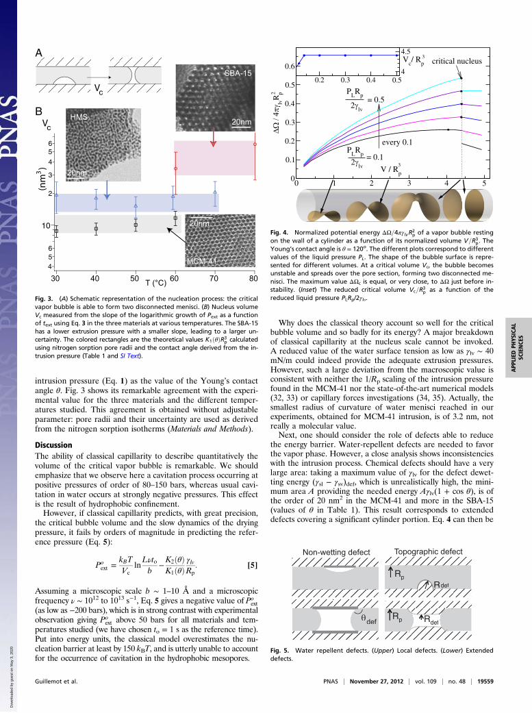
intrusion pressure (Eq. 1) as the value of the Young’s contactangle θ. Fig. 3 shows its remarkable agreement with the experi-mental value for the three materials and the different temper-atures studied. This agreement is obtained without adjustableparameter: pore radii and their uncertainty are used as derivedfrom the nitrogen sorption isotherms (Materials and Methods).
DiscussionThe ability of classical capillarity to describe quantitatively thevolume of the critical vapor bubble is remarkable. We shouldemphasize that we observe here a cavitation process occurring atpositive pressures of order of 80–150 bars, whereas usual cavi-tation in water occurs at strongly negative pressures. This effectis the result of hydrophobic confinement.However, if classical capillarity predicts, with great precision,
the critical bubble volume and the slow dynamics of the dryingpressure, it fails by orders of magnitude in predicting the refer-ence pressure (Eq. 5):
Poext =
kBTVc
lnLνtob
−K2ðθÞK1ðθÞ
γlvRp
: [5]
Assuming a microscopic scale b ∼ 1–10 Å and a microscopicfrequency ν ∼ 1012 to 1013 s−1, Eq. 5 gives a negative value of Po
ext(as low as −200 bars), which is in strong contrast with experimentalobservation giving Po
ext above 50 bars for all materials and tem-peratures studied (we have chosen to = 1 s as the reference time).Put into energy units, the classical model overestimates the nu-cleation barrier at least by 150 kBT, and is utterly unable to accountfor the occurrence of cavitation in the hydrophobic mesopores.
Why does the classical theory account so well for the criticalbubble volume and so badly for its energy? A major breakdownof classical capillarity at the nucleus scale cannot be invoked.A reduced value of the water surface tension as low as γlv ∼ 40mN/m could indeed provide the adequate extrusion pressures.However, such a large deviation from the macroscopic value isconsistent with neither the 1/Rp scaling of the intrusion pressurefound in the MCM-41 nor the state-of-the-art numerical models(32, 33) or capillary forces investigations (34, 35). Actually, thesmallest radius of curvature of water menisci reached in ourexperiments, obtained for MCM-41 intrusion, is of 3.2 nm, notreally a molecular value.Next, one should consider the role of defects able to reduce
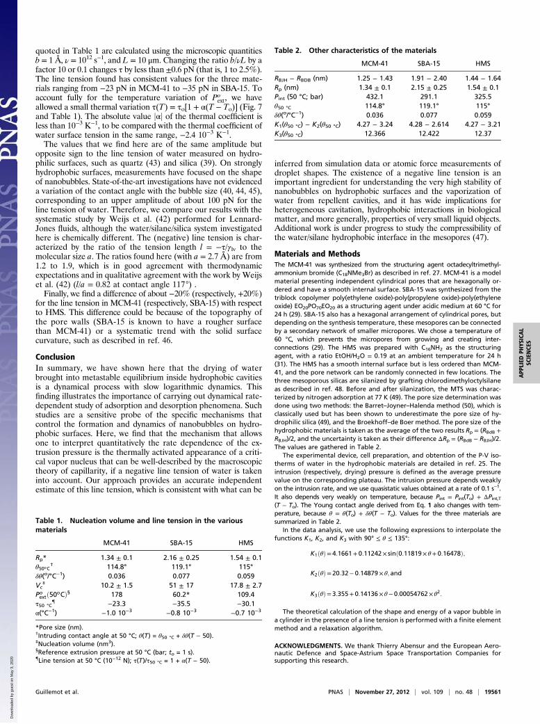
the energy barrier. Water-repellent defects are needed to favorthe vapor phase. However, a close analysis shows inconsistencieswith the intrusion process. Chemical defects should have a verylarge area: taking a maximum value of γlv for the defect dewet-ting energy (γsl − γsv)def, which is unrealistically high, the mini-mum area A providing the needed energy Aγlv(1 + cos θ), is ofthe order of 20 nm2 in the MCM-41 and more in the SBA-15(values of θ in Table 1). This result corresponds to extendeddefects covering a significant cylinder portion. Eq. 4 can then be
56
10
2
3
4
56
807060504030 T (°C)
SBA-15
Vc
3(n
m )
20nmHMS
20nm
MCM-41
20nm
4
Vc
A
B
Fig. 3. (A) Schematic representation of the nucleation process: the criticalvapor bubble is able to form two disconnected menisci. (B) Nucleus volumeVc measured from the slope of the logarithmic growth of Pext as a functionof text using Eq. 3 in the three materials at various temperatures. The SBA-15has a lower extrusion pressure with a smaller slope, leading to a larger un-certainty. The colored rectangles are the theoretical values K1ðθÞR3
p calculatedusing nitrogen sorption pore radii and the contact angle derived from the in-trusion pressure (Table 1 and SI Text).
0 1 2 3 4 50
0.1
0.2
0.3
0.4
0.5
0.6
ΔΩ /
4πγ lv
Rp
0.2 0.3 0.4 0.54
4.5
2
3V / R
p
PLR
p
2γlv
= 0.5
2γlv
= 0.1P
LR
p
every 0.1
critical nucleusVc/ R
p3
Fig. 4. Normalized potential energy ΔΩ=4πγlvR2p of a vapor bubble resting
on the wall of a cylinder as a function of its normalized volume V=R3p. The
Young’s contact angle is θ = 120o. The different plots correspond to differentvalues of the liquid pressure PL. The shape of the bubble surface is repre-sented for different volumes. At a critical volume Vc, the bubble becomesunstable and spreads over the pore section, forming two disconnected me-nisci. The maximum value ΔΩc is equal, or very close, to ΔΩ just before in-stability. (Inset) The reduced critical volume Vc=R3
p as a function of thereduced liquid pressure PLRp/2γlυ.
RpRdef
RdefRp
Non-wetting defect Topographic defect
θdef
RpRdef
RdefRpθdef
Fig. 5. Water repellent defects. (Upper) Local defects. (Lower) Extendeddefects.
Guillemot et al. PNAS | November 27, 2012 | vol. 109 | no. 48 | 19559
APP
LIED
PHYS
ICAL
SCIENCE
S
Dow
nloa
ded
by g
uest
on
May
3, 2
020

used to estimate self-consistently the energy barrier on the ex-tended defect, with a local contact angle θdef. Taking the MCM-41 for instance, ½Po
extð50oCÞ= 178 barsÞ], we need a local contactangle θdef ∼ 135° to provide the adequate barrier. However, thecalculated intrusion pressure on such extended defect should be750 bars, much higher than the maximum pressure around 500bars reached in the experiment. Such strong defects should notbe wetted, and no nucleation should be needed for emptying thepores hosting them (Fig. 5). A similar inconsistency is obtainedfor topographic defects, such as bumps on the pore walls andpores constrictions (SI Text, section II). Thus, the low nucleationbarrier shown by the drying pressure cannot be easily attributedto wall defects in the framework of classical capillarity.Finally, the effect of long-range interactions can be estimated
from the value of the disjoining pressure Aslυ/6πD3, where Aslυ is
the wall–vapor–liquid Hamaker constant, and D is the distanceof a meniscus portion to the wall. In the heart of the pore, witha typical Hamaker constant of 10−20 J, the disjoining pressure isof the order of 5 bars, which is not relevant. It is more importantclose to the contact line. This effect is, indeed, described by thethermodynamic concept of line tension introduced by Gibbsmore than a century ago to account for the excess energy causedby long-range interactions close to a three-phases contact line(36, 37). With an expected magnitude of the order τ ∼ γlva (abeing the molecular size, and τ ∼ 20 pN for water), line tensionplays a significant role only for liquid objects of nanometric sizein the three dimensions of space. In contrast to surface tension,it can be negative and thus, reduce the energy of a sessile nano-bubble. Experimental determinations of line tension are, how-ever, notoriously difficult, and values reported for water ondifferent substrates vary greatly in amplitude (from 10−11 to 10−6 N)and sign and tend to depend on the method used (37, 38). Themost direct methods, based on the size dependence of the contactangle of sessile drops/bubbles (39), are limited by the difficulty ofexact contact angle measurements at the required scale (1–100nm) (40) and the bias induced by surface heterogeneities (41).In a previous work, Lefevre et al. (22) attributed the low energybarrier for the drying of silanized MCM-41 to a negative line
tension and estimated an amplitude of some 10−11 N. In theirrecent numerical study of the evaporation kinetics of waterconfined between hydrophobic plates, Sharma and Debenedetti(19) also found nucleation energy governed by the line tensionbut with a positive value. The solid phases are, however, verydifferent in the two cases: the surfaces in ref. 19 are nonsup-ported 2D solid phases (a single layer of atoms), which shouldbehave quite differently from a 3D solid phase for long-rangeinteractions. In their systematic numerical study of 3D solid andfluid phases interacting with Lennard–Jones potentials, Weijset al. (42) found a negative line tension for contact angle valuesbetween 70° to 130° (42).We have studied (with a finite element method) the effect of
a line tension τ on the critical nucleus energy. The surprisingresult is that, although the line tension changes the shape of thecritical nucleus, the critical energy is simply given by the ex-pression (22) (Eq. 6)
ΔΩc ’ PLK1ðθÞR3p + γlvK2ðθÞR2
p + τK3ðθÞRp; [6]
where K1(θ) and K2(θ) are the very same functions as in Eq. 4(case without line tension) and K3(θ)Rp is equal to the contactline perimeter of the critical nucleus computed without line tension.This result reflects the almost exact compensation between the lineenergy gained in changing the shape of the nucleus and the asso-ciated losses in volume and surface energies (Figs. 4 and 6).In contrast to the previous effects, the impact of line tension
on the energy barrier is huge: a value of τ order γlva ∼ 20 pNchanges the energy barrier by hundreds of kBT and the dryingpressure by hundreds of bars. Thus, we interpret the high valueof the drying pressure in the hydrophobic mesopores as a strongsupport, if not a direct proof, of a negative line tension of wateron the C8-silanized silica. The value of the line tension can thenbe calculated from the experimental values of Po
ext using Eq. 6and the tabulated values of K1, K2, and K3 given in Materials andMethods (Eq. 7):
Poext =
kBTVc
lnLνtob
−K2ðθÞK1ðθÞ
γlυRp
−τ
R2p
K3ðθÞK1ðθÞ: [7]
The result is shown in Fig. 7 and Table 1. The precision of ±2bars on the drying pressure gives a relative precision better than10−2 on τ. This is a much higher resolution than the one pro-vided by currently available experimental methods. The values
0 0.1 0.2 0.3 0.4P
L R
p / 2γ
lv
-0.2
-0.1
0
0.1
0.2
0.3
0.4
0.5ΔΩ
c/4πγ
lv R
p2
τ = 0τγ
lv= 0.1 R
p
= - 0.1 Rp
τγ
lv
MCM-41
Fig. 6. Comparison of two different methods for calculating the normal-ized energy barrier ΔΩc=4πγlvR
2p for vapor nucleation in a cylinder. The dif-
ferent colors correspond to different values of the line tension τ. TheYoung’s contact angle is θ = 120°, and the x axis is the normalized liquidpressure. The continuous lines correspond to the first method used in ref. 22,leading to Eq. 6 in the text. The × symbol corresponds to the proper methoddeveloped here and described in SI Text, section III: the critical nuclei aredirectly calculated taking into account the finite line tension. The case ofMCM41 at 50 °C corresponds to τ/γlυ = −0.25Rp and has been plotted forillustration. The value of the energy barrier at the extruding pressure PL =178 bars is 48 kBT. (Inset) A nucleus shape for τ/γlυ = −0.3Rp.
200
150
100
50
0
Pex
t (ba
r)
807060504030T (°C)
MCM-41
HMS
SBA-15
o
Fig. 7. The reference extrusion pressure Poext as a function of temperature in
the three materials. The dashed line is the best fit obtained by fitting the linetension in Eq. 6. The pore radii, contact angles, water surface tension, andexpressions of K1, K2, and K3 are listed in SI Text, section I. In each material,we allow a small linear variation τ(T) = τo(1 + α(T − To)). The thermal co-efficient α is of the order −10−3 K−1 (Table 1). For comparison, the thermalcoefficient of water surface tension in the same range is −2.4 10−3 K−1.
19560 | www.pnas.org/cgi/doi/10.1073/pnas.1207658109 Guillemot et al.
Dow
nloa
ded
by g
uest
on
May
3, 2
020
quoted in Table 1 are calculated using the microscopic quantitiesb = 1 Å, ν = 1012 s−1, and L = 10 μm. Changing the ratio b/νL by afactor 10 or 0.1 changes τ by less than ±0.6 pN (that is, 1 to 2.5%).The line tension found has consistent values for the three mate-rials ranging from −23 pN in MCM-41 to −35 pN in SBA-15. Toaccount fully for the temperature variation of Po
ext, we haveallowed a small thermal variation τ(T) = τo[1 + α(T − To)] (Fig. 7and Table 1). The absolute value jαj of the thermal coefficient isless than 10−3 K−1, to be compared with the thermal coefficient ofwater surface tension in the same range, −2.4 10−3 K−1.The values that we find here are of the same amplitude but
opposite sign to the line tension of water measured on hydro-philic surfaces, such as quartz (43) and silica (39). On stronglyhydrophobic surfaces, measurements have focused on the shapeof nanobubbles. State-of-the-art investigations have not evidenceda variation of the contact angle with the bubble size (40, 44, 45),corresponding to an upper amplitude of about 100 pN for theline tension of water. Therefore, we compare our results with thesystematic study by Weijs et al. (42) performed for Lennard-Jones fluids, although the water/silane/silica system investigatedhere is chemically different. The (negative) line tension is char-acterized by the ratio of the tension length l = −τ/γlv to themolecular size a. The ratios found here (with a = 2.7 Å) are from1.2 to 1.9, which is in good agreement with thermodynamicexpectations and in qualitative agreement with the work by Weijset al. (42) (l/a = 0.82 at contact angle 117°) .Finally, we find a difference of about −20% (respectively, +20%)
for the line tension in MCM-41 (respectively, SBA-15) with respectto HMS. This difference could be because of the topography ofthe pore walls (SBA-15 is known to have a rougher surfacethan MCM-41) or a systematic trend with the solid surfacecurvature, such as described in ref. 46.
ConclusionIn summary, we have shown here that the drying of waterbrought into metastable equilibrium inside hydrophobic cavitiesis a dynamical process with slow logarithmic dynamics. Thisfinding illustrates the importance of carrying out dynamical rate-dependent study of adsorption and desorption phenomena. Suchstudies are a sensitive probe of the specific mechanisms thatcontrol the formation and dynamics of nanobubbles on hydro-phobic surfaces. Here, we find that the mechanism that allowsone to interpret quantitatively the rate dependence of the ex-trusion pressure is the thermally activated appearance of a criti-cal vapor nucleus that can be well-described by the macroscopictheory of capillarity, if a negative line tension of water is takeninto account. Our approach provides an accurate independentestimate of this line tension, which is consistent with what can be
inferred from simulation data or atomic force measurements ofdroplet shapes. The existence of a negative line tension is animportant ingredient for understanding the very high stability ofnanobubbles on hydrophobic surfaces and the vaporization ofwater from repellent cavities, and it has wide implications forheterogeneous cavitation, hydrophobic interactions in biologicalmatter, and more generally, properties of very small liquid objects.Additional work is under progress to study the compressibility ofthe water/silane hydrophobic interface in the mesopores (47).
Materials and MethodsThe MCM-41 was synthesized from the structuring agent octadecyltrimethyl-ammonium bromide (C18NMe3Br) as described in ref. 27. MCM-41 is a modelmaterial presenting independent cylindrical pores that are hexagonally or-dered and have a smooth internal surface. SBA-15 was synthesized from thetriblock copolymer poly(ethylene oxide)-poly(propylene oxide)-poly(ethyleneoxide) EO20PO70EO20 as a structuring agent under acidic medium at 60 °C for24 h (29). SBA-15 also has a hexagonal arrangement of cylindrical pores, butdepending on the synthesis temperature, these mesopores can be connectedby a secondary network of smaller micropores. We chose a temperature of60 °C, which prevents the micropores from growing and creating inter-connections (29). The HMS was prepared with C16NH2 as the structuringagent, with a ratio EtOH/H2O = 0.19 at an ambient temperature for 24 h(31). The HMS has a smooth internal surface but is less ordered than MCM-41, and the pore network can be randomly connected in few locations. Thethree mesoporous silicas are silanized by grafting chlorodimethyloctylsilaneas described in ref. 48. Before and after silanization, the MTS was charac-terized by nitrogen adsorption at 77 K (49). The pore size determination wasdone using two methods: the Barret–Joyner–Halenda method (50), which isclassically used but has been shown to underestimate the pore size of hy-drophilic silica (49), and the Broekhoff–de Boer method. The pore size of thehydrophobic materials is taken as the average of the two results Rp = (RBdB +RBJH)/2, and the uncertainty is taken as their difference ΔRp = (RBdB − RBJH)/2.The values are gathered in Table 2.
The experimental device, cell preparation, and obtention of the P-V iso-therms of water in the hydrophobic materials are detailed in ref. 25. Theintrusion (respectively, drying) pressure is defined as the average pressurevalue on the corresponding plateau. The intrusion pressure depends weaklyon the intrusion rate, and we use quasistatic values obtained at a rate of 0.1 s−1.It also depends very weakly on temperature, because Pint = Pint(To) + ΔPint,T(T − To). The Young contact angle derived from Eq. 1 also changes with tem-perature, because θ = θ(To) + δθ(T − To). Values for the three materials aresummarized in Table 2.
In the data analysis, we use the following expressions to interpolate thefunctions K1, K2, and K3 with 90° ≤ θ ≤ 135°:
K1ðθÞ= 4:1661+ 0:11242× sinð0:11819× θ+ 0:16478Þ;
K2ðθÞ= 20:32− 0:14879× θ; and
K3ðθÞ= 3:355+ 0:14136× θ− 0:00054762× θ2:
The theoretical calculation of the shape and energy of a vapor bubble ina cylinder in the presence of a line tension is performed with a finite elementmethod and a relaxation algorithm.
ACKNOWLEDGMENTS. We thank Thierry Abensur and the European Aero-nautic Defence and Space-Astrium Space Transportation Companies forsupporting this research.
Table 1. Nucleation volume and line tension in the variousmaterials
MCM-41 SBA-15 HMS
Rp* 1.34 ± 0.1 2.16 ± 0.25 1.54 ± 0.1θ50oC
† 114.8° 119.1° 115°δθ(o/°C−1) 0.036 0.077 0.059Vc
‡ 10.2 ± 1.5 51 ± 17 17.8 ± 2.7Poextð50oCÞ§ 178 60.2* 109.4
τ50 °C{ −23.3 −35.5 −30.1
α(°C−1) −1.0 10−3 −0.8 10−3 −0.7 10−3
*Pore size (nm).†Intruding contact angle at 50 °C; θ(T) = θ50 °C + δθ(T − 50).‡Nucleation volume (nm3).§Reference extrusion pressure at 50 °C (bar; to = 1 s).{Line tension at 50 °C (10−12 N); τ(T)/τ50 °C = 1 + α(T − 50).
Table 2. Other characteristics of the materials
MCM-41 SBA-15 HMS
RBJH − RBDB (nm) 1.25 − 1.43 1.91 − 2.40 1.44 − 1.64Rp (nm) 1.34 ± 0.1 2.15 ± 0.25 1.54 ± 0.1Pint (50 °C; bar) 432.1 291.1 325.5θ50 °C 114.8° 119.1° 115°δθ(o/°C−1) 0.036 0.077 0.059K1(θ50 °C) − K2(θ50 °C) 4.27 − 3.24 4.28 − 2.614 4.27 − 3.21K3(θ50 °C) 12.366 12.422 12.37
Guillemot et al. PNAS | November 27, 2012 | vol. 109 | no. 48 | 19561
APP
LIED
PHYS
ICAL
SCIENCE
S
Dow
nloa
ded
by g
uest
on
May
3, 2
020
1. Lum K, Chandler D, Weeks JD (1999) Hydrophobicity at small and large length scales.J Phys Chem B 103(22):4570–4577.
2. Ishida N, Inoue T, Miyahara M, Higashitani K (2000) Nano bubbles on a hydrophobicsurface in water observed by tapping-mode atomic force microscopy. Langmuir16(16):6277–6380.
3. Tyrrell JW, Attard P (2001) Images of nanobubbles on hydrophobic surfaces and theirinteractions. Phys Rev Lett 87(17):176104.
4. Meyer EE, Rosenberg KJ, Israelachvili JN (2006) Recent progress in understandinghydrophobic interactions. Proc Natl Acad Sci USA 103(43):15739–15746.
5. Seddon JRT, Lohse D (2011) Nanobubbles and micropancakes: Gaseous domains onimmersed substrates. J Phys Condens Matter 23(13):133001.
6. Christenson HK, Claesson PM (2001) Direct measurements of the force between hy-drophobic surfaces in water. Adv Colloid Interface Sci 91(3):391–436.
7. Chandler D (2002) Hydrophobicity: Two faces of water. Nature 417(6888):491.8. Berne BJ, Weeks JD, Zhou R (2009) Dewetting and hydrophobic interaction in physical
and biological systems. Annu Rev Phys Chem 60:85–103.9. ten Wolde PR, Chandler D (2002) Drying-induced hydrophobic polymer collapse. Proc
Natl Acad Sci USA 99(10):6539–6543.10. Patel AJ, et al. (2011) Extended surfaces modulate hydrophobic interactions of
neighboring solutes. Proc Natl Acad Sci USA 108(43):17678–17683.11. Zhou R, Huang X, Margulis CJ, Berne BJ (2004) Hydrophobic collapse in multidomain
protein folding. Science 305(5690):1605–1609.12. Giovambattista N, Lopez CF, Rossky PJ, Debenedetti PG (2008) Hydrophobicity of
protein surfaces: Separating geometry from chemistry. Proc Natl Acad Sci USA 105(7):2274–2279.
13. Mittal J, Hummer G (2008) Static and dynamic correlations in water at hydrophobicinterfaces. Proc Natl Acad Sci USA 105(51):20130–20135.
14. Borkent BM, Dammer SM, Schönherr H, Vancso GJ, Lohse D (2007) Superstability ofsurface nanobubbles. Phys Rev Lett 98(20):204502.
15. Ducker WA (2009) Contact angle and stability of interfacial nanobubbles. Langmuir25(16):8907–8910.
16. Bolhuis PG, Chandler D (2000) Transition path sampling of cavitation between mo-lecular scale solvophobic surfaces. J Chem Phys 113(18):8154–8160.
17. Leung K, Luzar A, Bratko D (2003) Dynamics of capillary drying in water. Phys Rev Lett90(6):065502.
18. Luzar A (2004) Activation barrier scaling for the spontaneous evaporation of confinedwater. J Phys Chem B 108(51):19859–19866.
19. Sharma S, Debenedetti PG (2012) Evaporation rate of water in hydrophobic con-finement. Proc Natl Acad Sci USA 109(12):4365–4370.
20. Liu D, et al. (2007) Observation of the density minimum in deeply supercooled con-fined water. Proc Natl Acad Sci USA 104(23):9570–9574.
21. Zhang Y, et al. (2011) Density hysteresis of heavy water confined in a nanoporoussilica matrix. Proc Natl Acad Sci USA 108(30):12206–12211.
22. Lefevre B, et al. (2004) Intrusion and extrusion of water in hydrophobic mesopores.J Chem Phys 120(10):4927–4938.
23. Lefevre B, et al. (2004) Intrusion and extrusion of water in highly hydrophobic mes-oporous materials: Effect of the pore texture. Coll and Surf A Physicochem Eng Asp241:265–272.
24. Eroshenko VA, Regis RC, Soulard M, Patarin J (2001) Energetics: A new field of ap-plications for hydrophobic zeolites. J Am Chem Soc 123(33):8129–8130.
25. Guillemot L, Galarneau A, Vigier G, Abensur T, Charlaix E (2012) New device tomeasure dynamic intrusion/extrusion cycles of lyophobic heterogeneous systems. RevSci Instrum 83(10):105105.
26. Kresge CT, Leonowicz ME, Roth WJ, Vartuli JC, Beck JS (1992) Ordered mesoporousmolecular sieves synthesized by a liquid-crystal template mechanism. Nature 359:710–712.
27. Martin T, Galarneau A, Di Renzo F, Brunel D, Fajula F (2004) Great improvement ofchromatographic performance using MCM-41 spheres as stationary phase in HPLC.Chem Mater 16(9):1725–1731.
28. Zhao D, et al. (1998) Triblock copolymer syntheses of mesoporous silica with periodic50 to 300 angstrom pores. Science 279(5350):548–552.
29. Galarneau A, et al. (2003) Microporosity and connections between pores in SBA-15mesostructured silicas as a function of the temperature of synthesis. New J Chem 27(1):73–79.
30. Tanev PT, Pinnavaia TJ (1996) Mesoporous silica molecular sieves prepared by ionicand neutral surfactant templating: A comparison of physical properties. Chem Mater8(8):2068–2079.
31. Di Renzo F, et al. (1999) Textural control of micelle-templated mesoporous silicates:The effects of co-surfactants and alkalinity. Microporous Mesoporous Mater 28(3):437–446.
32. Huang DM, Geissler PL, Chandler D (2001) Scaling of hydrophobic solvation free en-ergies. J Phys Chem B 105(28):6704–6709.
33. Dzubiella J, Swanson JMJ, McCammon JA (2006) Coupling hydrophobicity, dispersion,and electrostatics in continuum solvent models. Phys Rev Lett 96(8):087802.
34. Fisher LR, Israelachvili JN (1981) Direct measurement of the effect of meniscus forceon adhesion: A study of the applicability of macroscopic thermodynamis to micro-scopic liquid interfaces. Colloids Surf 3(4):303–319.
35. Christenson HK (1988) Adhesion between surfaces in undersaturated vapors - A re-examination of the influence of meniscus curvature and surface forces. J ColloidInterface Sci 121(1):170–178.
36. Gibbs JW (1957) The Scientific Papers (Dover, New York), Vol I.37. Drelich J (1996) The significance and magnitude of the line tension in three-phase
(solid-liquid-fluid) systems. Colloids Surf A Physicochem Eng Asp 116:43–54.38. Amirfazli A, Neumann AW (2004) Status of the three-phase line tension: A review.
Adv Colloid Interface Sci 110(3):121–141.39. Pompe T, Herminghaus S (2000) Three-phase contact line energetics from nanoscale
liquid surface topographies. Phys Rev Lett 85(9):1930–1933.40. Borkent BM, de Beer S, Mugele F, Lohse D (2010) On the shape of surface nano-
bubbles. Langmuir 26(1):260–268.41. Checco A, Guenoun P, Daillant J (2003) Nonlinear dependence of the contact angle of
nanodroplets on contact line curvature. Phys Rev Lett 91(18):186101.42. Weijs JH, Marchand A, Andreotti B, Lohse D, Snoeijer JH (2011) Origin of line tension
for a lennard-jones nanodroplet. Phys Fluids 23(2):022001.43. Zorin Z, Platikanov D, Kolarov T (1987) The transition region between aqueous
wetting films on quartz and the adjacent meniscus. Colloids Surf 22:147–159.44. Zhang XH, Maeda N, Craig VSJ (2006) Physical properties of nanobubbles on hydro-
phobic surfaces in water and aqueous solutions. Langmuir 22(11):5025–5035.45. Zhang XH, Quinn A, Ducker WA (2008) Nanobubbles at the interface between water
and a hydrophobic solid. Langmuir 24(9):4756–4764.46. Bauer C, Dietrich S (2000) Shapes, contact angles, and line tensions of droplets on
cylinders. Phys Rev E Stat Phys Plasmas Fluids Relat Interdiscip Topics 62(2):2428–2438.47. Sarupria S, Garde S (2009) Quantifying water density fluctuations and compressibility
of hydration shells of hydrophobic solutes and proteins. Phys Rev Lett 103(3):037803.48. Martin T, et al. (2001) Towards total hydrophobisation of MCM-41 type silica surface.
Stud Surf Sci Catal 135:4621–4628.49. Galarneau A, Desplantier D, Dutartre R, Di Renzo F (1999) Micelle-templated silicates
as a test-bed for methods of mesopore size evaluation. Microporous MesoporousMater 27:297–308.
50. Barrett EP, Joyner LG, Halenda PP (1951) The determination of pore volume and areadistributions in porous substances. I. Computations from nitrogen isotherms. J AmChem Soc 73(1):373–380.
19562 | www.pnas.org/cgi/doi/10.1073/pnas.1207658109 Guillemot et al.
Dow
nloa
ded
by g
uest
on
May
3, 2
020