adrenocortical activity and emotion...
TRANSCRIPT

Adrenocortical Activity and Emotion RegulationAuthor(s): Kathy Stansbury and Megan R. GunnarSource: Monographs of the Society for Research in Child Development, Vol. 59, No. 2/3, TheDevelopment of Emotion Regulation: Biological and Behavioral Considerations (1994), pp. 108-134Published by: Wiley on behalf of the Society for Research in Child DevelopmentStable URL: http://www.jstor.org/stable/1166141 .
Accessed: 12/05/2013 16:39
Your use of the JSTOR archive indicates your acceptance of the Terms & Conditions of Use, available at .http://www.jstor.org/page/info/about/policies/terms.jsp
.JSTOR is a not-for-profit service that helps scholars, researchers, and students discover, use, and build upon a wide range ofcontent in a trusted digital archive. We use information technology and tools to increase productivity and facilitate new formsof scholarship. For more information about JSTOR, please contact [email protected].
.
Wiley and Society for Research in Child Development are collaborating with JSTOR to digitize, preserve andextend access to Monographs of the Society for Research in Child Development.
http://www.jstor.org
This content downloaded from 130.132.173.181 on Sun, 12 May 2013 16:39:32 PMAll use subject to JSTOR Terms and Conditions
ADRENOCORTICAL ACTIVITY AND EMOTION REGULATION
Kathy Stansbury and Megan R. Gunnar
As repeatedly noted by the contributors to the first part of this volume, emotions consist of. physiological, behavioral, and subjective-experiential components. Accordingly, an understanding of the physiology of emotion is essential to understanding emotion processes, and the concept of emotion regulation also requires an understanding of the relations between physiol- ogy and behavior. Thus, like Campos, Campos, and Barrett (1989), we view emotion regulation not as a response to emotion but rather as a process- or systems-oriented perspective on the study of emotions. From this perspec- tive, an understanding of emotions requires an understanding of the dy- namic, reciprocal relations between the behavioral expression of emotions- including action tendencies and coping processes (see Dawson, in this volume; Lazarus, 1991)-and the physiological processes associated with emotion experience.
Given that physiological processes are integral to emotions and their regulation, investigators working in this domain have become increasingly interested in identifying emotion-relevant physiological systems that can be studied in healthy, normal children (see Dawson, in this volume; Fox, in this volume; and Porges, Doussard-Roosevelt, & Maiti, in this volume). One of these is the hypothalamic-pituitary-adrenocortical (HPA) system. Corti-
Portions of this essay were supported by an Alcohol and Drug Administration Mental Health Association (ADAMHA) Postdoctoral Fellowship in Child Development (5T32- MH15755) to Kathy Stansbury and by a National Institutes of Health research grant (HD-16494) and a National Institute of Mental Health research scientist award (MH- 00946) to Megan R. Gunnar. We wish to thank Seymour Levine for reviewing a draft of this essay, Kaye O'Geay for secretarial help, and the many graduate and undergraduate students from our research group whose work we review. Please direct correspondence to Megan Gunnar, Institute of Child Development, 51 East River Road, University of Minnesota, Minneapolis, MN 55455.
108
This content downloaded from 130.132.173.181 on Sun, 12 May 2013 16:39:32 PMAll use subject to JSTOR Terms and Conditions
THE DEVELOPMENT OF EMOTION REGULATION
sol, the hormonal product of this system, can now be measured easily and noninvasively in small samples of saliva. In addition, the adrenocortical system plays a major role in stress resistance (Selye, 1950), and many believe that emotions mediate the magnitude of the adrenocortical response to environmental challenge (Mason, 1975). The combination of ease of mea- surement and hypothesized sensitivity to emotion processes makes assess- ment of adrenocortical activity a highly attractive option in studies of early emotion development. In the following essay, we first provide an overview of the psychobiology of the HPA system and then relate the activity of this system to processes of emotion regulation.
THE PSYCHOBIOLOGY OF THE HPA SYSTEM
Cortisol is the primary hormonal product of the HPA system in hu- mans. The adrenocortical system is a neuroendocrine system, which means that the production of cortisol is regulated, in large part, by centers in the central nervous system (CNS). The hypothalamus is the principle brain region involved in the regulation of cortisol, with centers in the limbic sys- tem serving to modulate and coordinate hypothalamic activity with percep- tual and cognitive inputs from higher centers in the brain (de Kloet, 1991). Cortisol and its precursor hormones also affect the CNS and influence emo- tion and cognition.
Control of Cortisol Production
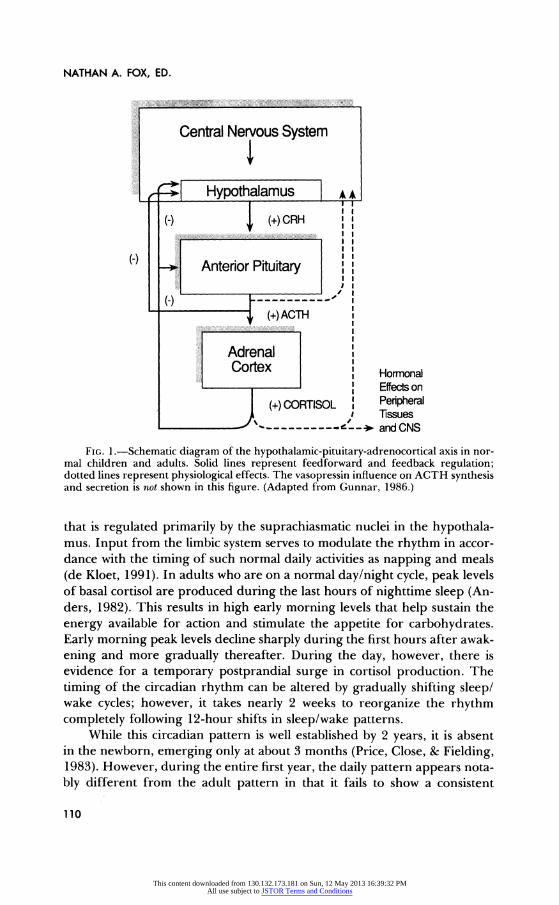
Figure 1 shows a schematic of the HPA system. The production of cortisol is under the control of cortisol-releasing hormone (CRH) and vaso- pressin, which are produced in the hypothalamus. Vasopressin (whose ef- fects are not schematized in the figure) potentiates and acts synergistically with the action of CRH on the cells in the anterior pituitary that produce adrenocorticotrophic hormone (ACTH) (Palkovits, 1987). ACTH is then released into general circulation and stimulates cells in the cortex of the adrenal glands to produce cortisol and release it into the bloodstream. Once in circulation, most of the cortisol immediately binds to cortisol binding globulin (CBG). It is the unbound fraction of the cortisol in circulation that is biologically active. Activity in the HPA system occurs in pulses; increases in circulating cortisol reflect an increase in the frequency of pulses of CRH.
Basal Activity
A certain amount of cortisol is essential for normal, nonstressed activity. These basal levels of cortisol follow a daily or circadian rhythm in adults
109
This content downloaded from 130.132.173.181 on Sun, 12 May 2013 16:39:32 PMAll use subject to JSTOR Terms and Conditions
NATHAN A. FOX, ED.
~;$iiiii ~~ ;:::I:--- ----- ------- ---- ;;i ?i: Central Nervous System
I Hypothalamus A• (-) (+) CRH
II
Anterior Pituitary I
~--- --Woo#
(+) ACTH
Adrenal Cortex Hormonal
Effects on
(+) CORTISOL Peripheral "I Tissues
""- --- and CNS
FIG. 1.-Schematic diagram of the hypothalamic-pituitary-adrenocortical axis in nor- mal children and adults. Solid lines represent feedforward and feedback regulation; dotted lines represent physiological effects. The vasopressin influence on ACTH synthesis and secretion is not shown in this figure. (Adapted from Gunnar, 1986.)
that is regulated primarily by the suprachiasmatic nuclei in the hypothala- mus. Input from the limbic system serves to modulate the rhythm in accor- dance with the timing of such normal daily activities as napping and meals (de Kloet, 1991). In adults who are on a normal day/night cycle, peak levels of basal cortisol are produced during the last hours of nighttime sleep (An- ders, 1982). This results in high early morning levels that help sustain the energy available for action and stimulate the appetite for carbohydrates. Early morning peak levels decline sharply during the first hours after awak- ening and more gradually thereafter. During the day, however, there is evidence for a temporary postprandial surge in cortisol production. The timing of the circadian rhythm can be altered by gradually shifting sleep/ wake cycles; however, it takes nearly 2 weeks to reorganize the rhythm completely following 12-hour shifts in sleep/wake patterns.
While this circadian pattern is well established by 2 years, it is absent in the newborn, emerging only at about 3 months (Price, Close, & Fielding, 1983). However, during the entire first year, the daily pattern appears nota- bly different from the adult pattern in that it fails to show a consistent
110
This content downloaded from 130.132.173.181 on Sun, 12 May 2013 16:39:32 PMAll use subject to JSTOR Terms and Conditions

THE DEVELOPMENT OF EMOTION REGULATION
0.50 -
0.45 -
=L 0.40 -
o 0.35 S0.35 -
0.30
0.25 -
0.20
PRENAP WAKEUP 45 MINUTE POSTNAP
Sampling Period
FIG. 2.-Salivary cortisol concentrations in 24 8-1 1-month-olds obtained immediately before, immediately after, and 45 min after their normal morning nap. (From Larson, Gunnar, & Hertsgaard, 1991.)
decrease over the middle portion of the day (Spangler, Meindl, & Gross- man, 1988). This infant pattern may reflect the coordination of basal cortisol
activity with napping and feeding schedules. Indeed, morning naps do ap- pear to be associated with a transient decrease in cortisol (see Fig. 2), which rebounds to prenap concentrations 45 min after the nap (Larson, Gunnar, & Hertsgaard, 1991).
Stress Activity
Three major pathways mediate the HPA stress response (de Kloet, 1991). One pathway involves direct stimulation of the pituitary and hypo- thalamus by biochemicals arriving from general circulation. Another in- volves direct visceral and sensory stimulation, including pain and blood pressure changes, traveling to the hypothalamus through brain-stem path- ways. The third pathway involves the transmission of psychological stimuli reaching the hypothalamus from the cerebral cortex via the limbic circuits. It is believed that the amygdala plays a major role in facilitating the initiation of the stress response while the hippocampus plays a major role in terminat- ing the response and returning the HPA system to basal regulation (de Kloet, 1991; Smuts & Levine, 1977). Following the onset of a stressor, it takes about 10-15 min to produce a rise in circulating cortisol levels and
111
This content downloaded from 130.132.173.181 on Sun, 12 May 2013 16:39:32 PMAll use subject to JSTOR Terms and Conditions

NATHAN A. FOX, ED.
20-30 min for cortisol to reach peak stress concentrations in plasma. Even if the response is acute, it may take several hours for the "extra" cortisol to be cleared from circulation.
Adequate Measurement of Basal Levels
This brief discussion of the basal and stress-induced activity of the HPA
system should serve to alert researchers to the importance of adequately measuring basal levels of cortisol. A determination of whether a change from basal to stress regulation of the system has been produced requires knowledge of what the concentrations of cortisol would have been in the absence of the stressor (Levine & Coe, 1985). When the system is function-
ing under basal regulation, decreases in cortisol are expected over the day. During the morning hours, when basal levels are declining rapidly, a small cortisol response may not yield increases in cortisol over prestimulation levels. The significance of the response might be apparent only when corti- sol levels are compared to levels obtained under similar conditions at the same time of day but in the absence of the stressor. This can be seen in the data on separation from mother shown in Figure 3. The effects of a 30-min separation were evident only when cortisol levels of babies separated from their mothers were compared to levels obtained from the same babies when their mothers stayed with them during the 30-min period (Gunnar, Larson, Hertsgaard, Harris, & Brodersen, 1992).
Cortisol and Stress Resistance
Although stress is often thought of as bad, the ability to mount a stress response is necessary for survival. An individual without a functioning HPA system could not survive even the normal perturbations of daily life and could exist only in physically and emotionally protected environments (Selye, 1950). The adrenocortical system appears to perform three major functions in stress resistance. First, along with other hormones and systems, it participates in the mobilization of energy resources that are needed for action. Second, cortisol serves a homeostatic function in regulating the activ- ity of other stress-sensitive systems including the central and peripheral catecholamine systems, the endogenous opiate system, and the immune sys- tem (Munck, Guyre, & Holbrook, 1984). Third, cortisol, ACTH, and CRH act in the brain and can affect memory, learning, and emotions (McEwen, de Kloet, & Rostene, 1986). Clearly, healthy adaptation requires the capacity to produce increased cortisol under conditions of threat and to return its production to basal levels as soon as the threat has passed.
112
This content downloaded from 130.132.173.181 on Sun, 12 May 2013 16:39:32 PMAll use subject to JSTOR Terms and Conditions

THE DEVELOPMENT OF EMOTION REGULATION
--,-- Caretaker Condition with Babysitter "- Caretaker Condition with Mother
0.6
0.5
S 0.4
S 0.3 o
"0.2
0.1
0.0 PRESESSION POSTSESSION
Time of Saliva Sample
FIG. 3.-Salivary cortisol concentrations in 19 infants, aged 9 months, tested twice: once separated for 30 min from their mothers and once playing in the mother's presence for 30 min (order counterbalanced). Salivary cortisol measures were obtained immediately before and after the 30-min periods. Standard errors were approximately 0.03 jxg/dl. (Adapted from Gunnar, Larson, et al., 1992.)
CNS Effects
The CNS effects of glucocorticoids (e.g., cortisol, corticosterone, and related steroid hormones) on behavior are of most relevance to research on emotion regulation. Most of the information that we have about the CNS effects of glucocorticoids has been derived from animal research, a fact that the reader should keep in mind in reading the following section. Because in rodents the steroid produced by the HPA system is corticosterone and not cortisol, the more general term glucocorticoids will be used in discussing these data.
It has been hypothesized that the effects of glucocorticoids on behavior are largely mediated by their actions on steroid receptors in the brain-stem reticular formation, the hippocampus, and the frontal cortex (for more extensive reviews, see de Kloet, 1991; McEwen et al., 1986). Glucocorticoids do not have unitary excitatory or inhibitory influences on neural tissue. Instead, the effect of these steroid hormones depends on the type of steroid receptor that is stimulated and on the point in the process at which the
113
This content downloaded from 130.132.173.181 on Sun, 12 May 2013 16:39:32 PMAll use subject to JSTOR Terms and Conditions

NATHAN A. FOX, ED.
response is being measured. Glucocorticoids have both fast membrane and slow gene-mediated effects on neuronal activity (de Kloet, 1991). The fast effects of glucocorticoids take milliseconds to seconds to develop, while the slow effects take minutes to hours to emerge and may last hours after hor- mone concentrations have returned to basal levels.
Elevations in glucocorticoids in response to stress initially stimulate cor- tical arousal via excitatory action on brain-stem reticular activity. In emo-
tionally stable adults, these excitatory brain-stem effects are associated with
feelings of increased energy and ability to concentrate (Born, Hitzler, Pie- trowsky, Pauschinger, & Fehm, 1989). In contrast, the more slowly devel-
oping effects of glucocorticoids largely function to lower neuronal excita- tion, which may produce a reversal of the initial stimulatory influences (de Kloet, 1991). Thus, in normal subjects who are maintained on increased
glucocorticoids for several days, there is some evidence for a decrease in
energy (hypomania), a loss of ability to concentrate, and an increase in depressive affect (Wolkowitz et al., 1988).
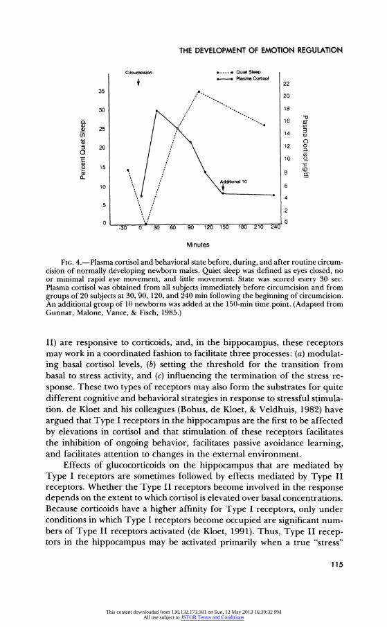
These inhibitory effects develop even when elevations in glucocorti- coids are relatively short lived, and they may influence the consolidation and storage of memories for the events transpiring during and after the stressor. They are also involved in the inhibitory effect of glucocorticoids on REM sleep, which persists for hours after stress levels of cortisol have been cleared from circulation (see Fig. 4; and also Gunnar, Malone, Vance, & Fisch, 1985). Thus, in response to a noxious stimulus, glucocorticoids may initially help support the increased cortical arousal needed to sustain heightened behavioral and emotion activity and then facilitate the behav- ioral withdrawal and quiescence that may be needed to help recover from the physical and emotional effects of the stressor. This dual role of glucocor- ticoids has clear implications for our understanding of emotion regulation.
Another major effect of glucocorticoids is to raise sensory thresholds via decreasing evoked sensory potentials (Born et al., 1989). At the same time, increased corticoids facilitate the integration and interpretation of incoming sensory information (Henkin, 1970). Thus, once the slow-action effects of glucocorticoids have developed, elevated corticoid levels may be associated with a reduction in an individual's actual sensitivity to auditory, tactile, or visual stimuli, including pain stimuli, along with improved abilities to interpret the meaning of stimulation and to integrate information about the stressor arriving from different sensory modalities.
Elevated glucocorticoid levels also have complex effects on learning and memory. de Kloet (1991) has hypothesized that these effects serve to facilitate the elimination of behavior that is no longer of relevance and to consolidate behavioral strategies that serve to reduce real or perceived threat. Many of the effects on learning and memory may involve corticoid receptors in the hippocampus. Two types of receptors (Type I and Type
114
This content downloaded from 130.132.173.181 on Sun, 12 May 2013 16:39:32 PMAll use subject to JSTOR Terms and Conditions
THE DEVELOPMENT OF EMOTION REGULATION
Circumcision e----4 Quiet Sleep S- Plasma Cortisol 22
35 . 20
30 %18
5 25 I) 14
20 12 o a c 10 O
o 151
Additional 10 10 6 4
5 2
0 0 -30 0 30 60 90 120 150 180 210 240
Minutes
FIG. 4.-Plasma cortisol and behavioral state before, during, and after routine circum- cision of normally developing newborn males. Quiet sleep was defined as eyes closed, no or minimal rapid eye movement, and little movement. State was scored every 30 sec. Plasma cortisol was obtained from all subjects immediately before circumcision and from groups of 20 subjects at 30, 90, 120, and 240 min following the beginning of circumcision. An additional group of 10 newborns was added at the 150-min time point. (Adapted from Gunnar, Malone, Vance, & Fisch, 1985.)
II) are responsive to corticoids, and, in the hippocampus, these receptors may work in a coordinated fashion to facilitate three processes: (a) modulat- ing basal cortisol levels, (b) setting the threshold for the transition from basal to stress activity, and (c) influencing the termination of the stress re-
sponse. These two types of receptors may also form the substrates for quite different cognitive and behavioral strategies in response to stressful stimula- tion. de Kloet and his colleagues (Bohus, de Kloet, & Veldhuis, 1982) have argued that Type I receptors in the hippocampus are the first to be affected by elevations in cortisol and that stimulation of these receptors facilitates the inhibition of ongoing behavior, facilitates passive avoidance learning, and facilitates attention to changes in the external environment.
Effects of glucocorticoids on the hippocampus that are mediated by Type I receptors are sometimes followed by effects mediated by Type II receptors. Whether the Type II receptors become involved in the response depends on the extent to which cortisol is elevated over basal concentrations. Because corticoids have a higher affinity for Type I receptors, only under conditions in which Type I receptors become occupied are significant num- bers of Type II receptors activated (de Kloet, 1991). Thus, Type II recep- tors in the hippocampus may be activated primarily when a true "stress"
115
This content downloaded from 130.132.173.181 on Sun, 12 May 2013 16:39:32 PMAll use subject to JSTOR Terms and Conditions

NATHAN A. FOX, ED.
response of the HPA system has been elicited. The exact levels of corticoids that denote the "stress" cutoff will vary during the day because the availabil- ity of Type I receptors varies with the daily cortisol rhythm (de Kloet, 1991). There are no clear guidelines for determining these levels; however, it is estimated that Type I receptors are 80%-90% occupied under basal condi- tions. Thus, perhaps a 10%-20% increase in cortisol can be accommodated by Type I receptors, with larger increases affecting Type II receptors and constituting a qualitatively different response.
Once Type II receptors in the hippocampus have been activated, their effects may modulate hippocampal-mediated integration of newly learned behaviors with previously learned material. Thus, activation of Type II receptors in the hippocampus may be associated with changes in response to the stressor over time or with repetitions of the stressful event. These well-modulated effects on brain activity and behavior tend to characterize the effect of low to moderate stress levels of glucocorticoids. When the response to a stressor is intense and corticoids rise rapidly to extreme levels, disruptions in learning and adaptive behavior may be observed. Individual differences related to both genetic and experiential factors also influence the balance between Type I and Type II receptor-mediated behaviors. All these effects are clearly relevant to our understanding of emotion regulation processes.
Negative Feedback and Stress Threshold
Levels of glucocorticoids in circulation are regulated by negative feed- back. The hypothalamus appears to be the major site of negative feedback regulation, but other structures, including the pituitary and hippocampus, also play a role. Three types of negative feedback have been distinguished: fast, intermediate, and slow. Fast feedback involves multisynaptic control of ACTH release. It occurs within 10 min of the rise in glucocorticoids (i.e., about 20-25 min after stress activation). Fast feedback appears to be desen- sitized or "overridden" with chronic stress exposure and may play a role in increasing HPA reactivity in chronically stressed individuals (Young, Akana, & Dallman, 1990). Intermediate feedback may operate at the limbic and forebrain system level to alter reactivity to stimulation, thus dampening continued activation of limbic stress circuits in prolonged stressful encoun- ters. Finally, slow feedback involves the blockade of the CRH-vasopressin system in the hypothalamus and ACTH production in the pituitary. This feedback action takes 30-60 min to begin to develop (i.e., 45 min or more after activation), and its effects may not become pronounced for hours (de Kloet, 1991; Jacobson & Sapolsky, 1991).
116
This content downloaded from 130.132.173.181 on Sun, 12 May 2013 16:39:32 PMAll use subject to JSTOR Terms and Conditions

THE DEVELOPMENT OF EMOTION REGULATION
de Kloet and his colleagues (de Kloet, 1991; McEwen et al., 1986) have hypothesized that regulation of the HPA stress response is related to Type I and Type II receptor density in the hippocampus. According to these researchers, Type I receptor density may set the threshold for HPA reactiv- ity, with a greater density being associated with lower reactivity (and possibly higher basal cortisol levels). Under this model, Type II receptors modulate negative feedback, with greater density being associated with a greater ca- pacity to terminate the HPA stress response.
There is evidence in the animal literature for individual differences in the number of Type I and Type II receptors in the hippocampus (de Kloet, 1991). In rodents, stressful stimulation experienced during infancy increases, while chronic elevation in cortisol experienced after infancy decreases, the number of Type II receptors (Meaney, Aitken, Van Berkel, Bhatnager, & Sapolsky, 1988). Several different strains of rats have also been identified that differ in number of Type I and Type II receptors. Strains with more Type I and Type II receptors show a higher threshold for the lifting of tonic inhibition of the hypothalamic CRH neurons and a lower threshold for the termination of the stress response (Walker, Rivest, Meaney, & Au- bert, 1989). These strains also show reduced fearfulness or negative emo- tionality and increased capacity to respond to and learn under stressful environmental circumstances. These behavioral differences have been shown both for receptor differences resulting from prior stress experiences and strain and for genetic differences (Meaney et al., 1988; Walker et al., 1989).
EMOTION REGULATION AND PERIODS OF THE HPA RESPONSE
This overview of the psychobiology of the HPA system clearly points to the relevance of this system for the study of emotion regulation. It also points to the need to consider mutual and reciprocal relations between emotion processes and HPA activity. Furthermore, it is clear that time is an important parameter in relating HPA activity to the behavioral regulation of affect. As an aid to investigators new to the use of cortisol measures in their research, in what follows we briefly consider the major "periods" or "phases" of the HPA response to a stressor as they relate to emotion regula- tion processes. Our time estimations are only rough approximations. The actual timing of events would depend on numerous factors, including the magnitude and the rate of the HPA response. A consideration of these response periods is relevant to research on both adults and children; how- ever, in what follows, we focus on children.
117
This content downloaded from 130.132.173.181 on Sun, 12 May 2013 16:39:32 PMAll use subject to JSTOR Terms and Conditions

NATHAN A. FOX, ED.
Initial Reaction
Before a psychological stressor can activate an HPA stress response, the stressor must be perceived and interpreted, at least to some extent (Zajonc, 1984). If after this rudimentary processing the event is perceived as threatening, stress circuits in the limbic system and hypothalamus may be activated. This period of initial reaction probably encompasses only the first moments after the child alerts to the event. One critical issue to consider in relating HPA activity to emotion regulation for this "period" is the delin- eation of types of emotion reactions that are associated with elevations in cortisol.
Preelevation
If a stress response is activated, the physiological changes set in motion begin to create the substrates necessary to sustain increased physical and psychological activity. For the first 5-10 min following stress activation, cortisol levels will still be at, or close to, basal concentrations, and thus the effects of cortisol on the CNS will not be a major factor influencing behavior. Increases in CRH, vasopressin, and ACTH, however, may begin to influ- ence CNS activity at this time.
While these physiological changes are occurring, we can assume that the child is engaged in a more complete appraisal of the emotional meaning of the event, perhaps incorporating the immediate subjective experience of her altered physiological state (Lazarus & Folkman, 1984). The child may also begin to assess his or her coping and emotion regulation resources. The processes involved in both continued emotion appraisal and emotion regulation may either increase or decrease the activity in the limbic circuits involved in the stress response. As a result of this continued appraisal, the child may decide that the event is not actually threatening, thereby decreas- ing the stimulation of limbic stress circuits and terminating the stress re- sponse. Conversely, the child may experience the event as even more threat- ening, and stimulation of the limbic stress circuits may increase.
For many of the emotionally distressing situations that we are able to study in the laboratory, psychological and central nervous system processes occurring in these first two periods may not be sufficient to produce a significant, detectable cortisol stress response in enough subjects to permit an adequate examination of hormone-behavior relations. For ethical rea- sons, the stressors that we use are mild, the situations brief, and the supports provided to the child sufficient to prevent demonstrable elevations in corti- sol in most subjects. For example, it is now clear that even such seemingly stressful events as separation from the mother may be insufficient to pro-
118
This content downloaded from 130.132.173.181 on Sun, 12 May 2013 16:39:32 PMAll use subject to JSTOR Terms and Conditions

THE DEVELOPMENT OF EMOTION REGULATION
duce elevated cortisol concentrations when separation occurs in a supportive environment with other caregivers available to respond to the baby's needs (Gunnar, Larson, et al., 1992; Tennes, Downey, & Vernadakis, 1977). Fur- thermore, it has been shown that, for monkeys, the presence and availability of the mother buffers the HPA stress response to many potentially stressful events (Levine, 1970), and in the laboratory the human mother is typically present unless separation is the stressor being examined. The problem of identifying sufficiently salient stressors in research on children has led many researchers to seek naturally occurring or real-life stressful contexts in which to examine the relations between HPA activity and emotion regu- lation.
Period of CNS Effects: Fast-developing Influences
Once cortisol levels have risen sufficiently above basal values-about 10-15 min after stress activation-cortisol can begin to have effects on CNS activity. The initial effects will involve those that can occur quickly, and, as discussed earlier, should include changes leading to a sense of increased energy and concentration, facilitation of passive avoidance, and attention to changes in the environment. These effects may facilitate emotion regulation through facilitating avoidance of noxious or threatening elements of the situation. Avoiding behaviors that lead to increased distress may, in turn, increase the likelihood that the child will try other behaviors that may be more adaptive. Fast negative feedback effects may also begin to exert influ- ence on continued CRH-vasopressin and ACTH production at this time.
Period of CNS Effects: More Slowly Developing Influences
The neuronal effects that develop more slowly take at least 15 min after cortisol has risen above basal levels (i.e., after the onset of period 3). Thus, the earliest "slow gene-mediated effects" may not occur until about 20-30 min after stress activation of the HPA system. Furthermore, most of these influences will take much longer, including the slow negative feedback effects described earlier. The point is that these more slowly developing effects are probably taking place after the child has left the laboratory and/ or an acute stress experience is over. Nonetheless, because many of the gene-mediated effects influence memory processes and the integration of new information, they will be important to the study of emotion regulation and may help explain how emotion regulation experiences alter the child's responses to arousing events on subsequent exposures.
119
This content downloaded from 130.132.173.181 on Sun, 12 May 2013 16:39:32 PMAll use subject to JSTOR Terms and Conditions

NATHAN A. FOX, ED.
TABLE 1
PLASMA CORTISOL AND PERCENTAGE OF CRYING IN RESPONSE TO CIRCUMCISION AND HEEL-STICK BLOOD SAMPLING IN NEONATES
Time Point
Stimulus N (min)a r pb
Circumcision ........................... 9 30 +.71 < .05 Circumcision (with soothing)c ............. 20 30 +.44 < .05 Circumcision (with soothing) .............. 20 90 +.50 < .05 Circumcision (no soothing) ............... 13 20 + .35 N.S. Heel stick (no soothing) .................. 18 20 +.54 < .05 Heel stick (with soothing) ................ 13 20 +.50 N.S. Heel stick (no soothing) .................. 12 20 +.55 < .05
SouRcE.-Adapted from Gunnar, Marvinney, Isensee, & Fisch (1989). NOTE.-N.S. = not significant. a Time point indicates minutes between onset of stimulation and poststimulation blood sampling. Behavior was
always obtained during stimulation. b Z = 7.47 for the combined significance of the seven correlation coefficients presented in this table. c
Soothing indicates that all subjects were given pacifiers.
ILLUSTRATIONS FROM RESEARCH ON CHILDREN
Most of the work on normally developing human children examines acute, brief stressors and changes in cortisol at approximately 20-30 min after stressor onset. Because of this, our data on children deal primarily with questions relating behavior to HPA activity during the first two to three periods outlined above. These questions include asking which emotions are related to activation of the HPA stress response, which emotion regulation processes are involved in mediating the stress response, and how individual differences might be understood in the context of these first two questions.
Emotions Associated with Activation
It has long been believed that negative emotions play a primary role in activating the HPA stress response (Mason, 1971). This view finds support in data showing that crying during infancy is at times correlated with post- stressor cortisol concentrations (see Table 1). These data on newborns in Table 1 conform well with data on adults showing that conditions of work stress and military training lead to correlated increases in cortisol and self- reports of boredom, impatience, irritation, fear, and anxiety (Franken- haeuser, 1980; Ursin, Baade, & Levine, 1978).
While states of negative affect do appear to be associated with elevations in cortisol, it is not clear that they are sufficient to cause activation of the HPA stress circuits. The literature on HPA activity and emotion behavior is replete with instances of statistical dissociations (Levine, Wiener, Coe,
120
This content downloaded from 130.132.173.181 on Sun, 12 May 2013 16:39:32 PMAll use subject to JSTOR Terms and Conditions

THE DEVELOPMENT OF EMOTION REGULATION
Repeated Stimulation n=49
.90
.85 80
.80 -
70 - .75 . I Base
m 60 - .70 - r-- Response x
cm .65 -
50- *-- .60
- OD +C
aO 40 o .55 -
0 ( .45 -
20 .40
.35 - 10
.30 ?
0Day 1 Day 2 Day 1 Day 2 Day1 Day 2 Day1 Day 2
Behavioral Response Salivary Cortisol
FIG. 5.-Behavioral distress and HPA activity in response to two physical exams
separated by 24 hours in normally developing human newborns. Behavioral distress is defined as the weighted average of the percentage of 30-sec coding intervals in the states of active awake or crying. Salivary cortisol in Rg/dl obtained before and 30 min after the
beginning of a 6-min physical exam. (Adapted from Gunnar, Connors, & Isensee, 1989.)
Bayart, & Hayashi, 1987). For example, on repeated exposure to a psycho- logical stressor, we found that the newborn HPA stress response habituated (Gunnar, Connors, & Isensee, 1989; Gunnar, Hertsgaard, Larson, & Riga- tuso, 1992) but that crying did not (see Fig. 5). Similarly, during a prolonged separation, cortisol levels in rhesus monkey infants returned to basal con- centrations within 24 hours, while behavioral distress continued for days (Gunnar, Gonzales, Goodlin, & Levine, 1981). A decade ago, in a review of the adult research, Rose (1980) pointed out that rapid adaptation was also highly characteristic of the human adult HPA stress response.
Dissociations between HPA activity and a negative emotion response have led many to question how closely negative emotions are linked to stress
121
This content downloaded from 130.132.173.181 on Sun, 12 May 2013 16:39:32 PMAll use subject to JSTOR Terms and Conditions

NATHAN A. FOX, ED.
1.20 - Discharge Exams Heelsticks
1.00-
S 0.80 -
o 0.60
o
0 0.40
0.20
0.00 1 2 1 2
N=71 N=-18
Test Day
FIG. 6.-Salivary cortisol in response to physical exams and heel sticks in normally developing human newborns. Trials were separated by 24 hours. (Adapted from Gunnar, Hertsgaard, et al., 1992.)
activation of the HPA system. Levine (Hennessy & Levine, 1979), for exam-
ple, has suggested that cortisol is not the "emotion juice." Instead, novelty, uncertainty, discrepancy, and/or incongruity serve as the primary psycho- logical factors that activate the HPA response. This, of course, might explain why, with repetitions of a stressor, one can see adaptation of the HPA
response. To add to the complexity, data also exist that appear to contradict the
novelty/uncertainty hypothesis. First, when stressors are repeated and thus become more familiar, stress elevations in cortisol sometimes increase rather than decrease. Sensitization of the cortisol stress response occurs with more noxious or intense stressors (Groves & Thompson, 1970; Natelson et al., 1988). This is demonstrated in Figure 6 using data from a study comparing the neonatal HPA response to repeated heel sticks (more noxious) with the
response to repeated physical exams (less noxious). Second, novel experi- ences occurring in pleasant emotion contexts do not elevate cortisol. At times, in fact, pleasurable situations have been associated with a lowering of cortisol below basal concentrations (Wadeson, Mason, Hamburg, & Handlon, 1963). In a study that we conducted with 6-13-month-olds, we contrasted the novelty of a first-time swimming experience with the positive
122
This content downloaded from 130.132.173.181 on Sun, 12 May 2013 16:39:32 PMAll use subject to JSTOR Terms and Conditions
THE DEVELOPMENT OF EMOTION REGULATION
emotion typically engendered in mother-baby swim classes (Hertsgaard, Gunnar, Larson, Brodersen, & Lehman, 1992). Rather than elevating corti- sol levels, as the uncertainty hypothesis would predict, swimming lowered cortisol below baseline, as the emotion hypothesis would predict.
To summarize, neither the emotion nor the uncertainty hypothesis ap- pears sufficient to explain the various types of results that have been re- ported. Perhaps one reason the relative contributions of emotions and un- certainty are still unclear is that these factors need to be considered within an emotion regulation framework. As we have suggested previously, uncer- tainty may play a role in mediating the effect of emotion on the HPA system. However, the critical aspect of uncertainty may not be novelty or unfamiliar- ity but rather uncertainty about how to control or influence the stressful event and one's behavioral and emotion reactions to it (Gunnar, Marvinney, Isensee, & Fisch, 1989). Thus, uncertainty about the effectiveness of emo- tion regulation processes may be the critical mediating factor. This idea is not new. It permeates the literature on coping and HPA regulation (Rose, 1980) and to some extent the literature on "ego defenses" and HPA activity (Wolff, Friedman, Hofer, et al., 1964).
Emotion Regulation Processes Mediating the HPA Response
We are viewing emotion regulation behaviors as those actions initiated by the child that function to reduce negative emotions or sustain positive emotions and their associated physiological processes (see also Masters, 1991). Many of the regulatory behaviors described below correspond to those discussed by Thompson (in this volume). They are also, of course, similar to those discussed by Lazarus and Folkman (1984) and others study- ing stress and coping in adults.
Control
The controllability of stimulation is among the most important factors determining the effect of noxious stimulation on HPA activity. There is now good evidence that the HPA stress response is affected more by the perception or expectation of control than by the actual "fact" of control (Weiss, 1971). Thus, when individuals believe that they can effectively con- trol potentially threatening events, even noxious stimuli such as electric shocks (Weiss, 1971) and loud noises (Hanson, Larson, & Snowden, 1976) produce only a small HPA response compared to the responses observed in yoked subjects, who experience the same stimulation but believe that they are unable to control it.
Not only does perceived control reduce elevations in cortisol in the
123
This content downloaded from 130.132.173.181 on Sun, 12 May 2013 16:39:32 PMAll use subject to JSTOR Terms and Conditions

NATHAN A. FOX, ED.
24 .---. Distress Responses 4.0 23- Plasma Corlisol Z
23 "
22 3 22 ~ cr
CD
S 21 ,
0 20 3.0 0 o CD
19 /
C,, 18 /
0 17 - " S 162.0 - o 0
C) 15 CA
cu
E 14 CD cn CA
_u 13 C . 12 1.0 M
CA) 11
'
%% 0 10 "%C
% CD 9 %
8 8 week- l2 mo.- 2yrs.- 3 yrs.- 5 yrs.- 7 yrs.- 11 mo. 2 yrs. 3 yrs. 4 yrs. 6 yrs. 8 yrs.
Age
FIG. 7.-Observer-rated distress and plasma cortisol in a cross-sectional study of chil- dren with PKU (phenylketonuria) during anticipation of and reaction to venipuncture (Gunnar, Marvinney, et al., 1988).
context of noxious stimulation, but it also appears to lower cortisol below basal levels in the context of pleasant and interesting stimulation. Franken- haeuser, Lundberg, and Forsman (1978) allowed adults to work at a com-
plex task at their own, preferred work pace and found that, compared to adults who performed the task at the experimenter's set pace, those who had control actually showed a lowering of cortisol below their normal basal levels. In addition, these subjects reported feelings of increased interest and effort combined with reduced boredom and impatience.
In human children, control also plays an important role in emotion regulation (e.g., Gunnar, 1980). Although no studies have directly exam- ined the effect of control on children's HPA activity, the results of one study do suggest that developmental changes in children's knowledge of emotion control may influence the HPA stress response. These results come from a cross-sectional examination of plasma cortisol and behavioral distress in children with PKU (phenylketonuria), from whom blood samples were re- peatedly taken as part of the treatment for this disorder (Gunnar, Marvin- ney, et al., 1989). These children were developing normally and had good dietary control of phenylalanine concentrations.
As shown in Figure 7, age changes in distressed behavior were dissoci- ated from age changes in cortisol concentrations. When crying was low, cortisol was high. In interviews with these children, we found that elevations in cortisol and reductions in crying were noted for those who verbalized the display rule that children should "act tough" or "not cry" when they
124
This content downloaded from 130.132.173.181 on Sun, 12 May 2013 16:39:32 PMAll use subject to JSTOR Terms and Conditions

THE DEVELOPMENT OF EMOTION REGULATION
come to the clinic (Saarni, 1979) but who could not generate strategies that allowed them to maintain such self-control (Harris, Olthof, & Meerum Terwogt, 1981; McCoy & Masters, 1985). The older children who exhibited low levels of crying and of cortisol, on the other hand, both verbalized the display rule and generated a number of strategies for controlling their overt displays of distress. One interpretation is that these older children were more certain about their ability to maintain self-control whereas the younger ones were uncertain about their emotion control.
Distraction and Attention Regulation
Distraction and/or attention regulation is another emotion regulation strategy that may influence HPA activity (see Thompson, in this volume). Rothbart (e.g., Rothbart & Derryberry, 1981) has suggested that the devel- opment of attention regulation underlies the changes in emotion behavior and self-regulation abilities that are noted between infancy and childhood. There is currently no evidence that distraction buffers or lowers the cortisol stress response in young children. However, there is evidence that attention to pleasant, relaxing stimulation under nonstressful conditions lowers corti- sol below basal levels. Activities like watching nature films, meditation, and hypnosis lower cortisol in human adults (Handlon et al., 1962; Sachar, Fish- man, & Mason, 1965; Wadeson et al., 1963). In infants, we have found that riding in the car lowers cortisol below home basal levels. Furthermore, ba- bies who stayed awake during the whole car trip showed decreases in cortisol similar to the decreases observed for babies who got drowsy and fell asleep (Larson et al., 1991). Thus, the effect appeared to be related to the car trip itself, not to sleeping or napping. The lowering of cortisol during parent- infant swim classes may be another instance in which attending to pleasant, interesting stimulation operated to reduce HPA activity (Hertsgaard et al., 1992).
Self-Soothing and Adjunctive Behavior
Self-grooming, nonnutritive sucking, rocking, and rhythmic stroking are all behaviors that increase under noxious conditions and that have been hypothesized to play a role in emotion regulation (Kopp, 1989). The oppor- tunity to perform these behaviors reduces the HPA stress response to some noxious stimuli in rodents (Brett & Levine, 1979). Furthermore, Levine (e.g., Brett & Levine, 1979) has shown that how often the animal performs these behaviors is not related to how effective the behavior is in buffering the adrenal response. Instead, what appears to matter is that the environ- ment supports the opportunity to engage in the behavior. Thus, the data
125
This content downloaded from 130.132.173.181 on Sun, 12 May 2013 16:39:32 PMAll use subject to JSTOR Terms and Conditions

NATHAN A. FOX, ED.
on these behaviors are similar to the data on control; it is the opportunity to use the behavior, not the performance of the behavior, that appears to modify the stress reaction. The effects of these types of behaviors on HPA activity have not been studied in children.
Social Companionship
Social support plays a major role in modulating the effect of noxious life events (Lazarus & Folkman, 1984). In infant monkeys, familiar conspe- cifics lower the HPA response to separation from the mother (Levine, John- son, & Gonzales, 1985). We also found that a playful baby-sitter buffered the HPA response of 9-month-olds to 30-min separations from their mothers (Gunnar, Larson, et al., 1992). And, of course, one reason that swimming did not elevate cortisol among our 6-13-month-old infants may have been because the babies were with their mothers.
The proximal mechanisms for the effects of social companions may include all the emotion regulation strategies described above as well as oth- ers. Certainly, the presence of a responsive adult increases infants' personal control over the environment and over their own internal state. A playful baby-sitter also supports distraction and attention regulation. Additionally, interactions with caregivers provide a rich context for learning regulation strategies, including cognitive restructuring and other complex cognitive strategies.
In summary, the behaviors and/or psychological processes that are known to modify HPA activity are the same processes and behaviors that are studied by emotion regulation researchers. Positive social relationships, pleasant, challenging tasks, and self-stimulatory actions are known to be part of the strategies that children use to regulate emotion states (Kopp, 1989; McCoy & Masters, 1985; Stansbury, 1991; Thompson, in this vol- ume). Although the effects of emotion regulation behaviors on HPA activity have not been studied extensively in human children, this will be an impor- tant avenue of inquiry.
Individual Differences
There are large individual differences in neuroendocrine responsivity. While some stressors are intense enough to activate the HPA and other stress systems in the majority of individuals, most stimulate increases in HPA activity in only some. Many factors may contribute to these differences in response, including such factors as experiential history, developmental level, current resources and strategies, and physiological differences in the HPA system. Models relating these various factors to differences in HPA
126
This content downloaded from 130.132.173.181 on Sun, 12 May 2013 16:39:32 PMAll use subject to JSTOR Terms and Conditions

THE DEVELOPMENT OF EMOTION REGULATION
reactivity in children (and adults) have not been worked out. However, temperamental variations among children may reflect the combined influ- ence of many of these factors and thus may serve as a useful heuristic in approaching the study of individual differences in HPA reactivity and emo- tion regulation.
To organize our discussion of individual variation, we use two major temperament factors that have also been considered as bases for childhood emotion problems. These dimensions are a fear of novelty and distress in response to limitations (Rothbart & Derryberry, 1981). In considering fear of novelty, we also include other temperamental and emotion dispositions that seem to involve inhibition in the face of arousing stimulation and that have been viewed by others as composing negative orientations associated with anxiety, depression, and emotion and behavioral withdrawal (David- son, 1984b; Dawson, in this volume; Roth & Cohen, 1986). These are emo- tions associated with internalizing behavior disorders (Kagan, Reznick, & Snidman, 1988; Rosenbaum et al., 1988). Distress in response to limitations, in contrast, has been viewed as a disposition toward externalizing behavior problems (Bates, 1989; Magnusson, 1988). This affective orientation in- cludes anger, aggressiveness, and a low tolerance of frustration. While these are also negative emotions, they can be contrasted with the negative affects listed earlier because they involve an outward orientation and approach (Davidson, 1984b). In discussing these individual differences, we consider variations in both the normal and the pathological range because such a joint consideration can be informative for both normally developing and at-risk populations (Cicchetti, 1989a). Having said this, however, we recog- nize that, although in some instances pathology may reflect extremes in normal functioning, in others it may indicate a reorganization of the rela- tions between physiological and behavioral processes.
Behavioral Inhibition and Withdrawal
There is a strong theoretical basis for expecting HPA reactivity to be associated with behavioral inhibition, anxiety, fearfulness, internalizing be- havior disorders, and depression (e.g., Henry & Stephens, 1977). Indeed, much of the current research on the HPA system in humans is motivated by an interest in identifying the physiological substrate of negative emotions associated with withdrawal (e.g., Young, Haskett, Murphy-Weinberg, Wat- son, & Akil, 1991). This orientation is reflected in Kagan's theory of behav- ioral inhibition (Kagan et al., 1988), according to which extreme behavioral inhibition in the face of the unfamiliar is the result of a constitutionally lower threshold for activation of the stress circuits in the limbic lobes. This lower threshold for reactivity should be reflected in greater HPA and sym-
127
This content downloaded from 130.132.173.181 on Sun, 12 May 2013 16:39:32 PMAll use subject to JSTOR Terms and Conditions
NATHAN A. FOX, ED.
pathetic reactivity. Kagan and his colleagues have repeatedly demonstrated that extremely inhibited children show higher and less variable heart-rate responses to mildly novel stimulation than do extremely bold children (see, e.g., Kagan, Reznick, & Snidman, 1987).
HPA activity has been less clearly linked to behavioral inhibition. Kagan did report higher home and laboratory salivary cortisol concentrations among extremely inhibited children when compared to bold children (Ka- gan et al., 1987). Suomi (Scanlan, Suomi, Higley, & Kraemer, 1982) also noted that monkeys bred for inhibition showed greater HPA reactivity than did those bred for boldness. On the other hand, research by Tennes and Kreye (1985) showed that second graders with higher basal cortisol levels were more socially outgoing rather than inhibited.
The relations between behavioral inhibition and HPA reactivity are likely to be complex. We expect that they may depend on the amount of social support provided to the child. Indeed, we recently found that fearful toddlers showed elevations in cortisol during testing with arousing, novel stimuli only if they were insecurely attached to the parent who accompanied them for testing. If the relationship was secure, then the fearful children did not show any increase in cortisol (see Nachmias, Gunnar, Mangelsdorf, Parritz, & Buss, 1993).
In addition to social support, a child's willingness to take risks may also play a role. In a study of nursery school children, we found that cortisol reactivity during the first weeks of school was greater for children whose parents described them as highly active and attracted to highly stimulating activities. Shy children did not show high reactivity during this time. The more inhibited children, however, avoided the kinds of social and physical activities that would elicit elevations in cortisol. Thus, social support and the inhibited child's options to choose safe, less risky activities and events may be important moderators of the relations between inhibition and HPA activity (see Gunnar, 1992).
Anxiety and Ego Defenses
There are relatively few studies of the relations between generalized trait anxiety and HPA reactivity in children. The few studies that do exist suggest that, for children as well as for adults, measures of anxiety are relatively poor predictors of children's HPA activity even when they are more specific to the stressor (i.e., test anxiety) (see McBurnett et al., 1991; Tennes & Kreye, 1985). Dissociations between anxiety measures and HPA activity typically have been explained in the clinical literature by reference to the mediating effects of ego defenses (Wolff et al., 1964). Strong or effective ego defenses are expected to prevent the experience of anxiety,
128
This content downloaded from 130.132.173.181 on Sun, 12 May 2013 16:39:32 PMAll use subject to JSTOR Terms and Conditions

THE DEVELOPMENT OF EMOTION REGULATION
maintain the individual's ability to engage in normal cognitive and interper- sonal functioning, and buffer stress-reactive physiological systems. The adult research on ego defense effectiveness has largely supported these predictions (Mason, Sachar, Fishman, et al., 1965; Wolff et al., 1964). Simi- larly, both of the two studies conducted with school-age children have also shown that strong ego defenses, as measured by Rorschach tests and clinical interviews, were associated with lower cortisol during hospitalization for minor surgery (Knight et al., 1979) and, in hemophiliacs, during hospitaliza- tions for observation and following a major bleed (Mattsson, Gross, & Hall, 1971).
Given these data, it would seem that ego defense effectiveness may be an important construct to measure in studies relating anxiety-eliciting events to emotion regulation and HPA activity in children. However, there are at least two reasons for caution at this point. First, in correlating the compo- nents of ego defenses with cortisol, Knight et al. (1979) found that only the affective distress component predicted HPA activity. Children who scored higher on affective distress had higher cortisol levels. This association could have been predicted independently of the ego defense construct. Second, strong ego defenses are, at times, associated with immature or maladaptive emotion regulation behaviors. Thus, the children with hemophilia who were judged to be coping effectively with the trauma of the condition and the hospitalization were more poorly defended according to the ego defense measures and also showed higher cortisol levels (Mattsson et al., 1971). Thus, at least with regard to normal children, once coping or emotion regulation processes are taken into consideration, strength of ego defenses may not add significantly to our understanding of the relations between anxiety- eliciting events and HPA activity. It is possible that measures of intrapsychic defense processes may become important in understanding emotion pro- cesses and neuroendocrine activity in clinically disturbed children.
Depression
Some of the earliest interest in cortisol and psychological functioning focused on clinical depression; many depressed patients have elevated basal cortisol levels and fail to suppress HPA activity in response to dexametha- sone, a synthetic corticosteroid (Carroll et al., 1981). Researchers now sus- pect that disturbed regulation of the HPA system in clinical depression and other, possibly related, clinical syndromes (e.g., anorexia nervosa) may be the result of feedback mechanism dysregulation. Young and her colleagues (Young et al., 1991) demonstrated weakened fast feedback in depressed adults; depressed subjects did not show the expected suppression of precur- sor hormones in response to exogenous increases in cortisol. As noted, the
129
This content downloaded from 130.132.173.181 on Sun, 12 May 2013 16:39:32 PMAll use subject to JSTOR Terms and Conditions
NATHAN A. FOX, ED.
animal literature is fairly clear in showing that chronic or repeated stress- elevated concentrations of glucocorticoids in the brain down-regulate the receptors involved in the fast negative feedback studied by Young and her colleagues (Jacobson & Salposky, 1991; Young et al., 1990).
The links between weakened fast feedback and depression in childhood and adolescence are not clear at this point. Cortisol hypersecretion has sometimes been shown among clinically depressed children and adolescents
(Puig-Antich, 1982), but at least one methodologically sound study of de- pressed adolescents has shown that these subjects have normal levels of cortisol over nearly all the daily cycle (Dahl et al., 1991). Furthermore, although failure to suppress cortisol with dexamethasone is noted in a large percentage of adult patients with major depression, it is less characteristic in childhood and adolescent depression (Goodyer, Herbert, Moor, & Altham, 1991). Indeed, in one recent study, adolescents with more severe depression actually showed greater suppression of cortisol in response to dexametha- sone (Birmaher et al., 1992). There is now evidence that ACTH may be down-regulated in response to CRH challenge in a subset of clinically de- pressed children by the age of 8-12 years (Ryan & Dahl, 1993). Down- regulation of ACTH may explain the apparently normal concentrations of cortisol observed in these studies of depressed children and adolescents.
Internalizing Behavior Problems
Several research groups have reported that depressed adults frequently exhibited internalizing behavior problems as children, and this pathway has also now been demonstrated in several prospective studies as well (Robins, 1966, 1986; Rutter, 1970). Thus, it may be useful to seek antecedents of adult depression in the relations between internalizing behavior problems and HPA reactivity in children. These relations have been examined in two studies. Granger and Stansbury examined preschool children who scored in the clinical range for internalizing and externalizing behavior problems. In response to novel social settings, which should theoretically elicit distress in internalizing children, greater increases in cortisol were associated with higher scores on measures of social withdrawal, unhappiness, and anxiety (see Granger, Stansbury, & Henker, 1994).
In contrast, McBurnett et al. (1991) did not find that 8-16-year-old boys with anxiety disorders alone (i.e., in the absence of conduct disorder) exhibited higher cortisol levels. However, boys with both anxiety and con- duct disorders had elevated cortisol levels relative to the other clinically diagnosed subjects in the study. This finding suggests that a focus on emo- tion behavior associated with affective withdrawal, fear, and anxiety may be too exclusive. HPA reactivity during childhood may also be related to nega-
130
This content downloaded from 130.132.173.181 on Sun, 12 May 2013 16:39:32 PMAll use subject to JSTOR Terms and Conditions

THE DEVELOPMENT OF EMOTION REGULATION
tive affects associated with approach, anger in response to loss of control, externalizing behavior, and conduct disorder.
Distress in Response to Limitations and Approach Emotions
Behavioral tendencies toward distress in response to limitations, a low tolerance of frustration, anger, and aggression are considered to be orthog- onal to those behavioral tendencies discussed in relation to emotion with- drawal. Rothbart (1981) has shown that there is only a low correlation in infancy between temperaments characterized by a fear of novelty and those characterized by distress in response to limitations; however, in older chil- dren, internalizing and externalizing behavior problems are often exhibited by the same child (Puig-Antich, 1982; Wolff, 1971). Therefore, although we have just discussed evidence indicating a relation between adrenocortical reactivity and behavioral inhibition, it is also possible that similar reactivity forms part of the physiological substrate for anger, aggression, and other negative dispositions associated with approach in response to psychological stress.
The evidence that HPA reactivity is associated with a temperament prone to greater distress in response to limitations comes from research on the effects on infants of separation from the mother. In several studies of 9-month-old infants, Rothbart's (1981) Infant Behavior Questionnaire was used to predict behavioral and salivary cortisol responses to a 30-min separa- tion from the mother. A temperament prone to distress in response to limitations predicted distress and cortisol responses on separation, while one characterized by a fear of novelty did not (Gunnar, Larson, et al., 1992). Thus, babies who showed greater HPA responses on losing control over access to their mothers were also the ones who cried and fussed more when forbidden objects were taken away, when they had to wait for food, when they were told no, and so on. A tendency to resist or react against threats to personal control may be the common factor in these results (Franken- haeuser, 1980).
Angry reactions to loss of control are also a core component of the Type A, coronary-disease-prone behavior pattern (Glass, 1977). Impatience, time urgency, high standards, aggressivity, and hostility are characteristic of Type A individuals (Rosenman & Chesney, 1980). Although most often studied in relation to the activity of the sympathetic nervous system, HPA activity and Type A behavior have been directly examined in several studies (Frankenhaeuser & Lundberg, 1985). As long as Type A individuals experi- ence personal control, they do not have higher cortisol levels or greater cortisol reactivity than Type B individuals, even though they frequently choose to work harder, faster, and to higher standards. Type As do show
131
This content downloaded from 130.132.173.181 on Sun, 12 May 2013 16:39:32 PMAll use subject to JSTOR Terms and Conditions

NATHAN A. FOX, ED.
greater HPA reactivity than Type Bs, however, when they must perform tedious, boring tasks and have little control over the work environment.
It is not clear that Type A behavior patterns can be identified in chil- dren under the age of 5 years (Matthews & Angulo, 1980). In preschool children, behavior labeled as Type A primarily reflects aggression and a low tolerance of frustration, and it is not clear that these behaviors are the antecedents of later Type A coronary-prone patterns. Furthermore, Lundberg (1986) has found few relations in preschoolers between these behaviors and either sympathetic or adrenocortical measures.
Externalizing Behaviors and Conduct Disorder
Aggression and a low tolerance of frustration during the preschool years have more often been examined as antecedents of later externalizing behavior disorders than as antecedents of Type A behavior patterns (see Hinshaw, 1987). In the studies described earlier, the children identified as displaying clinically significant externalizing behavior patterns (Granger et al., 1994) or conduct disorders (McBurnett et al., 1991) were not found to show elevated levels of cortisol in response to novel social settings. If any- thing, children scoring in the clinical range for the use of anger, aggression, hostility, and destructiveness in their social interactions (unless coupled with an anxiety disorder) exhibited significantly lower cortisol concentrations than did the other children. These data suggest that, rather than being associated with elevations in cortisol, externalizing behavior disorders may be linked to a buffering of the HPA system in social situations.
If children with externalizing behavior disorders are buffered from experiencing HPA activation in many social settings, how might this be integrated with an understanding of HPA activity and emotion regulation? In animal research, there is some evidence that a reflexive kind of fighting that is elicited under stressful conditions in rats can lower or buffer the HPA response to foot shock (Connor, Vernikos-Danellis, & Levine, 1971). Thus, some kinds of aggressive behavior may be akin to adjunctive activities known to lower the HPA response to otherwise stressful stimulation. Alter- natively, Sroufe (1983) has reported that, for some children (those classified insecure/avoidant), hurting other children appears to be pleasurable. For such children, bullying others may be a form of diversion that they can use to make themselves feel good and to "tune out" other emotionally dis- tressing stimuli. Finally, children who use aggression frequently and who are "good" at it may also believe that aggressive, externalizing behaviors give them control over events. If extremely aggressive, externalizing behaviorally disordered children are shown to be buffered from HPA reactions to stress when they have the opportunity to engage in externalizing behaviors, any
132
This content downloaded from 130.132.173.181 on Sun, 12 May 2013 16:39:32 PMAll use subject to JSTOR Terms and Conditions

THE DEVELOPMENT OF EMOTION REGULATION
or all of these explanations might account for the pattern of HPA activity observed.
Although children exhibiting externalizing behaviors in the clinical range appear to be somewhat buffered from HPA responding in their peer relations, the opposite may be true of children exhibiting normal or nonclin- ical levels of these behaviors. In several pilot studies, we have examined HPA activity in a laboratory preschool by sampling children's saliva for cortisol determination repeatedly just prior to snack time. Most children exhibited a narrow range of values over days, with variation within the error of measurement of the cortisol assay. Some, however, showed greater variation and more days when cortisol was elevated. These "high reactor" children did not score higher on scales of internalizing or externalizing behavior problems. Indeed, all the children scored well within the nonclini- cal range on these scales. However, for boys especially, scoring higher on items specifically related to peer conflict and aggression was associated with HPA reactivity. These data suggest that, for normally developing children, conflict and aggression may activate the HPA system. Why these associations reverse for children with clinically relevant externalizing behavior is not clear but may be an important avenue of inquiry.
SUMMARY AND CONCLUSIONS
Activity of the HPA system does appear to be related to emotion regula- tion processes in children. The conditions known to modulate HPA activity in animals, adults, and children correspond well to the behavioral strategies often discussed in the domain of emotion regulation. Individual differences in emotion processes related to negative emotion temperaments appear to be associated with individual differences in HPA reactivity among normally developing children, with both fearful, inhibited temperaments and dis- tressed, angry temperaments being associated with greater HPA reactivity. Among children exhibiting behavior problems in the clinical range, how- ever, it may be the "internalizing" patterns that are associated with greater HPA reactivity.
The body of research concerning the psychobiology of the HPA system strongly suggests that associations between emotion regulation styles and HPA activity are not merely correlations, that they do indeed reflect poten- tial causal connections. HPA activation and regulation has been shown in animals both to influence and to be influenced by emotions and their corre- sponding behavioral and psychological processes. Despite a reasonable body of research that now exists on children, many questions regarding the rela- tions between HPA activity and emotion processes remain to be examined. In addressing these questions, it may be useful to consider several periods of
133
This content downloaded from 130.132.173.181 on Sun, 12 May 2013 16:39:32 PMAll use subject to JSTOR Terms and Conditions

NATHAN A. FOX, ED.
the HPA response. Most of the work on children involves the interrelations between emotion and adrenocortical systems during the first 10-15 min of the stress response. This would include the initial activation of the system and the subsequent emotion and physiological processes involved in contin- uing or terminating the response. Little attention has been paid to more slowly developing effects of HPA activity on the central nervous system in children, particularly with regard to its influence on children's memories for stressful events and the emotion regulation strategies that they em- ployed during the event. Studies of change or continuity of these intercon- nections over several exposures to a stressor, as well as between earlier and later points in the activation and regulation process, will be especially important to our understanding of the regulation of affective behavior.
134
This content downloaded from 130.132.173.181 on Sun, 12 May 2013 16:39:32 PMAll use subject to JSTOR Terms and Conditions