amyloid-b-dependent compromise of microvascular structure - brain
TRANSCRIPT
BRAINA JOURNAL OF NEUROLOGY
Amyloid-b-dependent compromise ofmicrovascular structure and function in a modelof Alzheimer’s diseaseAdrienne Dorr,1 Bhupinder Sahota,1 Lakshminarayan V. Chinta,1,4 Mary E. Brown,2 Aaron Y. Lai,2
Keran Ma,2 Cheryl A. Hawkes,3 JoAnne McLaurin2 and Bojana Stefanovic1,4
1 Sunnybrook Research Institute, 2075 Bayview Avenue, Toronto, Ontario, Canada M4N 3M5
2 Department of Laboratory Medicine and Pathobiology, University of Toronto, 1 King’s College Circle Toronto, Ontario, Canada M5S 1A8
3 Division of Clinical Neurosciences, Southampton General Hospital, University of Southampton, South Lab and Pathology Block, LD66
(Mailpoint 806), Tremona Road Southampton, Hampshire SO16 6YD, UK
4 Department of Medical Biophysics, University of Toronto, 610 University Avenue, Toronto, Ontario, Canada M5G 2M9
Correspondence to: JoAnne McLaurin, PhD
Department of Laboratory Medicine and Pathobiology,
University of Toronto,
1 King’s College Circle Toronto,
Ontario,
Canada M5S 1A8
E-mail: [email protected]
The majority of patients with Alzheimer’s disease have cerebral amyloid angiopathy, thus showing deposition of amyloid-b
peptides in the walls of leptomeningeal and cortical arterioles. These deposits are believed to result from impaired clearance of
parenchymal amyloid-b peptides. In the current work, we examined the changes in cortical microvascular structure and function
in situ in TgCRND8, a transgenic mouse model of Alzheimer’s disease. In contrast to venules, cortical arterioles were shown to
increase in tortuosity and decrease in calibre with amyloid-b peptide accumulation. These structural changes were accompanied
by progressive functional compromise, reflected in higher dispersion of microvascular network transit times, elongation of the
transit times, and impaired microvascular reactivity to hypercapnia in the transgenic mice. Moreover, inhibition of amyloid-b
peptide oligomerization and fibrillization via post-weaning administration of scyllo-inositol, a naturally occurring stereoisomer
of myo-inositol, rescued both structural and functional impairment of the cortical microvasculature in this Alzheimer’s disease
model. These results demonstrate that microvascular impairment is directly correlated with amyloid-b accumulation and high-
light the importance of targeting cerebrovascular amyloid angiopathy clearance for effective diagnosis, monitoring of disease
progression and treatment of Alzheimer’s disease.
Keywords: cerebral amyloid angiopathy; vessel tortuosity; microvasculature; two-photon fluorescence microscopy; scyllo-inositol
IntroductionAlzheimer’s disease, the most common cause of dementia, in-
volves a progressive decline in memory and cognition that
correlates with synaptic and neuronal dysfunction and loss
(Querfurth and LaFerla, 2010). In addition to amyloid-b
peptide-containing extracellular plaques, intracellular neurofibril-
lary tangles and atrophy in select brain areas (Collie and Maruff,
doi:10.1093/brain/aws243 Brain 2012: 135; 3039–3050 | 3039
Received March 1, 2012. Revised May 31, 2012. Accepted July 13, 2012
� The Author (2012). Published by Oxford University Press on behalf of the Guarantors of Brain. All rights reserved.
For Permissions, please email: [email protected]
Dow
nloaded from https://academ
ic.oup.com/brain/article-abstract/135/10/3039/299079 by guest on 04 April 2019
2000; Citron, 2002), Alzheimer’s disease is associated with cere-
brovascular changes that precede clinical symptoms of the disease,
worsen over the course of degeneration, and exacerbate cognitive
decline (Nicolakakis and Hamel, 2011). The latter are evidenced by
amyloid deposition on the walls of leptomeningeal and cortical
penetrating arteries (cerebral amyloid angiopathy), inhibition of
angiogenesis, impaired vascular tone, resting hypoperfusion and
reduced haemodynamic responses to stimulation (Miao et al.,
2005; Girouard and Iadecola, 2006; Shin et al., 2007; Takano
et al, 2007). Post-mortem histology has shown various changes
in blood vessel morphology in the Alzheimer’s disease brain:
decreased vascular density, increased vessel curvature (Fischer
et al., 1990), degeneration of smooth muscle cells, vascular endo-
thelium alterations (Kalaria, 1996, 2002), capillary fragmentation
and abnormal blood–brain barrier permeability (Perlmutter, 1994).
Clinically, the degree of cerebral amyloid angiopathy is associated
with cognitive impairment (Pfeifer et al., 2002), white matter
hyperintensities (Holland et al., 2008) and cortical infarcts
(Greenberg, 2002). Although cerebral amyloid angiopathy-related
vascular impairments have been reported in both patients and
mouse models, it is still unclear whether this relationship is cor-
relative or causative. Furthermore, the development of cerebral
amyloid angiopathy in a brain region specific manner suggests
that mechanisms identified to date do not account for the speci-
ficity of deficits in human patients.
Deposition of amyloid-b within the vascular space is the result
of an age- and/or disease-associated failure of elimination of
amyloid-b from the parenchyma to the periphery for degradation
(Weller et al., 2009). There are multiple mechanisms proposed for
efflux of amyloid-b, including the active transport of amyloid-bacross the blood–brain barrier to the plasma by lipoprotein
receptor-related protein 1 (Deane et al., 2004; Bell et al., 2009).
Alternatively, other studies have shown active ingestion of
amyloid-b by perivascular macrophages, smooth muscle and endo-
thelial cells within the vessel wall, followed by amyloid-b degrad-
ation within the lysosomal pathway (Deane et al., 2004; Hawkes
and McLaurin, 2009). In addition, perivascular lymphatic drainage
of amyloid-b along the basement walls of arteries and capillaries to
peripheral lymph nodes is another route for peripheral degradation
(Weller et al., 2009).
In light of the wealth of evidence that the aggregation of sol-
uble amyloid-b peptides contributes to neuronal dysfunction in
Alzheimer’s disease, numerous compounds have been developed
to prevent amyloid-b oligomerization and/or fibrillization (LeVine,
2007). In particular, incubation of amyloid-b42 with scyllo-inositol
blocks fibril formation, stabilizes small amyloid-b oligomers, res-
cues amyloid-b42-induced cell toxicity and attenuates amyloid-boligomer-induced inhibition of hippocampal long-term potenti-
ation (McLaurin et al., 2000; Townsend et al., 2006). Moreover,
both prophylactic and therapeutic oral administration of
scyllo-inositol reduces high-molecular-weight amyloid-b oligomers
and cortical plaque load in the TgCRND8 mouse model of
Alzheimer’s disease (McLaurin et al., 2006). Magnetic resonance
spectroscopy demonstrated region-specific delivery of scyllo-
inositol in two mouse models of Alzheimer’s disease after dietary
supplementation, with scyllo-inositol delivery to the cortex and
hippocampus in both models (Choi et al., 2010). Therefore, this
CNS bioavailable small molecule can now be utilized to probe the
relationship between amyloid-b accumulation and the cerebral
vasculature changes seen in post-mortem Alzheimer’s disease
brains (Fenili et al., 2011). Previous studies have demonstrated
cerebrovascular dysfunction in young and old transgenic models
of Alzheimer’s disease as a function of amyloid-b (Miao et al.,
2005; Shin et al., 2007; Han et al., 2008). To date, however,
these changes have not been correlated with in situ determination
of vessel morphological alterations. Furthermore, the implications
of the potential cerebral amyloid angiopathy treatments on vessel
morphology and function have not been fully elucidated. Vascular
side effects have been reported for most amyloid-b peptide tar-
geting strategies in short-term treatment of patients with
Alzheimer’s disease; however, the long-term effects on the vascu-
lar structure and function, once cerebral amyloid angiopathy is
cleared, are still unknown.
The present work examined the progression of changes in the
cortical microvascular morphology and function with and without
scyllo-inositol treatment in the TgCRND8 mouse model of
Alzheimer’s disease, which exhibits a pattern of vascular amyloid
deposition typically found in human cerebral amyloid angiopathy.
The amyloid-b deposition in capillary beds leading to degenerated
string capillaries (Hunter et al., 2012) versus amyloid-b deposition
surrounding larger arterioles and arteries may have differential
effects on vascular function. The use of a mouse model of over-
production of amyloid-b allows us to address the role of amyloid-bin effecting changes in arteriolar morphology that lead to func-
tional deficits. In vivo two-photon laser scanning microscopy and
ex vivo immunohistochemistry were used to elucidate the influ-
ence of disease progression on the cortical penetrating vessels
morphology, microvascular network efficiency and vascular re-
activity to hypercapnia. To demonstrate a direct role for amyloid-bin vascular alterations, we utilized scyllo-inositol treatment to
remove amyloid-b at various stages of amyloid deposition.
Materials and methods
MiceTgCRND8/129/SvJ (n = 42) overexpressing human Swedish (KM670/
671NL) and Indiana (V717F) amyloid precursor protein mutations
under a hamster prion protein promoter were maintained on a 129
background (Chishti et al., 2001). Early onset amyloid-b deposition
and cognitive deficits have been shown in transgenic mice expressing
a double mutant form of amyloid precursor protein. Mice were
matched for age and sex and allowed food and water ad libitum.
The transgenic and their non-transgenic littermates (n = 32) were
divided into three cohorts based on the amyloid-b accumulation in
the transgenic group: 2–3 months of age with early soluble amyloid-band little to no plaque load; 4–6 months of age with moderate sol-
uble amyloid-b and plaque load; and 7–12 months of age with exten-
sive soluble amyloid-b and heavy plaque load similar to end-stage
human Alzheimer’s disease (henceforth referred to as early, mid and
late stage, respectively). A subset of transgenic mice (n = 15) were
given scyllo-inositol in drinking water (10 mg/ml) for 4 weeks (for
the mid stage cohort) and ad libitum from weaning (for the late
stage cohort) up to the imaging time point. Non-transgenic, transgenic
3040 | Brain 2012: 135; 3039–3050 A. Dorr et al.
Dow
nloaded from https://academ
ic.oup.com/brain/article-abstract/135/10/3039/299079 by guest on 04 April 2019
or scyllo-inositol-treated transgenic mice were imaged at the latter two
stages. All experimental protocols were approved by Sunnybrook
Research Institute Animal Care Committee and comply with the re-
quirements set forth by the Canadian Council for Animal Care.
Brain tissue preparation and thioflavinS stainingTransgenic and non-transgenic mice were deeply anaesthetized with
an overdose of sodium pentobarbital and perfused intracardially with
PBS (0.01 M), followed by 10% formalin. Brains were sectioned
(20 mm), washed in PBS, and incubated for 5 min at room temperature
with 1% thioflavin S (Sigma-Aldrich). Tissues were differentiated twice
in 70% ethanol, washed in PBS and mounted under a coverslip with
anti-fading agent. Photomicrographs were captured using a Coolsnap
digital camera (Photometrics) mounted on a Zeiss Axioscope 2 Plus
microscope and exported to Photoshop CS.
Cortical blood vessel isolation andbiochemical analysesCortical blood vessel isolation was performed as previously described
(Hawkes and McLaurin, 2009). Briefly, brain homogenates were cen-
trifuged (100 000g, 1 h, 4�C), and pellets resuspended in 500 ml of 0.1
M ammonium carbonate + 7% SDS (plus protease inhibitor cocktail)
and stirred for �4 h. Tissues were filtered through 100 mm and 40 mm
mesh filters to isolate blood vessel tufts from cortical filtrate. Tissues
were sonicated in 70% formic acid, centrifuged (100 000g, 1 h, 4�C)
and neutralized. Neutralized samples were diluted and analysed using
commercially available sandwich ELISA kits, following manufacturer’s
instructions (BioSource).
Surgical preparationSurgical procedures were similar to those described in detail previously
(Lindvere et al., 2010). Briefly, mice were anaesthetized via inhalation
of isoflurane [5% induction 1.5–2% maintenance in oxygen enriched
medical air (32% oxygen, balance nitrogen)]. Body temperature was
kept at 37.5�C, using a rectal probe and a feedback-controlled heating
pad (TC-1000, CWE Inc.). Hydration was maintained with subcuta-
neous injections of 0.5–1.0 ml Ringer’s solution every hour. Animals
were placed in a stereotaxic frame, with the head secured by ear and
incisor bars. A small cranial window, �3 mm in diameter, was centred
at 1.5 mm medial–lateral and �0.5 mm anterior–posterior relative to
bregma. The dura was removed and the space between the skull sur-
face and brain surface filled with 1% agarose in PBS (Sigma-Aldrich).
The cranial window was closed with a 5-mm circular glass coverslip
(World Precision Instruments) secured to the surrounding skull with
cyanoacrylate adhesive, and the dental cement-based well around
the cranial window filled with double-distilled water, for the water-
dipping objective. A subset of animals (n = 17) were tracheotomized
and mechanically ventilated (SAR 830/P, CWE Inc.) for the bolus-
tracking experiments, so as to allow estimation of vascular transit
times during a hypercapnic challenge (10% carbon dioxide in the
inspired gas mixture) and thus provide a measure of vascular reactivity
and function. End-tidal respiratory pressure, temperature, oxygen
saturation, breath and pulse distention and heart rate were recorded
throughout surgery and imaging (Biopac MP150, Biopac Systems Inc.;
MouseOx, Starr Life Sciences Corp.).
Image acquisitionTo allow imaging of the vasculature, a tail vein catheter was implanted
for the injection of 1.5–2.0 ml of fluorescent dextran (70 kDa Texas
Red or 70 kDa Oregon Green 33 mg/kg, dissolved in PBS; Invitrogen).
The animal was positioned under a �25 1.05 NA objective with a
working distance of 2 mm (Olympus), and the animal and stage
rotated to render the exposed cortical surface horizontal. Scanning
was performed with an FV1000MPE multiphoton laser scanning micro-
scope (Olympus). A Mai Tai Titanium Sapphire tunable laser (690–
1040 nm; Newport Corp.) was used to excite the fluorescent dextran
at 810 nm. An external photomultiplier tube (Hamamatsu) collected
the resulting fluorescent emissions.
Four bolus injections of 30 ml (8.25 mg/kg, for a total of 33 mg/kg)
of the fluorescent dextran were administered via the tail vein catheter
to estimate vascular transit times both during normocapnia and in the
course of hypercapnia (Lindvere et al., 2010). Time series scans were
acquired during each bolus at a single transverse plane at varying
depths (256 pixels � �128 pixels, 2 mm/pixel, 2 ms/pixel, for a total
scan time per bolus tracking acquisition of 60 s). A stack of XY scans
(0.5–1.0 mm � 0.5–1.0 mm), every 1.5 mm, spanning 300–650 mm of
the cortex were obtained for each mouse over a 512 mm � 512mm
field of view parallel to the cortical surface (dwell time 4–8 ms/pixel,
total scan time of 10–75 min depending on imaging parameters and
the range of cortical depths imaged). Plaques were visualized using the
intrinsic autoflourescent emissions (Kwan et al., 2009).
Morphological data analysisPenetrating vessels were identified as those being 10 mm in diameter or
more that descended into the cortex roughly perpendicular to the
surface of the brain. Semi-automatic intensity-based vessel segmenta-
tion was performed using Imaris (Bitplane). The degree of curvature
(tortuosity) was defined by the length along the vessel between begin-
ning and endpoints, divided by the 3D Euclidian distance between
beginning and endpoints (Bullitt et al., 2003). A higher value thus
indicated a more tortuous vessel, while a perfectly straight vessel
would have been assigned a tortuosity value of 1. The average
cross-sectional area of the vessels was determined by dividing the
vessel volume by vessel length, and the average vessel radius calcu-
lated from this average cross-sectional area. Penetrating vessels were
designated arteries or veins based on their morphological features and
branching pattern (Scharrer, 1940; Duvernoy et al., 1981; Kasischke
et al., 2011). Those cortical penetrating vessels exhibiting few
branches across the cortical depth, having a large capillary-free
space in their surround and having a fairly constant diameter through-
out their length were designated as arteries. In turn, vessels showing
more branches, a smaller capillary free space in their surround, and a
diameter that increased toward the cortical surface were deemed
venules. Validation of vessel designation was done using the bolus-
tracking data since venules exhibit delayed bolus arrival times relative
to the arterioles.
Bolus-tracking data analysisThe bolus time series data acquired during normocapnia and hyper-
capnia were 2D median filtered (3 � 3 voxel kernel in x and y).
Maximum intensity projection was performed on the filtered time
series and the vessels of interest segmented semi-automatically. For
every vessel thus identified, the corresponding average vessel signal
intensity time series were computed, normalized to the peak signal
intensity, and integrated over time. The bolus passage, during both
Cerebral amyloid angiopathy and microvascular compromise Brain 2012: 135; 3039–3050 | 3041
Dow
nloaded from https://academ
ic.oup.com/brain/article-abstract/135/10/3039/299079 by guest on 04 April 2019

normocapnia and hypercapnia, was next modelled using the gamma
variate function (Kim et al., 2010; Kershaw and Cheng, 2011), follow-
ing our earlier work (Stefanovic et al., 2008), the average vessel signal
intensity changes with time were thus modelled as
SðtÞ ¼ Aðt=TTPÞa expð�ðt � TTP=�ÞÞ
with
� ¼ TTP2=FWHM2 � 8 log 2
� ¼ FWHM2=TTP=8= log 2
where A is a scaling constant, TTP is the time to peak, FWHM is the
full-width at half-maximum, and t is the time.
Statistical analysisAll data were analysed using linear mixed effects analysis (lme function
in nlme package, R), with subjects treated as random variables, thus
allowing for within-subject errors to be correlated. This modelling pro-
duces sensible restricted maximum likelihood estimates from the unba-
lanced allocation of subjects by factor (Pinheiro and Bates, 2000). For
the ELISA measurements, we thus investigated the dependence of the
amyloid-b40,42 load on the two fixed effects of interest: tissue type
(blood vessel versus vessel-depleted cortex) and age (in days). For the
vessel morphology measurements, tortuosity, average radius and
vessel length were modelled as linear mixed effects functions of
state (non-transgenic, transgenic and scyllo-inositol-treated transgenic)
and age (in days) or stage of amyloid-b accumulation (early, middle or
late) for each vessel type (arterioles and venules). Similarly, we inves-
tigated the effect of state and age on vascular transit time (in s) and
the hypercapnia-elicited changes in vascular transit time (in s). In
these, subjects, vessels and bolus repetition were treated as nested
random effects. Finally, we used an F-test to compare the variance
(i.e. dispersion) of the vascular transit times between non-transgenic,
transgenic and scyllo-inositol-treated transgenic cohorts.
Results
Amyloid-b40,42 vessel load progressivelyincreases with ageWe have previously shown that stable, soluble oligomeric amyloid-
b40 and amyloid-b42 accumulate in the cortex of TgCRND8 mice
(transgenic) with age, in association with extracellular plaque
deposition (Ma et al., 2011). To evaluate the temporal develop-
ment of cerebral amyloid angiopathy in this mouse model, brain
tissue sections from 2- to 3- (early stage cerebral amyloid angio-
pathy), 4- to 6- (mid-stage cerebral amyloid angiopathy) and 7- to
12-month-old (late-stage cerebral amyloid angiopathy) transgenic
mice were stained with thioflavin S dye. As shown in Fig. 1,
amyloid-b deposition in the young mice was detected predomi-
nantly in parenchymal plaques, with very little thioflavin S
observed on the blood vessels (Fig. 1A and D). By 4 months of
age, numerous amyloid-positive cortical blood vessels were visible
in the field of view (�25 magnification, Fig. 1B and E). In the
aged mice, both the number of vessels with cerebral amyloid
angiopathy and the extent of amyloid-b deposition along the
vessel wall were increased compared with mice in the early and
mid-stage cerebral amyloid angiopathy (Fig. 1C and F). To quan-
tify the age-dependent increase in cerebral amyloid angiopathy in
relation to amyloid-b accumulation in the parenchyma, levels of
total amyloid-b40 and amyloid-b42 in isolated blood vessels and
vessel-depleted cortex were analysed by sandwich ELISAs. The
purity of the blood vessel preparation was confirmed by western
blot analyses for the enrichment of smooth muscle actin and the
relative absence of the neuronal markers microtubule-associated
protein-2 and NeuN (Supplementary Fig. 1). Both amyloid-b42
(P = 0.0001) and amyloid-b40 (P = 0.0001) accumulated in trans-
genic mice with age. Whereas there was no difference between
the amounts of amyloid-b42 in the vessel-depleted cortex versus
on the vessels (P = 0.37), there was significantly more amyloid-b40
deposition on the vessels relative to cortex (P = 0.0031) (Table 1).
This predominance of amyloid-b40 on vessels is in concordance
with cerebral amyloid angiopathy composition in patients with
Alzheimer’s disease (Herzig et al., 2006).
Arteriolar tortuosity and radius changeTo determine the effect of cerebral amyloid angiopathy develop-
ment on the morphology of cortical blood vessels in vivo, two
photon fluorescence microscopy was used to image live cortical
vessels in the brains of transgenic and non-transgenic littermates
with early, mid- and late-stage cerebral amyloid angiopathy. Since
previous studies have utilized pathological markers to demonstrate
vessel degeneration (Christie et al., 2001; Paul et al., 2007), their
findings may be confounded by changes in vessel geometry
induced during perfusion and fixation. A complete segmentation
of the microvascular network in a 3-month-old non-transgenic
littermate mouse overlaid on the maximum intensity projection
of the corresponding raw data is shown in Fig. 2: the vessels
are encoded by their average radius, on a logarithmic scale.
Figures 3–5 display the segmentation-derived tubular model of
cortical penetrating vessels overlaid on maximum intensity projec-
tions of cortical microvasculature at the early, mid and late stage
of amyloid-b accumulation in transgenic mice and in the age-
matched non-transgenic mice. Arteriolar tortuosity was affected
by both transgene (P50.0002) and age (P = 0.0043). Figure 6
shows the arteriolar tortuosity as a function of time in the trans-
genic versus non-transgenic mice. The transgenic mice had higher
arteriolar tortuosity than the non-transgenic mice (P = 0.0014);
moreover, the arteriolar tortuosity in transgenic mice increased
with age (P = 0.022). Specifically, arteriolar curvature of the trans-
genic group was larger than that of non-transgenic cohort at the
mid stage (P = 0.0080) and trended toward being greater than
non-transgenic arteriolar curvature at the late stage (P = 0.14).
Whereas imaged arteriolar length was not affected by age
(P� 0.37), arteriolar radius tended to decrease with age in the
transgenic group (P = 0.11) but not in the non-transgenic group
(P = 0.81). The arteriolar radius of the transgenic group was smal-
ler than that of non-transgenic mice in the mid stage (P = 0.02)
and trended toward being smaller than the non-transgenic group’s
radius in the late stage (P = 0.09) (Fig. 7). While arteriolar tortu-
osity did not depend on arteriolar length (P = 0.56), higher
3042 | Brain 2012: 135; 3039–3050 A. Dorr et al.
Dow
nloaded from https://academ
ic.oup.com/brain/article-abstract/135/10/3039/299079 by guest on 04 April 2019

arteriolar tortuosity was associated with smaller arteriolar radius
(P = 0.0012).
Since vascular amyloid-b deposition was shown to correlate with
increased tortuosity in the transgenic mice, we examined the
effects of an amyloid-b oligomer-specific treatment on vascular
morphology. We utilized prophylactic treatment of transgenic
mice with scyllo-inositol to target amyloid-b oligomers in vivo
(McLaurin et al., 2006). Our results demonstrate that tortuosity
was significantly improved after scyllo-inositol treatment of trans-
genic mice in comparison with untreated transgenic mice
(P = 0.028), whereas the arteriolar tortuosity of scyllo-inositol-
treated transgenic mice was indistinguishable from that of the
non-transgenic mice (P = 0.80) (Figs 3–5). Moreover, the arteriolar
radius of the scyllo-inositol-treated transgenic mice could not be
distinguished from that of the non-transgenic mice at any stage
(Pearly = 0.18, Pmid = 0.19, Plate = 0.13). These results demonstrate
that prevention of amyloid-b oligomerization and deposition pre-
vents these morphological vessel changes in the transgenic mice.
Previous studies have suggested that the lack of amyloid-b clear-
ance in transgenic mouse models is the result of serum response
factor and myocardin-induced down regulation of lipoprotein
receptor-related protein 1 in blood vessel walls (Deane et al.,
2004; Bell et al., 2009). However, in the TgCRND8 mouse
model used in these studies, lipoprotein receptor-related protein
1 expression was not down regulated with disease progression or
scyllo-inositol treatment in cortical blood vessels (Supplementary
Fig. 2). In agreement, we have previously reported that plasma
amyloid-b levels do not significantly change with disease progres-
sion or scyllo-inositol treatment further suggesting that normal
brain to blood pathways are not altered in this mouse model
(McLaurin et al., 2006; Fenili et al., 2007).
In contrast to arteriolar findings, but in concert with arteriolar-
specific amyloid-b deposition, venular tortuosity did not change
with either age (P = 0.87) or transgene and treatment (P = 0.24).
Venular length was not affected by either transgene and treat-
ment (P = 0.47) or age (P = 0.52), and likewise, venular radius was
unaffected by transgene and treatment (P = 0.74) or age
(P = 0.27).
Microvascular transit time andhypercapnic reactivity are impairedTo determine the effect of vessel tortuosity on vascular function,
non-transgenic mice, untreated transgenic and those transgenic
treated with scyllo-inositol were assessed for vascular transit
times and reactivity to hypercapnia. Vascular transit times were
estimated by the amount of time it took a bolus of intravascular
fluorescent dye to pass through the vascular network, normalized
to the bolus arrival time within the imaged slice place (Stefanovic
et al., 2008). Overall, the vascular transit time was significantly
affected by state (P = 0.05) but not by age (P = 0.50). The vas-
cular transit time measurements, in the late stage of amyloid-baccumulation, are shown in Fig. 8A. The transit times in transgenic
mice were longer than those of non-transgenic mice (P = 0.029),
Figure 1 Cortical cerebrovascular amyloid load in the transgenic mice as a function of age. Photomicrographs of cortical tissue sections of
transgenic mice demonstrate a significant increase in the number of thioflavin S-positive cortical blood vessels with age: 2-month old (A,
D), 4-month old (B, E) and 8-month old (C, F) mice. The extent of vessel amyloid coverage also increases with age (D–F). Scale bars: A–
C = 75 mm; D–F = 35 mm.
Table 1 Mean amyloid-b40 and amyloid-b42 accumulationwith age on cortex and vessels (pg/ml)
Early Mid Late
Amyloid-b40
Cortex 0.13 � 0.08 38.63 � 11.89 518.89 � 51.70
Vessel 0.38 � 0.19 108.36 � 30.11 1179.23 � 107.62
Amyloid-b42
Cortex 0.29 � 0.13 131.25 � 15.62 818.81 � 237.82
Vessel 0.50 � 0.21 2.95 � 0.33 751.45 � 72.17
Mean � SEM, n = 6 for early and for mid, and n = 5 for late.
Cerebral amyloid angiopathy and microvascular compromise Brain 2012: 135; 3039–3050 | 3043
Dow
nloaded from https://academ
ic.oup.com/brain/article-abstract/135/10/3039/299079 by guest on 04 April 2019
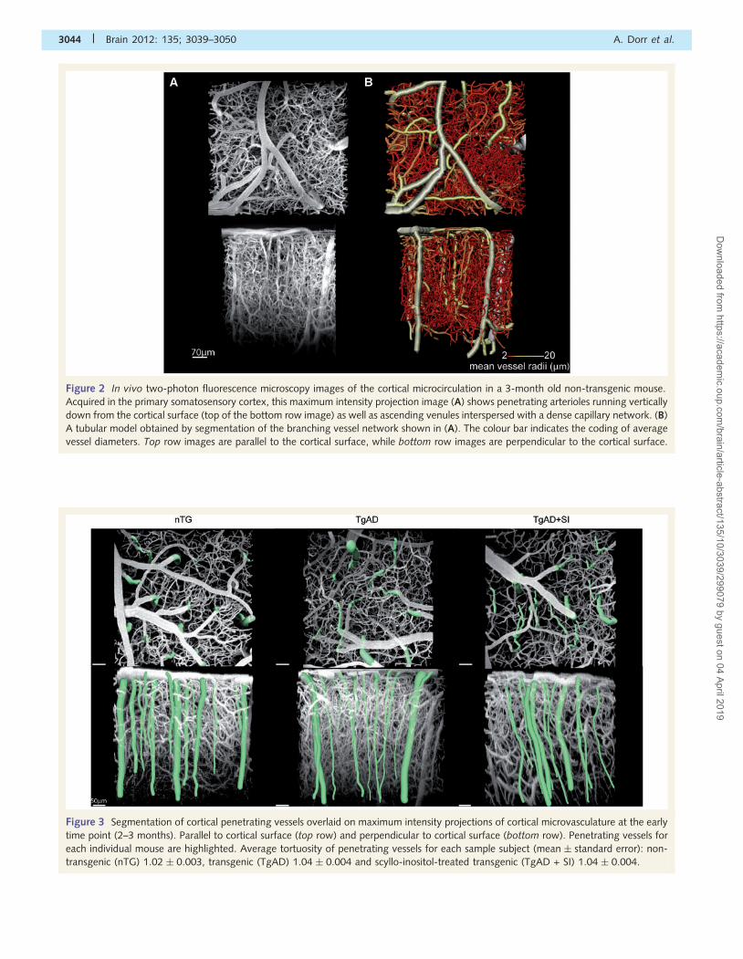
Figure 2 In vivo two-photon fluorescence microscopy images of the cortical microcirculation in a 3-month old non-transgenic mouse.
Acquired in the primary somatosensory cortex, this maximum intensity projection image (A) shows penetrating arterioles running vertically
down from the cortical surface (top of the bottom row image) as well as ascending venules interspersed with a dense capillary network. (B)
A tubular model obtained by segmentation of the branching vessel network shown in (A). The colour bar indicates the coding of average
vessel diameters. Top row images are parallel to the cortical surface, while bottom row images are perpendicular to the cortical surface.
Figure 3 Segmentation of cortical penetrating vessels overlaid on maximum intensity projections of cortical microvasculature at the early
time point (2–3 months). Parallel to cortical surface (top row) and perpendicular to cortical surface (bottom row). Penetrating vessels for
each individual mouse are highlighted. Average tortuosity of penetrating vessels for each sample subject (mean � standard error): non-
transgenic (nTG) 1.02 � 0.003, transgenic (TgAD) 1.04 � 0.004 and scyllo-inositol-treated transgenic (TgAD + SI) 1.04 � 0.004.
3044 | Brain 2012: 135; 3039–3050 A. Dorr et al.
Dow
nloaded from https://academ
ic.oup.com/brain/article-abstract/135/10/3039/299079 by guest on 04 April 2019

Figure 5 Segmentation of cortical penetrating vessels overlaid on maximum intensity projections of cortical microvasculature at the late
time point (6.5–12 months): parallel to cortical surface (top row) and perpendicular to cortical surface (bottom row). Penetrating vessels
for each individual mouse are highlighted. Average tortuosity of penetrating vessels for each sample subject (mean � standard error): non-
transgenic (nTG) 1.03 � 0.003, transgenic (TgAD) 1.10 � 0.006 and scyllo-inositol-treated transgenic (TgAD + SI) 1.03 � 0.005.
Figure 4 Segmentation of cortical penetrating vessels overlaid on maximum intensity projections of cortical microvasculature at the mid
time point (2–3 months): parallel to cortical surface (top row) and perpendicular to cortical surface (bottom row). Penetrating vessels for
each individual mouse are highlighted. Average tortuosity of penetrating vessels for each sample subject (mean � standard error): non-
transgenic (nTG) 1.03 � 0.004, transgenic (TgAD) 1.06 � 0.019 and scyllo-inositol-treated transgenic (TgAD + SI) 1.04�0.004.
Cerebral amyloid angiopathy and microvascular compromise Brain 2012: 135; 3039–3050 | 3045
Dow
nloaded from https://academ
ic.oup.com/brain/article-abstract/135/10/3039/299079 by guest on 04 April 2019

Figure 8 (A) The transit time (in s) across the different groups, at the late stage. The transit time was both longer (P = 0.029) and more
variable in the transgenic (nTg) group (P52.2 �10�16) when compared to non-transgenic (TgAD) group. The scyllo-inositol-treated
transgenic (TgAD + SI) group transit times could not be distinguished from the non-transgenic ones (P = 0.82). (B) The hypercapnia
induced changes in the absolute transit time across the different groups, at the late stage. Whereas the hypercapnia induced transit time
shortening in non-transgenic animals (P = 0.031), it resulted in transit time elongation in transgenic mice (P = 0.0001). (C) The hyper-
capnia induced changes in the absolute transit time in the transgenic group, across time. With age, the transit time in the transgenic group
tended to increase (P = 0.12).
Figure 6 The arteriolar tortuosity as a function of time in non-transgenic (A), transgenic (B) and scyllo-inositol-treated transgenic (C)
animals. Transgenic (nTg) mice had higher arteriolar tortuosity than the non-transgenic (TgAD) mice (P = 0.001), whereas the scyllo-
inositol-treated transgenic (TgAD + SI) mice were indistinguishable from non-transgenic (P = 0.80). The arteriolar tortuosity in transgenic
mice increased with age (P = 0.022): arteriolar curvature of the transgenic was larger than that of non-transgenic at the mid stage
(P = 0.008) and trended toward being greater than non-transgenic arteriolar curvature at the late stage (P = 0.14).
Figure 7 The arteriolar radius as a function of time in non-transgenic (A), transgenic (B) and scyllo-inositol-treated transgenic (C) animals.
Arteriolar radius tended to decrease with age in the transgenic (nTg) (P = 0.11) but not in non-transgenic (TgAD) (P = 0.81) or scyllo-
inositol-treated transgenic (TgAD + SI) (P = 0.74). The arteriolar radius of the transgenic was smaller than that of non-transgenic in the mid
stage (P = 0.02) and trended toward being smaller than the non-transgenic group’s radius in the late stage (P = 0.09).
3046 | Brain 2012: 135; 3039–3050 A. Dorr et al.
Dow
nloaded from https://academ
ic.oup.com/brain/article-abstract/135/10/3039/299079 by guest on 04 April 2019

whereas scyllo-inositol-treated transgenic mice transit times were
not distinguishable from those of non-transgenic mice (P = 0.82).
The transit time dispersion, taken to reflect the efficiency of the
microvascular network (lower dispersion being a putative marker
of a more efficient vascular network), was higher in the transgenic
group, relative to that in non-transgenic, during both air breathing
(P = 0.039) and hypercapnia (P = 0.030). In contrast, scyllo-inosi-
tol-treated transgenic transit time dispersion was not distinguish-
able from non-transgenic during either air breathing (P = 0.8787)
or hypercapnia (P = 0.6597). These data suggest that the trans-
genic vasculature is functionally inefficient as reflected in the larger
dispersion of transit times in the transgenic mice. Furthermore,
prevention of amyloid-b aggregation and deposition by scyllo-ino-
sitol is shown to successfully resolve this impairment.
The hypercapnic challenge had a significant effect on vascular
transit times across the three groups (P = 0.0003), as shown in
Fig. 8B. In the non-transgenic group, hypercapnia induced the
expected transit time shortening (P = 0.031) as a result of
carbon-dioxide-induced vessel dilatation. In contrast, the hyper-
capnic challenge resulted in transit time elongation in the
transgenic group (P = 0.0001). Figure 8C displays the hypercap-
nia-induced changes in the transit time in the transgenic group,
across time. There was a strong trend toward carbon dioxide-
induced transit time increase with amyloid-b accumulation in the
transgenic animals (P = 0.12). Given the previously discussed
changes in arteriolar, but not venular vascular networks and the
preferential accumulation of amyloid-b on the arterioles over
venules (Weller and Nicoll, 2003; Shin et al., 2007; Serrano-
Pozo et al., 2011; Fig. 9), we hypothesize that this paradoxical
response to hypercapnia resulted from compromised carbon-
dioxide-induced dilatation of the supply vasculature in the pre-
sence of preserved carbon-dioxide-elicited venous dilatation; the
increase in the drainer calibre alone thus elicited elongation of the
vascular transit time upon hypercapnia, providing further evidence
of the profound compromise of vascular function in this transgenic
mouse model.
DiscussionIn recent years, the vascular compromise in Alzheimer’s disease
dementia has garnered increased research attention. Amyloid-bpeptides have been demonstrated to elicit vasoconstriction and
cause vascular degeneration, elevating the risk of haemorrhage
and stroke (Perlmutter, 1994; Miao et al., 2005; Tian et al.,
2006). While the significance of vascular dysfunction has thus
become widely appreciated, the pathogenesis of cerebral amyloid
angiopathy and its implications have yet to be fully elucidated. In
the present work, we examined, in situ, the changes in cortical
penetrating vessel morphology and function with progressive
Figure 9 Maximum intensity projection image of the cortical microvasculature in a 9.5-month-old transgenic mouse. The autofluores-
cence signal is shown in blue (false coloured), with the intravascular fluorophore signal displayed in red. Cerebral amyloid angiopathy
appears as striped rings around the arterioles (yellow arrow); this ring pattern is not present around the venules (green arrow). The vessels
have been categorized based on the bolus arrival time estimates from the bolus tracking data, venules showing delayed bolus arrivals
relative to arterioles.
Cerebral amyloid angiopathy and microvascular compromise Brain 2012: 135; 3039–3050 | 3047
Dow
nloaded from https://academ
ic.oup.com/brain/article-abstract/135/10/3039/299079 by guest on 04 April 2019

accumulation of amyloid-b in the TgCRND8 mouse model of
Alzheimer’s disease. Under physiological conditions, cortical pene-
trating arterioles have been shown to be key for the healthy
supply of the cortex with metabolites (Nishimura et al., 2007)
and clearance of toxins, such as amyloid-b, from the brain
(Nicoll et al., 2004). In TgCRND8 mice, we found the progressive
accumulation of amyloid-b42 and particularly amyloid-b40 asso-
ciated with transgene-dependent increase in the tortuosity of the
penetrating arterioles, with the largest change observed in mid-
stage of the disease followed by the saturation of this effect in the
later stage. At the same time, the intraluminal arteriolar radii have
been shown to progressively decrease, probably exacerbating the
impairment of the normal cortical blood supply. These effect sizes
are consistent with the morphological changes reported in the
quantitative examinations of Alzheimer’s disease patient microvas-
cular beds (Fischer et al., 1990; Thore et al., 2007). In concert
with earlier reports of preferential deposition of amyloid-b on
arteriolar over venular walls (Weller and Nicoll, 2003; Shin
et al., 2007; Serrano-Pozo et al., 2011), we observed neither
tortuosity nor calibre changes in the venules. Moreover, we
found these morphological changes accompanied by progressive
compromise of the microvascular function. The latter was reflected
in a greater variability in vascular transit times across the cortical
microvasculature of the transgenic mice, longer vascular transit
times and impaired response of the microvascular bed to the
global vasodilatory challenge posed by elevated carbon dioxide
content in the inspired gas mixture. Transgenic mice not only
showed a progressive compromise in hypercapnia-induced vascu-
lar reactivity, but also exhibited transit time elongation rather than
shortening in response to hypercapnia in the late stage of the
disease. We believe this paradoxical hypercapnic response results
from differential effects of the disease process on arteriolar versus
venular side of the microvascular network, with largely preserved
carbon dioxide-induced venular vasodilatation accompanying a
profoundly compromised arteriolar dilatation.
We have previously shown that scyllo-inositol, administered
post-weaning, rescues the age-progressive accumulation of amy-
loid-b40/42 in both parenchyma and on blood vessels in this trans-
genic model (McLaurin et al., 2006). We presently found that
administration of scyllo-inositol resolved the vascular morphologi-
cal and functional changes observed in the transgenic mice. The
scyllo-inositol-treated TgCRND8 mice were thus found to be indis-
tinguishable from their non-transgenic littermates with respect to
arteriolar tortuosity, arteriolar calibre, transit time dispersion and
vascular reactivity to hypercapnia. These data thus provide
evidence that the vascular impairment in the TgCRND8 mice is
consequent to amyloid-b accumulation as both soluble oligomers
and insoluble deposits and that these structural and functional
compromises of the cortical microvasculature may be resolved by
preventing amyloid-b oligomerization and/or fibrillization. Since
scyllo-inositol decreases both soluble and insoluble amyloid-b
levels, the compromise in vascular function may be related not
only to the development of cerebral amyloid angiopathy but
also to amyloid-b-induced vasoactivity, endothelial and pericyte
damage or oxidative stress (Thomas et al., 1996; Verbeek et al.,
1997; Butterfield, 2002; Carrano et al., 2011).
Of note, in contrast to previous reports (Deane et al., 2004; Bell
et al., 2009), we did not find a disease-associated decrease in
lipoprotein receptor-related protein 1 expression in isolated cortical
blood vessels nor changes in plasma amyloid-b levels, suggesting
that brain to plasma efflux pathways are unaffected in this mouse
model. Hence, accumulation of amyloid-b and development of
cerebral amyloid angiopathy observed in this model may result
from decreased microvascular function and motive force for effec-
tive perivascular amyloid-b drainage (Weller et al., 2009). In addi-
tion, in light of the recent anti-amyloid-b immunization studies
that reported strong associations between reduced parenchymal
plaque load and increased cerebral amyloid angiopathy severity
and brain microhaemorrhages (Boche et al., 2008), we inspected
the two photon fluorescent data on the microvascular architecture
in the scyllo-inositol-treated animals at each stage for indication of
fluorescent dextran extravasation. We did not, however, observe
any such occurrences, arguing against an increase in bleeding from
cortical microvessels subsequent to scyllo-inositol treatment.
Overall, this work reports on the implications of cerebral amy-
loid angiopathy on the morphology and function of brain micro-
vessels over the course of disease progression, measured by
in vivo two photon fluorescence microscopy so as to avoid con-
founds from tissue processing techniques. Our findings shed new
light on the correlation between amyloid-b accumulation and
microvascular impairment in the Alzheimer’s disease brain, under-
score the importance of cerebral amyloid angiopathy in the aetiol-
ogy of Alzheimer’s disease and demonstrate the resolution of the
progressive structural and functional impairment of the brain
microvasculature by prevention of amyloid-b aggregation and
deposition. The translation of our results remains to be determined
as mouse models of Alzheimer’s disease do not recapitulate all
aspects of the disease process nor the normal ageing-associated
changes seen in humans.
AcknowledgementsWe thank Jianfei He and Kun Zhang for the surgical work as well
as Lynsie Thomason for technical support.
FundingSunnybrook Research Institute and the Canadian Institutes of
Health Research (MOP-37857 to J.M., MOP-94376 to J.M.,
C.A.H., and B.S. and MOP-10246 to J.M. and B.S.). A.D., B.S.,
L.V.C., M.E.B., A.Y.L., K.M., C.A.H., and B.S. declare no conflict
of interest. J.M. is named inventor on patents and patent applica-
tions relating to scyllo-inositol.
Supplementary materialSupplementary material is available at Brain online.
3048 | Brain 2012: 135; 3039–3050 A. Dorr et al.
Dow
nloaded from https://academ
ic.oup.com/brain/article-abstract/135/10/3039/299079 by guest on 04 April 2019

ReferencesBell RD, Deane R, Chow N, Long X, Sagare A, Singh I, et al. SRF and
myocardin regulate LRP-mediated amyloid-b clearance in brain vascu-
lar cells. Nat Cell Biol 2009; 11: 143–53.Boche D, Zotova E, Weller RO, Love S, Neal JW, Pickering RM, et al.
Consequence of A immunization on the vasculature of human
Alzheimer’s disease brain. Brain 2008; 131: 3299–310.
Bullitt E, Gerig G, Pizer SM, Lin W, Aylward SR. Measuring tortuosity of
the intracerebral vasculature from MRA images. IEEE Trans Med
Imaging 2003; 22: 1163–71.
Butterfield DA. Amyloid-b-peptide (1–42)-induced oxidative stress
and neurotoxicity: implications for neurodegeneration in
Alzheimer’s disease brain. A review. Free Radic Res 2002; 36:
1307–13.
Carrano A, Hoozemans JJM, van der Vies SM, Rozemuller AJM, van
Horssen J, de Vries HE. Amyloid beta induces oxidative stress-
mediated blood–brain barrier changes in capillary amyloid angiopathy.
Antioxid Redox Signal 2011; 15: 1167–78.
Chishti MA, Yang DS, Janus C, Phinney AL, Horne P, Pearson J, et al.
Early-onset amyloid deposition and cognitive deficits in transgenic mice
expressing a double mutant form of amyloid precursor protein 695. J
Biol Chem 2001; 276: 21562–70.Choi JK, Carreras I, Dedeoglu A, Jenkins BG. Consequence of Ab immu-
nization on the vasculature of human Alzheimer’s disease brain.
Neuropharmacology 2010; 59: 353–7.
Christie R, Yamada M, Moskowitz M, Hyman B. Structural and func-
tional disruption of vascular smooth muscle cells in a transgenic mouse
model of amyloid angiopathy. Am J Pathol 2001; 158: 1065–71.
Citron M. Alzheimer’s disease: treatments in discovery and development.
Nat Neurosci 2002; 5 (Suppl): 1055–7.
Collie A, Maruff P. The neuropsychology of preclinical Alzheimer’s dis-
ease and mild cognitive impairment. Neurosci Biobehav Rev 2000; 24:
365–74.Deane R, Wu Z, Sagare A, Davis J, Yan SD, Hamm K, et al. LRP/
Amyloid-b-peptide interaction mediates differential brain efflux of A-
b-isoforms. Neuron 2004; 43: 333–44.
Duvernoy HM, Delon S, Vannson JL. Cortical blood vessels of the human
brain. Brain Res Bull 1981; 7: 519–79.
Fenili D, Brown ME, Rappaport RV, McLaurin J Properties of
scyllo-inositol as a therapeutic treatment of AD-like pathology. J Mol
Med 2007; 85: 603–12.Fenili D, Weng YQ, Aubert I, Nitz M, McLaurin J. Sodium/myo-inositol
transporters: an examination of substrate transport requirements and
regional brain expression in the TgCRND8 mouse model of
Alzheimer’s disease. PLoS One 2011; 6: e24032.
Fischer VW, Siddiqi A, Yusufaly Y. Altered angioarchitecture in selected
areas of brains with Alzheimer’s disease. Acta Neuropathol 1990; 79:
672–9.Girouard H, Iadecola C. Neurovascular coupling in the normal brain and
in hypertension, stroke, and Alzheimer disease. J Appl Physiol 2006;
100: 328–35.
Greenberg SM. Cerebral amyloid angiopathy and vessel dysfunction.
Cerebrovasc Dis 2002; 13 (Suppl 2): 42–47.
Han BH, Zhou M-l, Abousaleh F, Brendza RP, Dietrich HH,
Koenigsknecht-Talboo J, et al. Cerebrovascular dysfunction in amyloid
precursor protein transgenic mice: contribution of soluble and insoluble
amyloid-b peptide, partial restoration via �-secretase inhibition. J
Neurosci 2008; 28: 13542–50.
Hawkes CA, McLaurin J. Selective targeting of perivascular macrophages
for clearance of beta-amyloid in cerebral amyloid angiopathy. PNAS
2009; 106: 1261–6.
Herzig MC, Van Nostrand WE, Jucker M. Mechanism of cerebral
beta-amyloid angiopathy: murine and cellular models. Brain Pathol
2006; 6: 40–54.
Holland CM, Smith EE, Csapo I, Gurol ME, Brylka DA, Killiany RJ, et al.
Spatial distribution of white-matter hyperintensities in Alzheimer
disease, cerebral amyloid angiopathy, and healthy aging. Stroke
2008; 39: 1127–33.
Hunter JM, Kwan J, Malek-Ahmadi M, Maarouf CL, Kokjohn TA,
Belden C, et al. Morphological and pathological evolution of the
brain microcirculation in aging and Alzheimer’s disease. Plos ONE
2012; 7: e36893.
Kalaria RN. Cerebral vessels in ageing and Alzheimer’s disease.
Pharmacol Ther 1996; 72: 193–214.
Kalaria RN. Small vessel disease and Alzheimer’s dementia: pathological
considerations. Cerebrovasc Dis 2002; 13 (Suppl2): 48–52.
Kasischke KA, Lambert EM, Panepento B, Sun A, Gelbard HA,
Burgess RW, et al. Two-photon NADH imaging exposes boundaries
of oxygen diffusion in cortical vascular supply regions. J Cereb Blood
Flow Metab 2011; 31: 68–81.
Kershaw L, Cheng H. A general dual-bolus approach for quantitative
DCE-MRI. Magn Reson Imaging 2011; 29: 160–6.
Kim J, Leira E, Callison R, Ludwig B, Moritani T, Magnotta V, et al.
Toward fully automated processing of dynamic susceptibility contrast
perfusion MRI for acute ischemic cerebral stroke. Comput Methods
Programs Biomed 2010; 98: 204–13.
Kwan AC, Duff K, Gouras GK, Webb WW. Optical visualization
of Alzheimer’s pathology via multiphoton-excited intrinsic fluores-
cence and second harmonic generation. Opt Express 2009; 17:
3679–89.
LeVine H III. Small molecule inhibitors of Abeta assembly. Amyloid 2007;
14: 185–97.
Lindvere L, Dorr A, Stefanovic B. Two-photon fluorescence microscopy
of cerebral hemodynamics. Cold Spring Harb Protoc 2010 (9):pdb.
prot5494.
Ma K, Mount HT, McLaurin J. Region-specific distribution of b-amyloid
peptide and cytokine expression in TgCRND8 mice. Neurosci Lett
2011; 492: 5–10.
McLaurin J, Golomb R, Jurewicz A, Antel JP, Fraser PE. Inositol stereo-
isomers stabilize an oligomeric aggregate of Alzheimer amyloid beta
peptide and inhibit abeta-induced toxicity. J Biol Chem 2000; 275:
18495–502.
McLaurin J, Kierstead ME, Brown ME, Hawkes CA, Lambermon MH,
Phinney AL, et al. Cyclohexanehexol inhibitors of Abeta aggregation
prevent and reverse Alzheimer phenotype in a mouse model. Nat Med
2006; 12: 801–8.
Miao J, Xu F, Davis J, Otte-Holler I, Verbeek MM, Van Nostrand WE.
Cerebral microvascular amyloid beta protein deposition induces vascu-
lar degeneration and neuroinflammation in transgenic mice expressing
human vasculotropic mutant amyloid beta precursor protein. Am J
Pathol 2005; 167: 505–15.
Nicolakakis N, Hamel E. Neurovascular function in Alzheimer’s disease
patients and experimental models. J Cereb Blood Flow Metab 2011;
31: 1354–70.
Nicoll JA, Yamada M, Frackowiak J, Mazur-Kolecka B, Weller RO.
Cerebral amyloid angiopathy plays a direct role in the pathogenesis
of Alzheimer’s disease. Pro-CAA position statement. Neurobiol Aging
2004; 25: 589–97.
Nishimura N, Schaffer CB, Friedman B, Lyden PD, Kleinfeld D.
Penetrating arterioles are a bottleneck in the perfusion of neocortex.
Proc Natl Acad Sci USA 2007; 104: 365–70.
Paul J, Strickland S, Melchor JP. Fibrin deposition accelerates neurovas-
cular damage and neuroinflammation in mouse models of Alzheimer’s
disease. J Exp Med 2007; 204: 1999–2008.
Perlmutter LS. Microvascular pathology and vascular basement mem-
brane components in Alzheimer’s disease. Mol Neurobiol 1994; 9:
33–40.Pfeifer LA, White LR, Ross GW, Petrovitch H, Launer LJ. Cerebral amyl-
oid angiopathy and cognitive function: The HAAS autopsy study.
Neurology 2002; 58: 1629–34.
Pinheiro JC, Bates DM. Mixed-effects models in S and S-PLUS. New
York: Springer; 2000.
Querfurth HW, LaFerla FM. Alzheimer’s disease. N Engl J Med 2010;
362: 329–44.
Cerebral amyloid angiopathy and microvascular compromise Brain 2012: 135; 3039–3050 | 3049
Dow
nloaded from https://academ
ic.oup.com/brain/article-abstract/135/10/3039/299079 by guest on 04 April 2019

Scharrer E. Arteries and veins in the mammalian brain. Anat Rec 1940;78: 173–96.
Serrano-Pozo A, Frosch MP, Masliah E, Hyman BT. Neuropathological
alterations in Alzheimer disease. Cold Spring Harb Perspect Med 2011;
1: a006189.Shin HK, Jones PB, Garcia-Alloza M, Borrelli L, Greenberg SM,
Bacskai BJ, et al. Age-dependent cerebrovascular dysfunction in a
transgenic mouse model of cerebral amyloid angiopathy. Brain 2007;
130 (Pt 9): 2310–19.Stefanovic B, Hutchinson E, Yakovleva V, Schram V, Russell JT,
Belluscio L, et al. Functional reactivity of cerebral capillaries. J Cereb
Blood Flow Metab 2008; 28: 961–72.Takano T, Han X, Deane R, Zlokovic B, Nedergaard M. Two-photon
imaging of astrocytic Ca2 + signaling and the microvasculature in ex-
perimental mice models of Alzheimer’s disease. Ann NY Acad Sci
2007; 1097: 40–50.Thomas T, Thomas G, McLendon C, Sutton T, Mullan M.
b-amyloid-mediated vasoactivity and vascular endothelial damage.
Nature 1996; 380: 168–71.
Thore CR, Anstrom JA, Moody DM, Challa VR, Marion MC, Brown WR.Morphometric analysis of arteriolar tortuosity in human cerebral white
matter of preterm, young, and aged subjects. J Neuropathol ExpNeurol 2007; 66: 337–45.
Tian J, Shi J, Smallman R, Iwatsubo T, Mann DM. Relationships in
Alzheimer’s disease between the extent of Abeta deposition in cerebral
blood vessel walls, as cerebral amyloid angiopathy, and the amount ofcerebrovascular smooth muscle cells and collagen. Neuropathol Appl
Neurobiol 2006; 32: 332–40.
Townsend M, Cleary JP, Mehta T, Hofmeister J, Lesne S, O’Hare E, et al.
Orally available compound prevents deficits in memory caused bythe Alzheimer amyloid-beta oligomers. Ann Neurol 2006; 60:
668–76.
Verbeek MM, De Waal RM, Schipper JJ, Van Nostrand WE. Rapid de-generation of cultured human brain pericytes by amyloid beta protein.
J Neurochem 1997; 68: 1135–41.
Weller RO, Nicoll JA. Cerebral amyloid angiopathy: pathogenesis and
effects on the ageing and Alzheimer brain. Neurol Res 2003; 25:611–6.
Weller RO, Preston SD, Subash M, Carare RO. Cerebral amyl-
oid angiopathy in the aetiology and immunotherapy of
Alzheimer disease. Alzheimer’s Res Ther 2009; 1: 6(doi:10.1186/alzrt6).
3050 | Brain 2012: 135; 3039–3050 A. Dorr et al.
Dow
nloaded from https://academ
ic.oup.com/brain/article-abstract/135/10/3039/299079 by guest on 04 April 2019