anion binding vs deprotonation in colorimetric ... filethis journal is (c) ... anion binding vs...
TRANSCRIPT

Supplementary Material (ESI) for Chemical Communications This journal is (c) The Royal Society of Chemistry 2006
Anion binding vs deprotonation in colorimetric pyrrolylamido(thio)urea based anion sensors
Louise S. Evans, Philip A. Gale*, Mark E. Light and Roberto Quesada* School of Chemistry, University of Southampton, Southampton, UK SO17 1BJ. Fax: +44 (0)23 8059 6805; Tel: +44 (0)23 8059 3332; E-mail: [email protected]; [email protected]
Supplementary information
Experimental procedures
5-Methyl-3,4-diphenyl-1H-pyrrole-2 carbohydrazide 6.
NMe
Ph Ph
O
NHNH2H
5-Methyl-3,4-diphenyl-1H-pyrrole-2 carboxylic acid ethyl ester (1g, 3.46mmol) was
added to a large excess of hydrazine hydrate (5mL, 100eq). To this was then added
enough ethanol to produce dissolution. The reaction was heated to reflux for 48 hrs,
and then allowed to cool to room temperature. Water was added inducing
precipitation of the product. This was collected by filtration, washed with water (3 x
10mL) and dried. A white crystalline solid was produced, 717mg, 71% yield.
Solubility precluded the acquisition of a 13C NMR spectrum, however the structure
was confirmed by X-ray crystallography (Table S1).
1H (CDCl3): δ 9.67 (br. s, 1H, pyrrole NH), δ 7.26 – 6.91 (m, 10H, Ar. H), δ 6.62 (br.
s, 1H, NH), δ 3.79 (br. s, 2H, NH2), δ 2.29 (s, 3H, CH3).
MS (ES+): 605.1 (2M + Na+)

Supplementary Material (ESI) for Chemical Communications This journal is (c) The Royal Society of Chemistry 2006
Table S1. Crystal data and structure refinement details for compound 6.
Empirical formula C18H17N3O Formula weight 291.35 Temperature 120(2) K Wavelength 0.71073 Å Crystal system Monoclinic Space group P21/n Unit cell dimensions a = 10.990(4) Å b = 18.366(9) Å β = 96.53(2)° c = 15.123(4) Å Volume 3033(2) Å3 Z 8 (2 molecules in the asymmetric unit) Density (calculated) 1.276 Mg / m3 Absorption coefficient 0.081 mm−1 F(000) 1232 Crystal Block; Colourless Crystal size 0.2 × 0.1 × 0.1 mm3 θ range for data collection 2.93 − 27.12° Index ranges −13 ≤ h ≤ 14, −22 ≤ k ≤ 23, −19 ≤ l ≤ 19 Reflections collected 27627 Independent reflections 6610 [Rint = 0.1582] Completeness to θ = 27.12° 98.5 % Absorption correction Semi−empirical from equivalents Max. and min. transmission 0.9919 and 0.9839 Refinement method Full-matrix least-squares on F2 Data / restraints / parameters 6610 / 0 / 432 Goodness-of-fit on F2 0.941 Final R indices [F2 > 2σ(F2)] R1 = 0.0685, wR2 = 0.1249 R indices (all data) R1 = 0.2330, wR2 = 0.1765 Extinction coefficient 0.0043(7) Largest diff. peak and hole 0.267 and −0.335 e Å−3
Diffractometer: Nonius KappaCCD area detector (φ scans and ω scans to fill asymmetric unit ). Cell determination: DirAx (Duisenberg, A.J.M.(1992). J. Appl. Cryst. 25, 92-96.) Data collection: Collect (Collect: Data collection software, R. Hooft, Nonius B.V., 1998). Data reduction and cell refinement: Denzo (Z. Otwinowski & W. Minor, Methods in Enzymology (1997) Vol. 276: Macromolecular Crystallography, part A, pp. 307−326; C. W. Carter, Jr. & R. M. Sweet, Eds., Academic Press). Absorption correction: Sheldrick, G. M. SADABS - Bruker Nonius area detector scaling and absorption correction - V2.10 Structure solution: SHELXS97 (G. M. Sheldrick, Acta Cryst. (1990) A46 467−473). Structure refinement: SHELXL97 (G. M. Sheldrick (1997), University of Göttingen, Germany). Graphics: Cameron - A Molecular Graphics Package. (D. M. Watkin, L. Pearce and C. K. Prout, Chemical Crystallography Laboratory, University of Oxford, 1993).

Supplementary Material (ESI) for Chemical Communications This journal is (c) The Royal Society of Chemistry 2006
2-(5-Methyl-3,4-diphenyl-1H-pyrrole-2-carbonyl)-N-phenylhydrazinecarboxamide 1.
NMe
Ph Ph
NH
OHHN
O
HNPh
Phenyl isocyanate (76µL, 0.70mmol) was dissolved in chloroform, and the solution
degassed for 10 mins. Then, 5-methyl-3,4-diphenyl-1H-pyrrole-2 carbohydrazide
(203mg, 0.70mmol) was added, and the reaction stirred at room temperature for 24
hours. A precipitate is formed, which was collected by filtration, washed with DCM
(3 x 10mL) and dried. A white solid was produced, 137mg, yield of 46%.
1H (DMSO): δ 11.67 (s, 1H, NH), δ 8.72 (s, 1H, NH), δ 8.30 (br. s, 1H, NH), δ 8.10
(s, 1H, NH), δ 7.48 (m, 2H, Ar. H), δ 7.25 (m, 10H, Ar. H), δ 7.04 (m, 2H, Ar. H), δ
2.31 (s, 3H, CH3). 13C (DMSO): δ 180.5, 160.6, 138.9, 134.8, 134.6, 130.7, 129.8, 128.9, 128.2, 127.9,
127.0, 126.6, 125.6, 124.9, 122.4, 118.7, 11.7.
MS ES+: 433.2 (M + Na+), 843.4 (2M + Na+), 1253.8 (3M + Na+). ES-: 409.3 (M - H).
Microanalysis: Calc. for C25H22O2N4.H2O: C: 70.08; H: 5.65; N: 13.08. Found C:
70.11; H: 5.20; N: 13.11.
2-(5-methyl-3,4-diphenyl-1H-pyrrole-2-carbonyl)-N-(4-nitrophenyl)
hydrazinecarboxamide 2.
NMe
Ph Ph
NH
OHHN
O
HN
NO2
4-Nitrophenyl isocyanate (115mg, 0.70mmol) was dissolved in chloroform (10mL)
and degassed for 10 mins. Then, 5-methyl-3,4-diphenyl-1H-pyrrole-2 carbohydrazide
(204mg, 0.70mmol) was added and the reaction stirred at room temperature for 24

Supplementary Material (ESI) for Chemical Communications This journal is (c) The Royal Society of Chemistry 2006
hours. A precipitate formed, which was collected by filtration, washed with DCM (3
x 10mL) and dried. A cream powder was produced, 280mg, 87% yield.
1H (DMSO): δ 11.62 (s, 1H, NH), δ 9.43 (br. s, 1H, NH), δ 8.43 (br. s, 1H, NH), δ
8.33 (br. s, 1H, NH), δ 8.17 (d, 2H, Ar. H, J=9.15Hz), δ 7.71 (d, 2H, Ar. H,
J=9.15Hz), δ 7.18 (m, 8H, Ar. H), δ 6.99 (m, 2H, Ar. H), δ 2.27 (s, 3H, CH3). 13C (DMSO): δ 160.9, 146.2, 141.1, 134.8, 134.7, 130.6, 129.8, 129.0, 127.8, 126.8,
126.6, 125.6, 125.0, 122.3, 118.6, 117.7, 11.7.
MS ES+: 478.2 (M + Na+), 933.5 (2M + Na+). ES-: 454.3 (M - H), 909.5 (2M - H).
Microanalysis: Calc. for: C25H21N5O4: C: 65.93; H: 4.65; N: 15.37. Found: C: 65.54;
H: 4.61; N: 15.05.
2-(5-methyl-3,4-diphenyl-1H-pyrrole-2-carbonyl)-N-phenylhydrazinecarbothioamide
3:
NMe
Ph Ph
NH
OHHN
S
HNPh
The pyrrole carbonyl hydrazine (200mg, 0.686mmol), was added to a degassed
solution of phenylisothiocyanate (82µL, 0.69 mmol) in chloroform (10mL). The
reaction was stirred at room temperature for 72 hours after which time a white
precipitate had formed. This was collected by filtration, washed with DCM and dried
to produce 214mg of product, 73% yield.
1H (DMSO-d6): δ 11.60 (s, 1H, pyrrole NH), δ 9.62 (br. s, 1H, NH), δ 9.52 (br. s, 1H,
NH), δ 8.86 (br.s, 1H, NH), δ 7.46 (d, 2H, CH, J = 7.92Hz), δ 7.33 (m, 2H, Ar. H), δ
7.19 (m, 9H, Ar. H), δ 6.97 (d, 2H, Ar. H, J = 6.78), δ 2.26 (s, 3H, CH3). 13C (DMSO-d6): δ 180.5, 160.6, 138.9, 134.8, 134.6, 130.7, 129.8, 128.9, 128.2, 127.9,
127.0, 126.6, 125.6, 124.9, 122.4, 118.7, 11.7.
Microanalysis: Calc. for: C25H22N4OS: C: 70.40; H: 5.20; N: 13.14. Found: C: 70.64;
H: 5.31; N: 13.05.

Supplementary Material (ESI) for Chemical Communications This journal is (c) The Royal Society of Chemistry 2006
2-(5-methyl-3,4-diphenyl-1H-pyrrole-2-carbonyl)-N-(4-nitrophenyl)
hydrazinecarbothioamide 4:
NMe
Ph Ph
NH
OHHN
S
HN
NO2
Pyrrole carbonyl hydrazine (400mg, 1.37mmol) was added to a degassed solution of
4-nitrophenylisothiocyanate (248mg, 1.38mmol) in chloroform (15mL). The reaction
was stirred at room temperature for 24 hours during which time a yellow precipitate
formed. The solution was filtered and the precipitate washed with DCM and dried to
produce 550mg of product, 85% yield.
1H (Acetone-d6): δ 11.92 (s, 1H, pyrrole NH), δ 9.60 (s, 1H, NH), δ 9.02 (s, br, 1H,
NH), δ 8.09 (m, 5H, CH and NH), δ 7.21 (m, 10H, CH), δ 2.37 (s, 3H, CH3). 13C (DMSO): δ 161.4, 146.3, 144.6, 135.8, 135.7, 131.7, 131.5, 131.0, 129.7, 128.8,
128.5, 128.0, 126.7, 124.8, 124.5, 123.3, 119.7, 12.0.
MSES+: 471.9 (M+), 535.0 (M + Na+ + CH3CN), 965.1 (2M + Na+).
Microanalysis: Calc. for: C25H21N5O3S: C: 63.68; H: 4.49; N: 14.85. Found: C: 63.66;
H: 4.47; N: 14.86.

Supplementary Material (ESI) for Chemical Communications This journal is (c) The Royal Society of Chemistry 2006
Figure S1 1H NMR spectra of the TBA salt of deprotonated compound 4 in DMSO-d6 .

Supplementary Material (ESI) for Chemical Communications This journal is (c) The Royal Society of Chemistry 2006
Figure S2 a) UV–vis absorption spectrophotometric titration of compound 2 with
TBA fluoride in DMSO at 25 ºC. b) Variation of absorbance at 390 nm versus
concentration of anion. The trend line is the result of the non linear least-square fit of
the experimental data according to A-A0=B×[F-]/(1+(K×[F-])

Supplementary Material (ESI) for Chemical Communications This journal is (c) The Royal Society of Chemistry 2006
Figure S3 a) UV–vis absorption spectrophotometric titration of compound 2 with
TBA acetate in DMSO at 25 ºC. b) Variation of absorbance at 390 nm versus
concentration of anion. The trend line is the result of the non linear least-square fit of
the experimental data according to A-A0=B×[G-]/(1+(K×[G-])
Supplementary Material (ESI) for Chemical Communications This journal is (c) The Royal Society of Chemistry 2006
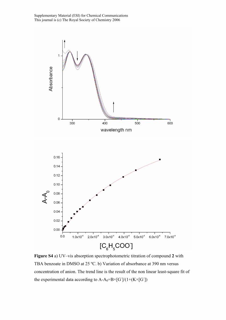
Figure S4 a) UV–vis absorption spectrophotometric titration of compound 2 with
TBA benzoate in DMSO at 25 ºC. b) Variation of absorbance at 390 nm versus
concentration of anion. The trend line is the result of the non linear least-square fit of
the experimental data according to A-A0=B×[G-]/(1+(K×[G-])

Supplementary Material (ESI) for Chemical Communications This journal is (c) The Royal Society of Chemistry 2006
Figure S5 a) UV–vis absorption spectrophotometric titration of compound 2 with
TBA dihydrogenphosphate in DMSO at 25 ºC. b) Variation of absorbance at 390 nm
versus concentration of anion. The trend line is the result of the non linear least-
square fit of the experimental data according to A-A0=B×[G-]/(1+(K×[G-])
Supplementary Material (ESI) for Chemical Communications This journal is (c) The Royal Society of Chemistry 2006
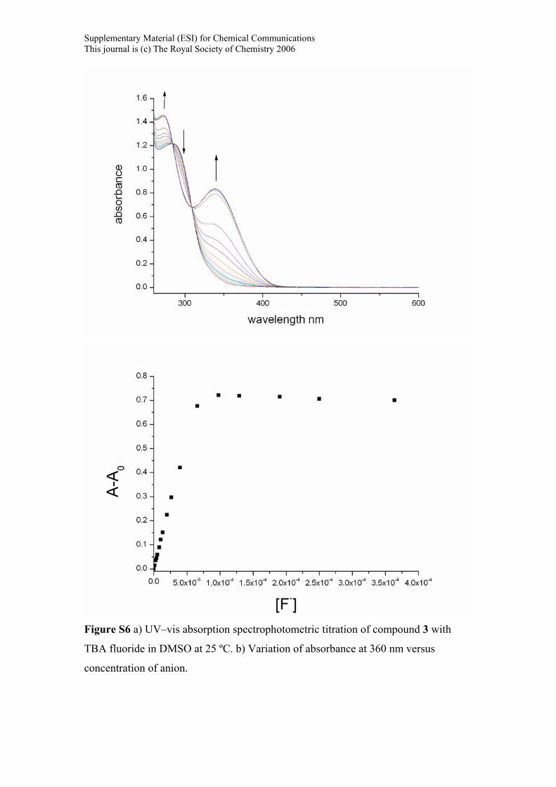
Figure S6 a) UV–vis absorption spectrophotometric titration of compound 3 with
TBA fluoride in DMSO at 25 ºC. b) Variation of absorbance at 360 nm versus
concentration of anion.

Supplementary Material (ESI) for Chemical Communications This journal is (c) The Royal Society of Chemistry 2006
Figure S7 a) UV–vis absorption spectrophotometric titration of compound 3 with
TBA dihydrogenphosphate in DMSO at 25 ºC. b) Variation of absorbance at 360 nm
versus concentration of anion.

Supplementary Material (ESI) for Chemical Communications This journal is (c) The Royal Society of Chemistry 2006
Figure S8 a) UV–vis absorption spectrophotometric titration of compound 4 with
TBA fluoride in DMSO at 25 ºC. b) Variation of absorbance at 360 nm versus
equivalents of fluoride.

Supplementary Material (ESI) for Chemical Communications This journal is (c) The Royal Society of Chemistry 2006
Figure S9 a) UV–vis absorption spectrophotometric titration of compound 4 with
TBA acetate in DMSO at 25 ºC. b) Variation of absorbance at 450 nm versus
equivalents of acetate.

Supplementary Material (ESI) for Chemical Communications This journal is (c) The Royal Society of Chemistry 2006
Figure S10 a) UV–vis absorption spectrophotometric titration of compound 4 with
TBA acetate in DMSO/ water 9:1 at 25 ºC. b) Variation of absorbance at 450 nm
versus equivalents of acetate.

Supplementary Material (ESI) for Chemical Communications This journal is (c) The Royal Society of Chemistry 2006
Figure S11 Stack plot of 1H NMR spectra of compound 2 in the presence of increasing amounts of TBAF recorded in DMSO-d6 .
Figure S12 Stack plot of 1H NMR spectra of compound 4 in the presence of increasing amounts of TBA Benzoate recorded in DMSO-d6 .

Supplementary Material (ESI) for Chemical Communications This journal is (c) The Royal Society of Chemistry 2006
Figure S13 X-Ray crystal structure of compound 2. Thermal ellipsoids are drawn at the 50% probability level.
Figure S14 X-Ray crystal structure of tetrabutylammonium (4-H+)-. Thermal ellipsoids are drawn at the 35% probability level. From the asymmetric unit one anion and the tetrabutylammonium cation in the general position are shown.