ap biology eukaryotic cell components part 2. mitochondria (nicknamed the “power house”)...
DESCRIPTION
MitochondriaTRANSCRIPT
AP Biology
Eukaryotic Cell ComponentsPart 2
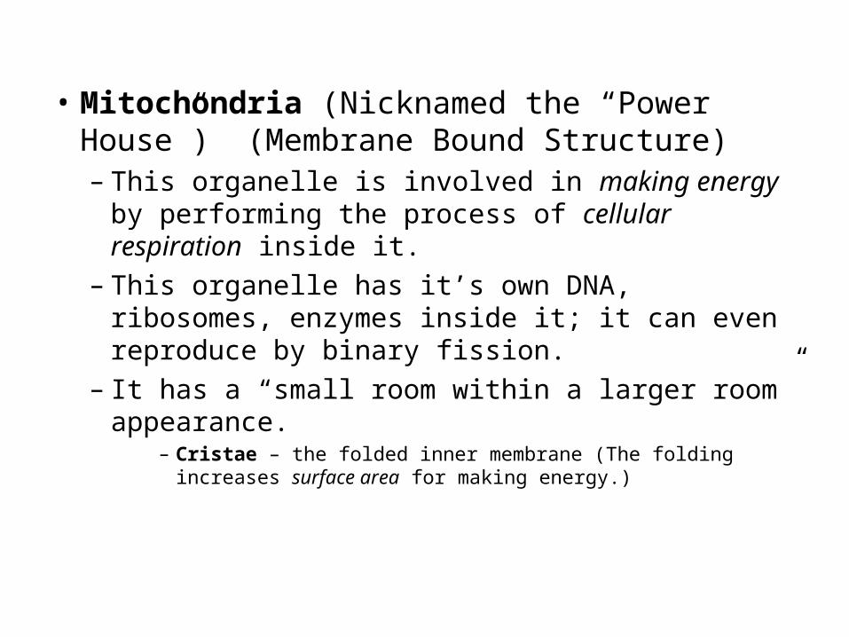
• Mitochondria (Nicknamed the “Power House”) (Membrane Bound Structure)– This organelle is involved in making energy by
performing the process of cellular respiration inside it.– This organelle has it’s own DNA, ribosomes, enzymes
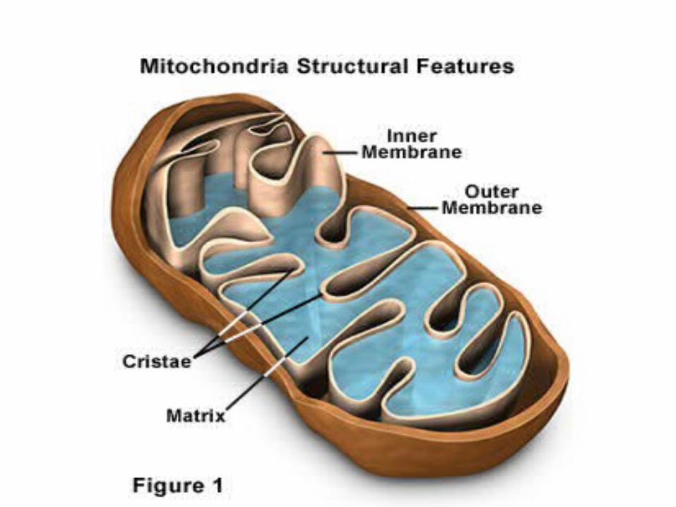
inside it; it can even reproduce by binary fission.– It has a “small room within a larger room” appearance.
– Cristae – the folded inner membrane (The folding increases surface area for making energy.)

Mitochondria

.
Mitochondrion
Intermembrane space
Outer membrane
Inner membrane
Cristae
Matrix
100 nmMitochondrialDNA
Freeribosomes in themitochondrialmatrix

• Evolutionary Significance? (They were believed to have been purple bacteria. Remember bacteria are prokaryotes. They entered into a symbiotic relationship with a larger prokaryote that could provide protection in return for extra energy. Together they would have an evolutionary advantage over other bacteria. The advantage allowed them to survive and reproduce and eventually lead to Eukaryotic cells.)

Prokaryotic Cell (Bacteria)

• Chloroplasts (Membrane Bound Structure)– These organelles are the site of Photosynthesis in plants
and algae.– They are a type of Plastid. (Plastid is a Pigment Container.
These contain the green pigment chlorophyll.)(“phyll” means “pigment”)
– Has it’s own DNA, ribosomes, and enzymes too! Reproduces by binary fission too!
– It has a “small room within a larger room” appearance too!
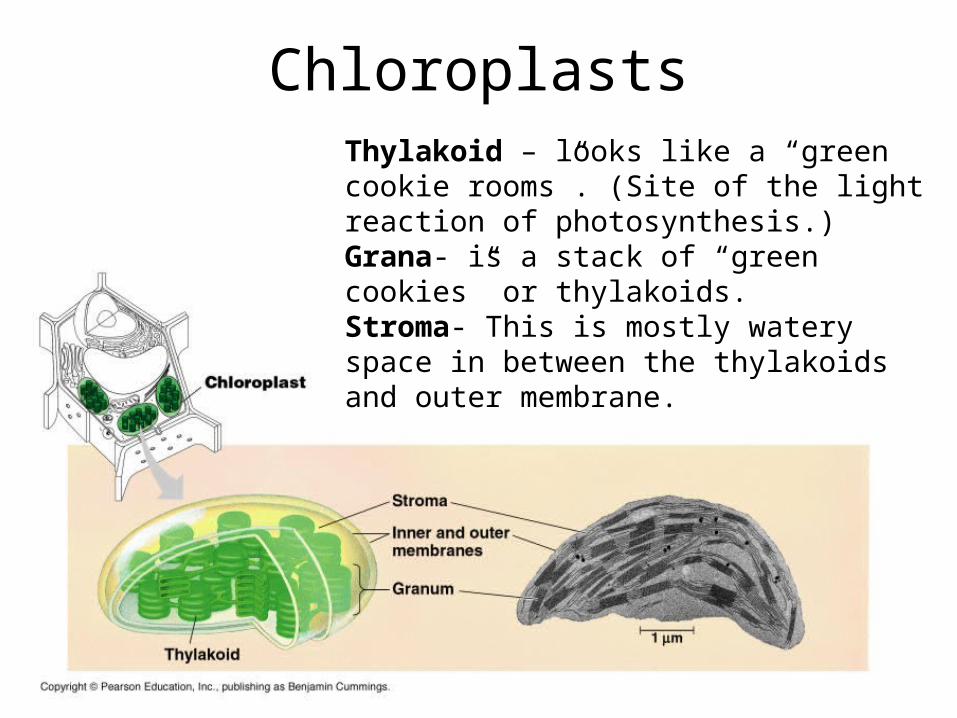
ChloroplastsThylakoid – looks like a “green cookie rooms”. (Site of the light reaction of photosynthesis.)Grana- is a stack of “green cookies” or thylakoids.Stroma- This is mostly watery space in between the thylakoids and outer membrane.

– Evolutionary Significance? (They too were believed to have been blue-green bacteria that entered into a symbiotic relationship for protection in return for energy.)
•

Prokaryotic Cell (Bacteria)

• Endosymbiont Hypothesis, remember, tries to scientifically explain these relationships.– This hypothesis was proposed by Lynn Margulis in the 1960’s.– It basically hypothesized that Prokaryotes came to live
together in a symbiotic relationship, the smaller living inside the larger, to gain a survival advantage over other prokaryotes and eventually they evolved into Eukaryotic cells over many generations that spanned hundreds of thousands of years.
• Smaller organism gained protection.• Larger organism gained energy production or faster motility.
Lynn Margulis

Endosymbiotic Hypothesis

Modern Day Eukaryotic Cells
Animal Plants

The Cytoskeleton!

• Cytoskeleton– These structures help support and protect the cell. (Much
like your skeleton does for you.)– It also helps to keep inner organelles organized. (Much like
your skeleton does for you.)– It also helps in cell motility or cell organelle movement
(Much like your skeleton helps you move.)– The cytoskeleton is composed of various sized protein
fibers (Your skeleton has different sized structures too. (Largest – bones, middle – Ligament and tendons, smallest- muscle fibers)
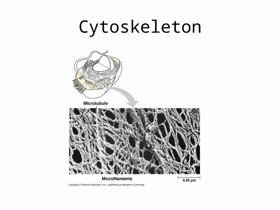
Cytoskeleton

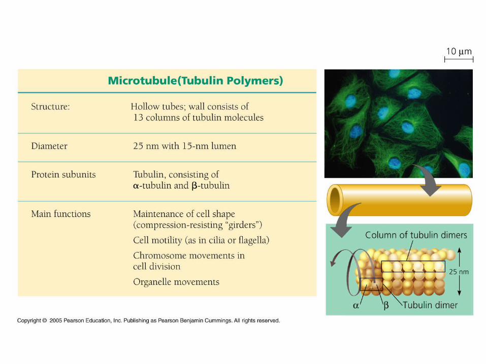
– Microtubules (These are the largest structures in the cytoskeleton.)
• These are large, hollow tubes.• They are composed of Tubulin protein.• There main function is support or movement of the cell or
organelle.• Important structures made of microtubules within a cell:
– Centrosomes/Centrioles (These act as anchors during cell division.)– Spindle Fibers (These act as guides or “tow ropes” for chromosomes
during division.– Cilia-These help with cell movement. Cells usually have a lot and
they are small in length.– Flagella- These are also for movement. Usually few on a cell and
they are very long in length.

Centrioles

Cellular Movement

• Microfilaments (These are the smallest structures in the cytoskeleton.)– These are solid rods.– Composed of Actin or Myosin protein.– They provide a pulling force.– They are abundant in muscle tissue cells in
animals.


Microfilaments in muscle tissue

Muscle Tissue under the Microscope

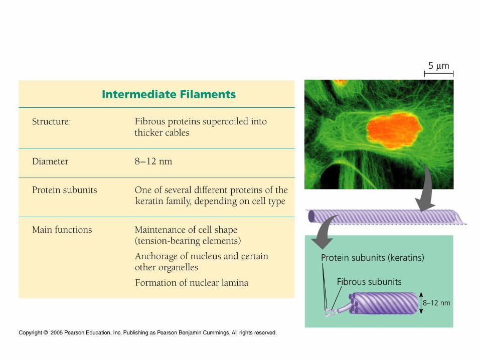
• Intermediate Filaments (These are medium sized structures.)(“inter” means “between”)
– These are permanent, solid rods.– They are mostly composed of keratin protein.– They help to reinforce and brace the large
microtubules.


• Protective or weight bearing structures for cells– Cell Wall of Plant Cells (composed of cellulose
primarily)– Cell Walls of Fungus (Composed of the
carbohydrate called Chitin. )– Extra Cellular Matrix (ECM)
• This is the outer protective “skeleton” of the cell plasma membrane in animal cells. (“extra” means “outside of”; “matrix” means “skeleton”)
• It also functions in communication with other cells.
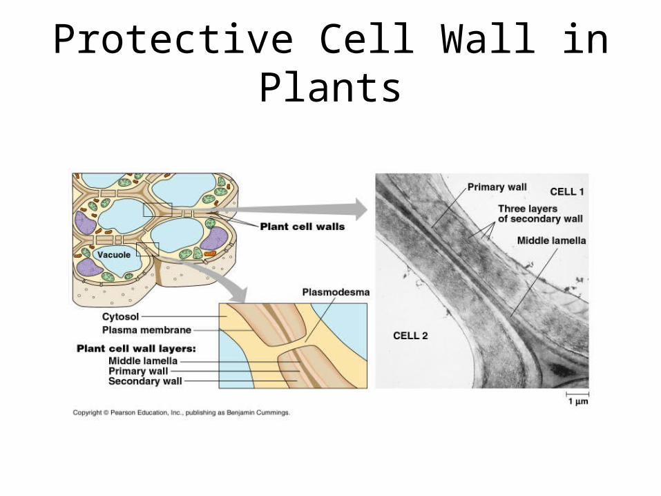
Protective Cell Wall in Plants
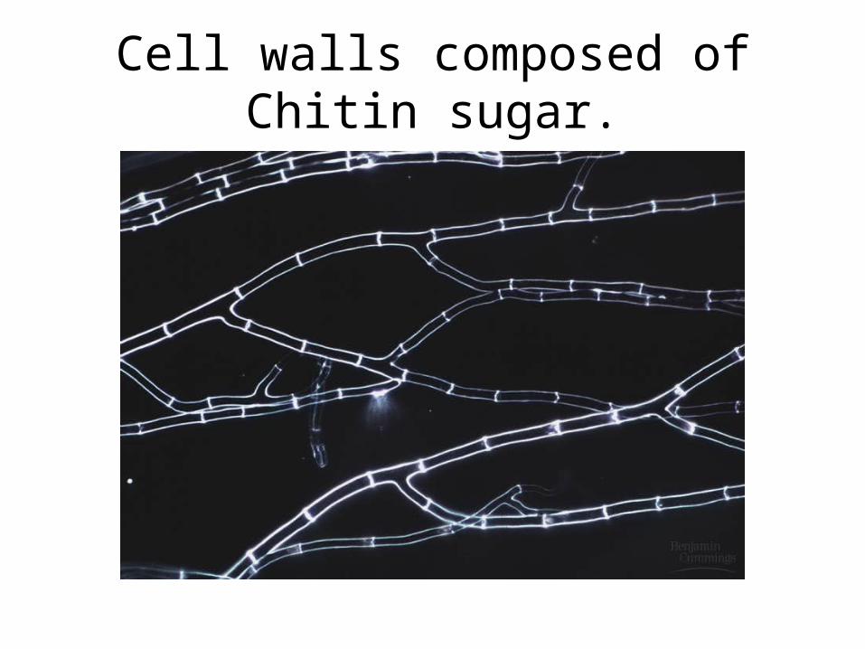
Cell walls composed of Chitin sugar.
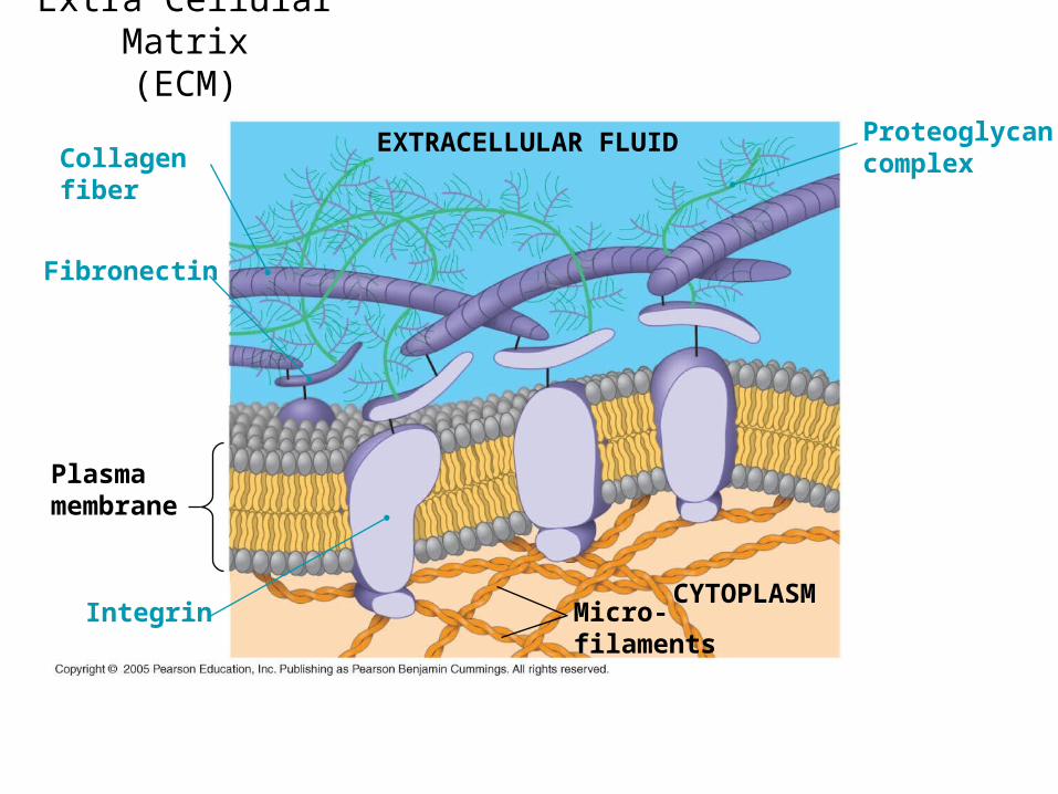
Extra Cellular Matrix(ECM)
EXTRACELLULAR FLUID ProteoglycancomplexCollagen
fiber
Fibronectin
Integrin Micro-filaments
CYTOPLASM
Plasmamembrane

• A CELL IS THE SUM OF IT”S PARTS! It is the basic unit of life only when all the parts work together to make “LIFE” possible.
A cell is an example of an Emergent Property.
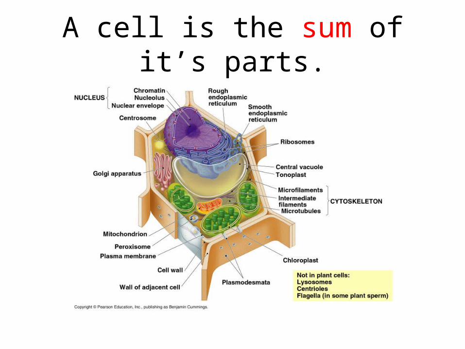
A cell is the sum of it’s parts.