arrocase nasopharynx cancer
TRANSCRIPT
ARROCaseNasopharynx Cancer
Kara D. Romano, MD
Daniel M. Trifiletti, MD
Paul W. Read, MD
University of Virginia
October 20, 2016

Case: HistoryH&P:
• 37 yo male presents with left-sided neck mass, ear pressure, otalgia, and pain radiating down the ipsilateral neck.
• ROS:
– + nose bleeds, numbness on the left cheek
– - trismus, dysphagia, odynophagia, diplopia, or changes in vision
• Pertinent physical exam findings:
– ECOG: (0) Fully active
– HEENT: No mucosal lesions on direct/indirect examination. Poor dentition - no molars. Perforated left TM with erythematous auditory canal. Tenderness to palpation of the left post-auricular space.
– Lymphatics: Firm, matted adenopathy of the left level II-III nodes, with palpable ipsilateral supraclavicular nodes.
• Flexible nasal laryngoscopy: ulcerated lesion of the left nasopharynx obstructing the left Eustachian tube orifice, fullness of the left fossa of rosenmuller, extending toward the soft palate and posterior pharyngeal wall with partial obstruction of the pharynx.
October 20, 2016

Common presenting signs & symptoms
• Neck mass
• Epistaxis
• Middle ear effusion/otalgia/decreased hearing
• Headache/pain
• Nasal congestion and drainage
• Trismus
• Cranial nerve deficits• Petro-sphenoidal syndrome (CN III-IV, VI): Oculomotor signs/symptoms
• Retro-parotidian syndrome (CN IV-XII): Enophthalmos, ptosis, miosis
October 20, 2016

Case: Imaging
October 20, 2016
CT Neck MRI Orbit, Face, & Neck
Nasopharyngeal mass
Enlarged
retro-
pharyngeal
lymph node
Enlarged contralateral
retro-pharyngeal lymph node
Multiple enlarged bilateral cervical lymph
nodes (levels II – V), including supraclavicular
node (level IV)
Work-Up & Evaluation1
• H&P including complete head & neck physical exam– Tobacco history
• Flexible nasopharyngeal fiber-optic laryngoscopy
• Labs
– EBV titers
• Biopsy of the primary site or FNA of neck
• MRI w/ contrast – include skull base, nasopharynx, and neck to clavicles
• CT Neck & Chest, as clinically indicated
• PET-CT (especially for non-keratinizing histology, endemic phenotype, N2 or N3, and stage III – IV)
• Medical oncology consultation
• Dental evaluation
• Nutrition/GI evaluation
• Speech and swallow evaluation
• Audiology evaluationOctober 20, 2016

Differential Diagnosis of malignancy for nasopharyngeal mass
If small nasopharyngeal mass and confined to the mucosa:
• Prominent, but normal adenoidal tissue
• Nasopharyngeal lymphoma
• Early primary nasopharyngeal malignancy
If larger nasopharyngeal mass +/- involvement of the skull base:
• Primary nasopharyngeal malignancy
• Adenoid cystic carcinoma
• Papillary adenocarcinoma
• Melanoma
• Plasmacytoma
• Lymphoma
• Chordoma/Chondrosarcoma
• Meningioma
• Rhabdomyosarcoma/other sarcoma
• Metastases
October 20, 2016

Case: Pathology• He underwent US-guided FNA of the left cervical neck mass
• Pathology: poorly differentiated carcinoma– Pleomorphic epithelioid cells in a background of lymphocytes
– + cytokeratin
– p16 negative
– EBV in-situ hybridization negative
– Neuron specific enolase negative
October 20, 2016
Epithelioid
carcinoma
Lymphocyte

Nasopharynx Cancer
Incidence:
• 5 per 1 million in the USA
• 100 – 400 per 1 million in Asia/Africa
Risk Factors:
• Salted or pickled foods
• Metal dust
• Epstein-Barr virus (EBV)
• Smoking
• HLA/Genetic
Presenting signs/symptoms:
• Palpable cervical adenopathy
– 70% cN+
– 90% pN+ (50% bilateral)
• Nasal discharge
• Hearing loss
• Trismus
• Cranial neuropathy (CN II – XII)
October 20, 2016

Anatomy: Skull Base & Nasopharynx
October 20, 2016
Pterygopalatine Fossa (PPF)
Sphenoid Sinus Clivus*
Soft Palate*
Choanae*
Sphenoid bone*
C1 – C2*
*anatomic boundaries of the NPx
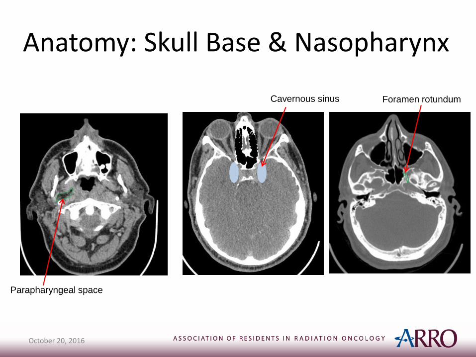
Anatomy: Skull Base & Nasopharynx
October 20, 2016
Parapharyngeal space
Cavernous sinus Foramen rotundum

WHO Grading SystemNasopharyngeal Carcinoma
• WHO I: keratinizing squamous cell carcinoma
– 20% prevalence
– Associated with smoking, HPV
– Poor LC
– Lower risk of DM
• WHO II: non-keratinizing, squamous cell carcinoma
– (A) Differentiated type
– (B) Undifferentiated type
– 30 – 40% prevalence
• WHO III: undifferentiated, lympho-epithelial, or basaloid squamous cell carcinoma
– 40 – 50% prevalence
– Most strongly associated with EBV
– Better LC
– Higher risk of DM
October 20, 2016

AJCC Staging System2
Nasopharyngeal CarcinomaTumorT1: confined to NPx, OPx, and/or nasal cavity without parapharyngeal extension
T2: parapharyngeal extension
T3: bony structures of skull case of paranasal sinuses
T4: intra-cranial extension, cranial nerves, HPx, orbit, or with extension to infra-temporal fossa/masticator space
NodesN1: unilateral, ≤ 6 cm, above SCLV fossa; or uni/bilateral RP nodes, ≤ 6 cm
N2: bilateral, ≤ 6 cm, above SCLV fossa
N3a: > 6cm
N3b: SCLV fossa*
MetastasesM1: any distant metastasis
*SCLV fossa is the triangular region defined by the 3 points:
(1) Superior margin of the sternal end of clavicle
(2) Superior margin of lateral end of clavicle
(3) Point where neck meets shoulder
Abbreviations:
NPx: nasopharynx, OPx: oropharynx, HPx: hypopharynx, SCLV: supraclavicular
October 20, 2016
32
1

AJCC Staging System2
Nasopharyngeal Carcinoma
October 20, 2016

Case: Management• cT1 cN3b cM0, stage IVB, p16- EBV- poorly differentiated
nasopharyngeal carcinoma
• Consultation with Otolaryngology, Medical Oncology, Radiation Oncology
• Treatment options for T2-4 or N+ include:
– Concurrent chemoradiation +/- adjuvant chemo
– Induction chemo concurrent chemoradiation
October 20, 2016

Case: Management
• This patient received:
– Concurrent CRT to 70 Gy in 33 fractions with weekly cisplatin 40 mg/m2
– Adjuvant/consolidation cisplatin/5-FU
October 20, 2016

Radiation Planning• Simulation:
– Supine, long mask, IV contrast:
– Co-register with MRI +/- PET-CT
• Radiation Dose/Fractionation:
– PTV1: 69.96 Gy in 33 fractions (2.12 Gy/Fx)
– PTV2: 59.4 Gy in 33 fractions (1.8 Gy/Fx)
– PTV3: 54 Gy in 33 fractions (1.63 Gy/Fx)
– Consider hyper-fractionation if dose to optic structures/brainstem would be otherwise unacceptable
October 20, 2016
Radiation Technique
• IMRT
• Proton beam radiotherapy
• Brachytherapy – Intracavitary boost
– Consider for recurrent disease
– Possible dose escalation of early/advanced primary tumors
October 20, 2016

Radiation Contouring• PTV1 (69.96 Gy)
– Gross disease
• PTV2 (59.4 Gy)
– High risk subclinical disease:
• Sphenoid sinus
• Cavernous sinus
• Skull base
• Clivus
• Posterior 1/3 of maxillary sinus
• Posterior 1/3 of nasal cavity
• Pterygopalatine fossae
• Parapharyngeal space
• Retropharyngeal space
• Soft palate
• PTV3 (54 Gy):
– Elective nodal coverage:
• Retrostyloid space
• Bilateral IB – V (can omit IB, if N0)
Contouring references:• e-contour http://econtour.org/cases/2
• http://www.nyp.org/pdf/imrt_2010_talk2_lee.pdf
October 20, 2016
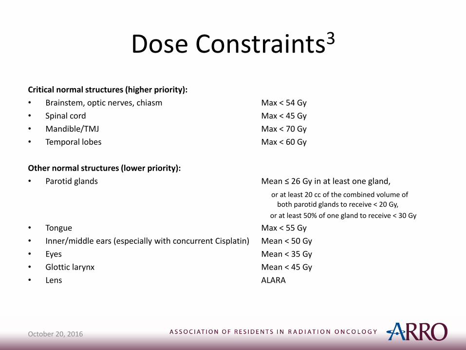
Dose Constraints3
Critical normal structures (higher priority):
• Brainstem, optic nerves, chiasm Max < 54 Gy
• Spinal cord Max < 45 Gy
• Mandible/TMJ Max < 70 Gy
• Temporal lobes Max < 60 Gy
Other normal structures (lower priority):
• Parotid glands Mean ≤ 26 Gy in at least one gland,
or at least 20 cc of the combined volume of both parotid glands to receive < 20 Gy,
or at least 50% of one gland to receive < 30 Gy
• Tongue Max < 55 Gy
• Inner/middle ears (especially with concurrent Cisplatin) Mean < 50 Gy
• Eyes Mean < 35 Gy
• Glottic larynx Mean < 45 Gy
• Lens ALARA
October 20, 2016
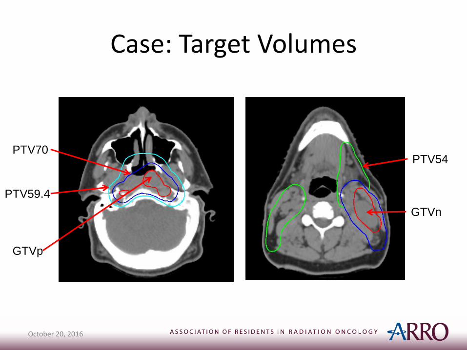
Case: Target Volumes
October 20, 2016
GTVp
PTV54
GTVn
PTV70
PTV59.4

Case: Isodose Distribution
October 20, 2016

Case: Isodose Distribution
October 20, 2016

Case: Isodose Distribution
October 20, 2016
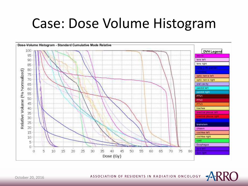
Case: Dose Volume Histogram
• Images of Treatment Plan
• Images of DVH
October 20, 2016

Treatment Algorithm
October 20, 2016
Chemotherapy
October 20, 2016
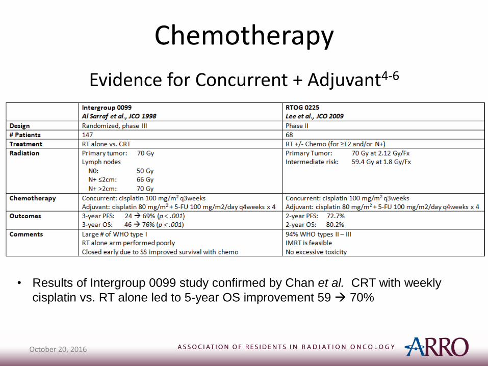
Evidence for Concurrent + Adjuvant4-6
• Results of Intergroup 0099 study confirmed by Chan et al. CRT with weekly
cisplatin vs. RT alone led to 5-year OS improvement 59 70%
Chemotherapy
October 20, 2016
Evidence for Induction:
• No demonstrated benefit of induction chemo
• NCCN Guidelines category 3 recommendation
• No demonstrated benefit for adjuvant chemo following definitive RT or CRT, although long term data not yet available
• NCCN Guidelines category 2B recommendation
Evidence for Adjuvant7:
Follow Up for Nasopharyngeal Cancer
• H&P + complete H&N physical exam +/- mirror/fiberoptic exam
– q1-3 months for year 1
– q2-6 months for year 2
– q4-8 months for years 3 – 5
– Yearly for years > 5
• Imaging for signs/symptoms
• TSH yearly (if irradiated)
• Speech/swallowing/dental/hearing evaluations
• Consider EBV-DNA monitoring
October 20, 2016

Nasopharynx Clinical Pearls
• Uncommon in the United States
• High likelihood of distant metastases
• Rarely, parotid nodal relapses can occur
• Due to the anatomic location, RT traditionally used over surgery
• Local control with RT alone of early (T1 – T2) tumors 80 – 90%
• Local control with RT alone of T3 – T4 tumors 30 – 65%
• Concurrent cisplatin-based chemo + RT has been shown to improve OS
• No evidence to support induction chemotherapy
• Little evidence to support adjuvant chemotherapy, although awaiting further results of Chen et al. phase 3 randomized trial
• RT technique is IMRT with simultaneous integrated boosts to 70 Gy, 59.4 Gy, and 54 Gy
October 20, 2016

References
1. National Comprehensive Cancer Network (NCCN) Clinical Practice Guidelines. Head and Neck Cancers, version 1.2015, Available at: http://www.nccn.org/professionals/physician_gls/pdf/head-and-neck.pdf, Accessed: March 28, 2016.
2. Edge SB, Byrd DR, Compton CC, et al. American Joint Committee on Cancer (AJCC) Cancer Staging Manual (Seventh Edition)New York, NY: Springer; 2010.
3. Lee N, RTOG 0225: A Phase II Study of Intensity Modulated Radiation Therapy (IMRT) +/ Chemotherapy for Nasopharyngeal Cancer, Available at: https://www.rtog.org/ClinicalTrials/ProtocolTable/StudyDetails.aspx?study=0225, Accessed: September 14, 2016.
4. Al-Sarraf M, LeBlanc M, Giri PG, et al. Chemoradiotherapy versus radiotherapy in patients with advanced nasopharyngeal cancer: phase III randomized Intergroup study 0099. J Clin Oncol 1998;16:1310-1317.
5. Lee N, Harris J, Garden AS, et al. Intensity-modulated radiation therapy with or without chemotherapy for nasopharyngeal carcinoma: radiation therapy oncology group phase II trial 0225. J Clin Oncol 2009;27:3684-3690.
6. Chan AT, Leung SF, Ngan RK, et al. Overall survival after concurrent cisplatin-radiotherapy compared with radiotherapy alone in locoregionally advanced nasopharyngeal carcinoma. J Natl Cancer Inst 2005;97:536-539.
7. Chen L, Hu CS, Chen XZ, et al. Concurrent chemoradiotherapy plus adjuvant chemotherapy versus concurrent chemoradiotherapy alone in patients with locoregionally advanced nasopharyngeal carcinoma: a phase 3 multicentrerandomised controlled trial. Lancet Oncology 2012;13:163-171.
October 20, 2016