contrast radiography of the nasopharynx
TRANSCRIPT
Postgrad. med. J. (November 1968) 44, 825-829.
Contrast radiography of the nasopharynx
GORDON EVISONM.B.Ch.B., D.M.R.D., F.F.R.
Department of Diagnostic Radiology,United Bristol Hospitals*
SummaryA technique of contrast radiography of the
nasopharynx is described and indications for theprocedure are reviewed. The main advantage ofusing this method is the simple and effective wayit outlines the tumour mass and shows its responseto therapy and any localized recurrences. Charac-teristic features of tumours at different sites areillustrated. The potential usefulness of this tech-nique in detecting early lesions is emphasized.
IntroductionCarcinoma of the nasopharynx is often diag-
nosed only late and with difficulty. Mortality ishigh, and the results of treatment have hardlyimproved over the past two decades. Because ofits inaccessibility, indirect methods of examiningthe nasopharynx, such as radiography, wouldseem to be specially valuable. However, plainfilms of the nasopharynx, while essential fordemonstrating bone destruction by the tumourif this is present, do not always clearly definethe soft-tissue mass. Attempts to use positivecontrast media in the nasopharynx were firstmade in 1934 by Ruedi & Zuppinger, usingLipiodol. More recently barium has been used(Lederman, 1961 ; Khoo, Chia & Nalpon, 1967a;Khoo, Kanagrasuntheram & Chia, 1 967b), andoily Dionosil (Jing & McGraw, 1965).The following is a description of the technique
used at the Bristol General Hospital, with a re-view of the normal anatomy of the contrast-filled nasopharynx, the indications for its use,and an assessment of its present usefulness asa diagnostic procedure.
TechniqueNo preliminary preparation of the patient is
required. With the patient supine, a submento-vertical (SMV) plain radiograph is taken on a12 x 10 in. film, using the Schonander head unit.A horizontal-beam lateral plain radiograph isalso taken with the patient's head in SMV posi-
*Present address: Royal United Hospital, Bath.
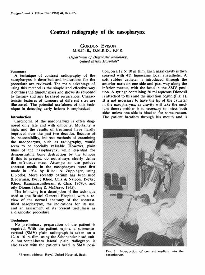
tion, on a 12 x 10 in. film. Each nasal cavity is thensprayed with 4% lignocaine local anaesthetic. Asoft rubber catheter is introduced through theanterior naris on one side and part way along theinferior meatus, with the head in the SMV posi-tion. A syringe containing 20 ml aqueous Dionosilis attached to this and the injection begun (Fig. 1).It is not necessary to have the tip of the catheterin the nasopharynx, as gravity will take the med-ium there; neither is it necessary to inject bothsides unless one side is blocked for some reason.The patient breathes through his mouth and is
FIG. 1. Introduction of contrast medium into thenasopharynx.

Gordon Evison
instructed not to swallow. When the patient feelsthe urge to swallow (but without swallowing), hegives a pre-arranged signal and the injection isstopped. Usually 8-15 ml are necessary and atthis point the nasopharynx is full of contrastmedium. Twelve x 10 in. SMV and horizontalbeam lateral SMV radiographs are then exposedas rapidly as possible. If it is desired to fill theEustachian tubes, the patient then swallows, withhis head still in the SMV position, and a further12 x 10 in. SMV radiograph is exposed. The Val-salva manoeuvre may be done to reinforce this,though it has been found that it has no addedadvantage over simple swallowing and is muchmore difficult for the patient. The contrastmedium is then blown out of the nose into paperhandkerchiefs by the patient, or swallowed aidedby a glass of fruit juice. The essence of obtainingsatisfactory radiographs is to fully explain to thepatient what is about to happen before starting,and if this is done then the procedure is verywell tolerated, even by small children.
Normal anatomyOn the SMV view, the contrast-filled naso-
pharynx appears somewhat dumb-bell shapedand normally quite symmetrical. The nasalseptum separates the two nasal cavities, and theinferior turbinates usually show as filling defectswithin these. The pharyngeal orifices of theEustachian tubes outline, bounded posteriorly bythe torus tubarius and behind this the fossa ofRosenmuller (Fig. 2) The cartilaginous portionsof the Eustachian tubes fill for about an inch,but we have never observed filling of the bonytube or middle ear, though this has been recor-ded (Wittenborg & Neuhauser, 1963). The lateralrecesses of the nasopharynx are subject to con-siderable individual variations, and these havebeen studied using positive contrast media andcine fluorography by Khoo et al. (1967a, b). Onthe lateral view (Fig. 3), the roof and posterior wallof the nasopharynx are usually smooth, thoughoccasionally in adults small irregularities may beseen as a normal feature due to adenoid remn-ants. The anterior margin is formed by the softpalate, which is usually clearly outlined.
Indications for nasopharyngographyThe following indications have been advanced
for contrast investigations of the nasopharynxby various authors:
(1) When a nasopharyngeal growth has beendetected clinically, or on a plain film, andit is desired.
(a) to see the extent and size of thegrowth,
-c-de
FIG. 2. Normal nasopharyngogram-submento-verti-cal. Note the bilateral symmetry, and the partial filling ofthe Eustachian tubes. (a) Inferior turbinate, (b) Nasalseptum, (c) pharyngeal orifice of the Eustachian tube,(d) torus tubarius and fossa of Rosenmuller, (e) posteriorwall of the nasopharynx.
b
FIG. 3. Normal nasopharyngogram-lateral view. (a)Sphenoid sinus, (b) postero-superior wall of the naso-pharynx, (c) soft palate.
826

Contrast radiography of nasopharynx
(b) to see if the Eustachian tubes areaffected.
(2) Where a nasopharyngeal growth is stillsuspected clinically, though plain films andendoscopy have failed to locate it.
(3) Any case of malignant enlargement ofcervical nodes where a primary is beingsought.
(4) To show the response of nasopharyngealneoplastic masses to treatment, e.g. radio-therapy or surgery.
(5) Occasionally, to demonstrate the patencyof the Eustachian tubes prior to opera-tions such as tympanoplasty.
(6) Occasionally, to show other lesions whichmay not be fully visible on endoscopy,e.g. hypertrophied posterior turbinates,choanal polypi, choanal atresia.
Nasopharyngeal tumoursThese may arise in the following sites:(1) The lateral wall, either the fossa of Rosen-
muller or the region of the torus tubarius.(2) The postero-superior wall or roof.(3) The nasopharyngeal surface of the soft
palate.(4) Undetermined site of origin.(5) Extrinsic tumours invading the naso-
pharynx.(1) Tumours arising from the lateral wall-
this is the most common site of origin, usuallyfrom one or other fossa of Rosenmuller. Theyare shown best on the submento-vertical projec-tion, appearing as an indentation or filling defecton the lateral margin of the contrast medium,obliterating the fossa of Rosenmuller or torustubarius. On the lateral view filling defects areusually shown on the postero-superior margin ofthe contrast medium. Fig. 4 shows a largeanaplastic carcinoma arising from the left lateralwall.
(2) Tumours arising from the postero-superiorwall or roof-these show as a soft tissue massprojecting into the cavity of the nasopharynxand causing filling defects on the postero-superiormargins of the contrast medium as shown onthe lateral view. If the tumour is small, the sub-mento-vertical view may appear normal, but withlarger tumours deficient filling of the posteriormargin of the contrast medium is usually evident,often more on one side than the other. Fig. 5(a)and (b) shows a large tumour arising from thepostero-superior wall and almost completelyoccluding the nasopharynx.
(3) Tumours arising from the nasopharyngealsurface of the soft palate-these show as an
increase in thickness of the soft palate, seen beston the lateral view (Fig. 6).
(4) Tumours whose site of origin cannot bedetermined-the larger tumours may fill thewhole of the nasopharynx and obstruct the pass-age of contrast beyond the posterior choanae. Inthese circumstances the exact site of origin can-not be determined.
FIG. 4. Anaplastic carcinoma (arrow) arising from theleft lateral wall, causing a large filling defect in the con-trast medium. (Reproduced by kind permission of theEditor of J. Laryng.)
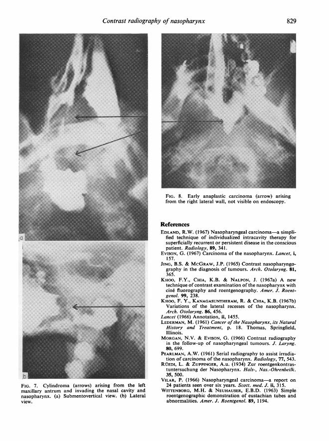
(5) Tumours arising in surrounding structuresmay invade the nasopharynx. Fig. 7(a) and (b)shows a large cylindroma which had spread fromthe left maxillary antrum into the nasal cavityand nasopharynx.
DiscussionThe technique of positive contrast radiography of
the nasopharynx described here is simple to per-form, requires only standard radiological equip-ment and does not cause undue discomfort to thepatient. The main value at the present time ofusing this method of investigation is the cleardemonstration it provides of the site of origin
827

828 Gordon Evison
aP
FIG. 5. Tumour (arrows) arising from the postero-superior wall, almost completely occluding the naso-pharynx. (a) Submento-vertical view. (b) Lateral view.(Reproduced by kind permission of the Editor ofJ. Laryng.)
and extent of nasopharyngeal tumours, and inshowing their response to radiotherapy (Pearl-
..~~ ~ ~ ~~~~~~~~.
FIG. 6. Carcinoma (arrow) arising from the naso-pharyngeal surface of the soft palate.
man, 1961; Morgan & Evison, 1966; Evison,1967). It is also valuable for showing localizedtumour recurrences (Edland, 1967). The most im-portant problem with these tumours is earlyrecognition, and the only way of increasing thenumber of survivors is to diagnose and treat theneoplasm at a much earlier stage (Lancet, 1966).Vilar (1966) found that the average time betweenthe onset of symptoms and a definite diagnosiswas 9 7 months, and in three of his twenty-fourcases direct examination of the post-nasal spaceand biopsy were at first negative. Most of ourcases were referred when the lesion was welladvanced, but in one case which presented withenlarged cervical glands and in whom endoscopyfailed to reveal a lesion, a nasopharyngogram(Fig. 8) showed a shallow filling defect on theright side which on biopsy proved to be ananaplastic carcinoma. Detection of small lesionsis necessarily difficult, but the procedure doesoffer a way of diagnosing at least some of theseat a much earlier stage than at present. It shouldbe considered whenever clinical suspicion of anasopharyngeal tumour arises.
AcknowledgmentsI am grateful to Professor J. H. Middlemiss and Dr E.
Rhys Davies for their help and advice and to the ConsultantE.N.T. and Radiotherapy staff of the United Bristol Hospitalsfor referral ofcases, to Mr J. E. Hancock for the reproductionsand to Miss A. Sainsbury for the typescript.
Contrast radiography of nasopharynx 829
r ..~~~~~~~~~~~~~~~~~~. .>l....~~~~~~~~~~~~~~~~~~~~~~~~~~~~~~~~~~~~~.;'.. ... . ... .I ~ ~ ~ ~ ~ ~ ~ ~ ~ ~ ~ ~ ~ ~ ~ ~ ~ ~ ~ ~ ~ ~ ~ ~ ~ ~ ~~~~~~~~....
I~~~~~~~~~~~~~~~~~~~~~~~~~~~~~~~~~~~~~~~~~~~~~~~~~~~~~~~
I~ ~~ .........I~~~~~~~~~~~~~~~~~~~~~~.... ...IlI*-I| _~~~~~~~~~~~~~~~~~~~........
FIG. 7. Cylindroma (arrows) arising from the leftmaxillary antrum and invading the nasal cavity andnasopharynx. (a) Submentovertical view. (b) Lateralview.
........
.. ~~~~~~~~~~~~......(:B gs .. : . 1~~~~~~~~~~~~~~~~~~~~~~~.......
FIG. 8. Early anaplastic carcinoma (arrow) arisingfrom the right lateral wall, not visible on endoscopy.
ReferencesEDLAND, R.W. (1967) Nasopharyngeal carcinoma-a simpli-
fied technique of individualized intracavity therapy forsuperficially recurrent or persistent disease in the consciouspatient. Radiology, 89, 341.
EvISON, G. (1967) Carcinoma of the nasopharynx. Lancet, i,157.
JING, B.S. & MCGRAW, J.P. (1965) Contrast nasopharyngo-graphy in the diagnosis of tumours. Arch. Otolaryng. 81,365.
KHOO, F.Y., CHIA, K.B. & NALPON, J. (1967a) A newtechnique of contrast examination of the nasopharynx withcin6 fluorography and roentgenography. Amer. J. Roent-genol. 99, 238.
KHOO, F. Y., KANAGASUNTHERAM, R. & CHIA, K.B. (1967b)Variations of the lateral recesses of the nasopharynx.Arch. Otolaryng. 86, 456.
Lancet (1966) Annotation, ii, 1455.LEDERMAN, M. (1961) Cancer of the Nasopharynx, its Natural
History and Treatment, p. 18. Thomas, Springfield,Illinois.
MORGAN, N.V. & EvISON, G. (1966) Contrast radiographyin the follow-up of nasopharyngeal tumours. J. Laryng.80, 699.
PEARLMAN, A.W. (1961) Serial radiography to assist irradia-tion of carcinoma of the nasopharynx. Radiology, 77, 543.
RUEDI, L. & ZUPPINGER, A.u. (1934) Zur roentgenkontras-tuntersuchung der Nasopharynx. Hals-, Nas.-Ohrenbeilk.35, 500.
VILAR, P. (1966) Nasopharyngeal carcinoma-a report on24 patients seen over six years. Scott. med. J. ii, 315.
WITTENBORG, M.H. & NEUHAUSER, E.B.D. (1963) Simpleroentgenographic demonstration of eustachian tubes andabnormalities. Amer. J. Roentgenol. 89, 1194.