assessment of the degradation rates and effectiveness of ... · scaffold, group b receiving the...
TRANSCRIPT

Journal of Materials Science: Materials in Medicine (2018) 29:138https://doi.org/10.1007/s10856-018-6145-2
CLINICAL APPLICATIONS OF BIOMATERIALS
Original Research
Assessment of the degradation rates and effectiveness of differentcoated Mg-Zn-Ca alloy scaffolds for in vivo repair of critical-sizebone defects
Nan Zhang 1,2● Dewei Zhao3
● Na Liu2● Yunfeng Wu4
● Jiahui Yang3● Yuefei Wang5
● Huanxin Xie1 ● Ye Ji1 ●
Changlong Zhou1● Jinpeng Zhuang1
● Yaming Wang4● Jinglong Yan1
Received: 24 January 2018 / Accepted: 1 August 2018 / Published online: 17 August 2018© The Author(s) 2018
AbstractSurgical repair of bone defects remains challenging, and the search for alternative procedures is ongoing. Devices made ofMg for bone repair have received much attention owing to their good biocompatibility and mechanical properties. Wedeveloped a new type of scaffold made of a Mg-Zn-Ca alloy with a shape that mimics cortical bone and can be filled withmorselized bone. We evaluated its durability and efficacy in a rabbit ulna-defect model. Three types of scaffold-surfacecoating were evaluated: group A, no coating; group B, a 10-μm microarc oxidation coating; group C, a hydrothermal duplexcomposite coating; and group D, an empty-defect control. X-ray and micro-computed tomography(micro-CT) images wereacquired over 12 weeks to assess ulnar repair. A mechanical stress test indicated that bone repair within each group improvedsignificantly over time (P < 0.01). The degradation behavior of the different scaffolds was assessed by micro-CT andquantified according to the amount of hydrogen gas generated; these measurements indicated that the group C scaffold betterresisted corrosion than did the other scaffold types (P < 0.05). Calcein fluorescence and histology revealed that greatermineral densities and better bone responses were achieved for groups B and C than for group A, with group C providing thebest response. In conclusion, our Mg-Zn-Ca-alloy scaffold effectively aided bone repair. The group C scaffold exhibited thebest corrosion resistance and osteogenesis properties, making it a candidate scaffold for repair of bone defects.
1 Introduction
Presently, treatment of critically sized and large defects inlong bones of humans caused by trauma, infection, or atumor remains a challenging undertaking for orthopedicsurgeons [1, 2]. Available repair modalities include auto-geneic, allogeneic [3, 4], and vascularized bone grafts [5].Although these treatments have achieved positive
outcomes, they are not without drawbacks, indicating theneed for improvement] [4, 6, 7]. To effectively repairdefects in long bones, Cobos et al. [8] described a simpleand effective technique that uses a titanium mesh cage incombination with a bone graft to repair critical-size bonedefects [9–11]; however, it is not possible to remove theinert titanium mesh after it becomes covered with bone, andthe continual presence of titanium mesh in a bone maycause stress shielding and secondary bone absorption [12].Thus, how to pair new materials with known surgicalgrafting options remains a major clinical problem.
Magnesium and its alloys have been extensively inves-tigated as possible materials for bone implants because oftheir excellent mechanical properties [13–15]. The mostintriguing advantage of a Mg alloy is that it can be tuned toresorb after completion of the bone-healing process, whichwould eliminate the need for a second surgery to remove it,thereby reducing the risk of infection, discomfort, and cost.However, the use of a Mg-alloy scaffold to repair a largesegmental bone defect has not been reported. Therefore, wefabricated a scaffold made of a Mg-Zn-Ca alloy that is
* Jinglong [email protected]
1 The Second Affiliated Hospital of Harbin Medical University,Harbin, Heilongjiang, People’s Republic of China
2 The Second Affiliated Hospital of Qiqihar Medical College,Qiqihar, Heilongjiang, People’s Republic of China
3 The Affiliated Zhongshan hospital of Dalian University,Dalian, Liaoning, People’s Republic of China
4 Harbin Institute of Technology, Harbin, Heilongjiang, People’sRepublic of China
5 Qiqihar Medical College, Qiqihar, Heilongjiang, People’sRepublic of China
1234
5678
90();,:
1234567890();,:

hollow and cylindrical, a shape that mimics a cortical boneand allows filling of the internal space with finely groundautologous morselized bone. We hypothesized that thescaffold could be used as a substitute for the titaniummesh–type scaffold currently used to repair bone defects.This scaffold would effectively reduce or obviate post-operative complications inherent in the use of the titaniummesh cage, which includes stress shielding and removalcomplications, and might promote bone regeneration relatedto the presence of Mg2+, which can stimulate more osteo-blastic response and enhance porosis [16], so that a bonedefect could be repaired. The purposes of the present studywere, therefore, as follows. To evaluate the durability ofdifferent scaffold compositions in vivo and to assess therelative efficacies of the scaffolds for repairing long-bonedefects.
2 Materials and methods
2.1 Design of different Mg-alloy scaffolds and thecoatings
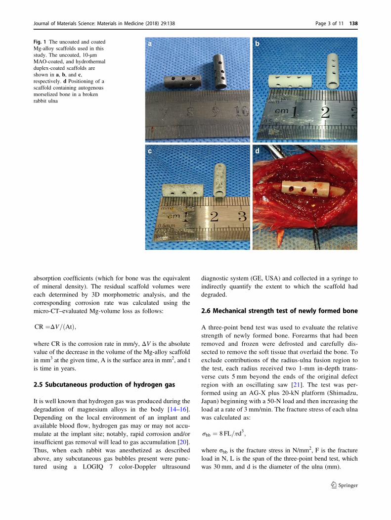
The basic Mg-alloy scaffold contained 2.5–3.0% (w/w) Zn,0.5–1.5% (w/w) Ca, and 0.5% (w/w) of a mixture of rareelements in pure Mg and was manufactured using a die-casttechnique as a cylinder of length 15 mm, outer diameter5 mm, and inner diameter 3 mm, with 12 holes (each 1-mmdiameter) drilled into the outside wall (Fig. 1a). The 10-μmmicroarc oxidation (MAO) bio-ceramic coat (Fig. 1b) and ahydrothermal duplex coat were synthesized (Fig. 1c) asdescribed previously [17]. Each scaffold underwentethylene-oxide gas sterilization before implantation.
2.2 Surgical procedure
All animal experiments were approved by the HarbinMedical University Institutional Animal Care and UseCommittee. Adult New Zealand White rabbits (36 male, 36female; mean age, 6.23 ± 0.37 months; mean weight 2.56 ±0.25 kg) were randomly assigned to one of four groups (18per group), with group A receiving the uncoated Mg-alloyscaffold, group B receiving the 10-μm MAO-coated scaf-fold, group C receiving the hydrothermal duplex layerscaffold, and group D (control) not receiving a scaffold. Therabbits were anesthetized intravenously through their earveins with 3% (w/v) pentobarbital sodium (30 mg/kg).Next, both forearms of the rabbits were shaved and disin-fected, and their ulnae exposed. A metal caliper was placedover each ulna to mark the break positions, and two ulnaosteotomies was performed ~15 mm apart. During thisprocedure, care was taken not to touch the radius. The bonefragments generated during the osteotomies were removed
and minced into small particles (diameter, 0.5–1 mm). Eachscaffold was filled with compacted, morselized bone andwas then carefully placed into the ulna at the position wherethe bone had been removed (Fig. 1d). The bone in thecontrol group was not filled with any material. The reasonfor using this defect model is that internal fixation of theulna is not needed because the adjacent radius is strongenough to support the animal when it moves [18]. Becauseeach adjacent radius was intact, and the scaffold wasembedded in one broken end of the ulna, the implant wasstable. In addition, the soft tissue and muscle adjacent toeach scaffold were carefully sutured to help stabilize thescaffold. Immediately following surgery, the animals wereallowed to resume their normal movement and behavior,including bearing their weight on their forearms.
The rabbits were housed for 4–12 weeks in stainless steelcages (one rabbit per cage) at 22 °C, 60% humidity, andunder a 12-h light/dark cycle in a ventilated room. Waterand food were available ad libitum. On each of the 3 daysfollowing the procedure, the rabbits were given a sub-cutaneous injection of 10 mg/kg penicillin. Each rabbit’sstitches were removed 10 days after the surgery. At 4, 8,and 12 weeks after surgery, six rabbits per group wereanesthetized and sacrificed by exsanguination. Their bilat-eral forearms (12 per group per time point) were collectedand frozen at −20 °C for further testing.
2.3 X-ray imaging
Scaffold placement and the forearm bone-healing processwere imaged using an X-ray scanner (Faxitron, USA;110 kV; anode current, 500 μA) at 4, 8, and 12 week aftersurgery. The lateral X-ray images of the forearm healingprocess were assessed and scored by three experienced,independent investigators who used the criteria reported in[19]. Due to the radius exit, which can induce new boneregeneration in the medial position of the bone defect, Thiscan lead to a false-positive scoring in X-ray image,There-fore, we only evaluated the X-ray images of the corticalbone opposite the radial side. Scores ranged from 0 (nohealing) to 12 (restoration of normal bone), a score of 7 wasconsidered the defect was preliminary reconstruction.
2.4 Micro-computed tomography (micro-CT)
Each rabbit’s forearms post-implant were scanned using anInveon Micro-CT system (Siemens Medical Solutions,Germany). Scans were performed with an X-ray voltage of80 kV, an anode current of 500 μA, and a 1000-ms exposuretime. Three-dimensional (3D) images were generated fromthe acquired 2D lateral projections using Inveon software.In each image, the scaffold could be distinguished from thesurrounding soft and hard tissues based on differences in
138 Page 2 of 11 Journal of Materials Science: Materials in Medicine (2018) 29:138
absorption coefficients (which for bone was the equivalentof mineral density). The residual scaffold volumes wereeach determined by 3D morphometric analysis, and thecorresponding corrosion rate was calculated using themicro-CT–evaluated Mg-volume loss as follows:
CR ¼ΔV= Atð Þ;
where CR is the corrosion rate in mm/y, ΔV is the absolutevalue of the decrease in the volume of the Mg-alloy scaffoldin mm3 at the given time, A is the surface area in mm2, and tis time in years.
2.5 Subcutaneous production of hydrogen gas
It is well known that hydrogen gas was produced during thedegradation of magnesium alloys in the body [14–16].Depending on the local environment of an implant andavailable blood flow, hydrogen gas may or may not accu-mulate at the implant site; notably, rapid corrosion and/orinsufficient gas removal will lead to gas accumulation [20].Thus, when each rabbit was anesthetized as describedabove, any subcutaneous gas bubbles present were punc-tured using a LOGIQ 7 color-Doppler ultrasound
diagnostic system (GE, USA) and collected in a syringe toindirectly quantify the extent to which the scaffold haddegraded.
2.6 Mechanical strength test of newly formed bone
A three-point bend test was used to evaluate the relativestrength of newly formed bone. Forearms that had beenremoved and frozen were defrosted and carefully dis-sected to remove the soft tissue that overlaid the bone. Toexclude contributions of the radius-ulna fusion region tothe test, each radius received two 1-mm in-depth trans-verse cuts 5 mm beyond the ends of the original defectregion with an oscillating saw [21]. The test was per-formed using an AG-X plus 20-kN platform (Shimadzu,Japan) beginning with a 50-N load and then increasing theload at a rate of 3 mm/min. The fracture stress of each ulnawas calculated as:
σbb ¼ 8 FL=πd3;
where σbb is the fracture stress in N/mm2, F is the fractureload in N, L is the span of the three-point bend test, whichwas 30 mm, and d is the diameter of the ulna (mm).
Fig. 1 The uncoated and coatedMg-alloy scaffolds used in thisstudy. The uncoated, 10-μmMAO-coated, and hydrothermalduplex-coated scaffolds areshown in a, b, and c,respectively. d Positioning of ascaffold containing autogenousmorselized bone in a brokenrabbit ulna
Journal of Materials Science: Materials in Medicine (2018) 29:138 Page 3 of 11 138

2.7 Calcein fluorescence
To characterize mineralization of newly formed bone, 3 or4 days prior to sacrifice, a solution of 1% (w/v) calcein in2% (w/v) NaHCO3 (10 mg/kg, Sigma-Aldrich, USA) wasinjected under the skin. Then, the harvested ulnae werefixed in 75% (v/v) ethanol. samples were embedded andpolymerized in methyl methacrylate resin (Technovit 7200,Kulzer, Germany). The resin blocks were cut using aEXAKT/E300CP microtome (Leica, Germany) with atungsten carbide blade into uncalcified sections of ~120 μm.Next, the sections were polished to a thickness of ~50 μmusing an cutting-grinding system (EXAKT/E400CS, Leica).Details of the bone sections were visualized through a
DM4000B fluorescence microscope (Leica), and the extentof mineral deposition evaluated according to their fluores-cence intensities.
2.8 Histology
Bone samples were histologically processed to micro-morphologically observe newly formed bone tissue sur-rounded the scaffolds. Samples were sectioned as describedin Section 2.7, decalcified, and stained with Van Gieson’spicrofuchsin solution (Sigma-Aldrich) to visualize osteo-cytes, chondrocytes, and surrounding soft tissue. The sec-tions were visualized and photographed through an opticalmicroscope (Model X71, Olympus, Japan).
Fig. 2 X-ray images of scaffoldsin rabbit ulnae at 4, 8, and12 weeks after implantation.Implanted uncoated, 10-μmMAO-coated, and hydrothermalduplex-coated scaffolds areshown in a, b, and c,respectively. d X-ray imagesshowing the empty voids in theulnae of the control group
138 Page 4 of 11 Journal of Materials Science: Materials in Medicine (2018) 29:138

2.9 Statistical analysis
Statistical analysis was performed using Prism 6.0 software(GraphPad, USA). All results are reported as the mean ± thestandard deviation. All groups were compared using one-way ANOVA followed by Tukey’s test. The X-ray scores,the corrosion rates, and mechanical stress test results werecompared at 4, 8, and 12 weeks (12 forearms per timepoint). In addition, the volumes of the gas bubbles collectedat weeks 2, 4, 8, and 12 were compared (12 forearms pertime). Statistical significance was defined as P < 0.05 or asP < 0.01.
3 Results
3.1 Evaluation of X-ray images
Figure 2 shows X-ray images of bone that reformed aroundthe scaffolds and in the empty void of the control group 4,8, and 12 weeks after surgery. The formation of new boneand bone remodeling at the implantation sites can be seen inall groups at week 4. The three types of scaffolds apparentlywere still mostly intact in the forearms at week 4. By week8, even more new bone was apparent within the defects andat the bone-defect junctions. The uncoated scaffolds hadbegun to degrade at that time. At week 12, abundant andmature bone tissue had formed in the regions of the defects;indeed bony bridges were apparent between the scaffoldsand the defects, and the fracture lines had disappeared whenthe integrity and continuity of the ulnae were restored.However, both types of coated scaffolds (Figs. 2b, c)showed better bone generation around the defect area atweek 12 than the uncoated scaffold (Fig. 2a). Conversely,the ulnae of the control group had not been repaired byweek 12 (Fig. 2d). As is well known, The X-ray scores forthe three groups are presented in Table 1. No significantdifference was apparent among the groups at week 4 (Fig.3); at weeks 8 and 12, however, the scores for groups B andC were significantly greater than for group A (P < 0.01 OR0.05 for B vs. A, and P < 0.01 for C vs. A). In addition, thescores for group C tended to be greater than for group B,although the scores for the two groups were not sig-nificantly different.
3.2 Micro-CT images
The corrosion rate (CR) and the volume-loss percentage foreach ulna containing a scaffold were calculated using its 3Dmicro-CT images (Fig. 4 and Table 2). By week 4, the meanCR value for the uncoated scaffolds was 2.58 ± 0.34 mm/y,and this value was the greatest among the three groups.Significant differences for the CR values were found amongthe three groups (P < 0.01). At week 8, the mean CR valuefor the uncoated scaffolds was 2.52 ± 0.08 mm/y and themean volume-loss percentage was 98.8 ± 3.22%, implyingthat the scaffolds had completely corroded (Fig. 4d). Themean CR value for group C had increased from 0.71 ±0.2 mm/y at 4 week to 1.03 ± 0.11 mm/y by 8 week. Sig-nificant differences were found among the three experi-mental groups (P < 0.01). By week 12, additional boneformation was found within and around the scaffolds,throughout the defects, and at the bone-defect junctions(Fig. 4). The scaffolds and defects were completely coveredwith new bone. Notably, more spongy callus formation inthe defect area was seen in the uncoated (Fig. 4g) andMAO-coated (Fig. 4h) scaffolds than those covered with thehydrothermal duplex coat (Fig. 4i). By week 12, the volumeloss for the scaffolds of group C was 74.06 ± 5.86%, and itsmean CR value was 1.26 ± 0.1 mm/y, which was sig-nificantly smaller than that for group B (P < 0.05). Inaccordance with the smaller CR value for group C than forgroup B, the scaffolds for group B were no longer obser-vable, whereas the scaffolds of group C had not completelydegraded. Interestingly, the mean CR value for the scaffoldsof group C gradually and significantly increased with time(P < 0.01).
3.3 Hydrogen gas formation as a measure ofscaffold corrosion
Gas bubbles that did not contain blood or other bodily fluidswere easily removed with a sterile syringe, and the bubbles,being sterile, had not caused infection. Gas volumes weremeasured at weeks 2, 4, 8, and 12 (Fig. 5), with the results
Table 1 X-ray image scores for different coated scaffold at week 4, 8,and 12 weeks after surgery
Week 4 Week 8 Week 12
Uncoated scaffold 1.01 ± 0.64 3.81 ± 1.28 7.14 ± 1.02
MAO coated scaffold 1.04 ± 0.6 5.64 ± 1.89 8.33 ± 1.35
Hydrothermal coated scaffold 1.65 ± 0.96 6.28 ± 1.56 9.4 ± 1.13
Fig. 3 X-ray images for the different coated scaffolds at weeks 4, 8,and 12 weeks after surgery. Group A, uncoated scaffold. Group B, 10-μm MAO-coated scaffold. Group C, hydrothermal duplex-coatedscaffold. *P < 0.05; **P < 0.01
Journal of Materials Science: Materials in Medicine (2018) 29:138 Page 5 of 11 138

indicating significant differences between all pairs of groupsat weeks 2 and 4 (P < 0.05). The gas volume was greater forgroup A at weeks 2 and 4 than for the other two groups, andthe gas volume was the least for group C at the same time.By week 8, the volumes of gas for groups A and B hadincreased dramatically, although the difference between thetwo groups was not significant. Conversely, the gas gen-erated by the group C scaffolds was significantly less thanthat for groups A and B (P < 0.05). At week 12, the gasvolumes from groups A and B were significantly less thanpreviously measured, and the gas from group C had slightlyincreased, although only the volumes of groups A and Bdiffered significantly (P < 0.05).
3.4 Biomechanical testing
The biomechanical testing were used to evaluate the healingconditions of different scaffold groups at different stages.The mechanical failure-stress results are shown in Fig. 6.Significant differences within each group were found atweeks 4, 8, and 12 (P < 0.01). For each time point, nosignificant differences were found when the values werecompared across the groups. This may be due to the factthat the healing rate of three groups is relatively close,although different, but it is not obvious in biomechanics.For instance, at 12 week, the ulna defect had been recon-structed effectively in three groups. so there was no sig-nificant difference to be found between groups. In addition,the results of mechanical experiments are affected by manyfactors [22], such as individual difference, ambient
conditions, applied/indentation load, and geometry anddimension of the specimen, these may induce the failure ofmechanical testing to accurately reflect the slight differencesin reality. However, Significant differences within eachgroup were found at weeks 4, 8, and 12 (P < 0.01). Thisresult suggests that the mechanical property of unla in allgroups had been recovered signally in the healing process,and demonstrated that this new Mg-Zn-Ca scaffold canrepair effectively critical-size bone defect.
3.5 Mineralization
The fluoroscopic calcein images of the bone tissue aroundthe scaffolds at weeks 4, 8, and 12 are shown in Fig. 7. Thebright green calcein fluorescence is indicative of the pre-sence of sedimentary calcium and newly formed bone [16].By week 4, the fluorescence intensity around the scaffoldswas similar for groups A, B, and C. At week 8, the fluor-escence intensity of every group was greater than it hadbeen at week 4. Interestingly, at week 12, the fluorescencearound the scaffolds of group A and B was more intensethan for the same two groups at 8 week. The mean intensitywas greater for group C than for groups A and B, and forgroup C the intensity was similar at weeks 8 and 12.
3.6 Histology
Figure 8 shows histological images of the tissues at theedges of the scaffolds at weeks 4, 8, and 12. Many chon-drocytes and new, small-island bone structures were
Fig. 4 Micro-CT images showing new bone formation and the degree of degradation of the three types of Mg-alloy scaffolds at weeks 4, 8, and 12after surgery
138 Page 6 of 11 Journal of Materials Science: Materials in Medicine (2018) 29:138

observed for the three groups with the new osteocytes beingimmature and arranged in an irregular manner at week 4. Atthe same time, large continuous fibroblast bands were seenaround the uncoated scaffolds (Fig. 7a). Again, at week 4,many chondrocytes and relatively small fibroblasts wereapparent with a relatively large amount of newly formedbone in group B (Fig. 7b), and the number of fibroblastbands was smallest for group C (Fig. 7c). By week 8, incomparison with week 4, a greater number of mature bonecells were observed for all groups, although the number ofchondrocytes had decreased. The fibroblast bands aroundthe group C scaffolds (Fig. 7f) were thinner than those of
groups A (Fig. 7d) and B (Fig. 7e). At week 12, all newlyformed bone cells had matured and were arranged sym-metrically around the scaffolds of groups A and B (Fig. 7g,h, respectively), and the numbers of chondrocytes sur-rounding the scaffolds had decreased further in both groups.In line with the aforementioned observations, the fibroblastsbands around the scaffolds of groups A and B were thinnerthan seen at week 8. Conversely, although correctly alignedmature bone cells and osteoid tissue contacts around thescaffolds of group C were seen, cartilage cells and immaturebone cells were also found (Fig. 7i), suggesting that boneremodeling around the group C scaffolds was still active atweek 12.
4 Discussion
4.1 Degradation of scaffolds
Because the Mg-Zn-Ca alloy has excellent mechanical andbiocompatibility properties, it may serve as a material forbone implants in the future [23–26]. However, a drawbackto its use is that its corrosion rate cannot be effectivelycontrolled once placed in a body. For this report, weshowed that, relative to the routine healing period for a bonefracture, our uncoated scaffold degraded much more rapidlyand had completely degraded by 8 weeks post implantation.The probable reason for the rapid degradation is the pre-sence of body fluids and soft tissue found in the region ofthe bone defect. Previous studies also demonstrated thatcorrosion of Mg alloys depends on their liquid environ-ments [19]. As an initial attempt to slow the degradation ofan implanted, uncoated scaffold, we coated scaffolds with a10-μm MAO bio-ceramic layer. The MAO layer containsMgSiO3, Mg2SiO4, CaSiO3, and MgO, which are expectedto improve the resistance of the scaffold to corrosion and itsbiocompatibility [27–29]. The CR values of the MAO-coated scaffolds were significantly smaller than those ofuncoated scaffolds, and significantly less hydrogen wascollected from the implanted MAO-coated scaffold sitescompared with the implanted uncoated scaffold sites atweeks 2 and 4, which demonstrated that the MAO coateffectively reduced degradation of the implanted device.Interestingly, no significant difference was observed for
Table 2 Percentage volume loss(DV) and corrosion rate (CR) forthe uncoated and coatedscaffolds at weeks 4, 8, and 12after surgery
Week 4 Week 8 Week 12
ΔV (%) CR(mm/y) ΔV (%) CR(mm/y) ΔV (%) CR(mm/y)
Group A 50.36 ± 6.75% 2.58 ± 0.34 98.8 ± 3.22% 2.52 ± 0.08 — —
Group B 33.72 ± 6.08% 1.75 ± 0.31 71.92 ± 4.84% 1.84 ± 0.12 98.6 ± 2.7% 1.68 ± 0.05
Group C 13.9 ± 3.87% 0.71 ± 0.2 40.15 ± 4.14% 1.03 ± 0.11 74.06 ± 5.86% 1.26 ± 0.1
Group A, uncoated scaffold. Group B, 10-μm MAO coated scaffold. Group C, hydrothermal coated scaffold
Fig. 5 Volumes of hydrogen gas extracted from the volumes sur-rounding each of the three types of Mg-alloy scaffolds at weeks 2, 4, 8,and 12 after surgery. *P < 0.05; **P < 0.01. Group A, uncoated scaf-fold. Group B, 10-μm MAO-coated scaffold. Group C, hydrothermalduplex-coated scaffold
Fig. 6 Mechanical stress test results for the uncoated and coatedscaffolds at weeks 4, 8, and 12 after surgery. *P < 0.05; **P < 0.01
Journal of Materials Science: Materials in Medicine (2018) 29:138 Page 7 of 11 138

hydrogen accumulation at the MAO-coated and uncoatedscaffolds at week 8, suggesting that the protective effect ofthe MAO layer no longer existed by then. This decrease ofprotection with time may be ascribed to the presence ofmicropores and crack defects in the MAO layer, whichinitially allow body-fluid ions to attack the scaffold butwith corrosion products subsequently blocking furthercorrosion [30, 31]. Consequently, degradation of the MAO-coated scaffold accelerated with time such that a meanvolume loss of 98.6 ± 2.7% was found at week 12, indi-cating that the corrosion resistance of the MAO coat nee-ded improvement.
To seal any micropores and cracks that open up to theMAO surface, which would improve the resistance of thescaffold to corrosion, a bioactive 60-μm thick hydrothermalduplex layer was placed over the MAO layer. We hadpreviously demonstrated that the hydrothermal compositelayer improved the corrosion resistance of the Mg-alloyscaffold in an in vitro test [17]. In this in vivo study, wefound that the hydrothermal duplex-coated scaffold volumeloss was 74.06 ± 5.86% by 12 weeks of implantation.
Consequently, the X-ray and micro-CT images of theforearms containing the hydrothermal duplex-coated scaf-folds indicated that partially degraded scaffolds were pre-sent at week 12. We also found that the CRs of thehydrothermal duplex-coated scaffolds were significantlyless than those of the MAO-coated scaffolds throughout theexperiment, which demonstrated that the hydrothermalduplex coating effectively delayed degradation of animplanted scaffold. However, we also found that the meanCR value for the hydrothermal duplex-coated scaffoldincreased significantly with time (Fig. 9; P < 0.05). None-theless, the amounts of hydrogen gas generated by thehydrothermal duplex-coated scaffolds were relatively smalland were constant with time; during the experiment; further,only a small amount of gas existed at the interface betweensoft tissue surfaces and the hydrothermal duplex-coatedscaffolds, suggesting that the gradually increasing corrosionrate found for the scaffolds may have not been caused by anincrease in hydrogen production as the produced hydrogenmay have been absorbed by the body. In summary, ourresults indicate that addition of the hydrothermal layer over
Fig. 7 Images of calcein fluorescence (green) around the scaffold-boneinterfaces at weeks 4, 8, and 12 after surgery. The fluorescenceidentifies sedimentary [Ca2+ OR calcium] and newly formed bone.
White arrows indicate the original positions of the scaffolds. Redarrows indicate the cut ends of the ulnae
138 Page 8 of 11 Journal of Materials Science: Materials in Medicine (2018) 29:138

that of the MAO-coated scaffold may significantly improveits corrosion resistance in vivo.
4.2 Osteogenesis promoted by the coated scaffolds
Previously, a Mg alloy was demonstrated to have goodosteoconduction and osteoinduction properties [32, 33].When released in the body, Mg2+ may promote osteoblastproliferation and differentiation [34, 35], may enhance Ca2+
mineralization surrounding the implant [16], and mayaccelerate osteoanagenesis and osseointegration [36]. Ourcurrent study reveals that, as the scaffolds of the threegroups degraded, new bone formation and remodelingoccurred. By week 12, the integrity and repair of the ulnaewere complete in the three groups, and these findings weresupported by the three-point bending test results and sug-gested that the scaffold with or without a coat probablycould effectively repair a large bone defect and recover themechanical strength of a repaired ulna. Our results are in
agreement with those of Smith and colleagues [37] whoused an AZ31-alloy scaffold to repair bone defects in rab-bits and who found that, at 12 weeks post-implantation, thedefects were essentially repaired. Even though ourmechanical test revealed no significant differences amongthe three types of scaffolds, the hydrothermal duplex-coatedscaffolds seemed to retain a more intact structural appear-ance than did the uncoated and MAO-coated scaffoldsaccording to the X-ray and micro-CT images. Improvedremodeling and less spongy callus formation of new bonewere observed around the hydrothermal duplex-coatedscaffolds, which indicated that the new bone-growthresponse to the hydrothermal duplex-coated scaffolds wasbetter than that of the MAO-coated scaffold.
The new bone-growth responses were perhaps better forthe hydrothermal duplex-coated scaffolds than for theuncoated and MAO-coated scaffold for the following rea-sons. Because the bio-mimetic, hydrothermal-duplex coatmore effectively inhibited corrosion of the underlyingscaffold, new bone growth around and within the scaffold(generated from the morselized bone) was more likely tooccur. In addition, the relatively intact scaffold was moreable to resist mechanical destabilization axially and linearlyduring the healing process. Stabilization would also havepromoted vascularization and morselized graft-bone inte-gration. Moreover, although the coatings decreased thedegradation rate of the Mg alloy, the steady release of Mg2+
into the surrounding soft tissues may have induced sub-stantial mineralization. In our study, the calcein fluores-cence intensity was similar in the images of bones from thethree groups at week 4, which indicated that the amount of
Fig. 8 Histological images of the scaffold/bone interfaces at weeks 4,8, and 12 after surgery. White arrows indicate the scaffold positions.Black arrows identify fibroblast bands. In each panel, an enlargement
of the area enclosed in the black square in the left image is shown onthe right to document new bone tissue. Cells colored blue are chon-drocytes, and cells colored red are osteocytes
Fig. 9 Corrosion rates of the three types of Mg alloy scaffolds at weeks4, 8, and 12 post-implantation. *P < 0.05; **P < 0.01. Group A,uncoated scaffold. Group B, 10-μm MAO-coated scaffold. Group C,hydrothermal duplex-coated scaffold
Journal of Materials Science: Materials in Medicine (2018) 29:138 Page 9 of 11 138

sedimented Ca2+ OR Ca2+ sediment present and newlyformed bone tissues in all three groups were similar at thattime. We found that the mineral densities around theuncoated and MAO-coated scaffolds were greatest at week8 and had decreased by week 12, whereas for the hydro-thermal duplex-coated scaffolds, the fluorescence intensitieswere similar at weeks 8 and 12, suggesting that thehydrothermal coating continued to stimulate mineralizationat least until week 12. More importantly, the hydrothermalduplex coat enhanced the biocompatibility of the Mg-alloyscaffold and reduced the potential for foreign-body rejectionmore so than did the MAO coat. Osteoblasts and chon-droblasts have been found to more easily proliferate andadhere to the bio-mimetic-type surface [38, 39]. Our his-tological study revealed that the number of mature osteo-cytes had increased, and the number of chondrocytes haddecreased around the uncoated and MAO-coated scaffoldsby week 8 and that mature osteocytes had arranged them-selves in a regular manner around the scaffolds. Conversely,more immature bone cells and chondrocytes were presentsurrounding the hydrothermal duplex-coated scaffolds atthat time. By week 12, more mature and regularly alignedosteocytes were found in the vicinity of the three types ofscaffolds than before, although few chondrocytes wereobserved surrounding the uncoated and MAO-coated scaf-folds. However, a small number of immature bone andcartilage cells were seen around the hydrothermal duplex-coated scaffolds, suggesting that bone formation was stilloccurring at week 12. Conversely, the fibroblast bands thatwere present near the hydrothermal duplex-coated scaffoldswere thinner than those of the other two groups throughoutthe experiment. The presence of a relatively larger numberof osteoblasts may reduce the number of fibroblast bandswhose appearance is a normal reaction to foreign bodies[40]. Fibroblast bands near a scaffold may attract lympho-cytes and allow for infiltration of inflammatory factors,thereby restricting osteoblast proliferation and differentia-tion and consequently inhibiting new bone formation sur-rounding the scaffold [35]. Our study suggests that thehydrothermal duplex coat could effectively stimulateosteoblast adhesion and reduce foreign-body reactions,thereby accelerating healing. Finally, hydrogen gas gen-eration may have been confined to local bone formation,and although most of the hydrogen bubbles were removedupon puncturing, residual hydrogen was observed in the X-ray images. Because the uncoated and MAO-coated scaf-folds were degraded more rapidly than the hydrothermalduplex-coated scaffolds, more hydrogen was released fromthe first two types of scaffold, and it could then accumulatebetween the newly formed bone and the surrounding softtissue. The residual gas may have increased the pH value ofthe local environment, thereby affecting the activity of theosteoblasts [33] and consequently the quality of new bone.
Although we did not find a negative effect on the osteoblastquality according to the mechanical stress test, the micro-CT images revealed that a large amount of hydrogen gasaccumulated in the space that might have been filled withnewly formed bone and that more spongy callus formed inthe defect areas of the uncoated and MAO-coated scaffolds.This suggests that the presence of gas may have affected thequality of the newly formed bone. In summary, our studydemonstrates that the hydrothermal duplex coat mayimprove the osteogenesis effect of the Mg-alloy scaffold.
Our study has certain shortcomings. First, we only mon-itored repair of the bone defect over a relatively short period.Consequently, we cannot comment on the long-term stabilityof the repair and further experimentation will be needed toaddress this issue. Second, micro-CT imaging was not used todetermine the ratio of bone volume to total volume(BV/TV)in the defect, which may be used as an additional measure ofnew bone formation. In addition, because the hydrothermalduplex-coated scaffold produced some hydrogen gas thataccumulated in the body, it cannot be used clinically. In thefuture, we intend to search for alloys of Mg that are moreclinically applicable and for coatings that afford better controlof the degradation of a scaffold.
Acknowledgements This study was carried out at the Central AnimalLaboratory of the Second Affiliated Hospital of Harbin MedicalUniversity, and the Key Laboratory of Myocardial Ischemia, HarbinMedical University, Ministry of Education, Heilongjiang Province,People’s Republic of China. We thank all laboratory personnel.
Funding This research was supported by the National Natural ScienceFoundation of China (Grant Nos. 81472107 and 51371071) and by theClinical Scientific Research Foundation of Qiqihar Medical College(Grant No. QY2015L-05).
Compliance with ethical standards
Conflict of interest The authors declare that they have no conflict ofinterest.
Open Access This article is distributed under the terms of the CreativeCommons Attribution 4.0 International License (http://creativecommons.org/licenses/by/4.0/), which permits use, duplication,adaptation, distribution, and reproduction in any medium or format, aslong as you give appropriate credit to the original author(s) and thesource, provide a link to the Creative Commons license, and indicate ifchanges were made.
References
1. Flierl MA, Smith WR, Mauffrey C, Irgit K, Williams AE, Ross Eet al. Outcomes and complication rates of different bone graftingmodalities in long bone fracture nonunions: a retrospective cohortstudy in 182 patients. J Orthop Surg Res. 2013;8:33.
2. Liang H, Li X, Shimer AL, Balian G, Shen FH. A novel strategy ofspine defect repair with a degradable bioactive scaffold preloadedwith adipose-derived stromal cells. Spine. 2014;14:445–54.
138 Page 10 of 11 Journal of Materials Science: Materials in Medicine (2018) 29:138

3. Brandoff JF, Silber JS, Vaccaro AR. Contemporary alternatives tosynthetic bone grafts for spine surgery. Am J Orthop.2008;37:410–4.
4. Feng W, Fu L, Liu J, Li D. The expression and distribution ofxenogeneic targeted antigens on porcine bone tissue. TransplantProc. 2012;44:1419–22.
5. Clemens MW, Chang EI, Selber JC, Lewis VO, Oates SD, ChangDW. Composite extremity and trunk reconstruction with vascu-larized fibula flap in postoncologic bone defects: a 10-yearexperience. Plast Reconstr Surg. 2012;129:170–8.
6. Gil-Albarova J, Gil-Albarova R. Donor site reconstruction in iliaccrest tricortical bone graft: surgical technique. Injury.2012;43:953–6.
7. Chadha M, Arora SS, Singh AP, Gulati D, Singh AP. Autogenousnon-vascularized fibula for treatment of giant cell tumor of distalend radius. Arch Orthop Trauma Surg. 2010;130:1467–73.
8. Cobos JA, Lindsey RW, Gugala Z. The cylindrical titanium meshcage for treatment of a long bone segmental defect: description ofa new technique and report of two cases. J Orthop Trauma.2000;14:54–59.
9. Segal U, Shani J. Surgical management of large segmentalfemoral and radial bone defects in a dog: through use of acylindrical titanium mesh cage and a cancellous bone graft. VetComp Orthop Traumatol. 2010;23:66–70.
10. Ostermann PA, Haase N, Rübberdt A, Ekkernkamp A. Manage-ment of a long segmental defect at the proximal meta-diaphysealjunction of the tibia using a cylindrical titanium mesh cage. JOrthop Trauma. 2002;16:597–601.
11. Lindsey RW, Gugala Z, Milne E, Sun M, Gannon FH, Latta LL.The efficacy of cylindrical titanium mesh cage for the recon-struction of a critical-size canine segmental femoral diaphysealdefect. J Orthop Res. 2006;24:1438–53.
12. Clements JR, Carpenter BB, Pourciau JK. Treating segmentalbone defects: a new technique. J Foot Ankle Surg. 2008;47:350–6.
13. Wu L, Feyerabend F, Schilling AF, Willumeit-Römer R,Luthringer BJ. Effects of extracellular magnesium extract on theproliferation and differentiation of human osteoblasts and osteo-clasts in coculture. Acta Biomater. 2015;27:294–304.
14. Witte F, Ulrich H, Palm C, Willbold E. Biodegradable magnesiumscaffolds: part II: peri-implant bone remodeling. J Biomed MaterRes A. 2007;81:757–65.
15. Castellani C, Lindtner RA, Hausbrandt P, Tschegg E, Stanzl-Tschegg SE, Zanoni G et al. Bone-implant interface strength andosseointegration: biodegradable magnesium alloy versus standardtitanium control. Acta Biomater. 2011;7:432–40.
16. Witte F, Kaese V, Haferkamp H, Switzer E, Meyer-Lindenberg A,Wirth CJ et al. In vivo corrosion of four magnesium alloys and theassociated bone response. Biomaterials. 2005;26:3557–63.
17. Guo JW, Sun SY, Wang YM, Zhou Y, Wei DQ, Jia DC.Hydrothermal biomimetic modification of micro-arc oxidizedmagnesium alloy for enhanced corrosion resistance and depositionbehaviors in SBF. Surf Coat Technol. 2015;269:183–90.
18. Schmitz JP, Hollinger JO. The critical size defect as an experi-mental model for craniomandibulofacial nonunions. Clin Orthop.1986;205:299–308.
19. Lane J, Sandhu H. Current approaches to experimental bonegrafting. Orthop Clin N Am. 1987;18:213–25.
20. Chaya A, Yoshizawa S, Verdelis K, Myers N, Costello BJ, ChouDT et al. In vivo study of magnesium plate and screw degradationand bone fracture healing. Acta Biomater. 2015;18:262–9.
21. Zaky SH, Lee KW, Gao J, Jensen A, Close J, Wang Y et al. Poly(glycerol sebacate) elastomer: a novel material for mechanicallyloaded bone regeneration. Tissue Eng Part A. 2014;20:45–53.
22. Passos SP, Nychka JA, Major P, Linke B, Flores-Mir C. In vitrofracture toughness of commercial Y-TZP ceramics: a systematicreview. J Prosthodont. 2015;24:1–11.
23. Guan F, Ma S, Shi X, Ma X, Chi L, Liang S et al. Biocompat-ibility of nano-hydroxyapatite/Mg-Zn-Ca alloy composite scaf-folds to human umbilical cord mesenchymal stem cells fromWharton’s jelly in vitro. Sci China Life Sci. 2014;57:181–7.
24. Hofstetter J, Martinelli E, Pogatscher S, Schmutz P, Povoden-Karadeniz E, Weinberg AM et al. Influence of trace impurities onthe in vitro and in vivo degradation of biodegradable Mg-5Zn-0.3Ca alloys. Acta Biomater. 2015;23:347–53.
25. Qu Y, Kang M, Dong R, Liu J, Liu J, Zhao J. Evaluation of a newMg-Zn-Ca-Y alloy for biomedical application. J Mater Sci MaterMed. 2015;26:5342.
26. Park RS, Kim YK, Lee SJ, Jang YS, Park IS, Yun YH et al.Corrosion behavior and cytotoxicity of Mg-35Zn-3Ca alloy forsurface modified biodegradable implant material. J Biomed MaterRes B Appl Biomater. 2012;100:911–23.
27. Ma WH, Liu YJ, Wang W, Zhang YZ. Improved biologicalperformance of magnesium by micro-arc oxidation. Braz J MedBiol Res. 2015;48:214–25.
28. Li H, Pan H, Ning C, Tan G, Liao J, Ni G. Magnesium withmicro-arc oxidation coating and polymeric membrane: an in vitrostudy on microenvironment. J Mater Sci Mater Med. 2015;26:147.
29. Xiong Y, Hu Q, Song R, Hu X. LSP/MAO composite bio-coatingon AZ80 magnesium alloy for biomedical application. Mater SciEng C Mater Biol Appl. 2017;75:1299–304.
30. Song G, Atrens A, Dargusch M. Influence of microstructure onthe corrosion of die cast AZ91D. Corros Sci. 1998;41:249–73.
31. Pan Y, He S, Wang D, Huang D, Zheng T, Wang S et al. In vitrodegradation and electrochemical corrosion evaluations of microaroxidized pure Mg, Mg-Ca and Mg-Ca-Zn alloys for biomedicalapplications. Mater Sci Eng C Mater Biol Appl. 2015;47:85–96.
32. Yoshizawa S, Brown A, Barchowsky A, Sfeir, Magnesium C. Ionstimulation of bone marrow stromal cells enhances osteogenicactivity, simulating the effect of magnesium alloy degradation.Acta Biomater. 2014;10:2834–42.
33. Guo Y, Ren L, Liu C, Yuan Y, Lin X, Tan L et al. Effect ofimplantation of biodegradable magnesium alloy on: BMP-2expression in bone of ovariectomized osteoporosis rats. MaterSci Eng C Mater Biol Appl. 2013;33:4470–4.
34. Fazel Anvari-Yazdi A, Tahermanesh K, Hadavi SM, Talaei-Khozani T, Razmkhah M, Abed SM. Cytotoxicity assessment ofadipose-derived mesenchymal stem cells on synthesized biode-gradable Mg-Zn-Ca alloys. Mater Sci Eng C Mater Biol Appl.2016;69:584–9.
35. Janning C, Willbold E, Vogt C, Nellesen J, Meyer-Lindenberg A,Windhagen H et al. Magnesium hydroxide temporarily enhancingosteoblast activity and decreasing the osteoclast number in peri-implant bone remodelling. Acta Biomater. 2010;6:1861–8.
36. Chen S, Guan S, Li W, Wang H, Chen J, Wang Y et al. In vivodegradation and bone response of a composite coating on Mg-Zn-Ca alloy prepared by microarc oxidation and electrochemicaldeposition. J Biomed Mater Res B Appl Biomater.2012;100:533–43.
37. Smith MR, Atkinson P, White D, Piersma T, Gutierrez G, RossiniG et al. Design and assessment of a wrapped cylindrical Ca-PAZ31 Mg alloy for critical-size ulna defect repair. J Biomed MaterRes B Appl Biomater. 2012;100:206–16.
38. Zou J, Shi Z, Xu H, Li X. In vitro studies on the degradability,bioactivity, and cell differentiation of PRP/AZ31B Mg alloyscomposite scaffold. Biomed Res Int. 2017;2017:5763173.
39. Yu W, Zhao H, Ding Z, Zhang Z, Sun B, Shen J et al. In vitro andin vivo evaluation of MgF2 coated AZ31 magnesium alloy porousscaffolds for bone regeneration. Colloids Surf B Biointerfaces.2017;149:330–40.
40. Gu XN, Xie XH, Li N, Zheng YF, Qin L. In vitro and in vivostudies on a Mg-Sr binary alloy system developed as a newkind ofbiodegradable metal. Acta Biomater. 2012;8:2360–74.
Journal of Materials Science: Materials in Medicine (2018) 29:138 Page 11 of 11 138