bacteria of dental caries in primary and permanent teeth...
TRANSCRIPT
JOURNAL OF CLINICAL MICROBIOLOGY, Apr. 2008, p. 1407–1417 Vol. 46, No. 40095-1137/08/$08.00�0 doi:10.1128/JCM.01410-07Copyright © 2008, American Society for Microbiology. All Rights Reserved.
Bacteria of Dental Caries in Primary and Permanent Teeth in Childrenand Young Adults�
Jørn A. Aas,1,5* Ann L. Griffen,3 Sara R. Dardis,3 Alice M. Lee,1 Ingar Olsen,5Floyd E. Dewhirst,1,2 Eugene J. Leys,4 and Bruce J. Paster1,2
Department of Molecular Genetics, The Forsyth Institute,1 and Department of Oral Medicine, Infection and Immunity,Harvard School of Dental Medicine,2 Boston, Massachusetts; Department of Pediatric Dentistry3 and Department of
Oral Biology,4 College of Dentistry, Ohio State University, Columbus, Ohio; and Faculty of Dentistry,University of Oslo, Oslo, Norway5
Received 13 July 2007/Returned for modification 2 October 2007/Accepted 14 January 2008
Although Streptococcus mutans has been implicated as a major etiological agent of dental caries, ourcross-sectional preliminary study indicated that 10% of subjects with rampant caries in permanent teethdo not have detectable levels of S. mutans. Our aims were to use molecular methods to detect all bacterialspecies associated with caries in primary and permanent teeth and to determine the bacterial profilesassociated with different disease states. Plaque was collected from 39 healthy controls and from intactenamel and white-spot lesions, dentin lesions, and deep-dentin lesions in each of 51 subjects with severecaries. 16S rRNA genes were PCR amplified, cloned, and sequenced to determine species identities. In areverse-capture checkerboard assay, 243 samples were analyzed for 110 prevalent bacterial species. Asequencing analysis of 1,285 16S rRNA clones detected 197 bacterial species/phylotypes, of which 50% werenot cultivable. Twenty-two new phylotypes were identified. PROC MIXED tests revealed health- anddisease-associated species. In subjects with S. mutans, additional species, e.g., species of the generaAtopobium, Propionibacterium, and Lactobacillus, were present at significantly higher levels than those ofS. mutans. Lactobacillus spp., Bifidobacterium dentium, and low-pH non-S. mutans streptococci were pre-dominant in subjects with no detectable S. mutans. Actinomyces spp. and non-S. mutans streptococci werepredominant in white-spot lesions, while known acid producers were found at their highest levels later indisease. Bacterial profiles change with disease states and differ between primary and secondary dentitions.Bacterial species other than S. mutans, e.g., species of the genera Veillonella, Lactobacillus, Bifidobacterium,and Propionibacterium, low-pH non-S. mutans streptococci, Actinomyces spp., and Atopobium spp., likelyplay important roles in caries progression.
Dental caries is one of the most common chronic infectiousdiseases in the world (2, 39). There are three major hypothesesfor the etiology of dental caries: the specific plaque hypothesis,the nonspecific plaque hypothesis, and the ecological plaquehypothesis (24, 26, 37). The specific plaque hypothesis hasproposed that only a few specific species, such as Streptococcusmutans and Streptococcus sobrinus, are actively involved in thedisease. On the other hand, the nonspecific plaque hypothesismaintains that caries is the outcome of the overall activity ofthe total plaque microflora, which is comprised of many bac-terial species (37). The ecological plaque hypothesis suggeststhat caries is a result of a shift in the balance of the residentmicroflora driven by changes in local environmental conditions(26).
Caries-associated bacteria traditionally have been identifiedby using culture-based methods, which exclude not-yet-culti-vated species. Molecular methods for bacterial identificationand enumeration now are performed routinely to more pre-cisely study bacterial species that are associated with dentalcaries, including those that are not presently cultivable (32, 33).
In a previous study, Becker et al. (3) compared the bacterialspecies found in early childhood caries to those found in caries-free children. Some species, such as Streptococcus sanguinis,were associated with health, while others, such as S. mutans,other Streptococcus spp., Veillonella spp., Actinomyces spp., Bi-fidobacterium spp., and Lactobacillus fermentum, were associ-ated with caries (3). These data also suggested that Actinomy-ces gerencseriae and other Actinomyces spp. play an importantrole in caries initiation. Munson et al. (29) used cultural andmolecular techniques similar to ours to determine those spe-cies associated with the middle and advancing front of dentalcaries in adults. The authors demonstrated a diverse bacterialcommunity, including S. mutans, Lactobacillus spp., Rothiadentocariosa, and Propionibacterium spp. (29). They also foundthat numerous novel taxa were present in carious lesions.Chhour et al. (8) used similar molecular techniques to deter-mine the microbial diversity in advanced caries in adults. Theydemonstrated an abundance of species of the genera Lactoba-cillus, Prevotella, Selenomonas, Dialister, Fusobacterium, Eu-bacterium, Olsenella, Bifidobacterium, Propionibacterium, andPseudoramibacter. S. mutans was not commonly detected.Corby et al. (10) examined the bacteria associated with dentalcaries and health in a subset of 204 twins aged 1.5 to 7 yearsold. A strain of an Actinomyces species, S. mutans, and Lacto-bacillus spp. were associated with disease. In contrast, bacterialspecies, including Streptococcus parasanguinis, Abiotrophia de-
* Corresponding author. Mailing address: Institute of Oral Biology,University of Oslo, Post Box 1052 Blindern, 0316 Oslo, Norway.Phone: (47) 22840343. Fax: (47) 22840305. E-mail: [email protected].
� Published ahead of print on 23 January 2008.
1407
on May 24, 2018 by guest
http://jcm.asm
.org/D
ownloaded from
fectiva, Streptococcus mitis, Streptococcus oralis, and S. sangui-nis, predominated in the indigenous bacterial flora of caries-free subjects (10). These findings concurred with those of ourearlier study (1), demonstrating that there is a distinctive mi-crobiota of the healthy oral cavity that is different from thatassociated with oral disease.
The purposes of this study were twofold: (i) to determine allbacterial species, cultivable as well as not yet cultivable, thatare associated with health and dental caries of permanent teethin children and young adults; and (ii) to describe the changesin bacterial profiles associated with the different states of thisdisease in primary and permanent teeth. Our major goal is toidentify all of the species associated with health and disease,especially early on in the infection, that would provide alter-native targets for biological intervention.
MATERIALS AND METHODS
Subject population. Subjects with severe dental caries and age-matched, car-ies-free controls were recruited from two different groups: (i) 15 subjects withcaries in primary teeth and 14 controls and (ii) 36 subjects with caries in sec-ondary teeth and 25 controls. Subjects were recruited from The ColumbusChildren’s Hospital Dental Clinic, Columbus, OH, and The Ohio State Univer-sity College of Dentistry Dental Clinic, Columbus. The subjects ranged in agefrom 2 to 21 years. Matching control subjects had no caries or existing restora-tions. Samples from primary teeth were previously analyzed (3) for 23 bacterialspecies or species groups. The actual samples were included in this study forcomparisons and for analysis in an expanded panel of 110 bacterial species.
Sampling. Plaque samples from the 39 healthy subjects were pooled from aminimum of four sites, including anterior and posterior teeth. From the 51 cariessubjects, plaque was collected separately from each of the following four differ-ent types of sites: (i) surfaces of intact enamel, (ii) surfaces of white-spot lesions,(iii) cavitated dentin lesions, and (iv) deep-dentin lesions. From the enamel,white-spot lesions, and cavitated lesions, the plaque was obtained by swiping thetooth surface with a dental explorer and transferring it onto a coarse sterileendodontic paper point. The carious dentin was excavated with either a spoonexcavator or a round burr at slow speed using a dental drill. Each sample wasobtained by pooling material collected from at least three different teeth. Sam-ples were placed in a sterile 1.5-ml microcentrifuge tube and transported to thelaboratory, where they were frozen until further analysis. Samples were collectedin a cross-sectional fashion, representing all of the defined disease states, todescribe the changes in the bacterial profiles of predominant bacteria.
Isolation of bacterial DNA. The DNA was isolated (3) and purified (22) aspreviously described and frozen at �20°C for later analysis.
Clonal analysis. The samples from permanent teeth of the following subjectswere selected for bacterial clonal analysis: three caries subjects with detectablelevels of S. mutans, two caries subjects without detectable levels of S. mutans, andtwo subjects from the healthy control group. Each of the disease states (asdescribed above) was represented among the five selected caries subjects and onepooled sample from the healthy subjects, for a total of 22 samples. The selectionof subjects for clonal analysis was based on results from preliminary checker-board analyses (see below).
Amplification of 16S rRNA genes by PCR and purification of PCR products.The 16S rRNA gene was amplified using a universal primer set (1) understandardized conditions, and the purification of PCR products was performed aspreviously described (1).
Cloning procedures. The cloning of PCR-amplified DNA was performed withthe TOPO TA cloning kit (Invitrogen, San Diego, CA) according to the instruc-tions of the manufacturer. Correct sizes of the inserts were determined in a PCRwith an M13 (�20) forward primer and an M13 reverse primer (Invitrogen).Prior to the sequencing of the fragments, the PCR-amplified 16S rRNA genefragments were purified and concentrated according to the methods of Paster etal. (33).
16S rRNA gene sequencing. Purified PCR-amplified 16S rRNA gene insertswere sequenced using an ABI Prism cycle sequencing kit (BigDye Terminatorcycle sequencing kit with AmpliTaq DNA polymerase FS; ABI, Foster City, CA)and a GeneAmp PCR system 9700 (ABI). The primers and sequencing reactionsused for sequencing have been described previously (33).
16S rRNA gene sequencing and data analysis of unrecognized inserts. Insertsof the correct size of approximately 1,500 bases were analyzed from 46 to 70
clones per sample (an average of 58.4 clones with a standard deviation of 6.3) fora total of 1,285 clones. Healthy controls and each of the four disease states(described above) were represented in the 22 samples analyzed. A sequence ofapproximately 500 bases was obtained first to determine the identity or approx-imate phylogenetic position. Full sequences of about 1,500 bases were achievedby using five to six additional sequencing primers (19) for those species deemednovel. Sequences of inserts were analyzed according to Aas et al. (1). Thesimilarity matrices were corrected for multiple-base changes at single positions(18) and were constructed from the aligned sequences by using only thosesequence positions for which data were available for 90% of the strains tested.The construction (34) and drawing (38) of phylogenetic trees were done accord-ing to Paster et al. (33). We are aware of the potential creation of 16S rRNAgene chimera molecules assembled during the PCR (23). The percentage ofchimeras in 16S rRNA gene libraries ranged from 1 to 15%. Chimeric sequenceswere identified by using the Chimera Check program in The Ribosomal Data-base Project (RDP-II; release 9.55; Center for Microbial Ecology, MichiganState University; http://rdp.cme.msu.edu/), by treeing analysis, or by base signa-ture analysis. The species identification of the chimeras was obtained, but thesequences were not examined for the phylogenetic analysis.
Reverse-capture checkerboard assay. Based on the sequencing data fromclonal analyses and previous results (3), 16S rRNA gene-based probes weredesigned for the most prevalent bacterial species associated with caries. Se-quences selected for use as probes were identified by the alignment of 16S rRNAgene sequences to detect regions where mismatches occurred (32). 16S rRNAgene probes were designed for a hybridization temperature of 55°C, and thestandard probe length was 18 to 20 nucleotides. New probes were quality testedagainst an extended clone library for cross-reactivity. From the 90 subjects, 1,000clinical samples were analyzed for 110 bacterial supragingival species in a DNA-DNA hybridization assay (Table 1). The reverse-capture checkerboard assay wascarried out as previously described (3, 32). DNA probes were synthesized withmultiple thymidines (T) at the 5� end of the oligonucleotide. The poly(T) tailswere cross-linked to a nylon membrane support via UV irradiation, leaving thespecific probe available for hybridization. The 16S rRNA genes from clinicalsamples were amplified by PCR with universal primers; the forward primer waslabeled at the 5� end with digoxigenin. The digoxigenin-labeled amplicon washybridized to the capture probes bound to the membrane, and the digoxigeninresidues were complexed to anti-digoxigenin antibody covalently bound to alka-line phosphatase. The levels of chemifluorescence were measured using a Stormsystem (Storm Technology Inc., Mountain View, CA).
Statistical analysis. Bacterial signals were measured by the intensity of spotson checkerboard membranes with ImageQuant TL (Amersham Biosciences,Piscataway, NJ) for array analysis as previously described (10). Statistical com-parisons were made by pairwise comparisons of the healthy controls to each ofthe sample groups (intact enamel, white-spot lesions, cavitated dentin lesions,and deep-dentin lesions) in subjects with caries by the PROC MIXED procedure(SAS, version 8; SAS Institute Inc., Cary, NC). The variance between the diseasestates is defined with the random statement of disease and dentition associationfor each of the bacteria tested (data not shown).
Nucleotide sequence accession numbers. The complete 16S rRNA gene se-quences of clones representing novel phylotypes defined in this study, sequencesof known species not previously reported, and published sequences are availablefor electronic retrieval from the EMBL (http://www.ebi.ac.uk/embl/), GenBank(http://www.ncbi.nlm.nih.gov/GenBank/), and DDBJ (http://www.ddbj.nig.ac.jp/)nucleotide sequence databases under the accession numbers shown in Fig. 1, 2,and 3.
RESULTS AND DISCUSSION
In our preliminary study, supragingival plaque from eachof the four defined disease states in 42 subjects with severedental caries in the secondary dentition was analyzed,screening for a selection of 28 caries-associated bacterialspecies using a reverse-capture DNA-DNA hybridizationassay. The results of the study demonstrated that 10% ofsubjects with rampant caries in the secondary dentition donot have detectable levels of S. mutans. In that study, S.mutans was not detected in intact enamel and white-spotlesions of diseased subjects, while most dentin lesions ordeep-dentin lesions harbored S. mutans.
The second step of the study was to assess those bacterial
1408 AAS ET AL. J. CLIN. MICROBIOL.
on May 24, 2018 by guest
http://jcm.asm
.org/D
ownloaded from
species associated with the progression of dental caries in per-manent teeth using pooled samples representing the diseasestates; intact enamel, white-spot lesions, dentin lesions, anddeep-dentin lesions in each of the five subjects with severecaries and pooled plaque from two healthy controls were se-lected for cloning and sequencing. Samples from subjects withand without detectable levels of S. mutans were included for
comparison, and the selection of subjects for the clonal analysiswas based on results from preliminary checkerboard analyses.Sequences from a total of 1,285 16S rRNA gene clones wereused to identify species and the closest relatives in 22 samples.Sequence analysis demonstrated a striking bacterial diversityof 197 species or phylotypes, representing eight bacterial phyla.Of this total, 99 (50%) strains have not yet been cultivated, 12
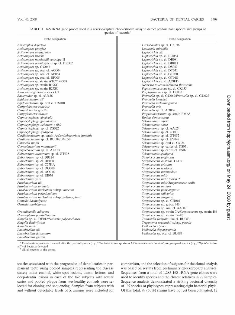
TABLE 1. 16S rRNA gene probes used in a reverse-capture checkerboard assay to detect predominant species and groups ofspecies of bacteriaa
Probe designation Probe designation
Abiotrophia defectiva Lactobacillus sp. cl. CX036Actinomyces georgiae Lautropia mirabilisActinomyces gerencseriae Leptotrichia allActinomyces israelii Leptotrichia sp. cl. BU064Actinomyces naeslundii serotype II Leptotrichia sp. cl. DE081Actinomyces odontolyticus sp. cl. DR002 Leptotrichia sp. cl. DR011Actinomyces sp. GU067 Leptotrichia sp. cl. DS049Actinomyces sp. oral cl. AG004 Leptotrichia sp. cl. DT031Actinomyces sp. oral cl. AP064 Leptotrichia sp. cl. GT020Actinomyces sp. oral cl. EP005 Leptotrichia sp. cl. GT018Actinomyces sp. strain ATCC 49338 Leptotrichia sp. cl. A39FDActinomyces sp. strain B19SC Neisseria mucosa/Neisseria flavescensActinomyces sp. strain B27SC Peptostreptococcus sp. cl. CK035Atopobium genomospecies C1 Porphyromonas sp. cl. DS033Bacteroides sp. cl. AU126 Prevotella sp. cl. GU069/Prevotella sp. cl. GU027Bifidobacterium allb Prevotella loescheiiBifidobacterium sp. oral cl. CX010 Prevotella melaninogenicaCampylobacter concisus Prevotella orisCampylobacter gracilis Prevotella sp. cl. AO036Campylobacter showae Propionibacterium sp. strain FMA5Capnocytophaga gingivalis Rothia dentocariosaCapnocytophaga granulosum Selenomonas infelixCapnocytophaga ochracea � 089 Selenomonas noxiaCapnocytophaga sp. cl. DS022 Selenomonas sp. cl. AA024Capnocytophaga sputigena Selenomonas sp. cl. GT010Cardiobacterium sp. strain A/Cardiobacterium hominis Selenomonas sp. cl. GT052Cardiobacterium sp. cl. BU084/BM058 Selenomonas sp. cl. EY047Catonella morbi Selenomonas sp. oral cl. Cs024Corynebacterium matruchotii Selenomonas sp. caries cl. DS051Corynebacterium sp. cl. AK153 Selenomonas sp. caries cl. DS071Eubacterium saburreum sp. cl. GT038 Selenomonas sputigenaEubacterium sp. cl. BB124 Streptococcus anginosusEubacterium sp. cl. BE088 Streptococcus australis T1-E5Eubacterium sp. cl. C27KA Streptococcus cristatusEubacterium sp. cl. DO008 Streptococcus gordoniiEubacterium sp. cl. DO016 Streptococcus intermediusEubacterium sp. cl. EI074 Streptococcus mitisEubacterium yurii Streptococcus mitis biovar 2Fusobacterium all Streptococcus mitis/Streptococcus oralisFusobacterium animalis Streptococcus mutansFusobacterium nucleatum subsp. vincentii Streptococcus parasanguinisFusobacterium periodonticum Streptococcus salivariusFusobacterium nucleatum subsp. polymorphum Streptococcus sanguinisGemella haemolysans Streptococcus sp. cl. CH016Gemella morbillorum Streptococcus sp. group H6
Streptococcus sp. oral cl. AA007Granulicatella adiacens Streptococcus sp. strain 7A/Streptococcus sp. strain H6Haemophilus parainfluenzae Streptococcus sp. strain T4-E3Kingella sp. cl. DE012/Neisseria polysaccharea Tannerella forsythia-like cl. BU063Kingella denitrificans Treponema socranskii subsp. paredisKingella oralis Veillonella atypicaLactobacillus all Veillonella dispar/parvulaLactobacillus fermentum Veillonella sp. oral cl. BU083Lactobacillus gasseri
a Combination probes are named after the pairs of species (e.g., “Cardiobacterium sp. strain A/Cardiobacterium hominis”) or groups of species (e.g., “Bifidobacteriumall”) of bacteria detected.
b all, all species of the genus.
VOL. 46, 2008 BACTERIA OF DENTAL CARIES 1409
on May 24, 2018 by guest
http://jcm.asm
.org/D
ownloaded from


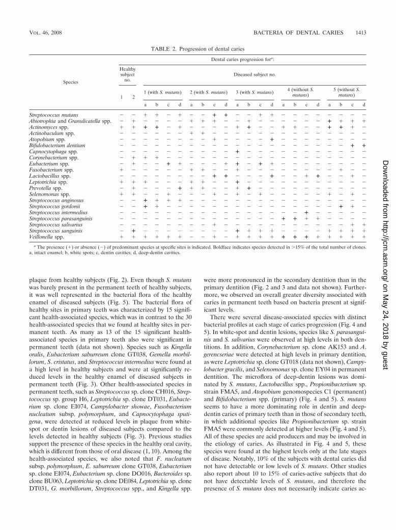
were previously uncharacterized strains, and 86 were knownspecies or phylotypes. Twenty-two novel phylotypes were iden-tified in this study. From each lesion, the number of taxaranged from 8 to 37 (Fig. 1). The phylogenetic relationships ofspecies detected in health and disease are shown in Fig. 1, andthe differences in bacterial profiles between healthy samplesand those from each of the four stages of the disease aresummarized in Table 2.
In subjects with S. mutans (subjects 1, 2, and 3) (Table 2),additional species, such as Atopobium genomospecies C1 orLactobacillus spp., were present at significantly higher levels. Insubject 1, Atopobium genomospecies C1 predominated amongall species, constituting 40% of the clones, while S. mutans wasdetected only at a low level (Fig. 1). In subjects without S.mutans, subject 4 had a predominance of Lactobacillus spp.,
whereas subject 5 had a predominance of Bifidobacterium den-tium and low-pH non-S. mutans streptococci, including S. mitis,S. sanguinis, Streptococcus gordonii, Streptococcus sp. cloneBW009, Streptococcus sp. clone EK048, and Streptococcus sali-varius (Fig. 1). The bacterial profiles during the initiation ofcaries (i.e., on intact enamel and white-spot lesions) (Fig. 1,sites a and b) were more distinct and complex than those inestablished caries (i.e., dentin and deep-dentin lesions) (Fig. 1,sites c and d, and Table 2). Actinomyces spp. and non-S. mutansstreptococci were detected at high levels in caries initiation(Fig. 1, sites a and b, and Table 2) compared to those of otherdisease states. Bacterial profiles of caries in the secondarydentition varied from subject to subject (Fig. 1, Table 2).
The goals of the clonal analyses were to first determine thebreadth of bacterial diversity in dental plaque samples and
FIG. 1. Phylogenetic relationships of species detected from dental caries in permanent teeth and from healthy controls. Subjects with (w Sm)and without (w/o Sm) detectable levels of S. mutans (Sm) were selected for clonal analysis. The distribution and levels of bacterial species/phylotypes among the five subjects and two healthy controls are shown by the columns of boxes to the right of the tree as either not detected (clearbox), �15% of the total number of clones assayed (shaded box), or �15% of the total number of clones assayed (darkened box). Fifteen percentwas chosen as the arbitrary level. GenBank accession numbers are provided. The marker bar represents a 5% difference in nucleotide sequences.
FIG. 2. Mean levels of health-associated species or species groups in primary teeth determined by a reverse-capture checkerboard assay for 15subjects with caries (green bars) and 14 healthy controls (white bars). *, P � 0.05 for pairwise comparisons of the healthy group to other groupsby PROC MIXED. P indicates the overall P value comparing all groups and sites by PROC MIXED. Strep., Streptococcus; Porphyrom.,Porphyromonas; Eubact., Eubacterium; Capnocyto., Capnocytophaga; Fusobact., Fusobacterium; cl., clone.
VOL. 46, 2008 BACTERIA OF DENTAL CARIES 1411
on May 24, 2018 by guest
http://jcm.asm
.org/D
ownloaded from

then assess specific bacterial associations with health and dis-ease. However, to better assess bacterial associations withhealth status, the final step of our study was to use a reverse-capture DNA-DNA hybridization assay using oligonucleotideprobes targeting 110 prevalent oral bacterial taxa. A total of243 clinical samples from primary and secondary teeth in atotal of 90 selected subjects were analyzed. Bacteria in pooledplaque samples from the enamel of healthy subjects (n � 39)were compared to those in samples from intact enamel, white-spot lesions, and dentin and deep-dentin lesions from each ofthe subjects with severe decay (n � 51). Pooled plaque samplesfrom a minimum of four sites in healthy individuals and pooledmaterial collected from at least three different teeth represent-ing four states of caries may dilute the number of bacteria that
dominate and alter the proportion of species present at a singlesite. Nevertheless, the predominant species present still will bedetected.
Based on these checkerboard analyses, differences in bacte-rial profiles between primary and secondary teeth, as well asthose of a number of health-associated and disease-associatedspecies, were revealed (Fig. 2 to 5, Table 3). Statistical analysesalso showed that bacterial profiles of the intact enamel ofhealthy subjects differed significantly from the bacterial pro-files of intact enamel from diseased subjects in both age groups(data not shown). In primary teeth, health-associated speciessuch as Capnocytophaga granulosa, Eubacterium sp. cloneDO016, and Streptococcus cristatus were detected at the samelevels in plaque from intact enamel of diseased subjects and
FIG. 3. Mean levels of health-associated species or species groups in permanent teeth determined by a reverse-capture checkerboard assay for36 subjects with caries (green bars) and 25 healthy controls (white bars). *, P � 0.05 for pairwise comparisons of the healthy group to other groupsby PROC MIXED. P indicates the overall P value comparing all groups and sites by PROC MIXED. Strep., Streptococcus; Eubact., Eubacterium;Eubact. sabur., Eubacterium saburreum; Capnocyto., Capnocytophaga; Fusobact. nuc. ss poly., Fusobacterium nucleatum subsp. polymorphum; Actino,Actinomyces; cl., clone.
1412 AAS ET AL. J. CLIN. MICROBIOL.
on May 24, 2018 by guest
http://jcm.asm
.org/D
ownloaded from
plaque from healthy subjects (Fig. 2). Even though S. mutanswas barely present in the permanent teeth of healthy subjects,it was well represented in the bacterial flora of the healthyenamel of diseased subjects (Fig. 5). The bacterial flora ofhealthy sites in primary teeth was characterized by 15 signifi-cant health-associated species, which was in contrast to the 30health-associated species that we found at healthy sites in per-manent teeth. As many as 13 of the 15 significant health-associated species in primary teeth also were significant inpermanent teeth (data not shown). Species such as Kingellaoralis, Eubacterium saburreum clone GT038, Gemella morbil-lorum, S. cristatus, and Streptococcus intermedius were found ata high level in healthy subjects and were at significantly re-duced levels in the healthy enamel of diseased subjects inpermanent teeth (Fig. 3). Other health-associated species inpermanent teeth, such as Streptococcus sp. clone CH016, Strep-tococcus sp. group H6, Leptotrichia sp. clone DT031, Eubacte-rium sp. clone EI074, Campylobacter showae, Fusobacteriumnucleatum subsp. polymorphum, and Capnocytophaga sputi-gena, were detected at reduced levels in plaque from white-spot or dentin lesions of diseased subjects compared to thelevels detected in healthy subjects (Fig. 3). Previous studiessupport the presence of these species in the healthy oral cavity,which is different from those of oral disease (1, 10). Among thehealth-associated species, we also noted that F. nucleatumsubsp. polymorphum, E. saburreum clone GT038, Eubacteriumsp. clone EI074, Eubacterium sp. clone DO016, Bacteroides sp.clone BU063, Leptotrichia sp. clone DE084, Leptotrichia sp. cloneDT031, G. morbillorum, Streptococcus spp., and Kingella spp.
were more pronounced in the secondary dentition than in theprimary dentition (Fig. 2 and 3 and data not shown). Further-more, we observed an overall greater diversity associated withcaries in permanent teeth based on bacteria present at signif-icant levels.
There were several disease-associated species with distinctbacterial profiles at each stage of caries progression (Fig. 4 and5). In white-spot and dentin lesions, species like S. parasangui-nis and S. salivarius were observed at high levels in both den-titions. In addition, Corynebacterium sp. clone AK153 and A.gerencseriae were detected at high levels in primary dentition,as were Leptotrichia sp. clone GT018 (data not shown), Campy-lobacter gracilis, and Selenomonas sp. clone EY04 in permanentdentition. The microflora of deep-dentin lesions was domi-nated by S. mutans, Lactobacillus spp., Propionibacterium sp.strain FMA5, and Atopobium genomospecies C1 (permanent)and Bifidobacterium spp. (primary) (Fig. 4 and 5). S. mutansseems to have a more dominating role in dentin and deep-dentin caries of primary teeth than in those of secondary teeth,in which additional species like Propionibacterium sp. strainFMA5 were commonly detected at higher levels (Fig. 4 and 5).All of these species are acid producers and may be involved inthe etiology of caries. As illustrated in Fig. 4 and 5, thesespecies were found at the highest levels only at the late stagesof disease. Notably, 10% of the subjects with dental caries didnot have detectable or low levels of S. mutans. Other studiesalso report about 10 to 15% of caries-active subjects that donot have detectable levels of S. mutans, and therefore thepresence of S. mutans does not necessarily indicate caries ac-
TABLE 2. Progression of dental caries
Species
Dental caries progression fora:
Healthysubject
no.Diseased subject no.
1 21 (with S. mutans) 2 (with S. mutans) 3 (with S. mutans) 4 (without S.
mutans)5 (without S.
mutans)
a b c d a b c d a b c d a b c d a b c d
Streptococcus mutans � � � � � � � � � � � � � � � � � � � � � �Abiotrophia and Granulicatella spp. � � � � � � � � � � � � � � � � � � � � � �Actinomyces spp. � � � � � � � � � � � � � � � � � � � � � �Actinobaculum spp. � � � � � � � � � � � � � � � � � � � � � �Atopobium spp. � � � � � � � � � � � � � � � � � � � � � �Bifidobacterium dentium � � � � � � � � � � � � � � � � � � � � � �Capnocytophaga spp. � � � � � � � � � � � � � � � � � � � � � �Corynebacterium spp. � � � � � � � � � � � � � � � � � � � � � �Eubacterium spp. � � � � � � � � � � � � � � � � � � � � � �Fusobacterium spp. � � � � � � � � � � � � � � � � � � � � � �Lactobacillus spp. � � � � � � � � � � � � � � � � � � � � � �Leptotrichia spp. � � � � � � � � � � � � � � � � � � � � � �Prevotella spp. � � � � � � � � � � � � � � � � � � � � � �Selenomonas spp. � � � � � � � � � � � � � � � � � � � � � �Streptococcus anginosus � � � � � � � � � � � � � � � � � � � � � �Streptococcus gordonii � � � � � � � � � � � � � � � � � � � � � �Streptococcus intermedius � � � � � � � � � � � � � � � � � � � � � �Streptococcus parasanguinis � � � � � � � � � � � � � � � � � � � � � �Streptococcus salivarius � � � � � � � � � � � � � � � � � � � � � �Streptococcus sanguinis � � � � � � � � � � � � � � � � � � � � � �Veillonella spp. � � � � � � � � � � � � � � � � � � � � � �
a The presence (�) or absence (�) of predominant species at specific sites is indicated. Boldface indicates species detected in �15% of the total number of clones.a, intact enamel; b, white spots; c, dentin cavities; d, deep-dentin cavities.
VOL. 46, 2008 BACTERIA OF DENTAL CARIES 1413
on May 24, 2018 by guest
http://jcm.asm
.org/D
ownloaded from
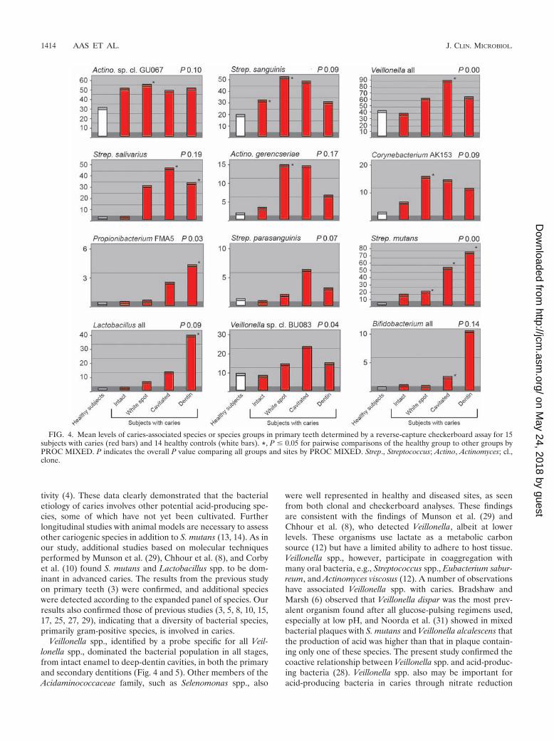
tivity (4). These data clearly demonstrated that the bacterialetiology of caries involves other potential acid-producing spe-cies, some of which have not yet been cultivated. Furtherlongitudinal studies with animal models are necessary to assessother cariogenic species in addition to S. mutans (13, 14). As inour study, additional studies based on molecular techniquesperformed by Munson et al. (29), Chhour et al. (8), and Corbyet al. (10) found S. mutans and Lactobacillus spp. to be dom-inant in advanced caries. The results from the previous studyon primary teeth (3) were confirmed, and additional specieswere detected according to the expanded panel of species. Ourresults also confirmed those of previous studies (3, 5, 8, 10, 15,17, 25, 27, 29), indicating that a diversity of bacterial species,primarily gram-positive species, is involved in caries.
Veillonella spp., identified by a probe specific for all Veil-lonella spp., dominated the bacterial population in all stages,from intact enamel to deep-dentin cavities, in both the primaryand secondary dentitions (Fig. 4 and 5). Other members of theAcidaminococcaceae family, such as Selenomonas spp., also
were well represented in healthy and diseased sites, as seenfrom both clonal and checkerboard analyses. These findingsare consistent with the findings of Munson et al. (29) andChhour et al. (8), who detected Veillonella, albeit at lowerlevels. These organisms use lactate as a metabolic carbonsource (12) but have a limited ability to adhere to host tissue.Veillonella spp., however, participate in coaggregation withmany oral bacteria, e.g., Streptococcus spp., Eubacterium sabur-reum, and Actinomyces viscosus (12). A number of observationshave associated Veillonella spp. with caries. Bradshaw andMarsh (6) observed that Veillonella dispar was the most prev-alent organism found after all glucose-pulsing regimens used,especially at low pH, and Noorda et al. (31) showed in mixedbacterial plaques with S. mutans and Veillonella alcalescens thatthe production of acid was higher than that in plaque contain-ing only one of these species. The present study confirmed thecoactive relationship between Veillonella spp. and acid-produc-ing bacteria (28). Veillonella spp. also may be important foracid-producing bacteria in caries through nitrate reduction
FIG. 4. Mean levels of caries-associated species or species groups in primary teeth determined by a reverse-capture checkerboard assay for 15subjects with caries (red bars) and 14 healthy controls (white bars). *, P � 0.05 for pairwise comparisons of the healthy group to other groups byPROC MIXED. P indicates the overall P value comparing all groups and sites by PROC MIXED. Strep., Streptococcus; Actino, Actinomyces; cl.,clone.
1414 AAS ET AL. J. CLIN. MICROBIOL.
on May 24, 2018 by guest
http://jcm.asm
.org/D
ownloaded from

(11). Silva Mendez et al. (36) demonstrated that acidity levelsbelow pH 7 and low concentrations of nitrite (0.2 mM) causedthe complete killing of S. mutans, with similar effects on theother organisms tested.
Actinomyces spp. play a major role in coaggregation inter-actions (9). Burt et al. (7) reported that Actinomyces-like or-ganisms usually predominate in noncariogenic plaques withhigh levels of S. mutans streptococci and lactobacilli. Actino-myces spp. are heterofermenters but tend to become homolac-tic producers under anaerobic conditions (16), thus contribut-ing to enamel demineralization. Strong coaggregation betweenActinomyces odontolyticus and Actinomyces israelii with strainsof either Veillonella parvula or Prevotella prevotii has been re-ported (35). Studies on coaggregation interactions betweenStreptococcus spp. and Actinomyces spp. have revealed thecomplementary adhesion-receptor mechanisms among the lat-ter organisms (9). A large proportion of positively coaggregat-ing pairs between either Prevotella intermedia ND8-9A orCampylobacter gracilis ND9-8A and strains of Streptococcus,Gemella, Peptostreptococcus, Lactobacillus, and Actinomycesindicate that the gram-negative obligate anaerobic rods play an
important role in the interactions leading to root caries (35).Nadkarni et al. (30) and Chhour et al. (8) found that novel anduncultured Prevotella and Prevotella-like bacteria dominate thediverse polymicrobial community in some cases of caries, sug-gesting an active role of Prevotella in caries progression. Fur-thermore, coaggregation between Fusobacterium nucleatumNT6-6A and six other bacterial species, i.e., Streptococcus bovisII/2 ND2-2, Streptococcus constellatus ND10-13A, Streptococ-cus sanguinis II ND7-3, Lactobacillus acidophilus ND7-2A, C.sputigena ND2-12A, and P. intermedia ND8-9A, shows thatthese bacteria have the ability to coaggregate with a largenumber of oral bacteria and possibly act as key organisms indental plaque formation during the later stages of plaque mat-uration and modulation of the climax community (20, 21).With further evidence that a large number of bacterial speciesare involved in the progression of dental caries, the interac-tions within different bacterial communities representing dif-ferent stages of the disease would be of considerable interest.
In light of recent efforts to develop vaccines specific for S.mutans, the study of the potential etiologic role of all speciesassociated with caries and caries progression is essential. The
FIG. 5. Mean levels of caries-associated species or species groups in permanent teeth determined by a reverse-capture checkerboard assay for36 subjects with caries (red bars) and 25 healthy controls (white bars). *, P � 0.05 for pairwise comparisons of the healthy group to other groupsby PROC MIXED. P indicates the overall P value comparing all groups and sites by PROC MIXED. Strep., Streptococcus; genomo.,genomospecies.
VOL. 46, 2008 BACTERIA OF DENTAL CARIES 1415
on May 24, 2018 by guest
http://jcm.asm
.org/D
ownloaded from

significance of our work is that the identification of additionalcaries pathogens provides alternative targets for biological in-terventions, and the identification of beneficial health-associ-ated species could provide the basis for therapeutic interven-tions to establish caries-resistant microbial communities.
Although we have shown that specific bacterial species areassociated with health, caries initiation, and caries production,there is subject-to-subject variation in the bacterial composi-tion. Munson et al. (29) demonstrated that many species ofLactobacillus were found overall in carious lesions, but onlyone or two Lactobacillus spp. were detected in each lesion in asubject. We and others have shown that 10 to 20% of subjectswith severe caries may not have detectable levels of S. mutansbut rather exhibit other acid-producing species. Furthermore,in some carious lesions S. mutans may be a minor bacterialcomponent of the dental plaque. These results support theecological plaque hypothesis (26), which suggests that caries isa result of a shift in the balance of the resident microfloradriven by changes in local environmental conditions, e.g., acidproduction by any number of species that cause tooth decay.
In summary, we have shown that half of the bacteria asso-ciated with dental caries have not yet been cultivated. Speciesin addition to S. mutans, e.g., species of Veillonella, Lactoba-cillus, Bifidobacterium, Propionibacterium, low-pH non-S. mu-tans streptococci, Actinomyces, and Atopobium, also may playan important role in caries production. Actinomyces spp. andnon-S. mutans streptococci may be involved in the initiation ofthe disease. Several specific species are associated with health,while others are associated with caries. Bacterial profileschange with the progression of disease and differ from theprimary to the secondary dentition. The present findings sup-port the ecological plaque hypothesis in caries disease, in thatchanges in ecologic factors require different bacterial qualitiesand stimulate alterations in the bacterial composition. Furtherstudies of the potential etiologic roles of these diverse bacterialcommunities, including additional and novel species, are rec-ommended. Identified cultivable and not-yet-cultivable organ-isms might provide additional targets for caries intervention.
ACKNOWLEDGMENTS
We thank Melvin Moeschberger and Kevin Tordoff for their contri-butions to the statistical analyses.
The study was supported by NIH grants DE11443 and DE016125,Omni Products, and the Faculty of Dentistry, University of Oslo,Norway.
REFERENCES
1. Aas, J. A., B. J. Paster, L. N. Stokes, I. Olsen, and F. E. Dewhirst. 2005.Defining the normal bacterial flora of the oral cavity. J. Clin. Microbiol.43:5721–5732.
2. Anusavice, K. J. 2002. Dental caries: risk assessment and treatment solutionsfor an elderly population. Compend. Contin. Educ. Dent. 23:12–20.
3. Becker, M. R., B. J. Paster, E. J. Leys, M. L. Moeschberger, S. G. Kenyon,J. L. Galvin, S. K. Boches, F. E. Dewhirst, and A. L. Griffen. 2002. Molecularanalysis of bacterial species associated with childhood caries. J. Clin. Micro-biol. 40:1001–1009.
4. Beighton, D. 2005. The complex oral microflora of high-risk individuals andgroups and its role in the caries process. Community Dent. Oral Epidemiol.33:248–255.
5. Bjørndal, L., and T. Larsen. 2000. Changes in the cultivable flora in deepcarious lesions following a stepwise excavation procedure. Caries Res. 34:502–508.
6. Bradshaw, D. J., and P. D. Marsh. 1998. Analysis of pH-driven disruption oforal microbial communities in vitro. Caries Res. 32:456–462.
7. Burt, B. A., W. J. Loesche, and S. A. Eklund. 1985. Stability of selected
TA
BL
E3.
Pred
omin
ant
spec
ies
inba
cter
ialp
rofil
esre
late
dto
cari
espr
ogre
ssio
nin
prim
ary
and
perm
anen
tde
ntiti
onsa
Spec
ies
dete
cted
inpr
imar
yde
ntiti
onSp
ecie
sde
tect
edin
perm
anen
tde
ntiti
on
Con
trol
Inta
cten
amel
Whi
te-s
pot
lesi
onD
entin
lesi
onD
eep-
dent
inle
sion
Con
trol
Inta
cten
amel
Whi
te-s
pot
lesi
onD
entin
lesi
onD
eep-
dent
inle
sion
Lep
totr
ichi
aal
lbA
ctin
omyc
essp
.cl
.GU
067
Vei
llone
llaal
lV
eillo
nella
all*
Stre
ptoc
occu
sm
utan
s*A
ctin
omyc
essp
.cl.
GU
067
Act
inom
yces
sp.c
l.G
U06
7V
eillo
nella
all
Vei
llone
llaal
lV
eillo
nella
all
Vei
llone
llaal
lV
eillo
nella
all
Act
inom
yces
sp.c
l.G
U06
7*St
rept
ococ
cus
mut
ans*
Vei
llone
llaal
lF
usob
acte
rium
nucl
eatu
msu
bsp.
poly
mor
phum
Vei
llone
llaal
lA
ctin
omyc
essp
.cl.
GU
067
Act
inom
yces
sp.c
l.G
U06
7*A
ctin
omyc
essp
.cl.
GU
067
Stre
ptoc
occu
san
gino
sus
Lep
totr
ichi
aal
lSt
rept
ococ
cus
sang
uini
s*A
ctin
omyc
essp
.cl
.GU
067
Act
inom
yces
sp.
cl.G
U06
7V
eillo
nella
all
Stre
ptoc
occu
ssa
ngui
nis
Stre
ptoc
occu
ssa
ngui
nis
Stre
ptoc
occu
ssa
livar
ius*
Stre
ptoc
occu
sm
utan
s*
Act
inom
yces
sp.
cl.G
U06
7St
rept
ococ
cus
sang
uini
s*L
epto
tric
hia
all
Stre
ptoc
occu
ssa
ngui
nis
Lac
toba
cillu
sal
l*St
rept
ococ
cus
sang
uini
sF
usob
acte
rium
nucl
eatu
msu
bsp.
poly
mor
phum
Lep
totr
ichi
asp
.cl.
GT
018
Stre
ptoc
occu
sm
utan
s*St
rept
ococ
cus
saliv
ariu
s*
Cap
nocy
toph
aga
gran
ulos
aSt
rept
ococ
cus
miti
s*St
rept
ococ
cus
saliv
ariu
sSt
rept
ococ
cus
saliv
ariu
s*St
rept
ococ
cus
saliv
ariu
sE
ubac
teriu
msp
.cl.
EI0
74E
ubac
teriu
msp
.cl.
EI0
74F
usob
acte
rium
nucl
eatu
msu
bsp.
poly
mor
phum
*St
rept
ococ
cus
sang
uini
sL
acto
baci
llus
all*
Fus
obac
teriu
mal
lC
apno
cyto
phag
asp
utig
ena
Cap
nocy
toph
aga
sput
igen
aV
eillo
nella
sp.
cl.B
U08
3St
rept
ococ
cus
sang
uini
sL
epto
tric
hia
sp.c
l.D
T03
1L
epto
tric
hia
sp.c
l.D
T03
1E
ubac
teriu
msp
.cl.
EI0
74St
rept
ococ
cus
miti
sSt
rept
ococ
cus
sang
uini
s
Stre
ptoc
occu
ssa
ngui
nis
Cap
nocy
toph
aga
gran
ulos
aSt
rept
ococ
cus
miti
sSt
rept
ococ
cus
miti
sSt
rept
ococ
cus
miti
sF
usob
acte
rium
anim
alis
Stre
ptoc
occu
sm
itis
Fus
obac
teriu
man
imal
isF
usob
acte
rium
anim
alis
Stre
ptoc
occu
sm
itis
Cam
pylo
bact
ergr
acili
sSt
rept
ococ
cus
cris
tatu
sSt
rept
ococ
cus
mut
ans
Sele
nom
onas
sp.c
l.E
Y04
7V
eillo
nella
sp.
cl.B
U08
3L
epto
tric
hia
all
Lep
totr
ichi
asp
.cl.
GT
018
Lep
totr
ichi
aal
lV
eillo
nella
sp.c
l.B
U08
3St
rept
ococ
cus
angi
nosu
s
Stre
ptoc
occu
scr
ista
tus
Act
inom
yces
sp.
stra
inB
19SC
Cor
yneb
acte
rium
sp.c
l.A
K15
3L
epto
tric
hia
all*
Act
inom
yces
sp.
stra
inB
19SC
Act
inom
yces
naes
lund
iise
roty
peII
Sele
nom
onas
sp.c
l.E
Y04
7*Se
leno
mon
assp
.cl.
EY
047
Lac
toba
cillu
sal
l*L
acto
baci
llus
gass
eri*
Cap
nocy
toph
aga
sput
igen
aSt
rept
ococ
cus
sp.c
l.C
H01
6A
ctin
omyc
esge
renc
seria
e*A
ctin
omyc
esge
renc
seria
eSe
leno
mon
assp
.cl.
EY
047
Cap
nocy
toph
aga
sput
igen
aC
apno
cyto
phag
agr
anul
osa
Stre
ptoc
occu
sm
itis
Ato
pobi
umge
nom
ospe
cies
C1*
Ato
pobi
umge
nom
ospe
cies
C1*
a%
The
resu
ltsar
eba
sed
onth
esi
gnal
leve
lin
are
vers
e-ca
ptur
ech
ecke
rboa
rdhy
brid
izat
ion
assa
y.*,
P�
0.05
for
pair
wis
eco
mpa
riso
nsof
the
heal
thy
grou
pto
othe
rgr
oups
byPR
OC
MIX
ED
test
s.cl
.,cl
one.
bal
l,al
lspe
cies
ofth
ege
nus.
1416 AAS ET AL. J. CLIN. MICROBIOL.
on May 24, 2018 by guest
http://jcm.asm
.org/D
ownloaded from

plaque species and their relationship to caries in a child population over 2years. Caries Res. 19:193–200.
8. Chhour, K. L., M. A. Nadkarni, R. Byun, F. E. Martin, N. A. Jacques, andN. Hunter. 2005. Molecular analysis of microbial diversity in advanced caries.J. Clin. Microbiol. 43:843–849.
9. Cisar, J. O., P. E. Kolenbrander, and F. C. McIntire. 1979. Specificity ofcoaggregation reactions between human oral streptococci and strains ofActinomyces viscosus or Actinomyces naeslundii. Infect. Immun. 24:742–752.
10. Corby, P. M., J. Lyons-Weiler, W. A. Bretz, T. C. Hart, J. A. Aas, T. Bou-menna, J. Goss, A. L. Corby, H. M. Junior, R. J. Weyant, and B. J. Paster.2005. Microbial risk indicators in early childhood caries. J. Clin. Microbiol.43:5753–5759.
11. Doel, J. J., N. Benjamin, M. P. Hector, M. Rogers, and R. P. Allaker. 2005.Evaluation of bacterial nitrate reduction in the human oral cavity. Eur.J. Oral Sci. 113:14–19.
12. Egland, P. G., R. J. Palmer, Jr., and P. E. Kolenbrander. 2004. Interspeciescommunication in Streptococcus gordonii-Veillonella atypica biofilms: signal-ing in flow conditions requires juxtaposition. Proc. Natl. Acad. Sci. USA101:16917–16922.
13. Fitzgerald, R. J., and P. H. Keyes. 1960. Demonstration of the etiologic roleof streptococci in experimental caries in the hamster. J. Am. Dent. Assoc.61:9–19.
14. Gibbons, R. J., and J. van Houte. 1975. Dental caries. Annu. Rev. Med.25:121–134.
15. Hahn, C.-L., W. A. J. Falkler, and G. E. Minah. 1991. Microbiological studiesof carious dentine from human teeth with irreversible pulpitis. Arch. OralBiol. 36:147–153.
16. Hogg, S. D. 1992. The lactic microflora of the oral cavity, p. 115–132. InB. J. B. Wood (ed.), The lactic acid bacteria. The lactic acid bacteria inhealth and disease, vol. 1. Elsevier Applied Science, London, United King-dom.
17. Hoshino, E. 1985. Predominant obligate anaerobes in human carious dentin.J. Dent. 64:1195–1198.
18. Jukes, T. H., and C. R. Cantor. 1969. Evolution of protein molecules, p.21–132. In H. N. Munro (ed.), Mammalian protein metabolism, vol. 3.Academic Press, Inc., New York, NY.
19. Kazor, C. E., P. M. Mitchell, A. M. Lee, L. N. Stokes, W. J. Loesche, F. E.Dewhirst, and B. J. Paster. 2003. Diversity of bacterial populations on thetongue dorsa of patients with halitosis and healthy patients. J. Clin. Micro-biol. 41:558–563.
20. Kolenbrander, P. E. 2000. Oral microbial communities: biofilms, interac-tions, and genetic systems. Annu. Rev. Microbiol. 54:413–437.
21. Kolenbrander, P. E., Y. Inouye, and L. V. Holdeman. 1983. New Actinomycesand Streptococcus coaggregation groups among human oral isolates from thesame site. Infect. Immun. 41:501–506.
22. Leys, E. J., A. L. Griffen, S. J. Strong, and P. A. Fuerst. 1994. Detection andstrain identification of Actinobacillus actinomycetemcomitans by nested PCR.J. Clin. Microbiol. 32:1288–1294.
23. Liesack, W., H. Weyland, and E. Stackebrandt. 1991. Potential risk of geneamplification by PCR as determined by 16S rDNA analysis of a mixed-culture of strict barophilic bacteria. Microb. Ecol. 21:191–198.
24. Loesche, W. J. 1992. The specific plaque hypothesis and the antimicrobialtreatment of periodontal disease. Dent. Update 19:68–74.
25. Loesche, W. J., and S. A. Syed. 1973. The predominant cultivable flora ofcarious plaque and carious dentine. Caries Res. 7:201–216.
26. Marsh, P. D. 1994. Microbial ecology of dental plaque and its significance inhealth and disease. Adv. Dent. Res. 8:263–271.
27. Martin, F. E., M. A. Nadkarni, N. A. Jacques, and N. Hunter. 2002. Quan-titative microbiological study of human carious dentine by culture and real-time PCR: association of anaerobes with histopathological changes inchronic pulpitis. J. Clin. Microbiol. 40:1698–1704.
28. Mikx, F. H., and J. S. Van der Hoeven. 1975. Symbiosis of Streptococcusmutans and Veillonella alcalescens in mixed continuous cultures. Arch. OralBiol. 20:407–410.
29. Munson, M. A., A. Banerjee, T. F. Watson, and W. G. Wade. 2004. Molecularanalysis of the microflora associated with dental caries. J. Clin. Microbiol.42:3023–3029.
30. Nadkarni, M. A., C. E. Caldon, K. L. Chhour, I. P. Fisher, F. E. Martin, N. A.Jacques, and N. Hunter. 2004. Carious dentine provides a habitat for acomplex array of novel Prevotella-like bacteria. J. Clin. Microbiol. 42:5238–5244.
31. Noorda, W. D., D. J. Purdell-Lewis, A. M. van Montfort, and A. H.Weerkamp. 1988. Monobacterial and mixed bacterial plaques of Streptococcusmutans and Veillonella alcalescens in an artificial mouth: development, me-tabolism, and effect on human dental enamel. Caries Res. 22:342–347.
32. Paster, B. J., I. M. Bartoszyk, and F. E. Dewhirst. 1998. Identification of oralstreptococci using PCR-based, reverse-capture, checkerboard hybridization.Methods Cell Sci. 20:223–231.
33. Paster, B. J., S. K. Boches, J. L. Galvin, R. E. Ericson, C. N. Lau, V. A.Levanos, A. Sahasrabudhe, and F. E. Dewhirst. 2001. Bacterial diversity inhuman subgingival plaque. J. Bacteriol. 183:3770–3783.
34. Saitou, N., and M. Nei. 1987. The neighbor-joining method: a new methodfor reconstructing phylogenetic trees. Mol. Biol. Evol. 4:406–425.
35. Shen, S., L. P. Samaranayake, and H. K. Yip. 2005. Coaggregation profilesof the microflora from root surface caries lesions. Arch. Oral Biol. 50:23–32.
36. Silva Mendez, L. S., R. P. Allaker, J. M. Hardie, and N. Benjamin. 1999.Antimicrobial effect of acidified nitrite on cariogenic bacteria. Oral Micro-biol. Immunol. 14:391–392.
37. Theilade, E. 1986. The non-specific theory in microbial etiology of inflam-matory periodontal diseases. J. Clin. Periodontol. 13:905–911.
38. Van de Peer, Y., and R. De Wachter. 1994. TREECON for Windows: asoftware package for the construction and drawing of evolutionary trees forthe Microsoft Windows environment. Comput. Appl. Biosci. 10:569–570.
39. World Health Organization. 2002. The world health report. Reducing risks,promoting healthy life. http://www.who.int/whr/2002/en/.
VOL. 46, 2008 BACTERIA OF DENTAL CARIES 1417
on May 24, 2018 by guest
http://jcm.asm
.org/D
ownloaded from