caries and the extraction of teeth
TRANSCRIPT

FACULTY OF DENTISTRYDepartment of Oral Diagnostics
Dr. Szabó Bence Tamás
DEPARTMENT OF ORAL DIAGNOSTICS
Caries
and the extraction of teeth
23TH September 2020

Caries and the extraction of teeth Dr. Szabó Bence Tamás
assistant lecturer
to be faithful to the title…
how to treat?

Caries and the extraction of teeth Dr. Szabó Bence Tamás
assistant lecturer
Acid
ge
ne
ratio
n
Caries on susceptible tooth surface
MULTIFACTORIAL

Caries and the extraction of teeth Dr. Szabó Bence Tamás
assistant lecturer
Streptococcus mutans, Actinomyces Viscosus
95% of the population
pH of 5,5 =>demineralization of enamel
predilection places
etiology

Caries and the extraction of teeth Dr. Szabó Bence Tamás
assistant lecturer
classification of carious lesions
occlusal, proximal, buccal, lingual, root surface
Course:
usually slow rampant caries
recurrent: next to the filling
arrested
Special cases:
After radiotherapy in case of head and neck tumors» xerostomia=dry mouth. destruction begins at cervical region and may encircle the
tooth very soon, leading to the loss of the entire crown
» X-ray film : we see radiolucent shadows at the neck of teeth.
Rampant caries: » acute, 6-8 year old children without proper toothbrushing, todays it is less common
due to education and fluorid. PREVENTION!!!!
» X-ray film: extensive interproximal and smooth surface caries

Caries and the extraction of teeth Dr. Szabó Bence Tamás
assistant lecturer
Rampant caries
3 2 1 1 2 3
acute course
uncontrollable teeth destuction.
deep approximal caries on the
maxillary incisors.

Caries and the extraction of teeth Dr. Szabó Bence Tamás
assistant lecturer
caries lesion on radiographs
caries=demineralizationradiolucent
on radiograph
It is possible to make mistake in two diffreent way: false positive diagnosis
or fals negative diagnosis
It is well known that caries progression is slow, so the dentist should be
conservative during caries diagnosis and treatment.
DON’T MISS THE CHANCE!
sound tooth

Caries and the extraction of teeth Dr. Szabó Bence Tamás
assistant lecturer
…but how to find?
How often should we perform radiographic examination?
Per
iap
ical
rad
iogr
aph • Is there any changes
in periapical and interradicular bone?
• Affected pulp:
• radicular cyst
• granuloma, etc.
Bit
ewin
gra
dio
grap
h • distal third of canineand the interproximaland occlusal surfacesof premolars and molars

Caries and the extraction of teeth Dr. Szabó Bence Tamás
assistant lecturer
bitewing films
the upper and lower teeth on the same film
approximal surfaces well visualized
good to know: more than half of all proximalsurface lesion can not be seen clinically and maybe detected only with radiograph

Caries and the extraction of teeth Dr. Szabó Bence Tamás
assistant lecturer
extension and the shape
ENAMEL
• D1: enamel caries penetratingless than half way throughenamel, notch shape
• D2: enamel caries penetratingmore than half way throughenamel, BUT not envolvingdentinoenamel junction, triangular shape
E+DENTIN
• D3: caries of enamel and dentin, extending less thanhalf way to pulp cavity, triangular shape (duo)
• D4: caries of enamel and dentin, penetrating more thanhalf way dentin toward pulpcavity

Caries and the extraction of teeth Dr. Szabó Bence Tamás
assistant lecturer
hystology, just a bit…

Caries and the extraction of teeth Dr. Szabó Bence Tamás
assistant lecturer
radiographic appearance of occlusal
caries
usually more extensive, borders not so well
defined.
pitfalls during the interpretation of the radiograph:
superimposition
buccal and occlusal caries.
location

Caries and the extraction of teeth Dr. Szabó Bence Tamás
assistant lecturer
radiographic appearance of proximal
caries
MAGNIFICATION!!!
actual depth of the caries is deeper than the
radiographically detected deepness.
develop slowly: 3 years to clinically apparent (white spot)
pitfalls:
cervical burnout
concavities produced by abrasion (flossing)
location
Caries and the extraction of teeth Dr. Szabó Bence Tamás
assistant lecturer
radiographic appearance of facial,
buccal and lingual caries
in enamel pits and fissures (premolars, molars, incisors,
foramen coecum!)
till small: round shape, later: elliptic/semilunar
sharp borders ( occlusal caries)
DANGER! USE MORE VIEWS! lesion can superimpose to the
CEJ or to the proximal surface => false occlusal or
proximal caries.
differentiation between buccal and lingual caries… more
than a piece of cake….
Clinical evaluation and probing are necessary!
location

Caries and the extraction of teeth Dr. Szabó Bence Tamás
assistant lecturer
vestibular surface cariesoval transparency projected in the middle
of the crown

Caries and the extraction of teeth Dr. Szabó Bence Tamás
assistant lecturer
coecum caries
(foramen coecum is usually on
the second incisors)
3 2 1
Caries and the extraction of teeth Dr. Szabó Bence Tamás
assistant lecturer
radiographic appearance of root
surface caries =cemental caries
Involves both CEMENTUM DENTIN !!!
Enamel is also affected in a special case: when the lesion extends into
the dentin under the enamel along the CEJ.
In elderly people it has a frequency of 40-70 %
Associated with gingival recession and horizontal bone loss
Affected surfaces in a decreasing order: buccal, lingual, proximal
On radiograph: ill defined, saucerlike or notched radiolucency
Pitfalls: false positive : cervical burn out
location

Caries and the extraction of teeth Dr. Szabó Bence Tamás
assistant lecturer
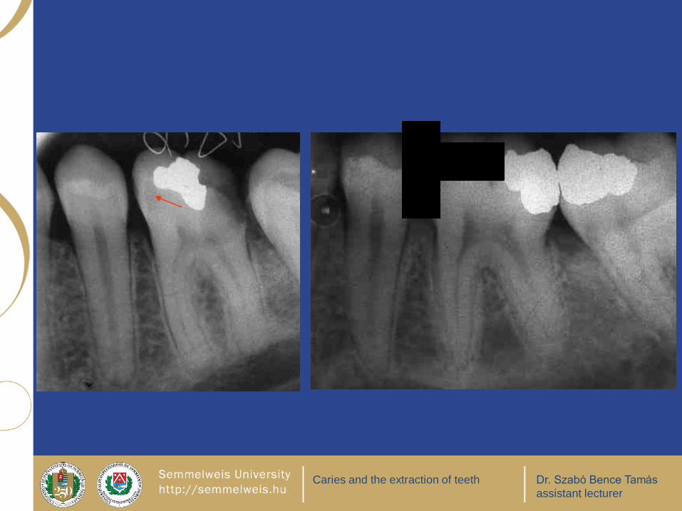
radiographic appearance of recurrent
caries next to the restoration
direction of the beam!
recurrent lesions at the mesiogingival, distogingival and occlusal margins of a restoration are frequently discoveredon radiograph.
BUT: we miss relativly big lesions around the buccal, lingualor facial restorations
causes:
poor adaptation of a restoration-marginal leakage
inadequate extension of a restoration
the original lesion is not completly evacuated
=> residual or recurrent caries???

Caries and the extraction of teeth Dr. Szabó Bence Tamás
assistant lecturer
8 7 6 5
8: mesioangular retention, 7: occlusal caries, 6: MO amalgam filling, sec. caries on the approximal and
lingual surface

Caries and the extraction of teeth Dr. Szabó Bence Tamás
assistant lecturer
When can you see the secondary caries under the filling?
Wherever the restoration is, we
will see the caries under it if the X-
ray beam is parallel with the
approximal surface.
B: The secondary caries will not
be visible if it is situated in middle
and the beam comes from above
or from beneath.
A: The secondary caries in the
buccal corner will not be visible if
the beam comes from beneath,
but we will see it if the beam
comes from above.
A:The secondary caries in the
lingual corner will not be visible if
the beam comes from above, but
we will see it if the beam comes
from beneath.

Caries and the extraction of teeth Dr. Szabó Bence Tamás
assistant lecturer
filling materials on X-ray film
atomic number thickness quality of X-ray
RADIOPAQUE = RADIODENS = WHITE:
amalgam
gold
calcium hydroxide
guttapercha
silver point
RADIOLUCENT = TRANSPARENT = BLACK: silicate
Composite resin
porcelain

Caries and the extraction of teeth Dr. Szabó Bence Tamás
assistant lecturer
DANGER: FAILURES
Dg.: no caries, but we discribe something as a caries (false positive):
cervical burnout
pseudotransparency
mach bands
Dg.: caries, but we miss it (false negative):
X-ray beam is not ortoradial, the crowns can overlap eachother (superimposition), and hide the approximalcaries.
external oblique ridge can superimpose to a lesion, orcusps can hide occlusal caries

Caries and the extraction of teeth Dr. Szabó Bence Tamás
assistant lecturer
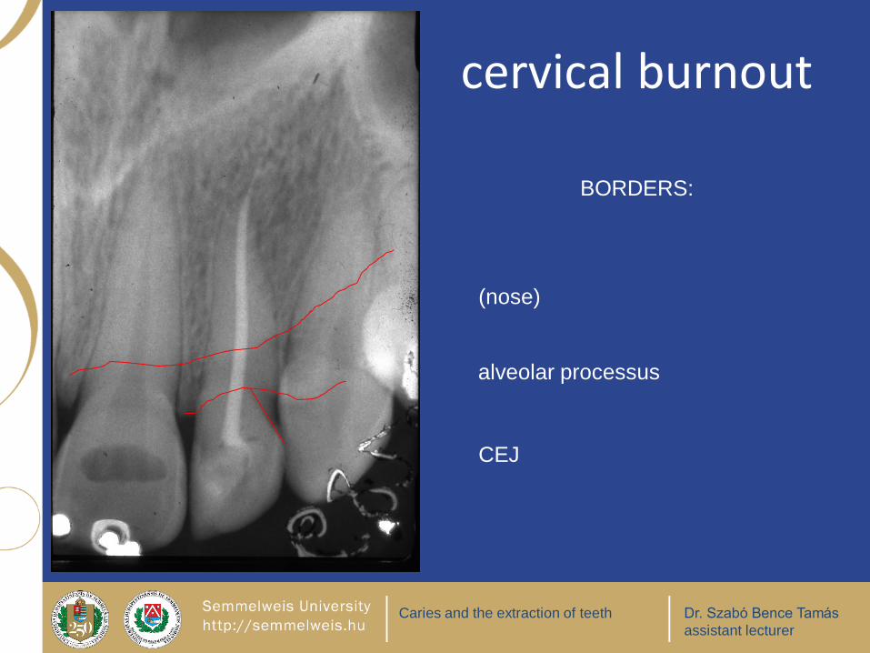
cervical burnout
beam is not ortoradial or the
tooth is in torsion
CEJ has a sinus wave shape
triangular shape transparency
Caries and the extraction of teeth Dr. Szabó Bence Tamás
assistant lecturer
(nose)
alveolar processus
CEJ
cervical burnout
BORDERS:

Caries and the extraction of teeth Dr. Szabó Bence Tamás
assistant lecturer
cervical burn out

Caries and the extraction of teeth Dr. Szabó Bence Tamás
assistant lecturer
pseudotransparency
• cervical part of the tooth is free due to
horizontal bone loss
• not covered by enamel,
=> is relatively darker
remember: X-ray summarizes

Caries and the extraction of teeth Dr. Szabó Bence Tamás
assistant lecturer
Little arrow: secondary caries, big arrow: pseudotransparency

Caries and the extraction of teeth Dr. Szabó Bence Tamás
assistant lecturer
1 2 3 4 5 and a
supernumerary tooth
pseudotransparency

Caries and the extraction of teeth Dr. Szabó Bence Tamás
assistant lecturer
Caries and the extraction of teeth Dr. Szabó Bence Tamás
assistant lecturer

Caries and the extraction of teeth Dr. Szabó Bence Tamás
assistant lecturer
ortoradial beam (goes parallel to the proximal surface of the tooth)
-> no overlapping of crowns.
mesioexcentric X-ray beam
-> there will be less overlapping between the crowns
mesioexcentricdistoexcentric
orthoradial

Caries and the extraction of teeth Dr. Szabó Bence Tamás
assistant lecturer
8 7 6
overlapping crowns: the x-ray beam is not ortoradial
failure possibility: approximal caries is not visible

Caries and the extraction of teeth Dr. Szabó Bence Tamás
assistant lecturer
anatomical landmarks: external oblique
ridge is superimposed to a caries

Caries and the extraction of teeth Dr. Szabó Bence Tamás
assistant lecturer
Transparent zone (hystologic terminus technicus):
opaque line/area under the occlusal caries = trasparent zone on histologic image
Cause: increased mineralization, narrowed dentin canaliculi
7

Caries and the extraction of teeth Dr. Szabó Bence Tamás
assistant lecturer
Use magnifying glass!D3 D1
3 4 5 6 7
3 4 5 6 7

Caries and the extraction of teeth Dr. Szabó Bence Tamás
assistant lecturer
On radiograph sometimes the caries does not seem to be continous.
Remember: X-ray summarizes!

Caries and the extraction of teeth Dr. Szabó Bence Tamás
assistant lecturer
deep occlusal caries
secondary
caries
cervical burn out
D3D3
5 6 7 8
5 6 7 8

Caries and the extraction of teeth Dr. Szabó Bence Tamás
assistant lecturer
8 7 6 5
5: total destruction of the crown, 6: distoapproximal D3 caries,the lesion penetrate
under CEJ-due to horizontal bone loss

Caries and the extraction of teeth Dr. Szabó Bence Tamás
assistant lecturer
5 7 8
deep occlusal caries (8): we examine whether the pulp is affected

Caries and the extraction of teeth Dr. Szabó Bence Tamás
assistant lecturer
4 5 6 7
deep occlusal caries
accessory radix

Caries and the extraction of teeth Dr. Szabó Bence Tamás
assistant lecturer
8 7 6deep occlusal caries
in the region of the contact
point D2 & D3 carieses in
the neighbouring teeth

Caries and the extraction of teeth Dr. Szabó Bence Tamás
assistant lecturer
3 4 5 6 7 3 4 5 6 7 D4
D3 D3
D1

Caries and the extraction of teeth Dr. Szabó Bence Tamás
assistant lecturer
4 5 6 7 8
first molar: D4 caries on occlusal and lingual surface, no periapical
lesion

Caries and the extraction of teeth Dr. Szabó Bence Tamás
assistant lecturer
on the base of X-ray film we can not know about the pulp even in the case
of very deep approximal caries. ODL D4 caries.
4 5 6 7

Caries and the extraction of teeth Dr. Szabó Bence Tamás
assistant lecturer
X-rays summarize!! A relatively smaller carious lesion can project to the
pulp a bigger lesion which really affect the pulp can missed because of the
big amount of sound enamel and dentin which surround it.
Be careful! It is not possible to saywhether the pulp is affected by alonethe radiograph. It is just a 2D image!!!

Caries and the extraction of teeth Dr. Szabó Bence Tamás
assistant lecturer
6 7 8OLD caries

Caries and the extraction of teeth Dr. Szabó Bence Tamás
assistant lecturer
8 6 5
Wisdom tooth: oclusal caries, external oblique ridge is
superimposed, mesioangular impaction, semilunar resorption,
pericoronitis (clinical sign could be: trismus)

Caries and the extraction of teeth Dr. Szabó Bence Tamás
assistant lecturer
8 7 5 Lingual caries, coecum caries

Caries and the extraction of teeth Dr. Szabó Bence Tamás
assistant lecturer
pseudotransparency
vestibular caries
approximal
caries
butterfly retention: not
useable technique any more
Caries and the extraction of teeth Dr. Szabó Bence Tamás
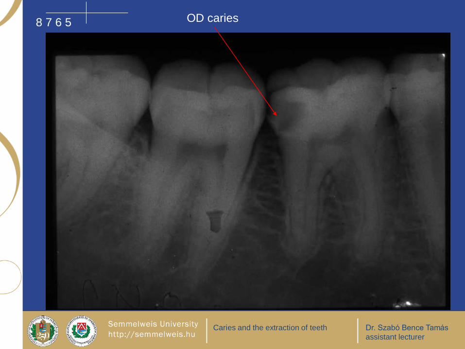
assistant lecturer
8 7 6 5 OD caries

Caries and the extraction of teeth Dr. Szabó Bence Tamás
assistant lecturer
4 1 1
root surface caries: in case of horizontal bone loss the root is not surrounded by
bone, usually not triangular shape.
Caries and the extraction of teeth Dr. Szabó Bence Tamás
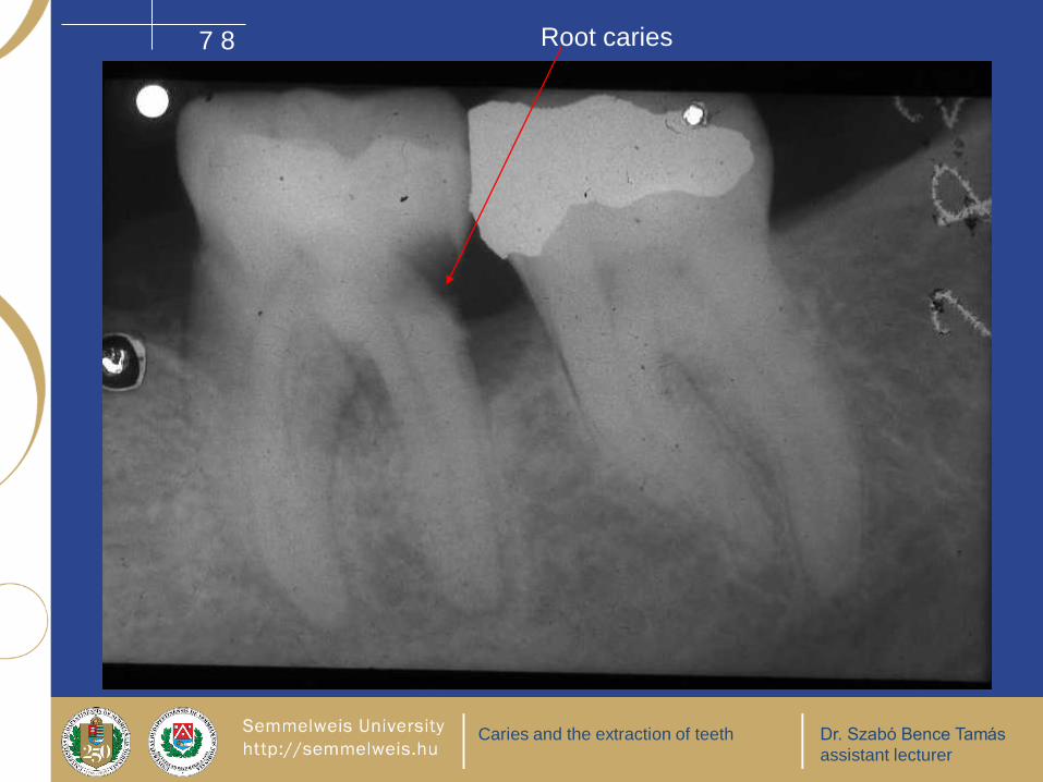
assistant lecturer
7 8 Root caries

Caries and the extraction of teeth Dr. Szabó Bence Tamás
assistant lecturer
4 3 2 1
Cervical burnout
borders of it:
nose and CEJ
Little arrows:
approximal
caries D3-4

Caries and the extraction of teeth Dr. Szabó Bence Tamás
assistant lecturer
4 5 6occlusal caries

Caries and the extraction of teeth Dr. Szabó Bence Tamás
assistant lecturer
7 8
secondary caries under
the occlusal filling

Caries and the extraction of teeth Dr. Szabó Bence Tamás
assistant lecturer
vestibular
surface
caries
3 2 1
Caries and the extraction of teeth Dr. Szabó Bence Tamás
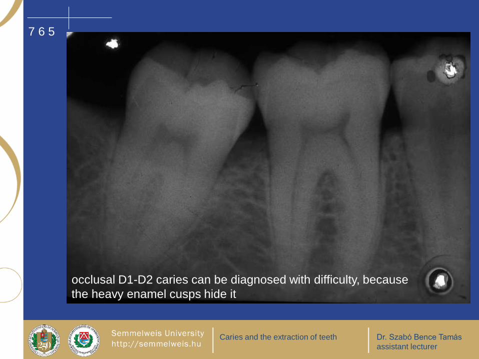
assistant lecturer
7 6 5
occlusal D1-D2 caries can be diagnosed with difficulty, because
the heavy enamel cusps hide it

Caries and the extraction of teeth Dr. Szabó Bence Tamás
assistant lecturer
secondary caries8 7 6 5

Caries and the extraction of teeth Dr. Szabó Bence Tamás
assistant lecturer
lingual cariessec. caries, OD
4: accessory radix, total destruction of the crown
4 5 6 7

Caries and the extraction of teeth Dr. Szabó Bence Tamás
assistant lecturer
8 7 6 5 4
5: 2 roots, 6: distal secondary caries, D3 , 7: mesiaoapproximal D3 caries

Caries and the extraction of teeth Dr. Szabó Bence Tamás
assistant lecturer
secondary caries which involve the root
4 6 7 8

Caries and the extraction of teeth Dr. Szabó Bence Tamás
assistant lecturer
remnants of the teeth: destruction because of caries

Caries and the extraction of teeth Dr. Szabó Bence Tamás
assistant lecturer
extraction

Caries and the extraction of teeth Dr. Szabó Bence Tamás
assistant lecturer
difficult extraction
what can we expect?
X-RAY!
long, slim and/or curved root
hypercementosis
splayed roots
total retention : the tooth is totally surrounded by bone
impacted tooth
predisposition for fracture: endodontic treatment, resorptio dentis
closeness of the sinus
closeness of the mandibular canal
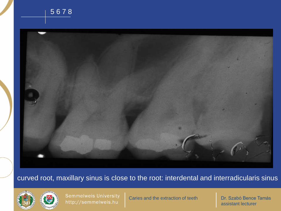
Caries and the extraction of teeth Dr. Szabó Bence Tamás
assistant lecturer
curved root, maxillary sinus is close to the root: interdental and interradicularis sinus
5 6 7 8

Caries and the extraction of teeth Dr. Szabó Bence Tamás
assistant lecturer
5 6 7 8
accessory radix: bigger chance to fracture

Caries and the extraction of teeth Dr. Szabó Bence Tamás
assistant lecturer
6: three rooted, accessory radix
4 5 6 7

Caries and the extraction of teeth Dr. Szabó Bence Tamás
assistant lecturer
curved roots
5 6 7 8
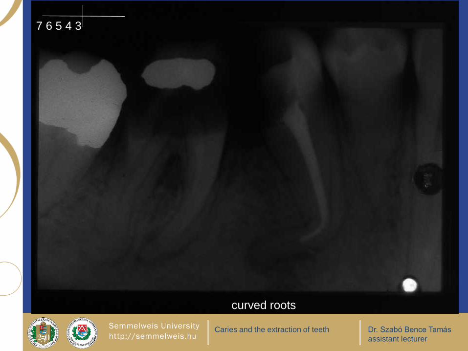
Caries and the extraction of teeth Dr. Szabó Bence Tamás
assistant lecturer
curved roots
7 6 5 4 3

Caries and the extraction of teeth Dr. Szabó Bence Tamás
assistant lecturer
after endodontic
treatment teeth have
predisposition to
fracture
2 3 4 5

Caries and the extraction of teeth Dr. Szabó Bence Tamás
assistant lecturer
1
7 5 4 3

Caries and the extraction of teeth Dr. Szabó Bence Tamás
assistant lecturer
bulge apex
hypercementosis:
extraction with a lot of
bone fragment??

Caries and the extraction of teeth Dr. Szabó Bence Tamás
assistant lecturer
internal resorption:
the frequency of the fracture is
bigger
6 5 4 3

Caries and the extraction of teeth Dr. Szabó Bence Tamás
assistant lecturer
during the extraction the
intraalveolar septum will
fracture out
5 6 7 8
Caries and the extraction of teeth Dr. Szabó Bence Tamás
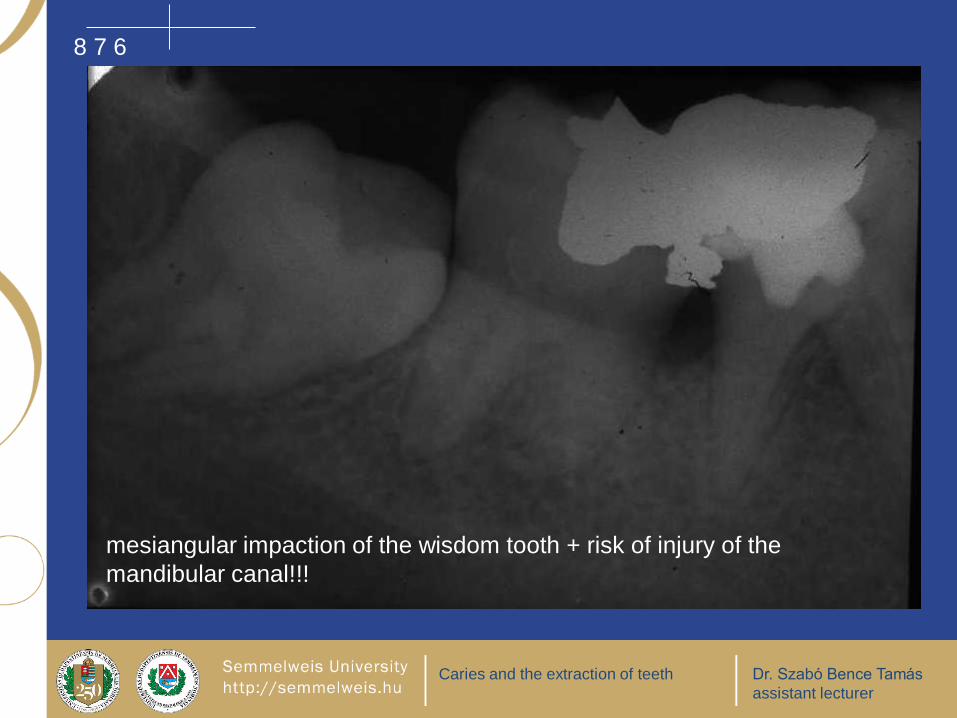
assistant lecturer
mesiangular impaction of the wisdom tooth + risk of injury of the
mandibular canal!!!
8 7 6

Caries and the extraction of teeth Dr. Szabó Bence Tamás
assistant lecturer
long roots
8 7 6 5

Caries and the extraction of teeth Dr. Szabó Bence Tamás
assistant lecturer
dilaceration, curved roots
1 1 2

Caries and the extraction of teeth Dr. Szabó Bence Tamás
assistant lecturer
easy extarction
thick, short root
piramid shape roots
milk tooth with partly absorbed root
partly absorbed apex or alveolar process

Caries and the extraction of teeth Dr. Szabó Bence Tamás
assistant lecturer
7 6 5
7: piramid shape root, 6: splayed roots

Caries and the extraction of teeth Dr. Szabó Bence Tamás
assistant lecturer
bone loss
partlially absorbed
alveolar process
Caries and the extraction of teeth Dr. Szabó Bence Tamás
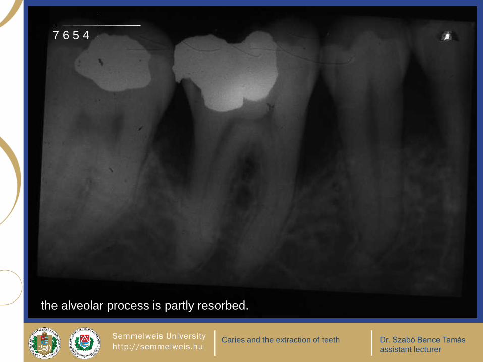
assistant lecturer
the alveolar process is partly resorbed.
7 6 5 4

Caries and the extraction of teeth Dr. Szabó Bence Tamás
assistant lecturer
pyramid shape root: easier extraction
5 6 7 8

Caries and the extraction of teeth Dr. Szabó Bence Tamás
assistant lecturer
extraction: complications
during the extraction: collapse root fracture – the fractured fragment can pass into the sinus, soft tissue,
mandibular canal aspiration of the root alveolar fracture, the bone fragment can pass into the maxillary sinus fracture of the maxillary tuberosity soft tissue damage the antagonistic or neighbouring tooth could tip out opening of the maxillary sinus during the local anesthesia could damage the plexus venosus,or vessels heavy bleeding from bone or soft tissue
after the extraction: periostitis, phlegmone bleeding pain

Caries and the extraction of teeth Dr. Szabó Bence Tamás
assistant lecturer
During the extraction you can make X-ray film and you
will see the complications
damage of anatomical structures
fracture of the alveolar process: more or less unavoidable
fracture of the root:
neck region: cervical fracture,
body of the root: median fracture,
apex region: apical fracture.
» Difficult situation if the zygomatic arch superimpose to that region, slim
apicis frequently hardly visible, it could seem like if it were in the sinus
damage of neighbouring tooth or developing tooth: accidental extraction
fracture of the mandibule
fracture of instruments
Caries and the extraction of teeth Dr. Szabó Bence Tamás
assistant lecturer
after the extraction
on the fourth day: ostitis alveolaris
after a few weeks-months: lamina dura yet visible
after months-years:
reossification: commonly it has a smaller density due to smaller calcium content, so it looks like less dens than the surrounding bone
enostosis (whiter than the surrounding bone)
Caries and the extraction of teeth Dr. Szabó Bence Tamás
assistant lecturer
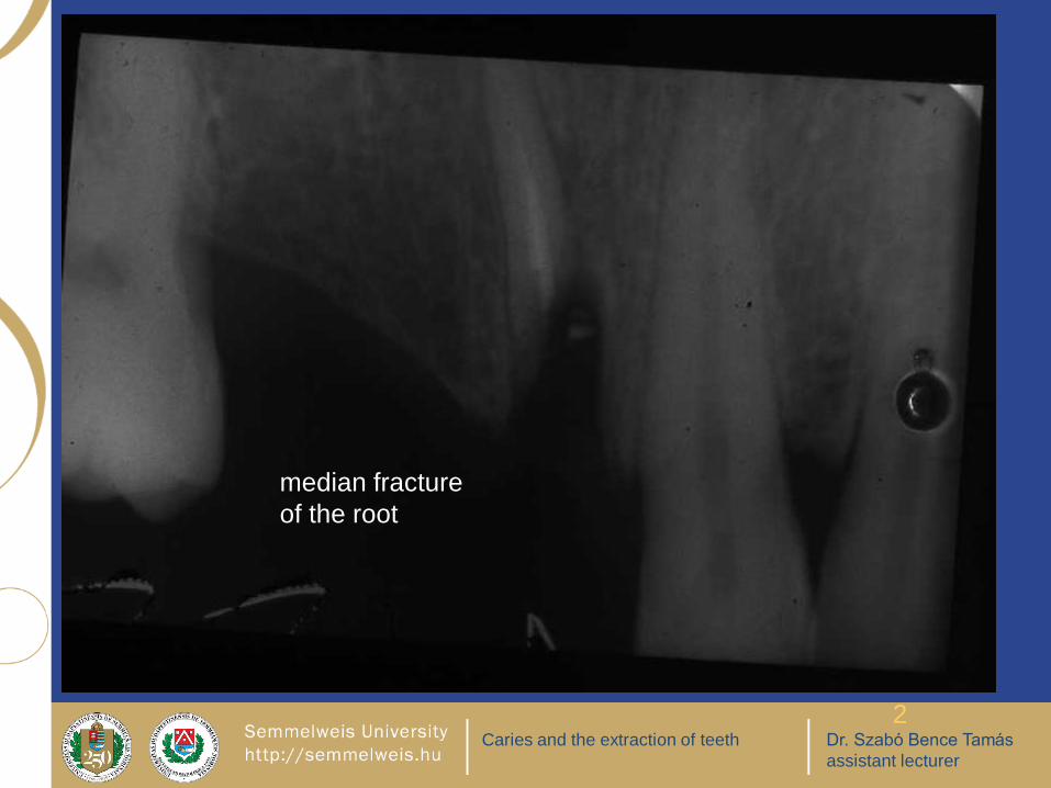
2
median fracture
of the root
Caries and the extraction of teeth Dr. Szabó Bence Tamás
assistant lecturer
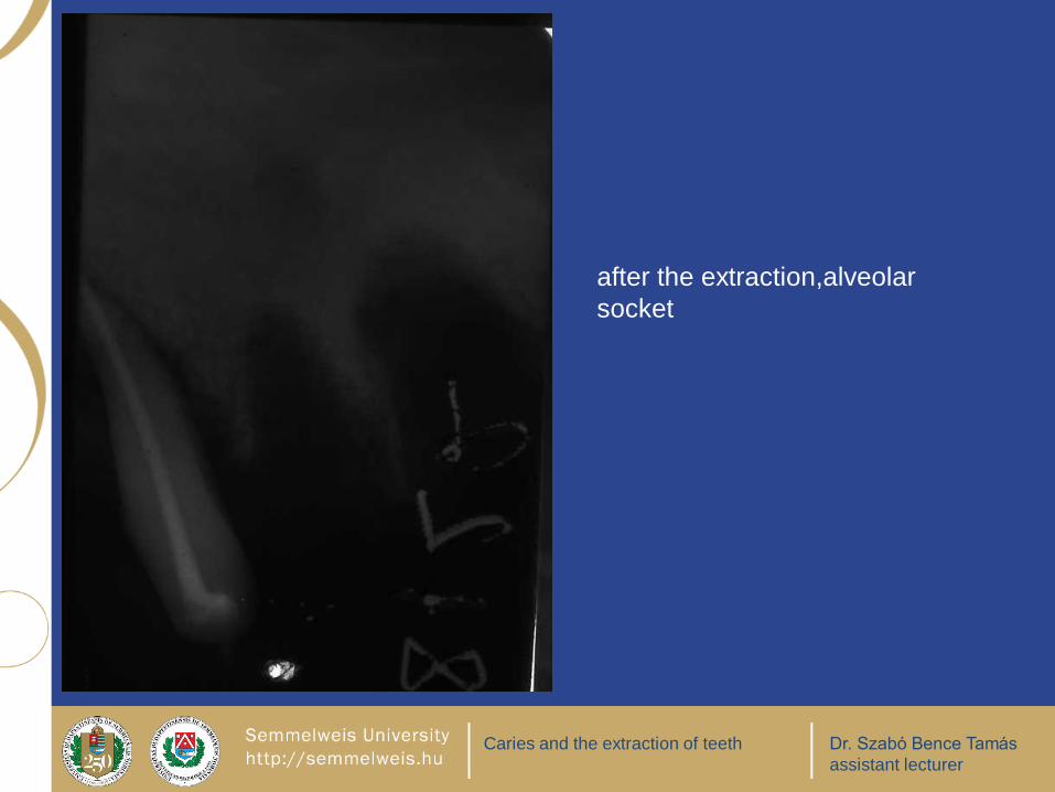
after the extraction,alveolar
socket

Caries and the extraction of teeth Dr. Szabó Bence Tamás
assistant lecturer
reossification in the previous
place of the two central incisors.
2 2

Caries and the extraction of teeth Dr. Szabó Bence Tamás
assistant lecturer
median fracture
6 7 8

Caries and the extraction of teeth Dr. Szabó Bence Tamás
assistant lecturer
median fracture

Caries and the extraction of teeth Dr. Szabó Bence Tamás
assistant lecturer
median fracture, bone fragments
3 (4) 5

Caries and the extraction of teeth Dr. Szabó Bence Tamás
assistant lecturer
apical fracture

Caries and the extraction of teeth Dr. Szabó Bence Tamás
assistant lecturer
fracture
It is visible on the
X-Ray film
previous the
extraction.

Caries and the extraction of teeth Dr. Szabó Bence Tamás
assistant lecturer
alveolar fracture

Caries and the extraction of teeth Dr. Szabó Bence Tamás
assistant lecturer
fracture

Caries and the extraction of teeth Dr. Szabó Bence Tamás
assistant lecturer
the injury of the maxillary sinus is most common during the
extraction of the first upper molar.
8 7 6 5 4

Caries and the extraction of teeth Dr. Szabó Bence Tamás
assistant lecturer
tuberal fracture, alveolar process fracture

Caries and the extraction of teeth Dr. Szabó Bence Tamás
assistant lecturer
8 7
mandibular and wisdom tooth root farcture with dislocation

Caries and the extraction of teeth Dr. Szabó Bence Tamás
assistant lecturer
radix relicta: long time after the extraction, the root remnant is
surrounded by bone

Caries and the extraction of teeth Dr. Szabó Bence Tamás
assistant lecturer
5 6 7 8
increased risk of tuber fracture: the maxillary sinus has a big tuberal recess.

Caries and the extraction of teeth Dr. Szabó Bence Tamás
assistant lecturer
maxillary sinus has a big tuberal recess

Caries and the extraction of teeth Dr. Szabó Bence Tamás
assistant lecturer
Radix in antro
Sinus aperta

Caries and the extraction of teeth Dr. Szabó Bence Tamás
assistant lecturer
radix in antro

Caries and the extraction of teeth Dr. Szabó Bence Tamás
assistant lecturer
fractured root passed in the sinus

Caries and the extraction of teeth Dr. Szabó Bence Tamás
assistant lecturer
opened sinus: SINUS APERTA! compacta line is not continous!

Caries and the extraction of teeth Dr. Szabó Bence Tamás
assistant lecturer
opened maxillary sinus

Caries and the extraction of teeth Dr. Szabó Bence Tamás
assistant lecturer
CAVE: developing teeth could damage even extracted during milk tooth extraction.
The developing teeth have not yet roots.
Caries and the extraction of teeth Dr. Szabó Bence Tamás
assistant lecturer
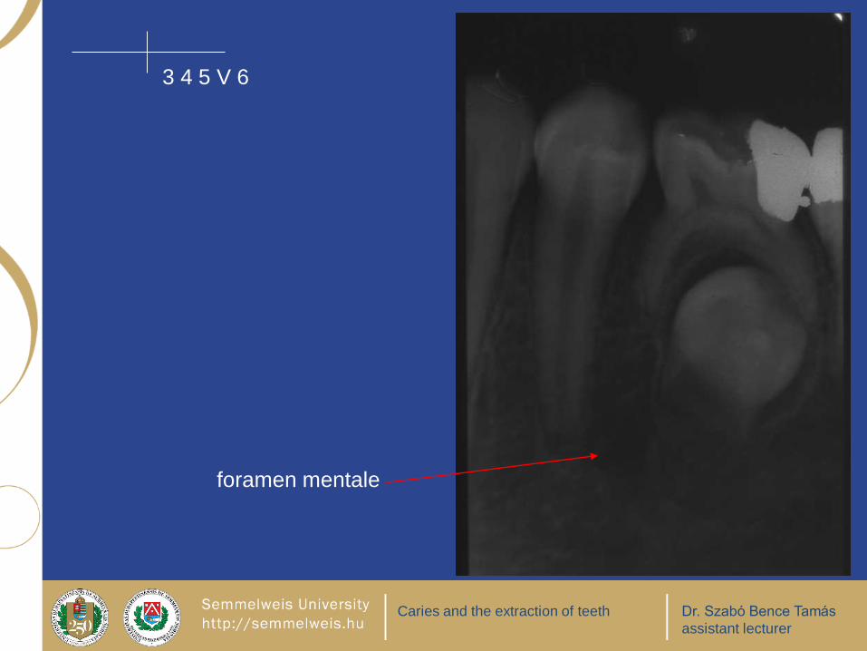
3 4 5 V 6
foramen mentale

Caries and the extraction of teeth Dr. Szabó Bence Tamás
assistant lecturer
milk molar with an accidentally
extracted premolar

Caries and the extraction of teeth Dr. Szabó Bence Tamás
assistant lecturer
2 3 4 IV 5 V 6
Milk molars’ crown are destruated. Amalgam fillings, 6: mesioapproximal D2

Caries and the extraction of teeth Dr. Szabó Bence Tamás
assistant lecturer
bone fragments

Caries and the extraction of teeth Dr. Szabó Bence Tamás
assistant lecturer
7 6
bone fragments

Caries and the extraction of teeth Dr. Szabó Bence Tamás
assistant lecturer
amalgam particules and apical root fracture
3 4 6 7

Caries and the extraction of teeth Dr. Szabó Bence Tamás
assistant lecturer
4 5 7 bone fragments
recent exraction socket, the lamina dura is visible: thin opaque layer

Caries and the extraction of teeth Dr. Szabó Bence Tamás
assistant lecturer
12
apical fracture

Caries and the extraction of teeth Dr. Szabó Bence Tamás
assistant lecturer
5 6 7 8
cervical fracture of the roots

Caries and the extraction of teeth Dr. Szabó Bence Tamás
assistant lecturer
amalgam particules,
apical fracture

Caries and the extraction of teeth Dr. Szabó Bence Tamás
assistant lecturer
apical root fracture

Caries and the extraction of teeth Dr. Szabó Bence Tamás
assistant lecturer
fractured instrument
(drill bit)

Caries and the extraction of teeth Dr. Szabó Bence Tamás
assistant lecturer
fractured needle

Caries and the extraction of teeth Dr. Szabó Bence Tamás
assistant lecturer
radix relicta
fractured instrument

Caries and the extraction of teeth Dr. Szabó Bence Tamás
assistant lecturer
2 1
root caries
fractured
instrument

Caries and the extraction of teeth Dr. Szabó Bence Tamás
assistant lecturer
THANK YOU VERY MUCH FOR YOUR
KIND ATTENTION!