bilateral arm-abduction shoulder radiography to...
TRANSCRIPT
)225( COPYRIGHT 2018 © BY THE ARCHIVES OF BONE AND JOINT SURGERY
Arch Bone Jt Surg. 2018; 6(3): 225-232. http://abjs.mums.ac.ir
the online version of this article abjs.mums.ac.ir
Fei Wu, MD; Amir R. Kachooei, MD; Mohammad H. Ebrahimzadeh, MD; Farshid Bagheri, MD; Ehsan Hakimi, MD; Babak Shojaie, MD, FA; Ara Nazarian, PhD
Research performed at Orthopedic Research Center, Mashhad University of Medical Sciences, Mashhad, Iran
Corresponding Author: Farshid Bagheri, Shahid Kamyab Emdadi Hospital, Orthopedic Research Center, Mashhad University of Medical Sciences, Mashhad, IranEmail: [email protected]
RESEARCH ARTICLE
Received: 18 November 2017 Accepted: 23 April 2018
Bilateral Arm-Abduction Shoulder Radiography to Determine the Involvement of the Scapulothoracic
Motion in Frozen Shoulder
Abstract
Background: We hypothesize that there is no difference in the motion of the scapula with respect to the thoracic wall (scapulothoracic interface) between the affected versus non-affected shoulder on 0° and 90° standard arm abduction radiography.
Methods: We enrolled 30 patients with the diagnosis of unilateral frozen shoulder after ruling out of other pathologies. Bilateral standard shoulder radiography was done in two position of 0° and 90° of arm abduction. Non-affected side was used as a control group.
Results: The mean scapulothoracic angle of the affected side was significantly larger than the non-affected side in both 0° and 90°of abduction in spite that the scapulohumeral angles were comparable in 0°, indicating potential alteration in scapular positioning.
Conclusion: Scapulothoracic motion and position can be affected in frozen shoulder along with other areas. All treatment modalities should be applied to this area as well if substantial difference was detected between the two sides.
Level of evidence: I
Keywords: Center equator distance, Frozen shoulder, Radiography, Scapulohumeral angle, Scapulothoracic
Introduction
The diagnosis of frozen shoulder is usually made by demonstrating a global decrease in shoulder range of motion, predominantly through testing
of the glenohumeral motion and not scapulothoracic
motion. Stiffness predominantly occurs after fibrosis in the glenohumeral capsule and the scapulohumeral interface (1-3). Taking the global involvement of the shoulder region into account, one can assume that
SCAPULOTHORACIC MOTION IN FROZEN SHOULDERTHE ARCHIVES OF BONE AND JOINT SURGERY. ABJS.MUMS.AC.IRVOLUME 6. NUMBER 3. MAY 2018
)226(
obtained. The non-affected side was considered as the internal control group. We followed the following guidelines to avoid discrepancies:1. Patient standing with the tube in front and 105 cm
away from the shoulder radiating perpendicular to the cassette in the back, both arms hanging aside the trunk in 0° of arm abduction, 0° of rotation and 0° of extension with the thumb facing forward to obtain true bilateral shoulder anteroposterior view. Voltage and exposure time differs as a function of each person’s body mass.
2. Holding the same position, the patient tries to elevate the arm to 90° of abduction. In patients that were not able to obtain the 90° abduction, we accepted the maximum possible abduction of the arm and we assumed that the scapulothoracic motion is compensating for the glenohumeral stiffness to reach to the maximum possible abduction.
We defined four radiographic parameters on shoulder radiography:1. Scapulohumeral angle (SHA): The angle formed
between the humeral shaft axis and the lateral border of the scapula starting from the inferior angle is called the scapulohumeral angle [Figure 1 a, b].
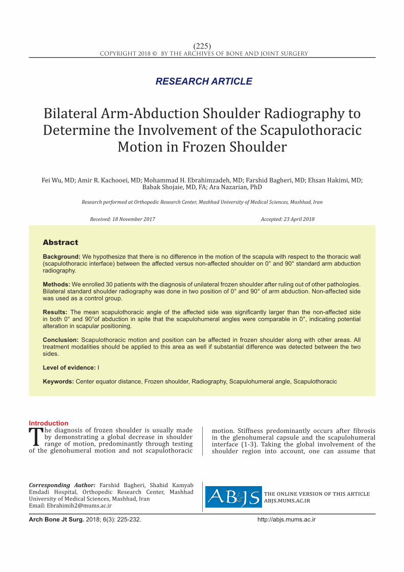
2. Scapulothoracic angle (STA): The angle between the lateral border of the ribs and the lateral border of the scapula is called the scapulothoracic angle [Figure 1 a, b]. In the affected shoulder with decreased glenohumeral motion, we normally expect the scapulothoracic motion to compensate for the terminal abduction. Therefore, this angle decreases as the arm abducts. Increase in this angle at 90° abduction may suggest that this articulation is also involved with either stiffness or muscle spasm [Figure 1 c, d].
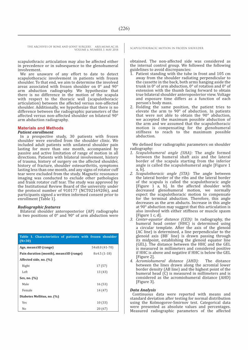
3. Center-equator distance (CED): In radiography, the humeral head center (HHC) is determined using a circular template. After the axis of the glenoid (AC line) is determined, a line perpendicular to the glenoid axis (BB´ line) is drawn passing through its midpoint, establishing the glenoid equator line (GEL). The distance between the HHC and the GEL is measured in millimeters and considered positive if HHC is above and negative if HHC is below the GEL [Figure 2].
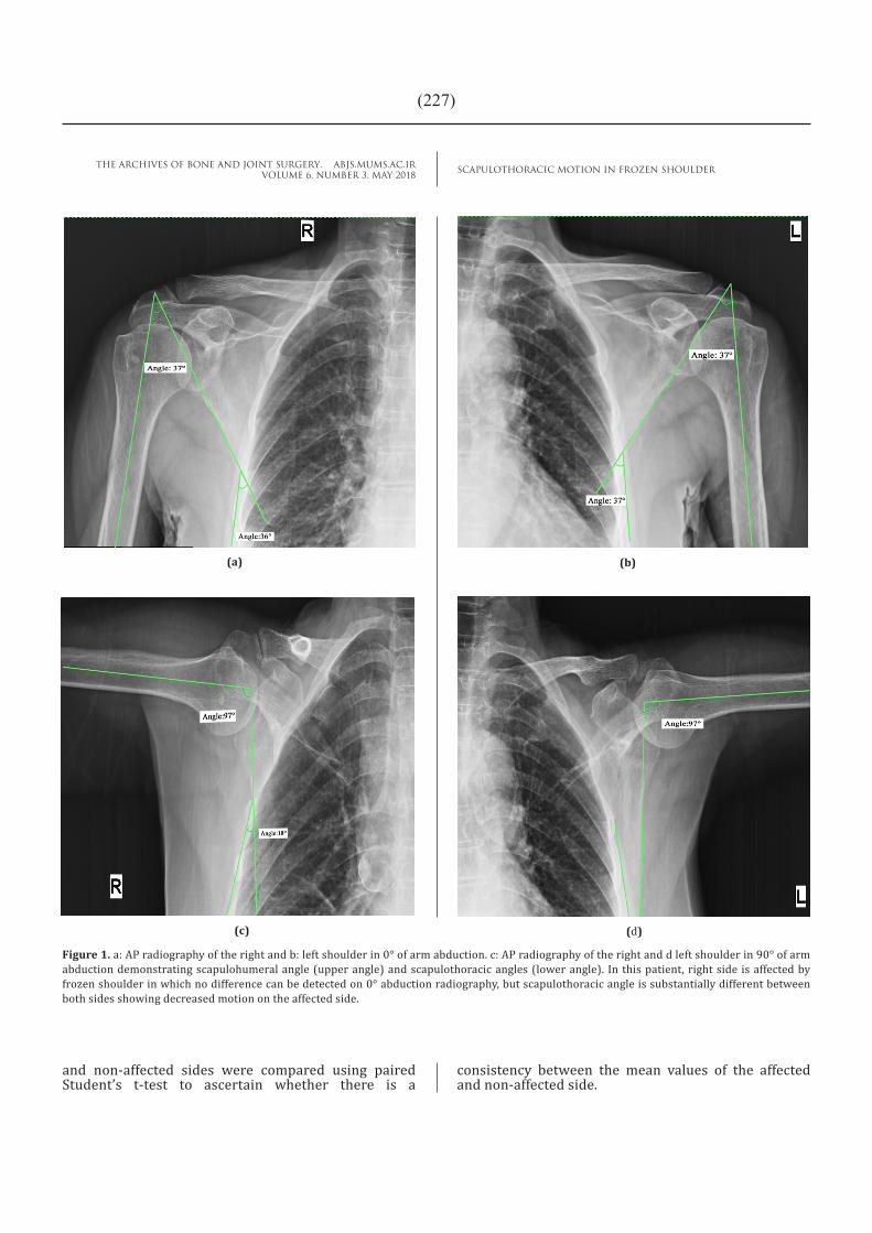
4. Acromiohumeral distance (AHD): The distance between the lines drawn along the acromial lower border density (AB line) and the highest point of the humeral head (C) is measured in millimeters and is considered as the acromiohumeral distance (AHD) [Figure 3].
Data AnalysisContinuous data were reported with means and
standard deviation after testing for normal distribution using the Kolmogorov-Smirnov test. Categorical data were presented as absolute values and percentages. Measured radiographic parameters of the affected
scapulothracic articulation may also be affected either in precedence or in subsequence to the glenohumeral involvement.
We are unaware of any effort to date to detect scapulothoracic involvement in patients with frozen shoulder. To that end, we aim to determine the involved areas associated with frozen shoulder on 0° and 90° arm abduction radiography. We hypothesize that there is no difference in the motion of the scapula with respect to the thoracic wall (scapulothoracic articulation) between the affected versus non-affected shoulder. Additionally, we hypothesize that there is no difference between the radiographic parameters of the affected versus non-affected shoulder on bilateral 90° arm abduction radiography.
Materials and MethodsPatient enrollment
In a prospective study, 30 patients with frozen shoulder were enrolled from the shoulder clinic. We included adult patients with unilateral shoulder pain lasting for more than one month, accompanied by passive and active limitation of range of motion in all directions. Patients with bilateral involvement, history of trauma, history of surgery on the affected shoulder, history of fracture, shoulder osteoarthritis, symptoms lasting less than one month, and any signs of rotator cuff tear were excluded from the study. Magnetic resonance imaging was conducted to exclude other pathologies and frank rotator cuff tear. The study was approved by the Institutional Review Board of the university under the protocol number of 910177 (NCT02169206), and participants signed a written informed consent prior to enrollment [Table 1].
Radiographic featuresBilateral shoulder anteroposterior (AP) radiographs
in two positions of 0° and 90° of arm abduction were
Table 1. Characteristics of patients with frozen shoulder (N=30)
Age, mean±SD (range) 54±8.0 (41-70)
Pain duration (month), mean±SD (range) 8±4.5 (1-18)
Affected side, no. (%)
Right 17 (57)
Left 13 (43)
Sex, no. (%)
Male 16 (53)
Female 14 (47)
Diabetes Mellitus, no. (%)
Yes 10 (33)
No 20 (67)
SCAPULOTHORACIC MOTION IN FROZEN SHOULDERTHE ARCHIVES OF BONE AND JOINT SURGERY. ABJS.MUMS.AC.IRVOLUME 6. NUMBER 3. MAY 2018
)227(
and non-affected sides were compared using paired Student’s t-test to ascertain whether there is a
consistency between the mean values of the affected and non-affected side.
Figure 1. a: AP radiography of the right and b: left shoulder in 0° of arm abduction. c: AP radiography of the right and d left shoulder in 90° of arm abduction demonstrating scapulohumeral angle (upper angle) and scapulothoracic angles (lower angle). In this patient, right side is affected by frozen shoulder in which no difference can be detected on 0° abduction radiography, but scapulothoracic angle is substantially different between both sides showing decreased motion on the affected side.
(a) (b)
(c) (d)

SCAPULOTHORACIC MOTION IN FROZEN SHOULDERTHE ARCHIVES OF BONE AND JOINT SURGERY. ABJS.MUMS.AC.IRVOLUME 6. NUMBER 3. MAY 2018
)228(
Figure 2. Anteroposterior shoulder radiography demonstrating center equator distance. Values were considered negative when humeral head center was below the glenoid equator line and positive when it was above this line.
Figure 3. Anteroposterior shoulder radiography showing acromiohumeral distance measurement.
SCAPULOTHORACIC MOTION IN FROZEN SHOULDERTHE ARCHIVES OF BONE AND JOINT SURGERY. ABJS.MUMS.AC.IRVOLUME 6. NUMBER 3. MAY 2018
)229(
ResultsThe mean scapulothoracic angle (STA) was
significantly larger on the affected side than on the non-affected side in both 0° and 90°of abduction, showing potential alteration in scapular positioning [Table 2]. The larger scapulothoracic angle on the affected side showed less scapular abduction motion and possible stiffness of the scapulothoracic interface [Figure 1 a-d; Figure 4]. Despite that the scapulohumeral angle (SHA) of the affected side was significantly less than the non-affected side on 90° arm abduction radiography, scapulothoracic angle (STA) remained significantly larger on the affected side, indicating the lack of
compensation at the scapulothoracic interface.In 0° of arm abduction, there were no differences in
SHA, CED, and AHD parameters between the affected and non-affected sides (P values = 0.49, 0.19, and 0.061, respectively). There was no difference in center-equator distance (CED) between affected and non-affected sides in 90° abduction radiography displaying no substantial change in humeral head position in respect to the glenoid [Table 2]. Acromiohumeral distance was comparable between the affected and non-affected sides, supporting the idea that the humeral head position remains unchanged in frozen shoulders [Tables 2; 3; Figure 3 a, b].
Table 2. Radiographic parameters of the affected and non-affected side in two different position of 0° and 90° of shoulder abduction
Mean±SD Min-Max ¥ P value
Scapulohumeral angle (SHA)
0° abduction
Affected 40±7.5° 27-54°0.49
Non-affected 39±7.0° 29-56°
90° abduction
Affected 79±16° 41-106°<0.001
Non-affected 89±11° 67-119°
Scapulothoracic angle (STA)
0° abduction
Affected 38±7.0° 29-54°0.0020
Non-affected 35±6.5° 24-50°
90° abduction
Affected 16±11° 0-34°0.035
Non-affected 12±11° 0-32°
Center-equator distance (CED), mm
0° abduction
Affected (-1.0)±2.4 (-7.3)-4.00.19
Non-affected (-1.7)±2.4 (-8.0)-4.0
90° abduction
Affected 1.0±1.2 (-0.8)-3.2 0.90
Non-affected 1.0±1.2 (-1.1)-3.4
Acromiohumaral distance (AHD), mm
0° abduction
Affected 9.3±1.3 7-120.061
Non-affected 9.7±1.4 7-12
¥ Paired samples t-test

SCAPULOTHORACIC MOTION IN FROZEN SHOULDERTHE ARCHIVES OF BONE AND JOINT SURGERY. ABJS.MUMS.AC.IRVOLUME 6. NUMBER 3. MAY 2018
)230(
DiscussionIn this study we aimed to evaluate radiographic
parameters on both affected and non-affected sides of patients with frozen shoulder at two different positions of 0° and 90° of arm abduction. We hypothesized that there was no difference between scapulothoracic motion of the affected and non-affected shoulders.
We rejected the null hypothesis, as we demonstrated significant differences between scapulothoracic angles of the affected and non-affected sides at both 0º and 90º of arm abduction.
There are limitations associated with this study that must be considered when interpreting the results. The
Figure 4. Scatter plot of the scapulothoracic angle on 90º arm abduction radiography showing larger angles on the affected side, which suggests the lack of compensatory movement in compare to the non-affected side.
Table 3. Range of movement from 0 to 90° of shoulder abduction on the affected andnon-affected side
Mean±SD Min-Max ¥ P value
Scapulohumeral angle change from 0 to 90°
Affected 37±17° (-8)-62°<0.001
Non-affected 49±10° 37-74°
Scapulothoracic angle change from 0 to 90°
Affected 20±9.5° 3-40°0.20
Non-affected 24±9.0° 2-39°
Center-equator distance change from 0 to 90°, mm
Affected 1.9±2.7 (-4)-9.80.21
Non-affected 2.5±2.2 (-1.8)-9.3
¥ Paired samples t-test
SCAPULOTHORACIC MOTION IN FROZEN SHOULDERTHE ARCHIVES OF BONE AND JOINT SURGERY. ABJS.MUMS.AC.IRVOLUME 6. NUMBER 3. MAY 2018
)231(
sample size is not large enough to enable generalizing the findings, therefore meriting future larger studies. Another limitation is that we were not able to analyze the subgroups by diabetes, sex, and stage of the disease, which requires further studies with larger sample size. We used radiographs as a 2 dimensional sketch of the shoulder in which we were not able to assess the influence of scapular rotation in space. We strived to demonstrate a baseline to provoke ideas for future studies on the areas of involvement in frozen shoulder.
At first, one can assume that glenohumeral abduction would be ceased at some point due to glenohumeral capsule contracture whereas scapulothoracic interface is potentially responsible to compensate for the remainder of abduction by further outward rotation against the thoracic wall. However, our results indicated that scapular motion decreased rather than being increased. Our findings showed that scapulothoracic motion can be affected by frozen shoulder and limitation of motion could be attributed to both scapulothoracic and scapulohumeral joints.
We are aware of a study reporting a patient with “adhesive scapulothoracic” as a differential diagnosis of frozen shoulder (4). In this study, the authors reported a middle-aged woman presenting with insidious onset of shoulder pain and limitation of motion diagnosed initially as frozen shoulder (4). In follow-up, shoulder abduction decreased to below 90° with no scapular rotation at all. The patient responded well to arthroscopic release of the intra-bursal fibrosis between the serratus muscle and the thorax. Problems with scapulothorac articulation have been shown to be a source of pain around the shoulder, chest, and even the breast (5). Studies of scapulothoracic bursitis and snapping scapular syndrome have shown that pain and motion limitation are the chief disabling complaints (6). Smooth scapular gliding requires congruent and free space between the scapula and the thoracic wall. In case of “adhesive scapulothoracic”, fibrosis hinders free scapular gliding, resulting in pain and compromised total shoulder motion (4-9). In addition, scapular orientation can also be affected so that the working muscles are influenced (10).
It may be counterintuitive that operative treatment solely on the glenohumeral and scapulohumeral enhances good clinical results. However, this is not in contrast to what we found since the postoperative physiotherapy can address both sites of involvement. We postulate two possible reasons for decreased scapulothoracic motion in frozen shoulder. One is that the nature of the disease is more general, which affects not only glenohumeral and scapulohumeral interface but also the scapulothoracic interface. We cannot comment on whether “adhesive scapulothoracic” starts in precedence or is the consequence of glenohumeral contracture. On the other hand, we can argue that impaired scapulothoracic motion
could be due to hyper protectiveness in patients with painful glenohumeral contracture trying to protect and avoid scapulothoracic motion. Considering these assumptions, we propose further studies to objectively assess what is joint stiffness and what is just guarding.
Our findings showed that scapulothoracic articulation is affected in frozen shoulder as well as scapulohumeral and glenohumeral articulation either by contracture or hyper protectiveness, which may consequently result in contracture. Therefore, all modalities of treatment including physical therapy, exercise, injection, and even arthroscopic release can be applied to scapoluthoracic articulation as well. Focus on scapular muscle strengthening and pain management during physical therapy could potentially result in further synchronized gliding during shoulder motion (11). It seems encouraging to assess scapulothoracic motion in frozen shoulder with 90° abduction radiography and consider this joint as a part of the treatment, especially in recalcitrant frozen shoulders when arthroscopic release is planned.
Scapulothoracic motion and position can be affected in frozen shoulder along with the other areas. We recommend that its potential motion and position be evaluated on bilateral 90° abduction radiography. All treatment modalities should be applied to this area as well if substantial difference was detected between the two sides.
Acknowledgements None.
Fei Wu MDDepartment of Orthopedic Surgery, Renmin Hospital of Wuhan University, Wuhan, China.
Amir R. Kachooei MD Mohammad H. Ebrahimzadeh MDEhsan Hakimi MDOrthopedic Research Center, Mashhad University of Medical Sciences, Mashhad, Iran
Farshid Bagheri MDShahid Kamyab Emdadi Hospital, Orthopedic Research Center, Mashhad University of Medical Sciences, Mashhad, Iran
Babak Shojaie MD FATraumatology , Hand and Orthopedic Surgery Department, st. Marien Medical Campus, Friesoythe, Germany
Ara Nazarian PhDCenter for Advanced Orthopaedic Studies, Department of Orthopaedic Surgery, Beth Israel Deaconess Medical Center, Harvard Medical School, Boston, USA

SCAPULOTHORACIC MOTION IN FROZEN SHOULDERTHE ARCHIVES OF BONE AND JOINT SURGERY. ABJS.MUMS.AC.IRVOLUME 6. NUMBER 3. MAY 2018
)232(
1. Ebrahimzadeh MH, Moradi A, Pour MK, Moghadam MH, Kachooei AR. Clinical outcomes after arthroscopic release for recalcitrant frozen shoulder. Arch Bone Jt Surg. 2014; 2(3):220-4.
2. Kachooei AR, Moradi A, Janssen SJ, Ring D. The influence of dominant limb involvement on DASH and QuickDASH. Hand (N Y). 2015; 10(3):512-5.
3. Ebrahimzadeh MH, Birjandinejad A, Golhasani F, Moradi A, Vahedi E, Kachooei AR. Cross-cultural adaptation, validation, and reliability testing of the Shoulder Pain and Disability Index in the Persian population with shoulder problems. Int J Rehabil Res. 2015; 38(1):84-7.
4. Lehtinen JT, Tetreault P, Warner JJ. Arthroscopic management of painful and stiff scapulothoracic articulation. Arthroscopy. 2003; 19(4):E28.
5. Boneti C, Arentz C, Klimberg VS. Scapulothoracic bursitis as a significant cause of breast and chest wall pain: underrecognized and undertreated. Ann Surg Oncol. 2010; 17(Suppl 3):321-4.
6. Warth RJ, Spiegl UJ, Millett PJ. Scapulothoracic bursitis and snapping scapula syndrome: a critical
review of current evidence. Am J Sports Med. 2014; 43(1):236-45.
7. Chang WH, Im SH, Ryu JA, Lee SC, Kim JS. The effects of scapulothoracic bursa injections in patients with scapular pain: a pilot study. Arch Phys Med Rehabil. 2009; 90(2):279-84.
8. Conduah AH, Baker CL 3rd, Baker CL Jr. Clinical management of scapulothoracic bursitis and the snapping scapula. Sports Health. 2010; 2(2):147-55.
9. Son SA, Lee DH, Lee YO, Lee SC, Kim KJ, Cho JY. Operative management in a patient with scapulothoracic bursitis. Korean J Thorac Cardiovasc Surg. 2013; 46(6):486-9.
10. Noguchi M, Chopp JN, Borgs SP, Dickerson CR. Scapular orientation following repetitive prone rowing: implications for potential subacromial impingement mechanisms. J Electromyogr Kinesiol. 2013; 23(6):1356-61.
11. Celik D. Comparison of the outcomes of two different exercise programs on frozen shoulder. Acta Orthop Traumatol Turc. 2010; 44(4):285-92.
References