biomaterials applications of chemical mechanical polishing
TRANSCRIPT

ICPT 2012, October, 15-17, 2012, Grenoble, France
Biomaterials Applications of Chemical Mechanical Polishing
G.Bahar Basim1, Zeynep Ozdemir1, Ozal Mutlu2
1Ozyegin University, Department of Mechanical Engineering, Nisantepe Mevki, Orman Sokak,
No 13, Alemdag, Cekmekoy, 34794, Istanbul, Turkey 2Marmara University, Department of Biology, Faculty of Arts and Sciences, Goztepe Campus,
Kadikoy, 34722, Istanbul, Turkey
Chemical Mechanical Polishing (CMP) is used in semiconductor industry to enable planarization of the interlayer dielectrics and metals. In this study, CMP is used as a polishing technique to modify the surface roughness of the bio-implant materials in a controlled manner. As an alternative technique to sand-blasting/etching or laser structuring on the implant surfaces, CMP results in formation of a protective oxide layer on the titanium surfaces that can limit surface contamination while enabling surface nano-structuring that is known to promote bioactivity.
Keywords: CMP, Titanium Implants, Biomaterials, Micro-Patterning.
1. Introduction
Biomaterials are known to be used as implants for dental, cardiovascular and orthopaedic applications [1]. Recently, their usage for advanced therapy medicinal products (ATMP) extending from simple bandages to in-vivo applications such as stents, resulted in an additional challenge since the functionality of these devices is as critical as the material selection. In addition to standard materials like metals, ceramics/glass, polymers and composite materials, new materials are also used like nanotubes, nanowires, fibers or nanoparticles to provide magnetic properties, mechanical control or other functionalities such as controlled drug delivery devices. Particularly the surface modification is studied to deliver the desired functionality which can be induced in many different ways including blasting, chemical etching, a combination of sandblasting and etching, powder coating, ion-sputtering, anodizing or laser pulse deposition of biomaterials like hydroxyapatite [2]. Among the materials available for bio-applications, metals have more advantages due to their non-degradability and manufacturing flexibility. Particularly titanium and its alloys are favoured as bio implants due to their surface characteristics which promote biocompatibility [3]. The surface oxide and porosity are known to affect the cell growth on the titanium surfaces. There are many studies characterizing the titanium metal surface for roughness and biocompatibility after treating with etching or blasting type structuring methods [4-5]. As an alternative technique, it was demonstrated that the application of CMP on Ti implants has created a better quality TiO2 oxide film on the surface and promoted biocompatibility [6]. In CMP process, a chemically altered top film is continuously formed through slurry chemicals and removed by the mechanical action of the slurry abrasive particles. These chemically altered top films have to be a protective oxide to enable planarization by stopping chemical corrosion on the recessed metal surfaces while the elevated structures are polished [7]. The self-protective nature of the metal oxide thin films are determined by Pilling-Bethworth ratio
385

ICPT 2012, October, 15-17, 2012, Grenoble, France
Figure 1. Optical micrographs (40X) of the anodized and CMP treated titanium surfaces with schematic representation of the formation of the surface oxide film and expected P-B ratios. in air, which compares the molar volume of the oxide film formed to the molar volume of its native metal [8]. Figure 1 demonstrates how the P-B ratios change for an anodized titanium surface as compared to a titanium surface post CMP. It is predicted that anodized oxide film on the titanium surface is porous due to the tensile stresses building on the thick oxide film grown on the surface. On the other hand, when the titanium plate is treated with CMP using 3 wt% H2O2 as an oxidizer, a nano-scale protective oxide film forms [9]. In this study, CMP technique is used to induce controlled nano/macro-roughness on the titanium surfaces to help increase the cell growth capability of the bio implants [10, 11]. It is aimed to create engineered interfaces for bio implant materials and for ATMPs with self-protective surfaces to minimize chemical reactivity, while promoting their biocompatibility through surface patterning.
2. Experimental Details The bio-compatibility of the titanium samples was studied through bacteria growth analyses to determine the affinity of the species towards the as is versus polished surfaces. Titanium plates were sterilized before they were placed into Cronobacter Sakazakii (Gram-) bacteria species. 100μl microorganisms from the nutrient broth microbial stock spread on nutrient agar plates. Ti samples were incubated at 37 ºC and bacteria colonization was recorded after a week time frame. Titanium foils with 1 mm thickness and 99.6% purity (TI000430) obtained from Goodfellow Cambridge Limited diced to 46 x 28 mm pieces for CMP. The original sample surface was annealed. CMP was performed on a desktop Tegrapol-31 polisher using 5 wt% Al2O3 slurry made of 50-nm size particles at pH 4 and at 7.88 psi pressure. Surfaces were polished with a Suba IV sub-pad stacked under a polytex buff pad to help retain a soft interface to conserve the original surface planarity for the implant material. In addition, silicon carbide 150C and P320 abrasive papers were used to create the micro-scale roughness. Samples ran with the polymeric CMP pads were polished for 5 minutes with ~3% oxidizer addition and samples ran by using the abrasive papers were polished by using 5%wt H2O2 for
386
ICPT 2012, October, 15-17, 2012, Grenoble, France
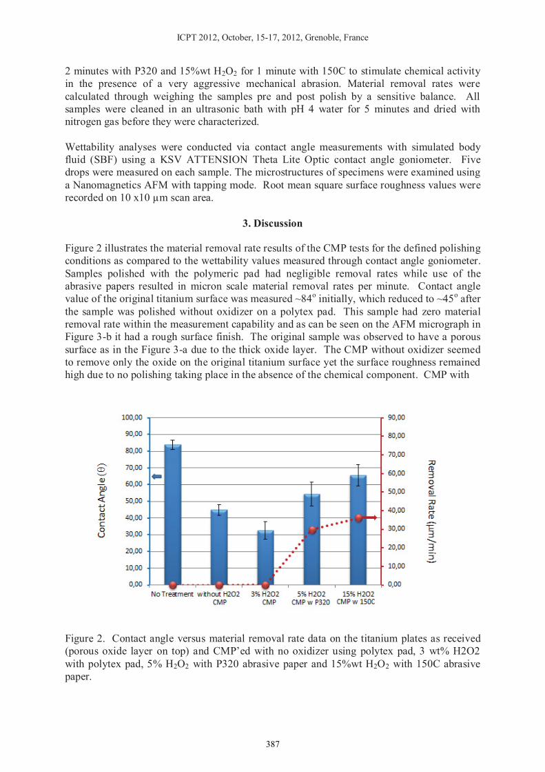
2 minutes with P320 and 15%wt H2O2 for 1 minute with 150C to stimulate chemical activity in the presence of a very aggressive mechanical abrasion. Material removal rates were calculated through weighing the samples pre and post polish by a sensitive balance. All samples were cleaned in an ultrasonic bath with pH 4 water for 5 minutes and dried with nitrogen gas before they were characterized. Wettability analyses were conducted via contact angle measurements with simulated body fluid (SBF) using a KSV ATTENSION Theta Lite Optic contact angle goniometer. Five drops were measured on each sample. The microstructures of specimens were examined using a Nanomagnetics AFM with tapping mode. Root mean square surface roughness values were recorded on 10 x10 μm scan area.
3. Discussion Figure 2 illustrates the material removal rate results of the CMP tests for the defined polishing conditions as compared to the wettability values measured through contact angle goniometer. Samples polished with the polymeric pad had negligible removal rates while use of the abrasive papers resulted in micron scale material removal rates per minute. Contact angle value of the original titanium surface was measured ~84o initially, which reduced to ~45o after the sample was polished without oxidizer on a polytex pad. This sample had zero material removal rate within the measurement capability and as can be seen on the AFM micrograph in Figure 3-b it had a rough surface finish. The original sample was observed to have a porous surface as in the Figure 3-a due to the thick oxide layer. The CMP without oxidizer seemed to remove only the oxide on the original titanium surface yet the surface roughness remained high due to no polishing taking place in the absence of the chemical component. CMP with
Figure 2. Contact angle versus material removal rate data on the titanium plates as received (porous oxide layer on top) and CMP’ed with no oxidizer using polytex pad, 3 wt% H2O2 with polytex pad, 5% H2O2 with P320 abrasive paper and 15%wt H2O2 with 150C abrasive paper.
387

ICPT 2012, October, 15-17, 2012, Grenoble, France
(a) (b) (c)
Figure 3. AFM micrographs of the titanium surfaces (a) original sample with a porous oxide layer (b) after polishing with polytex pad with no oxidizer addition (c) after CMP with polytex pad with 3 wt% H2O2 addition. the 3wt% oxidizer addition, on the other hand, resulted in 0.11 m/min material removal and also a smooth surface finish as can be seen in Figure 3-c. Contact angle value of the CMP smoothened surface was ~33o. As the surface roughness of the titanium plates increased through CMP with the abrasive papers, contact angle values increased to 54o and 66o for the P320 and 150C papers, respectively. In addition, the material removal rates reached to ~30 m/min range when the abrasive papers were used. The very high contact angle on the untreated sample can be explained through the different nature of the surface since the original sample was anodized creating a very thick oxide layer with a porous structure. In order to analyse the biocompatibility of the prepared surfaces, controlled bacteria growth analyses were conducted. Figure 4 illustrates the growth of bacteria after 7 days of plating the samples. It is clearly seen that the surfaces intentionally scratched tend to accumulate more bacteria colonies as compared to the smoother surfaces. Particularly the sample processes through proper CMP process allowed the least amount of bacteria growth around the titanium plate. This observation is in an agreement with the earlier studies demonstrating that nano and micro-scale patterns created on biomaterial surfaces promote the potential bacteria growth by increasing the capability of bacteria attachment in addition to promoting the cell growth/biocompatibility.
Figure 4. Bacteria growth thickness around the plated titanium samples after 7 days.
388

ICPT 2012, October, 15-17, 2012, Grenoble, France
4. Conclusions
It is known that the increased surface microstructure and surface oxidation can promote the adhesion of the bio-species on the implant surfaces. In this study, CMP is proposed as an alternative technique to induce microstructure to the titanium surfaces to enable more biocompatible surfaces by simultaneously forming a protective oxide layer. Wettability analyses through contact angle measurements were shown to be a valid and easy approach to detect the surface roughness that affects the bio-activity on the surfaces. Most importantly, CMP can be an economical and clean technique to induce controlled surface roughness on the AMTPs.
5. References [1] Shirkhanzadeh, M., Azadegan, M., Liu, G. Q., Mater. Lett., 24 (1995) 7-12. [2] Gupta, A., Dhanraj, M., Sivagami, G., The Internet Journal of Dental Science (2009) Vol. 7, No 1. [3] Shibata, K., Kamegai, A., Titanium in dentistry: Biocompatibility of titanium. Quintessence, Tokyo, 1988, pp.35-41. [4] Kim, H., Choi, S-H., Ryu, J-J., Koh, S-Y, Park, J-H., Lee, I-S., Biomed. Mater. 3, pp. 1-6. [5] Singh, R.G., Journal of Dental Implants, Vol 2, Iss. 1, (2012) pp. 15-18. [6] Chathapuram V.S., Du, T., Sundaram, K.B., Desai, V., Microelectronic Engineering 65 (2003) pp.478-488. [7] Kaufman, F.B., Thomson, D.B. Broadie R.E., Jaso, M.A., Guthrie, W.L., Pearson, M.B., Small, M.B., Journal of the Electrochemical Society, 138, (1991) 3460. [8] Basim, G.B., ECS Transactions, 25 (7), (2009) pp. 315-326. [9] Stanford C.M., Keller, J.C., Crit Rev Oral Biol Med, 2 (1991) pp. 83-101. [10] Kurella, A., Dahotre, N.B., Journal of Biomaterials Applications, 20 (2005) 4-50. [11] Martinez, E., Engel, E., Planell, J.A., Samiteier, J., Annals of Anatomy, 191 (2009) 126-135.
389