biomaterials for nanoparticle vaccine delivery systems …moonlab/james_moon_lab/publicatio… ·...
TRANSCRIPT

EXPERT REVIEW
Biomaterials for Nanoparticle Vaccine Delivery Systems
Preety Sahdev & Lukasz J. Ochyl & James J. Moon
Received: 13 February 2014 /Accepted: 12 May 2014 /Published online: 22 May 2014# Springer Science+Business Media New York 2014
ABSTRACT Subunit vaccination benefits from improved safetyover attenuated or inactivated vaccines, but their limited capabilityto elicit long-lasting, concerted cellular and humoral immuneresponses is a major challenge. Recent studies have demonstratedthat antigen delivery via nanoparticle formulations can significantlyimprove immunogenicity of vaccines due to either intrinsicimmunostimulatory properties of the materials or by co-entrapment of molecular adjuvants such as Toll-like receptoragonists. These studies have collectively shown that nanoparticlesdesigned to mimic biophysical and biochemical cues of pathogensoffer new exciting opportunities to enhance activation of innateimmunity and elicit potent cellular and humoral immuneresponses with minimal cytotoxicity. In this review, we presentkey research advances that were made within the last 5 years inthe field of nanoparticle vaccine delivery systems. In particular, wefocus on the impact of biomaterials composition, size, and surfacecharge of nanoparticles on modulation of particle biodistribution,delivery of antigens and immunostimulatory molecules, traffickingand targeting of antigen presenting cells, and overall immuneresponses in systemic and mucosal tissues. This review describesrecent progresses in the design of nanoparticle vaccine deliverycarriers, including liposomes, lipid-based particles, micelles andnanostructures composed of natural or synthetic polymers, andlipid-polymer hybrid nanoparticles.
KEY WORDS Nanoparticle . Vaccination . Subunit vaccine .Liposomes . Polymeric particles
ABBREVIATIONSaAPC Artificial antigen presenting cellAPC Antigen-presenting cellBSA Bovine serum albuminCFA Complete Freund’s adjuvantCpG Oligonucleotide with unmethylated
CpG motifscSLN Cationic solid lipid nanoparticlesCTL Cytotoxic T-cell lymphocyteDC Dendritic cellDC-Chol 3β-[N-(N’,N’-Dimethylaminoethane)-
carbamoyl] cholesterolDDA Dimethyl dioctadecyl-ammoniumdLNs Draining lymph nodesDOPE Dioleoyl phosphatidyl ethanolamineDOTAP 1,2-dioleoyl-3-trimethylammoninum
propaneDPPC 1,2-Dipalmitoyl-sn-glycero-3-phosphocholineDPTAP 1,2-Dipalmitoyl-3-trimethylammonium-
propanedsRNA Double stranded RNAeDPPC 1,2-Diacyl-sn-glycero-3-ethylphosphocholineHA Hyaluronic acidHIV Human immunodeficiency virusHla Staphylococcal α-haemolysinHPV Human papillomavirusICMVs Interbilayer-crosslinked multilamellar vesiclesLPNs Lipid-polymer hybrid nanoparticlesMHC-I Major histocompatibility complex class IMHC-II Major histocompatibility complex class IMPLA Monophosphoryl lipid ANK Natural killerNKT Natural killer T-cellNLR Nod-like receptor
P. Sahdev : L. J. Ochyl : J. J. Moon (*)Department of Pharmaceutical Sciences, College of PharmacyUniversity of Michigan 2800 Plymouth Road NCRCAnn Arbor, Michigan 48109, USAe-mail: [email protected]
P. Sahdev : L. J. Ochyl : J. J. MoonBiointerfaces Institute, University of MichiganAnn Arbor, Michigan 48109, USA
J. J. MoonDepartment of Biomedical Engineering, College of EngineeringUniversity of Michigan Ann Arbor, Michigan 48109, USA
Pharm Res (2014) 31:2563–2582DOI 10.1007/s11095-014-1419-y
OVA OvalbuminPAMP Pathogen associated molecular
patternPBAE Poly-(β-amino ester)PCL Poly(ε-caprolactone)PEG Poly(ethylene glycol)PEI PolyethyleneiminePGA Poly(glycolic acid)PHB Poly(hydroxybutyrate)PLA Poly(lactic acid)PLGA Poly(lactic-co-glycolic acid)PMMA PolymethylmethacrylatepolyI:C Polyinosinic:cytidylic acidPPAA Poly(propylacrylic acid)PPS Polypropylene sulfidePRR Pattern-recognition receptorR8 OctaarganineSIV Simian immunodeficiency virusTB Mycobacterium tuberculosisTDB Trehalose dibehenateTh1 T helper type 1Th2 T helper type 2TLR Toll-like receptorTMC Trimethyl chitosanα-galcer/αGC Alpha-galactosyl ceramideγ-PGA Gamma polyglutamic acid
INTRODUCTION
Vaccination is considered to be the most cost-effective strategyfor controlling infectious diseases (1). Ever since the first docu-mented case of vaccination performed by Edward Jenner in1796 against small pox, vaccination has been instrumental inreducing fatalities associated with life-threatening diseases includ-ing polio, measles, and diphtheria. Despite such huge success,there are still a large number of infectious pathogens, includinghuman immunodeficiency virus (HIV), Hepatitis C, andMycobacterium tuberculosis, for which effective vaccines are not yetavailable (2,3). Although traditional vaccine formulations, includ-ing live-attenuated or inactivated/killed pathogens, are veryeffective at generating high avidity and long-lasting immuneresponses, their clinical translation for fatal pathogens such asHIV have been challenging because of safety concerns and risksstemming from incomplete inactivation process, potential rever-sion to virulent form, and pre-existing anti-vector immunity (4,5).
In contrast, the new generation subunit vaccines, includingpeptides, recombinant proteins, and DNA, can potentiallyaddress these long-standing challenges. Subunit vaccines ei-ther synthesized or purified from pathogens are easy to man-ufacture and safe to administer with minimal toxicity. How-ever, their major drawbacks are weak immunogenicity and
short-term immune responses. This necessitates formulationof subunit vaccines with immunopotentiating adjuvants,which can be classified into two major categories; 1) deliveryvehicles (e.g. emulsions and micro/nanoparticles) that en-hance antigen delivery and presentation by antigen-presenting cells (APCs), and 2) immunostimulators (e.g. li-gands for Toll-like receptors (TLRs)) that activate innateimmunity and stimulate potent adaptive immune responses.Although numerous experimental adjuvants have been ex-plored over the last few decades, their clinical translation hasbeen extremely slow. Alum-based mineral salts that have beenused since the 1930’s still continue to be the major adjuvantsused in the United States (6). Alum-based adjuvants may besufficient for eliciting humoral immune responses with accept-able safety profiles, but they are poor immunostimulators ofCD4+ and CD8+ T cell-mediated immune responses, hencelimiting their potential use in vaccines designed forintracellular pathogens and cancer (2). Thus, there is anurgent need for novel adjuvants and formulations that caninduce long-term humoral and cellular immune responseswithout toxicity and virulence typically associated with tradi-tional microbe-based vaccines.
Recent advances in our understanding of antigen presen-tation by innate immune cells and their interaction withadaptive immune system have facilitated a rational approachfor design and development of vaccine delivery systems. Thekey elements of an effective vaccine are 1) an antigen againstwhich adaptive immune responses are elicited, 2) animmunopotentiator to stimulate the innate immune system,and 3) a delivery system to ensure targeting of antigen andimmunopotentiator to APCs. In this aspect, nanoparticle-based vaccine delivery systems engineered to meet thesecriteria have multi-fold advantages over traditional vaccines;1) encapsulation of antigens in particles prevents antigendegradation and increases their stability; 2) co-encapsulationof antigen and immunostimulatory agent in particles enhancesimmunogenicity and potency of vaccines; 3) APCs can readilyphagocytose and process particles; 4) particles designed forcytosolic delivery of antigens enhance cross-presentation andMHC-I presentation of antigens, thereby promoting cytotoxicT-cell lymphocyte (CTL) responses; 5) multivalent presenta-tion of antigens on the surfaces of particles allows crosslinkingof B cell receptor for enhanced humoral immune responses;and 6) surface modifications of particles with functional moi-eties and targeting ligands permit organ- and cell-specifictargeting to lymphoid organs and APCs.
As reflected by the substantial increase in the number ofpublications on this topic, immunization strategies based onsynthetic particulate carriers have gained considerable interestin recent years. Indeed, intense efforts in this research areahave advanced our understanding of the design criteria forsynthesis of biocompatible particle systems that can mimicbiophysical and biochemical characteristics of pathogens and
2564 Sahdev, Ochyl and Moon

elicit robust immune responses without toxicity and anti-vector immunity. In this review, we first outline innate andadaptive immune responses that nanoparticle-based vaccinesaim to modulate. We then focus our discussion on newemerging particle-based vaccine delivery systems categorizedby their biomaterial platforms. Since there are a number ofexcellent recent reviews that provide a broad overview of thisrapidly evolving field (7–11), we aim to highlight the latest keyresearch progresses that were published within the last 5 yearsand discuss their potential clinical utility and impact on futurevaccine design and development. Summary of our review withreferences to research articles is provided in Table I.
PRINCIPLES OF INNATE AND ADAPTIVEIMMUNITY
Themajor goal of vaccination is to provide long-term protectionagainst pathogen and infection by triggering the immune systemto eliminate infectious agents and toxic products from the body(1). The human immune system is broadly classified into innateand adaptive components. The innate immune system includesnonspecific, constitutive set of defenses that are activated quicklyafter infection (within minutes). Such responses are mediated bysoluble factors, such as complement proteins, and cellular effec-tors, such as granulocytes, mast cells,macrophages, dendritic cells(DCs) and natural killer (NK) cells. On the other hand, theadaptive immune responses take several days or weeks tobecome effective but provide antigenic specificity and
immunological memory, both of which are required forcomplete elimination of pathogens. The induction ofimmunological memory allows rapid immunological responseto subsequent exposures with the same antigen and forms thebasis for successful immunization (12).
Until recently, innate immunity was merely considered asthe first line of defense against pathogens. However, it is nowclear that the strength and type of adaptive immune responseshighly depend on the initial ‘danger’ signals recognized by theinnate immune system (13). The innate immune system recog-nizes signatures from pathogens called pathogen associatedmolecular patterns (PAMPs) (14). This detection is mediatedby diverse and evolutionarily conserved families of receptorscalled pattern-recognition receptors (PRRs) such as Toll-likereceptors (TLRs) and Nod-like receptors (NLRs), expressed ona wide variety of immune cells including DCs, neutrophils,macrophages, NK cells, and B cells (15,16). Danger signalsbound to PRRs activate macrophages and DCs and promoterelease of pro-inflammatory cytokines and chemokines andupregulation of CD80 and CD86 maturation markers, leadingto initiation of antigen-specific adaptive immune responses andfunctional differentiation of T cells. Synthetic danger signalsfrequently employed in particulate vaccine design include thefollowing molecular adjuvants. Polyinosinic:cytidylic acid(polyI:C) is a synthetic analogue of double-stranded viralRNA, known to promote strong T cell priming by activatingTLR3 inmacrophages andDCs (17,18).Monophosphoryl lipidA (MPLA) is a synthetic TLR4 agonist derived from lipopoly-saccharide, andMPLA adsorbed to aluminum salts, also known
Table I Recent Advances in Particle-Based Vaccines
Challenges Solutions References
Site of Administration Induction of mucosal immunity via oraland intranasal administration
Nanoparticles coated with or composed of mucoadhesivebiopolymers
68, 81, 102
Use of pH-responsive particles for protection of antigens fromstomach acids and subsequent release in lower digestive tract
61, 90
Antigen delivery to lymphnodes
Instability of particles in vivoIneffective lymphatic transport of particles
Increasing stability of lipid vesicles via interbilayer crosslinking 49–52
Design of small (<100 nm) and PEGylated nanoparticlesfor enhanced lymphatic drainage
35, 36, 117–120
Direct intranodal injection of particle vaccines 95
Activation of APCs Promotion of APC activation andmaturation to avoid immune tolerance
Co-delivery of antigens and danger signals within particles 57, 90, 91, etc.
Use of intrinsically immunostimulatory materials as building blocks 72, 103, 117, 118
Intracellular antigen release Antigens trapped in lysosomes are degraded Cell-penetrating peptides for cytosolic antigen delivery 40, 42, 44
pH-responsive polymers for endosome escape 45, 46, 113–115,125
Reduction-sensitive conjugation of antigens to particles 113–120
Vaccine preparation Loss of antigenicity and immunogenicityduring particle synthesis
Antigen loading into polymeric particles in aqueousconditions via self-healing process
99, 100
Use of cell membrane-decorated particles for adsorption andinactivation of bacterial toxin
130
Biomaterials for Nanoparticle Vaccine Delivery Systems 2565

as AS04, is currently licensed for the hepatitis B vaccineFendrix® and human papilloma virus vaccine Cervarix® (bothGlaxoSmithKline Biologicals) (13,19). Imidazoquinoline mole-cules such as imiquimod or gardiquimod are synthetic TLR7/8agonists (20). Imiquimod (Aldara® Imiquimod 5% cream) isapproved for external genital warts, superficial basal cell carci-noma and actinic keratosis, and a number of studies havedemonstrated its potential as a vaccine adjuvant and immuno-therapeutic drug (21–23). Synthetic oligonucleotide containingunmethylated CpG motifs (CpG) is a ligand for TLR9 and isknown to stimulate T helper type 1 (Th1)-based cellular im-mune responses by inducing IL-12 production in APCs (24,25).Alpha-galactosyl ceramide (α-galcer) is a glycolipid ligand forinvariant natural killer T-cells (NKT) when presented in thecontext of CD1d (26), and it has been shown to promote bothCD4+ andCD8+T-cell responses by inducing IFN-γ secretionfrom NKT cells (27).
The adaptive immune system is divided into twomain types:humoral and cell-mediated immunity. Humoral immunity ismediated by antibodies (IgG, IgA, IgE, IgM, and IgD) pro-duced by B lymphocytes and provides the principal defensemechanism against extracellular antigens. In contrast, the maincomponent of cell-mediated immune system is T cells, whoseactivation is dependent on antigen-presenting cells (APCs). DCsare themost efficient APCs that capture antigens, process them,and migrate to local lymph nodes where they present antigenicpeptides to T cells (28). T cells are subdivided into CD4+ Thelper cells (Th) and CD8+ cytotoxic T lymphocytes (CTLs).CD8+T cells directly kill infected cells and play a crucial role incombatting intracellular infections and cancer. CD8+ T cellsrecognize and respond to foreign antigens presented by majorhistocompatibility complex (MHC) class I molecules. On theother hand, CD4+ T-Helper cells (Th1 and Th2) recognizeforeign antigens presented by MHC class II molecules. IFN-γproduced by Th1 cells has an important role in the initiation ofcell-mediated immune responses and stimulates production ofIgG2 antibodies. In contrast, the cytokines secreted by Th2 cells(IL-4, IL-5, and IL-10) play an important role in controlling theactivation and differentiation of B cells and production of IgEand IgG1 antibodies.
LIPID-BASED DELIVERY VEHICLES
Liposomes
Liposomes have been investigated extensively for drug deliveryapplications due to their proven clinical safety, biocompatibil-ity, and ease of manufacturing and scale-up (29). Recent studieshave focused on the impact of physicochemical characteristicsof liposomal carriers on particle biodistribution, targeting oflymphoid organs and immune cells, and ultimately immuneactivation (30). Positively charged liposomes have been
associated with a depot effect via ionic interactions with nega-tively charged cellular membranes, leading to prolonged anti-gen release at the site of injection (31). Although antigenpersistence is important, co-delivery of high concentration ofantigen and adjuvant was determined to be necessary toprevent tolerance (32,33). Incorporation of 10–25% poly(eth-ylene glycol) (PEG) on the surface of positively charged lipo-somes has been shown to reduce the antigen depot by decreas-ing particle size, screening the surface charge, and increasingclearance from the site of injection and greater accumulation inthe local lymph nodes by at least 3-fold (34). Similarly, 1%PEGylation of liposomes consisting of 1,2-dioleoyl-3-trimethylammoninum propane (DOTAP) led to faster clear-ance from the site of injection and retention in draining lymphnodes (dLNs), resulting in 10- to 300-fold increase in IgG2aand IgG2b response compared with soluble antigenvaccination (35). Increasing PEG amount to 5% led to evenfaster clearance from the site of injection and dLNs, enhancingsystemic circulation and accumulation in the spleen. Com-pared with 1% PEG formulation, immunization with 5%PEG liposomes resulted in similar IgG2a and IgG2b primaryresponse (7 days post vaccination) but increased antibody titersby 1.5-fold three weeks after vaccination. Both formulations ledto a 2-fold increase in IFN-γ production following restimula-tion with antigen, compared with non-PEGylated liposomes.
Positively charged liposomes formed with cationic lipidDOTAP have also been extensively investigated for deliveryof plasmid DNA. DOTAP mixed with DNA spontaneouslyformed a virus-like structure with the condensed DNA locatedinside the lipid membranes, and the resulting structures pro-moted DC maturation and augmented anti-tumoral cellularimmune responses (36). The same liposomal platform was usedto deliver E7 protein and peptide antigens derived from humanpapillomavirus (37,38). These studies collectively have demon-strated immunogenicity of cationic liposomes. To identify cat-ionic liposomal formulations optimized for vaccine applica-tions, vaccinations were performed with liposomes composedof 1,2-Dipalmitoyl-sn-glycero-3-phosphocholine (DPPC) and acationic lipid component (Dimethyl dioctadecyl-ammonium(DDA), 1,2-Dipalmitoyl-3-trimethylammonium-propane(DPTAP), 1,2-Diacyl-sn-glycero-3-ethylphosphocholine(eDPPC), or 3β-[N-(N′,N′-Dimethylaminoethane)-carbamoyl]cholesterol (DC-Chol)). DC-Chol had the most pronouncedeffect on antibody titers and DCmaturation (39). Although thespecific cause for this improved action is still under investiga-tion, liposomes containing DC-Chol are distinctive from otherliposome formulations since DC-Chol contains a sterol groupand a tertiary amine head group, which affected the organiza-tion of the liposomal bilayer and prevented phase transition.
Aside from zeta potential, the size of liposomal carriers hasbeen shown to greatly affect biodistribution. For liposomesmodified with cell-penetrating peptide octaarganine (R8),PEGylation and size increase from 98 to 273 nm led to 4-
2566 Sahdev, Ochyl and Moon

fold increase in their localization to the spleen and IFN-γproduction (40). Additionally, immunization with liposomesgreater than 400 nm in diameter led to a 2-fold increase in theIgG2a/IgG1 ratio indicative of a Th1-skewed response, com-pared with 100 nm liposomes (41). On the other hand, studiesfrom another group have reported contradictory findings, indi-cating that size does not play a role, at least for cationic liposomeCAF01, composed of dimethyldioctadecylammonium (DDA)and immunostimulatory trehalose dibehenate (TDB). Regardlessof liposomal size (ranging from 300 nm to 4 μm), CAF01liposomes did not show any liposome-size dependent uptake orIgG1/2 responses in vivo, whereas size-dependent increase in cellproliferation was noted with larger particles possibly due to theirincreased retention in lymph nodes (31).
In addition to increased antigen uptake and protection, lipo-somal systems offer the opportunity to co-deliver antigen andimmunopotentiator for enhanced immune responses (32,33).R8-liposomes incorporated with polyinosinic:polycytidylic acid(polyI:C), a synthetic dsRNA which can signal through TLR3,promoted significantly higher CTL responses and reduced tu-mor growth, compared with antigen formulated with solublepolyI:C or complete Freund’s adjuvant (CFA) (42). Adsorptionof polyI:C onto DOTAP liposomes containing tumor lysate ledto TLR3-dependent DC activation, resulting in significant anti-tumor effects and higher frequency of CD8+ T cells infiltratingtumor (43).
Targeted intracellular antigen trafficking has been achievedby varying lipid composition of liposomes. Liposomes modifiedwith fusogenic lipid dioleoyl phosphatidyl ethanolamine(DOPE) and cell-penetrating peptide R8 released a significantlyincreased amount of antigen to the cytosol and induced a10-fold increase in antigen presentation via MHC-I pathwaywith at least 10-fold less dose of antigen, compared with pH-responsive or cationic liposomes (44). Further modifications ofthese liposomes with alpha-galactosyl-ceramide (αGC), a syn-thetic glycolipid and a potent activator of NKT cells, allowedefficient presentation ofαGCbyCD1d. Systemic administrationof these liposomes increased the frequency of NKT cells by atleast 3-fold comparedwith soluble αGC treatment group.Whenadministered intomice bearing B16F10 lung tumors, R8/αGC-liposomes drastically reduced the number of lung tumor metas-tases compared with soluble αGC treatment group, demonstrat-ing the therapeutic efficacy of liposome-based approach to boostNKT responses for cancer immunotherapy (40).
Overall, liposomes are among the most extensively investi-gated vaccine delivery systems owing to their biocompatible,biodegradable, and non-toxic nature. One of the main ad-vantages of liposomes is their ability to co-deliver both antigenand adjuvant to the same antigen presenting cell, which isconsidered crucial for inducing potent immune response. Thephysiochemical properties such as size, surface charge, lipidcomposition, and bilayer rigidity highly influence liposomebiodistribution and persistence, and these parameters can be
tuned to elicit a specific type and strength of the immuneresponse. In addition, liposomes can protect the antigens fromdegradation, enhance their uptake by APCs, promote releaseinto the cytosol, and even serve an immunostimulatory func-tion. The effectiveness of liposomes as vaccine delivery vehi-cles can be greatly enhanced by increasing their stability inphysiological condition, which is the focus of many recentdevelopments described below.
Polymer-Modified Lipid Vesicles
Post modification of preformed liposomes with polymers ofspecific function is a facile and convenient method to synthesizeliposomes with desired characteristics for vaccine delivery ap-plications. For example, liposomes have been surface-modifiedwith pH-sensitive fusogenic polymers, such as succinylatedpoly(glycidol) and 3-methylglutarylated poly(glycidol), to conferendosome disruptive capability to liposomes (45). Indeed, lipo-somes coated with 3-methylglutarylated poly(glycidol)enhanced cytosolic delivery of antigens in DCs and inducedMHC Class I antigen presentation, resulting in strong CTLactivation comparable to that induced by CFA, but withoutadverse side effects and inflammatory reactions associated withit (45). In a follow-up study, the authors further modified theirsystem by varying the structure of fusogenic poly(glycidol) de-rivatives to either linear or hyperbranched forms (Fig. 1) (46).Liposomes incorporated with linear polymer structures deliv-ered a model antigen ovalbumin (OVA) more efficiently intocytosols of DCs compared with liposomes modified withhyperbranched structures. Administration of these polymer-modified liposomes containing OVA induced strong OVA-specific CTL responses that rejected the engraftment of E.G7-OVA in a prophylactic condition and reduced tumor growth intumor-bearing mice. In a separate study, liposomes have alsobeen complexed with polyethyleneimine (PEI), which allowedsurface adsorption of proteins with high affinity. Thesenanocarriers saturated with protein antigens promoted antigenuptake and presentation by APCs and induced upregulation ofco-stimulatory markers and secretion of proinflammatorycytokines (47). Moreover, these liposome-polymer hybrid struc-tures preferentially activated Th1 immune responses, and in-clusion of immunomodulators, such as CpG andMPLA, withinliposomes dramatically enhanced the IgA and IgG2a responses.
Lipid-Based Nanoparticles
One of the major limitations of lipid vesicles for vaccineapplications has been their instability in the presence of serumcomponents (48); administration of liposomes in vivo results inrapid leakage of encapsulated macromolecules, leading topremature vesicle rupture and loss of antigens prior toreaching DCs in lymphoid organs. To address thislimitation, Moon et al. developed a new approach to stabilize
Biomaterials for Nanoparticle Vaccine Delivery Systems 2567
lipid vesicles by forming crosslinks between lipid headgroupswithin multilayered liposomes (49). The resulting lipid nano-particles, called interbilayer-crosslinked multilamellar vesicles(ICMVs), encapsulated a large of amount of protein antigen,exhibited good serum stability with zero-order antigen releasefor more than 30 days in serum-containing media, and dra-matically improved antigen delivery and uptake by DCs inlymphoid tissues compared with traditional liposomal vehi-cles. Importantly, ICMVs composed of crosslinked phospho-lipids underwent rapid degradation in an endolysosomal con-dition containing phospholipase, and this endosomal instabil-ity is postulated to enhance intracellular delivery of anti-gens and cross-presentation of antigens by DCs. Follow-ing a subcutaneous vaccination regimen consisting of aprime and two booster immunizations, ICMVs loadedwith OVA and MPLA expanded OVA-specific CD8+ Tcells to ~30% of the total CD8+ T cells in the systemiccompartment (49). In addition, ICMVs incorporatedwith a candidate malaria antigen, VMP001, derivedfrom Plasmodium vivax sporozoites, elicited significantlyhigher antibody titers lasting more than a year in micewith greater avidity and durability than soluble antigensmixed with conventional adjuvants, such as MPLA,alum, or Montanide (50). Stability of ICMVs alsoallowed deposition of these nanostructures on the sur-faces of microneedles via layer-by-layer approach fortranscutaneous vaccine delivery (51). Notably, non-invasive mucosal route of vaccination with ICMVs wasthe subject of a recent study by Li et al., who havedemonstrated that pulmonary ICMV vaccinationprimed 13-fold more CTLs than equivalent dose ofsoluble vaccine and generated CD8+ T cells with
mucosal homing phenotype (integrin α4β7+) (Fig. 2)
(52). CD8+ T cells expanded with ICMVs disseminatedto both local and distant mucosal tissues, includinglungs, cervico-vaginal and gastrointestinal tracts andestablished long-lived effector memory populations(Fig. 2b). To demonstrate the protective efficacy of thesememory CD8+ T cells, mice were immunized withICMVs carrying minimal CD8+ T cell epitope antigensderived from simian immunodeficiency virus (SIV) andchallenged with vaccinia virus expressing the target an-tigen. Mice immunized with ICMVs via pulmonaryroute were protected against the viral challenge andexhibited significantly reduced viral titers, whereas miceimmunized with soluble vaccines succumbed to the chal-lenge (Fig. 2c) (52). These studies have highlighted thepotency of ICMVs as a subunit vaccine platform forinduction of systemic and mucosal immunity, and effortsto test the clinical efficacy of this new vaccine technol-ogy are underway at Vedantra Pharmaceuticals.
There are other examples of lipid-based nanoparticles thatare offering an alternative drug delivery platform to colloidaldrug delivery carriers. For instance, solid lipid nanoparticleshave gained increasing interest for topical cosmetic andpharmaceutical applications (53). Formed by either high-pressure homogenization of lipid molecules or microemulsiontechnique, these solid lipid nanoparticles offer opportunities tosustain release of lipophilic and hydrophilic drugs. Cationicsolid lipid nanoparticles (cSLN) capable of adsorbing nega-tively charged DNA have been recently developed for deliveryof coding sequences for three different types of cysteine pro-teinases in Leishmania major (54). These lipid-based nanoparti-cles effectively protected the cargo from nucleases in vitro, and
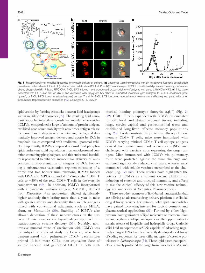
Fig. 1 Fusogenic polymer-modified liposomes for cytosolic delivery of antigens. (a) Liposomes were incorporated with pH-responsive, fusogenic poly(glycidol)derivatives in either a linear (MGlu-LPG) or hyperbranched structure (MGlu-HPG). (b)Confocal images of BMDCs treated with liposomes containing rhodamine-labeled phospholipid (Rh-PE) and FITC-OVA. MGlu-LPG induced more pronounced cytosolic delivery of antigens, compared with MGlu-HPG. (c) Mice wereinoculated with E.G7-OVA cells on day 0, and vaccinated with 50 μg of OVA either in unmodified liposomes (open triangles), MGlu-LPG-liposomes (opensquares), or MGlu-HPG-liposomes (closed squares) on days 7 and 14. MGlu-LPG-liposomes reduced tumor volume more effectively compared with otherformulations. Reproduced with permission (46). Copyright 2013, Elsevier.
2568 Sahdev, Ochyl and Moon

particles with~250 nm diameter and positive zeta potentialenhanced DC targeting, phagocytosis, and transfection effi-ciency. Additionally, augmented DC activation and maturationwere achieved by incorporation of a positively charged lipid withknown adjuvant characteristics (DOTAP) and CpG, which wereintegrated in pDNA. Administration of cSLN-pDNA led to1.76-fold greater IFNγ/IL-5 ratio compared with pDNA alone,indicating promotion of a Th1 response necessary for protectionagainst L. major. Enhanced IgG2a/IgG1 ratio with cSLN-pDNAvaccination also corroborated Th1-skewed response. In the end,cSLN-pDNA vaccine successfully decreased parasite burden by86.3%, which is a significant reduction compared with thepreviously reported mean average of parasite load reduction of59.2% for DNA alone vaccination.
Another class of emerging lipid-based nanoparticles for vac-cine delivery includes cubosomes, which consist of multiple lipidbilayers interrupted by numerous aqueous channels (55).Cubosomes have high loading capacity for both hydrophilicand hydrophobic bioactives due to their aqueous core and lipidmatrix with large surface area, and they have been shown toretard drug release in physiological condition. Cubosomes canbe prepared by high-energymechanical dispersion of lipids self-assembled into viscous cubic phase. Alternatively, they can beformed by solvent dilution method of dispersing liquid crystal-forming lipid, polymers, and hydrotrope in excess water.Cubosomes formed with minimal energy input with thesolvent dilution method have been recently investigated forvaccine applications (56,57). Cubosomes were shown to
efficiently encapsulate OVA along with MPLA andimiquimod (TLR4 and TLR7 agonists, respectively)and sustain their release over 1 week in vitro. Cubosomeswith adjuvants were more effective at generating OVA-specific humoral immune responses and CD8+ andCD4+ T cell responses, compared with conventionalliposomes or antigen adsorbed with alum (57).
The recent studies highlighted in this section focused onimproving physical stability of lipid-based vaccine deliverycarriers. These emerging nano-formulations have high serumstability, resistance to enzyme-mediated degradation,good drug loading efficiency, and strong immunogenicproperties, offering new versatile platform technologyfor vaccine development.
NATURAL POLYMER-BASED PARTICLES
Chitosan
Chitosan is a natural polyaminosaccharide derived primarilyfrom exoskeletons of crustaceans. It consists of copolymers ofβ-(1–4)-linked D-glucosamine (deacetylated unit) and N-acetyl-D-glucosamine (acetylated unit) monomers obtainedby deacetylation of chitin (58). Chitosan has attractive prop-erties for vaccine delivery applications, including high avail-ability, biodegradability, and biocompatibility. In addition,high density of amino groups in chitosan allows facile chemical
Fig. 2 Elicitation of potent mucosal CD8+ Tcell responses with pulmonary nanoparticle vaccination. (a) Intratracheal administration of ICMVs loaded withfluorescent OVA (red) resulted in efficient antigen delivery to mediastinal lymph nodes draining lungs by day 4. (b) Expansion and migration of OVA-specific OT-ICD8+ Tcells expressing luciferase was monitored after vaccination. Pulmonary administration of ICMVs led to robust expansion of OT-I CD8+ Tcells in thelungs by day 3, and their dissemination to the lungs (L), gastrointestinal tracts (G), Peyer’s patches (PP), and vaginal tract (V) by day 5. Mice immunized viapulmonary route with soluble antigen or subcutaneous route with particles had significantly reduced signals from OT-I Tcells in mucosal tissues. (c) Pulmonaryvaccination with ICMVs loaded with SIV gag antigen, AL11, and PADRE peptide on days 0 and 28 protected mice against challenge by intratracheal administrationof AL11-expressing vaccinia virus, whereas pulmonary vaccination with soluble antigens or subcutaneous vaccination with particles failed to protect the animals.Reproduced with permission (52). Copyright 2013, The American Association for Advancement of Science.
Biomaterials for Nanoparticle Vaccine Delivery Systems 2569

modification, complexation with negatively charged antigens,and muco-adhesiveness (59,60). Chitosan has been formulat-ed into nano/microparticles without the use of organic sol-vents or high shear stress via ionotropic gelation and self-assembly of polyelectrolytes, preserving immunogenicity ofantigens (61,62). Antigen-loaded chitosan nanoparticles signifi-cantly enhanced upregulation of co-stimulatory markers onAPCs and release of pro-inflammatory cytokines (e.g. IL-1β,IL-6, TNF-α) compared with soluble antigen. Chitosan particlescarrying OVA promoted antigen presentation via MHC-I andMHC-II pathways in DCs and induced higher proliferation ofantigen-specific CD4+ and CD8+ T cells than equivalent doseof soluble antigen (63). Interestingly, bothmacrophages andDCsexhibited maximum antigen uptake with 1 μm diameterchitosan particles, while 300 nm and 3 μm diameter particleshad decreased antigen uptake in vitro (63).
High density of amino groups in chitosan permits its inter-action with negatively charged antigens, plasmid DNA, andother anionic biopolymers. Exploiting these charge-mediatedionic interactions, a wide variety of proteins and DNA vac-cines have been incorporated into chitosan nano/micro par-ticles. Among these, chitosan nanoparticles coated with alginicacid have unique pH-responsive properties that make them anideal platform for oral vaccination (61). To synthesize theseparticles, amino groups within chitosan-plasmid DNA com-plexes were reacted with carboxyl groups in alginic acid,resulting in a crosslinked nanoparticle system stable at pH7.0. In acidic conditions of simulated gastric fluid (pH 1.5),the particles aggregated into micrometer-sized complexeswhile excess alginic acid, which is insoluble at pH 1.5, self-assembled into amorphous shell on the surfaces of particles,thereby protecting the encapsulated DNA against enzymaticand acidic degradation. As pH increased to 7.0, the aggregat-ed particles dispersed into nanoparticles. Oral administrationof particles carrying DNA for legumain, an asparaginyl endo-peptidase overexpressed in tumor cells, enhanced DNA up-take by macrophages and DCs in intestinal Peyer’s patches,and increased the frequency of legumain-specific cytotoxic Tcells (CD8+CD25+) while inhibiting regulatory T cells(CD4+CD25+). When mice bearing orthotopic 4T1 breasttumor were vaccinated with these particles via oral gavage,significant reduction in tumor volumes was observed, suggest-ing suitability of this system for oral DNA vaccination (61).Chitosan core nanogels coated with mannosylated alginate,which is an effective targeting ligand for C type lectin receptorexpressed by DCs have also been prepared (64). Surface ad-sorption of antigens on these particles significantly enhancedantigen uptake by DCs, while incorporation of adjuvantsallowed modulation of immune responses. Alginate-chitosannanogels loaded with Pam3Cys-SK4 (a TLR2 agonist) en-hanced IL-1β production, whereas particles admixed withCpG impaired production of IFN-α, IL-6, and TNF-α,demonstrating the importance of selecting optimal
combination of delivery vehicle and immunostimulatoryagents for ensuring appropriate immune activation. Highdensity of cationic charges on chitosan can also facilitateinteraction with negatively charged mucin on mucosalsurfaces, providing molecular attractive forces necessary formucoadhesion. The extent of mucoadhesion of chitosanmicrospheres was shown to be highly dependent upon theirzeta potential; chitosan microspheres formed by emulsificationand ionotropic gelation had higher cationic surface charge andsuperior mucoadhesiveness, compared with chitosan particlesformed by thermal crosslinking, glutaraldehyde crosslinking, ortripolyphosphate crosslinking (65).
Despite its salient features as a vaccine delivery platform,one of the major limitations of chitosan for biomedical appli-cations is its low solubility in physiological conditions. Chito-san with high density of acetylatedmonomers is soluble only inacidic medium with pH<5 (66). To overcome this limitation,several chitosan derivatives such as trimethyl chitosan andhydroxyethyl chitosan have been developed (59). Among the-se, trimethyl chitosan (TMC), formed by quaternization(methylation) of amino groups in chitosan, has been exten-sively investigated for vaccine applications. TMC nanoparti-cles co-encapsulating OVA antigen and immunostimulatoryagents, such as lipopolysaccharide, NOD-like receptor 2 li-gand muramyl dipeptide, and CpG, resulted in higher IgG,IgG1 and secretory IgA levels than non-adjuvanted TMC/OVA particles after intranasal or intradermal vaccination(67). TMC has also been further modified with thiol groups,which allowed self-assembly process with thiolated hyaluronicacid via ionic interactions (68). Spontaneous disulfide forma-tion between thiolated TCM and hyaluronic acid led toformation of chitosan/hyaluronic acid hybrid nanoparticles.These TMC-S-S-HA particles were coupled with maleimidePEG on their surfaces to shield the surface charge. Theresulting particles had higher stability in saline solutions, andvaccination with these particles via intranasal or intradermalroutes enhanced elicitation of IgG responses, compared withnon-stabilized particles (68).
Overall, these studies suggest great potential of chitosanbased nano/micro particles for vaccine delivery. In particular,their high bioavailabiliy, mucoadhesiveness, and biodegrad-able nature, combined with compatibility for oral andintransal route of administration, position chitosan-based par-ticles as an ideal vaccine carrier for mucosal vaccination. Weexpect that future studies focused on addressing their lowaqueous solubility at physiological conditions, irregular parti-cle size distribution, and low target specificity can lead tohighly effective vaccine formulations.
Gamma Polyglutamic Acid
Gamma polyglutamic acid (γ-PGA), a high molecular weightpolypeptide composed of linked glutamic acid units and
2570 Sahdev, Ochyl and Moon

carboxylate side chains, is produced by certain strains ofBacillus subtilis, which are natural components of a traditionalJapanese food item “natto” (69). The major advantages of γ-PGA polymer for biomedical applications include its watersolubility and biodegradability. Recent reports have describedmodification of γ-PGA with hydrophobic L-phenylalanineethyl ester, which converts γ-PGA into amphiphilic biopoly-mers with an ability to self-assemble into nanoparticles. Avariety of antigens, including HIV-1 p24, HIV-1 gp120, andJapanese encephalitis envelope protein have been encapsulat-ed and delivered via γ-PGA nanoparticles (69–73). Thesestudies have demonstrated that γ-PGA nanoparticles can po-tently activate both humoral and cellular immune responses.In addition, γ-PGA nanoparticles were efficiently internalizedby immature human monocyte-derived DCs and promotedtheir maturation and secretion of chemokines andinflammatory cytokines (74). Mice immunized with OVA-loaded γ-PGA nanoparticles had significantly higher frequen-cy of antigen-specific CD8+ T cells, compared with miceimmunized with soluble OVA alone, OVA+alum, orOVA+MPLA (75) . In a fol low-up study, γ-PGAnanoparticles without any additional adjuvant moleculeswere found to induce innate and adaptive immune responsesvia TLR4 and MyD88 signaling pathways, suggesting theirintrinsic immunostimulatory properties (72). Strategies to fur-ther enhance immunogenicity of γ-PGA nanoparticles includeincorporation of danger signals. Stable encapsulation of CpGinto poly(γ-glutamic acid)-graft-L-phenylalanine ethyl ester (γ-PGA-Phe) nanoparticles was achieved with polycationic prot-amine that stabilized CpG (76). This system allowed synergis-tic stimulation of TLR4 and TLR9 in macrophages by γ-PGA-Phe nanoparticles and induced potent antigen-specificcellular immunity.
Size of γ-PGA nanoparticles has been shown to influenceantigen uptake by APCs and their maturation and migrationto lymph nodes (76). γ-PGA nanoparticles with 40 nm diam-eter promoted highest activation of DCs and their migrationto lymph nodes as compared with 100 or 200 nm nanoparti-cles. Using a pH-insensitive, self-quenched OVA conjugate(DQ-OVA) that exhibits bright fluorescence upon proteolyticdegradation, the authors have shown that free DQ-OVAdegraded much faster than DQ-OVA delivered via γ-PGAnanoparticles. Interestingly, DQ-OVA formulated into larger(200 nm diameter) γ-PGA nanoparticles degraded at amuch faster rate, compared with protein delivered via40 nm nanoparticles. This is in contrast to a previousstudy that showed faster rate of degradation when antigenswere delivered via small polystyrene nanoparticles (50 nm)compared with larger 500 nm and 3 μm particles (77).Although the detailed mechanism is not clear yet, theauthors suggested that the unique amide bond in γ-PGAformed between α-amino and γ-carboxylic acid groupsmay have contributed to the polymer’s inherent resistance
to intracellular proteases and distinctive degradationkinetics.
γ-PGA nanoparticles also have been utilized as a vaccineplatform for cancer immunotherapy. Intranasal vaccinationwith OVA-loaded γ-PGA led to rapid particle uptake bynasopharyngeal-associated lymphoid tissue, antigen deliveryto the cervical lymph nodes, and induction of OVA-specificIFN-γ producing cells in the spleen and lymph nodes (78).Furthermore, mice vaccinated intranasally withOVA/γ-PGAnanoparticles were able to resist challenge by E.G7-OVAtumor cells and completely suppressed the formation of lungmetastasis by B16-OVA cells. In a separate study, γ-PGAnanoparticles complexed with benzalkonium chloride permit-ted efficient loading of OVA (79). Mice immunized with theseparticles generated balanced Th1 and Th2 antibody titers andwere protected against challenge with E.G7 cells expressingOVA. Although their efficacy in therapeutic tumor models isyet to be demonstrated, these studies have suggested thepotential clinical utility of γ-PGA nanoparticles for cancervaccine.
In short, γ-PGA nanoparticles have great potential not onlyas enhanced antigen delivery system but also as an effectivevaccine adjuvant that generates both humoral and cellularimmune response. For successful development of γ-PGA-based vaccines, more studies are warranted to better under-stand the mechanism for intracellular degradation of γ-PGAand their interaction with APCs.
Hyaluronic Acid
Hyaluronan (or hyaluronic acid, HA) is a linear polysaccha-ride composed of repeating disaccharide units of D-glucuronicacid andN-acetyl-D-glucosamine. HA is a normal componentof cartilage, and its high density of carboxylate groups helps toretain water and provide cartilage’s resistance to compression.In addition, HA plays critical roles in immune responses bymodulating leukocyte trafficking into inflamed skin, matura-tion and migration of epidermal dendritic cells, and T-cellactivation during antigen presentation (80). Due to its bio-compatibility, biodegradability, muco-adhesiveness, and goodsafety profile, HA-based delivery systems have been widelyexplored for vaccine applications. Intranasal immunizationwith HA-based microspheres loaded with influenza hemag-glutinin antigen and a mucosal adjuvant Escherichia Coli Heat-Labile Toxin (LTK63) was shown to enhance serum IgGlevels and hemagglutination inhibition titers (81).
HA, which is found in normal skin tissues, has been utilizedas a base material for preparation of transcutaneousmicroneedles. MicroHyala®, a microneedle patchcomprising of sodium hyaluronate has been used to deliver awide range of antigens derived from tetanus, diphtheria,malaria sporozoites, and influenza virus through skin (82).Antigen-loaded microneedle patches fabricated by
Biomaterials for Nanoparticle Vaccine Delivery Systems 2571

micromolding technologies were designed to undergo degra-dation after insertion into skin, effectively delivering antigensinto stratum corneum. Vaccination with microneedles gener-ated comparable IgG antibody titers as vaccination via sub-cutaneous injection. Biodegradable characteristic of HA-based microneedles offers a superior alternative to the con-ventional microneedles with regard to safety, offering a sim-ple, effective, and non-invasive vaccine delivery system (82).
An additional advantage of HA is its ability to reducegrowth of tumors, including colorectal carcinoma, which ispostulated to be mediated by activation of DCs (83). Admin-istration of DCs preconditioned with low molecular weightHA led to dramatically enhanced DC trafficking to tumor-regional lymph nodes in vivo (84). Vaccination of tumor-bearing mice with DCs preconditioned with HA and pulsedwith colorectal carcinoma tumor lysate resulted in significant-ly increased anti-tumor CTL responses and superior long-term protection against tumor recurrence, compared withadministration of DCs pulsed with tumor lysate alone.
In summary, HA and other natural polymers are attractivematerials for vaccine delivery applications due to their highabundance in nature, hydrophilicity, biodegradability, andtissue-specific interactions. Recent studies focused on improv-ing the physical stability of natural polymer-based nanoparti-cles have led to new vaccine formulations with enhancedefficiency of antigen delivery, especially for mucosal routes ofvaccination. Future studies should focus on utilization oftissue-targeting properties of these biopolymers and elabora-tion of mechanism of action, including inherent immunogenicproperties of these biopolymers.
SYNTHETIC POLYMER-BASED PARTICLES
Poly(Lactic-Co-Glycolic Acid)
Biodegradable polymeric nanoparticles have gained signifi-cant attention for their potential in drug and vaccine deliveryapplications. Synthetic polymers that are commonly used inbiomedical applications include aliphatic polyesters such aspoly(lactic acid), poly(glycolic acid), poly(ε-caprolactone),poly(hydroxybutyrate), and their copolymers. Among these,poly(lactic-co-glycolic acid) (PLGA) copolymer has beenexplored extensively for vaccine delivery due to its safety,biocompatibility, and biodegradability (85). PLGA polymerundergoes hydrolysis upon in vivo administration, formingbiologically compatible and metabolizable moieties i.e. lacticacid and glycolic acid, which are eventually removed from thebody by citric acid cycle (86). PLGA particles have beenutilized to deliver a wide range of protein and peptide anti-gens, including Hepatitis B surface antigen, VMP001 malariaprotein, HIV antigens, tumor-associated antigens, and whole
tumor lysate (87–93). The unique capability of PLGAparticles to sustain release of antigens and adjuvantmolecules for several weeks to months is thought to prolongand enhance immune stimulation (32). Long-term, potentimmune responses were achieved by single administration ofantigen-loaded PLGA particles via subcutaneous or intramus-cular route (94) or by direct injection of adjuvant-loadedPLGA partic les into regional lymph nodes (95) .Furthermore, the kinetics of antigen release from PLGAparticles can be easily controlled by modifying thecomposition and molecular weight of PLGA copolymers(96). Fine-tuning of their molecular composition led to thedevelopment of PLGA particles that can release antigens andadjuvants in several phases after a single vaccination, thusavoiding the need for multiple boost injections (97,98). Yet,one of the major drawbacks for the PLGA particle system hasbeen the potential exposure of antigens to harsh organicsolvents since conventional technique for loading antigens intoPLGA particles involves formation of single or double emul-sion (O/W or W/O/W) and solvent evaporation. However,with the recent advances in micro-encapsulation of biomole-cules into pre-formed, self-healing PLGA particles, antigenscan be loaded into PLGA vaccine particles in aqueouscondition with minimal loss of their integrity andantigenicity (99,100).
An important attribute of PLGA nanoparticles for vaccineapplications includes their ability to co-encapsulate antigensand adjuvants. Oral administration of PLGA nanoparticlesloaded with OVA antigen and MPLA adjuvant was shown toinduce significantly higher IgG and IgA antibody titers whencompared with OVA solution or PLGA nanoparticles loadedwith OVA alone (101). Primard et al. have demonstratedeffective induction of mucosal immunity after intranasal ad-ministration of PLGA nanoparticles coated with muco-adhesive chitosan and loaded with antigen (bovine serumalbumin - BSA) and TLR7 agonist (imiquimod) (Fig. 3a)(102). Co-encapsulating BSA and imiquimod into parti-cles prevented their enzymatic degradation and promot-ed their intracellular release, enhancing activation ofTLRs. Systemic and intranasal immunization withPLGA nanoparticles elicited similar titers of BSA-specific IgG, but interestingly, nasal immunization elic-ited significantly higher levels of IgA antibodies in nasalwashes, vaginal washes, and fecal samples, comparedwith systemic immunization, suggesting that the routeof vaccination has a critical role in induction of tissue-specific immune responses. Another study has examinedintranasal administration of PLGA nanoparticles coatedwith glycol chitosan (89). It was found that muco-adhesiveness of glycol chitosan coating prolonged thenasal residence time of PLGA nanoparticles, improvedantigen uptake, and induced potent mucosal and sys-temic immunity against Hepatitis B surface antigen.
2572 Sahdev, Ochyl and Moon
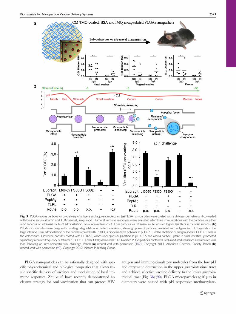
PLGA nanoparticles can be rationally designed with spe-cific physiochemical and biological properties that allows tis-sue specific delivery of vaccines and modulation of local im-mune responses. Zhu et al. have recently demonstrated anelegant strategy for oral vaccination that can protect HIV
antigen and immunostimulatory molecules from the low pHand enzymatic destruction in the upper gastrointestinal tractand achieve selective vaccine delivery to the lower gastroin-testinal tract (Fig. 3b) (90). PLGA microparticles (≥10 μm indiameter) were coated with pH responsive methacrylate-
Fig. 3 PLGA vaccine particles for co-delivery of antigens and adjuvant molecules. (a) PLGA nanoparticles were coated with a chitosan derivative and co-loadedwith bovine serum albumin and TLR7 agonist, imiquimod. Humoral immune responses were evaluated after three immunizations with the particles via eithersubcutaneous or intranasal route of administration. Local administration of PLGA particles via intranasal route induced higher IgA titers in mucosal surfaces. (b)PLGA microparticles were designed to undergo degradation in the terminal ileum, allowing uptake of particles co-loaded with antigens and TLR agonists in thelarge intestine. Oral administration of the particles coated with FS30D, a biodegradable polymer at pH>7.0, led to elicitation of antigen-specific CD8+ Tcells inthe colorectum. However, particles coated with L100-55, which undergoes degradation at pH>5.5 and allows particle uptake in small intestine, promotedsignificantly reduced frequency of tetramer+CD8+Tcells. Orally delivered FS30D-coated PLGA particles conferred Tcell-mediated resistance and reduced viralload following an intra-colorectal viral challenge. Panels (a) reproduced with permission (102). Copyright 2013, American Chemical Society. Panels (b)reproduced with permission (90). Copyright 2012, Nature Publishing Group.
Biomaterials for Nanoparticle Vaccine Delivery Systems 2573

based Eudragit FS30D21 polymer. This anionic tripolymercomposed of poly-(methyl acrylate, methyl methacrylate,methacrylic acid) is soluble only at pH>7.0, thus preventingencapsulated cargo materials from being released too early inthe stomach, while promoting content release in the largeintestine. PLGA particles were designed to release HIV pep-tide antigens together with poly(I:C) and CpG in the largeintestine, but not small intestine. Oral vaccination with theseparticles generated strong colorectal immunity and protectedmice against rectal and vaginal viral challenge. Importantly,by using particles with distinctive degradation properties, thisstudy for the first time has identified large intestine as thepotential target site after oral vaccination for elicitation ofprotective mucosal immune responses against mucosalpathogens.
To summarize, PLGA based micro/nanoparticles haveemerged as promising vaccine delivery candidates due to theircurrent clinical usage, biodegradable nature, ability to pro-long antigen release and co-encapsulate antigen andimmunopotentiator. We anticipate that the recent advancesin self-healing process of drug encapsulation into PLGA par-ticles, coupled with targeting capability of surface-modifiedPLGA particles, will allow effective delivery of vaccine com-ponents to lymphoid tissues with minimal loss of antigenicityand immunogenicity.
Polyethyleneimine
PEI is a cationic polymer, synthesized in various forms includ-ing linear or branched, and high or low molecular weightspecies. It is widely used as an agent for non-viral gene deliverydue to its ability to form nanoscale complexes (polyplexes)with nucleic acids by electrostatic interactions (103). Thesepolyplexes can bind heparan sulfate proteoglycans present onthe surface of cells including APCs and become internalizedvia endocytosis. During endosomal acidification, bufferingaction of PEI (i.e. proton sponge effect) results in osmoticrupture of endosomes, allowing cytosolic entry of polyplexesand release of cargo. Thus, PEI-DNA polyplexes are thoughtto increase uptake of plasmid DNA and improve its expres-sion, generating enhanced immune responses. Additional ad-vantages of PEI-based DNA vaccine delivery system includeits ease of preparation, stability, and high DNA loading effi-ciency. Indeed, it was shown that intravenous administrationof PEI-DNA plasmid complexes increased the levels of geneexpression by 20–400 fold and enhanced elicitation ofepitope-specific CD8+ T-cell responses by 10–25 fold withhigher frequency of antigen-specific Th1-helper cells (104).Memory cellular immune responses elicited by PEI-DNAcomplexes were able to respond rapidly and protect immu-nized animals against a lethal dose of recombinant Listeriamonocytogenes.
Several studies have exploited the ability of PEI to promotecross-presentation through the MHC Class I pathway(105,106). Treatment of mice with PEI complexed DNAencoding for OVA resulted in antigen-specific target cell lysisand activation of B3Z cells (OVA/Kb-specific CD8+ cyto-toxic T cell clone that recognizes the target cells through theclass I MHC molecules), indicative of greater rates of OVAcross-presentation (106). Mice immunized with DNA/PEIcomplexes either before or after tumor cell inoculation sup-pressed tumor growth with prolonged survival rate, suggestingthe efficacy of PEI-mediated DNA vaccination for anti-cancertherapy.
PEI-based vaccines have been investigated for their efficacyfor elicitation of mucosal immune responses. Intranasallyadministered PEI/DNA vaccine induced potent mucosaland systemic immune responses against hemagglutinin frominfluenza A H5N1 and H1N1 2009 viruses (107). RobustCD4+ and CD8+ memory T cell responses were observedin spleen and lungs, along with secretion of IFN-γ, TNF-α,and IL-2 from splenocytes as well as lung T cells. Moreover,the H5N1 vaccine elicited full protection against the parentalstrain and partial cross-protection against a distinct highlypathogenic H5N1 strain. Similarly, PEI-mediated intranasalimmunization with plasmid DNA for influenza hemagglutininor herpes simplex virus type-2 (HSV-2) glycoprotein D elicitedrobust IgG and IgA antibody titers and provided superiorprotection against lethal challenge with influenza virus,compared with a strong experimental mucosal adjuvantcholera toxin B (103). The authors have attributed the en-hanced immunogenicity of PEI-based vaccines to 1) increasedantigen uptake by APCs, 2) improved trafficking of DCs todLNs, 3) induction of non-proinflammatory cytokines includ-ing IL-4, IL-5 and IL-13, and 4) stimulation of Irf3-dependentsignaling.
To improve vaccine delivery, a multifunctional systemconsisting of a protein antigen and its encoding plasmid linkedtomaltosylated PEI nanocomplexes has been developed (108).High charge density on PEI enabled complex formation withplasmid DNA, while the maltosylated moieties induced high-affinity bonding with protein antigens. Immunization withPEI nanocomplexes triggered potent antibody responses andIFN-γ-producing CD8+ T cells against human papillomavi-rus. Since sugar receptors are abundantly expressed on APCs,modification of PEI with sugar moieties may have contributedto receptor-mediated endocytosis of PEI nanocomplexes(109). In a separate study, PEI complexed with alginatehas been synthesized into biodegradable and non-biodegradable nanogel formulations. The nanogels wereshown to increase in vitro antigen uptake by DCs ascompared to soluble antigen alone, with the biodegrad-able formulation being more effective at cytosolic anti-gen release (110). Additionally, this formulation waseffective at inducing DC maturation in vitro and
2574 Sahdev, Ochyl and Moon

significantly increased IgG and IFN-γ production andfrequency of antigen-specific CD8+ T-cells in vivo.
PEI-based systems, which have been traditionally used as atransfection agent, may provide an effective platform for DNAimmunogens, and recent studies indicating their potent mu-cosal adjuvant activity further highlight the potential of PEI invaccine applications. Future studies should be directed toaddress cytotoxicity associated with PEI for its clinical use inbiomedical applications.
Acrylic Acid Based Polymers
Po l y a l k y l a c r y l a t e b a s ed po l yme r s i n c l ud i n gpolymethylmethacrylate (PMMA), poly(ethylacrylic acid),poly(propylacrylic acid) and poly(butylacrylic acid) have beeninvestigated for vaccine delivery, and several studies havedemonstrated good adjuvant effect of polyacrylate-basednanoparticles with a considerable number of antigens(111,112). The ease of preparation, good safety record, andbiodegradable nature make them an excellent vaccine deliv-ery vehicle. Notably, Flanary et al. reported development of avaccine delivery system based on poly(propylacrylic acid)(PPAA) with pH-sensitive and membrane disruptive proper-ties (113). PPAA becomes protonated at low pH of endosomesand gains the ability to destabilize membranes - a uniquefeature that mimics membrane fusion induced by pathogenicproteins such as hemagglutinin and diphtheria toxin. OVAwas conjugated to PPAA polymer by a disulfide bond, whichallowed antigen release by glutathione reduction in cytoplasm.Antigen-polymer nano-conjugate increased intracellular ac-cumulation of OVA and enhanced MHC-I presentation andsubsequent CTL activation by 22-fold as compared with freeOVA. In a subsequent study, antigen-PPAA conjugate wascondensed into a particulate formulation by ionic complexa-tion of anionic PPAA-OVA conjugate with cationicpoly(dimethylaminoethyl methacrylate) (114). In-vivo adminis-tration of PPAA-containing formulations induced 8-fold in-crease in generation of OVA-specific CD8+ T cells and 11-fold increase in production of anti-OVA IgG as comparedwith saline or OVA solution. Using an EG.7-OVA mousetumor model, the authors showed that PPAA-containing car-riers suppressed tumor growth and increased tumor-free sur-vival rate by 3.5-fold compared with saline or OVA solution.
Building upon these results, Wilson et al. recently reporteddevelopment of pH-responsive, endosome-lytic polymer mi-celle structures designed for dual-delivery of antigen andimmunostimulatory DNA oligonucleotides (Fig. 4) (115). Toconstruct the micellar structure, amphiphilic diblock copoly-mer with two multifunctional modules was synthesized. Thefirst block consisted of cationic dimethylaminoethyl methac-rylate and pyridyl disulfide ethyl methacrylate for electrostaticinteraction with CpG and reversible disulfide bond formationwith thiol-bearing antigens, respectively. The second block
composed of PAA, DMAEMA, and butyl methacrylate hadhydrophobic and endosome-lytic properties that promotedmicelle assembly and cytosolic antigen delivery. The resultingmicelles were ~30 nm in diameter and exhibited potent pH-dependent membrane destabilizing activity. Conjugation ofOVA to the micellar structures significantly enhanced antigencross-presentation and elicited significantly higher CD8+ Tcell response as compared with free OVA or physical mixtureof components. Incorporation of CpG into the micelles fur-ther increased the CD8+ and CD4+ T cell responses by ~7-fold as compared with OVA conjugates and elicited balancedIgG1/IgG2c antibody responses.
In summary, acrylic acid based polymers with ‘smart’capability for pH-responsive membrane-destabilization allowsdelivery of therapeutic peptides, proteins, and nucleic acids
Fig. 4 Nanoparticle vaccine based on pH-responsive copolymers for cyto-solic delivery of antigen and immunostimulatory molecules. (a) Schematic forthe amphiphilic diblock copolymers with (i) hydrophilic and cationic block forconjugation of antigen and electrostatic complexation with CpG and (ii)hydrophobic and endosomolytic block for micelle assembly and cytosolicdelivery of antigens. (b) Mice were immunized with conjugates (ova-pol),ova mixed with free CpG (ova+CpG), dual-delivery vehicles (ova-pol/CpG),and free ova mixed with CpG/micelle complexes (ova+pol/CpG).Splenocytes were restimulated ex vivo with free OVA257–264 for IFN-γ pro-duction among CD8+ Tcells detected via intracellular cytokine staining (leftpanel) and ELISPOT (middle panel). ELISPOT quantification of IFN-γ pro-duction among CD4+ Tcells after splenocytes restimulation with OVA323–339(right panel). Reproduced with permission (115). Copyright 2013, AmericanChemical Society.
Biomaterials for Nanoparticle Vaccine Delivery Systems 2575
into the cytoplasm of targeted cells, and this unique featuremay be exploited for enhancing cross-presentation of antigensand elicitation of cellular immune responses.
Polypropylene Sulfide
Fully synthetic and biodegradable Pluronic-stabilized poly-propylene sulfide (PPS) nanoparticles have been developedas a new antigen nanocarrier system. (116). Surface of PPSnanoparticles can be precisely engineered to display specificchemical moieties or antigens in a disulfide-sensitive manner.It was shown that surface chemistry on PPS nanoparticles candirectly influence activation of complement cascade, therebymodulating generation of danger signals in situ and initiationof adaptive immune responses (117,118). The size of PPSnanoparticles has been shown to be a major factor determin-ing their lymphatic transport and uptake by APCs. Afterintradermal injection, PPS nanoparticles in the size range of30 nm drained rapidly through lymphatics to the dLNs,whereas lymphatic transport of 100 nm nanoparticles wasonly 10% as efficient (117). Similarly, intranasal vaccinationwith PPS nanoparticles resulted in penetration of particlesthrough the nasal epithelium, transition via M cells, andparticle uptake by APCs in the nasal-associated lymphoidtissue (119). Interestingly, for intranasal route of vaccination,as the size of PPS nanoparticles was increased from 30 to200 nm, antigen was more effectively delivered via bothMHC-I andMHC-II presentation pathways (119). Moreover,intranasal immunization with 200 nm nanoparticles signifi-cantly increased the frequency of antigen-specific lung CD4+T cells producing IFN-γ, TNF-α, or IL-2 and enhancedsystemic and mucosal humoral responses in the lungs com-pared with 30 nm particles. As noted by the authors of thisstudy, this seemingly contrasting impact of particle size on theresulting immune responses suggests that physiology of the siteof vaccination is the major determinant for draining of parti-cles to lymphoid tissues and antigen delivery to APCs.
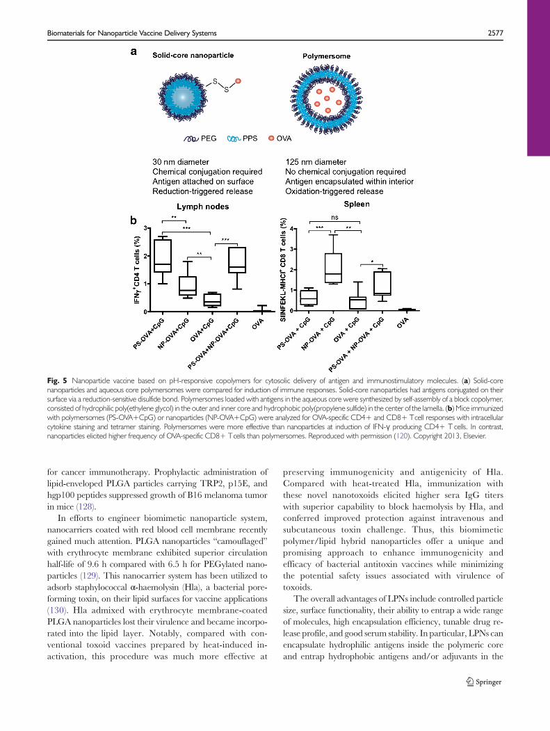
Two different platforms composed of PPS polymer werecompared for their potency as vaccine vehicles (Fig. 5a). Solid-core Pluronic-stabilized PPS nanoparticles were conjugatedwith protein antigens on their surface via a disulfide linker,while PEG17-bl-PPS30 polymersomes were loaded with anti-gens within their aqueous core (120). Polymersomes are stableself-assembled polymeric shells capable of releasing hydropho-bic as well as hydrophilic moieties (121,122). Mice immunizedwith polymersomes exhibited enhanced induction of IFN-γ-producing CD4+T cells in spleen, lymph nodes, and lungscompared with mice immunized with PPS nanoparticles(Fig. 5b). On the other hand, PPS nanoparticles surface-displaying OVA significantly enhanced the clonal expansionof antigen-specific CD8+ T cells than polymersomes. Takingadvantage of the distinct behaviors of two nanocarriers, theauthors combined the two systems (nanoparticles and
polymersomes) in a single vaccine formulation, which gener-ated concerted CD4+ and CD8+ T cell responses, thuspositioning PPS-based delivery systems as ideal vaccinenanocarriers for induction of robust cellular immune re-sponses (120).
LIPID COATED POLYMERIC PARTICLES
Lipid-polymer hybrid nanoparticles (LPNs) have emerged as apromising vaccine delivery platform in the past few years.LPNs combine the advantages of polymeric nanoparticleswith physical stability and liposomes with biomimeticcharacteristics (123,124). In general, LPNs have the followingthree components; 1) a polymer core, 2) phospholipid layerenveloping the polymer core, and 3) an outer lipid-PEG layer.Therapeutic drugs and proteins can be encapsulated insidethe polymeric core while lipid layer provides biocompatibilityand biomimetic properties to the polymeric core. The outer-most lipid-PEG coating provides steric stabilization and pro-longs the in-vivo circulation time of particles (123).
LPNs consisting of poly-(β-amino ester) (PBAE) coreenveloped by a phospholipid bilayer shell were prepared fordelivery of mRNA-based vaccines (125). PBAE is a pH-responsive polymer capable of promoting endosomal disrup-tion in acidic condition. Lipid-enveloped PBAE particles,surface-adsorbed with mRNA via electrostatic interactionwere shown to disrupt endosomes in DCs, allowing cytosolicdelivery of mRNA. Intranasal administration of these particlessignificantly enhanced the expression of reporter protein lu-ciferase within 6 h as compared to naked mRNA, suggestingtheir potential utility for mRNA-based in vivo transfection forvaccine applications.
Similarly, pathogen-mimicking polymeric vaccine deliverysystem consisting of PLGA nanoparticle core coated by a lipidmembrane has been reported (126,127). PLGA particles withantigen conjugated on the lipid envelope elicited high antigen-specific IgG titers (>106) that were sustained for over 100 daysafter two immunizations with just 2.5 ng of antigen. Insertionof lipophilic danger signals, such as MPLA and αGC furtherenhanced antibody titers by ~12-fold, thus providing a potentdose-sparing vaccine delivery system with reduced risk ofreactogenicity. These PLGA-lipid hybrid nanoparticles weresubsequently applied for vaccination against malaria Plasmo-dium vivax sporozoites (88). Nanoparticles co-delivering MPLAand malaria vaccine candidate, VMP001, on the outer lipidenvelope promoted germinal center formation and elicitedlong-lasting Th1/Th2 balanced humoral immune responses.Antibodies raised by particle vaccination were able to agglu-tinate live Plasmodium vivax sporozoites. In a separate study,PLGA-lipid hybrid nanostructures were developed for deliv-ery of multiple melanoma tumor-associated antigen peptides
2576 Sahdev, Ochyl and Moon
for cancer immunotherapy. Prophylactic administration oflipid-enveloped PLGA particles carrying TRP2, p15E, andhgp100 peptides suppressed growth of B16 melanoma tumorin mice (128).
In efforts to engineer biomimetic nanoparticle system,nanocarriers coated with red blood cell membrane recentlygained much attention. PLGA nanoparticles “camouflaged”with erythrocyte membrane exhibited superior circulationhalf-life of 9.6 h compared with 6.5 h for PEGylated nano-particles (129). This nanocarrier system has been utilized toadsorb staphylococcal α-haemolysin (Hla), a bacterial pore-forming toxin, on their lipid surfaces for vaccine applications(130). Hla admixed with erythrocyte membrane-coatedPLGA nanoparticles lost their virulence and became incorpo-rated into the lipid layer. Notably, compared with con-ventional toxoid vaccines prepared by heat-induced in-activation, this procedure was much more effective at
preserving immunogenicity and antigenicity of Hla.Compared with heat-treated Hla, immunization withthese novel nanotoxoids elicited higher sera IgG titerswith superior capability to block haemolysis by Hla, andconferred improved protection against intravenous andsubcutaneous toxin challenge. Thus, this biomimeticpolymer/lipid hybrid nanoparticles offer a unique andpromising approach to enhance immunogenicity andefficacy of bacterial antitoxin vaccines while minimizingthe potential safety issues associated with virulence oftoxoids.
The overall advantages of LPNs include controlled particlesize, surface functionality, their ability to entrap a wide rangeof molecules, high encapsulation efficiency, tunable drug re-lease profile, and good serum stability. In particular, LPNs canencapsulate hydrophilic antigens inside the polymeric coreand entrap hydrophobic antigens and/or adjuvants in the
Fig. 5 Nanoparticle vaccine based on pH-responsive copolymers for cytosolic delivery of antigen and immunostimulatory molecules. (a) Solid-corenanoparticles and aqueous core polymersomes were compared for induction of immune responses. Solid-core nanoparticles had antigens conjugated on theirsurface via a reduction-sensitive disulfide bond. Polymersomes loaded with antigens in the aqueous core were synthesized by self-assembly of a block copolymer,consisted of hydrophilic poly(ethylene glycol) in the outer and inner core and hydrophobic poly(propylene sulfide) in the center of the lamella. (b) Mice immunizedwith polymersomes (PS-OVA+CpG) or nanoparticles (NP-OVA+CpG) were analyzed for OVA-specific CD4+ and CD8+ Tcell responses with intracellularcytokine staining and tetramer staining. Polymersomes were more effective than nanoparticles at induction of IFN-γ producing CD4+ T cells. In contrast,nanoparticles elicited higher frequency of OVA-specific CD8+ Tcells than polymersomes. Reproduced with permission (120). Copyright 2013, Elsevier.
Biomaterials for Nanoparticle Vaccine Delivery Systems 2577

lipid envelope, mimicking features of pathogens and providinga versatile materials platform for vaccine delivery.
CONCLUSIONS AND FUTURE OUTLOOK
Subunit vaccines benefit greatly from nanoparticle formula-tions due to improved antigen uptake and targeting to APCsas well as enhanced immunogenicity stemming from eitherimmunostimulatory features of nanocarriers themselves oradjuvant molecules co-delivered by the particles. Most partic-ulate vaccine platforms in consideration are biodegradableand biocompatible with minimal toxicity, offering safe andeffective alternatives to traditional microorganism-based vac-cines. We covered a wide range of distinct nanoparticle for-mulations differing in composition, size, charge, and route ofadministration, all of which have been shown to play crucialroles in modulation of biodistribution, cellular trafficking,and overall immune response. In the end, the majority ofparticulate vaccines delivering subunit antigens andimmunostimulatory agents improved immune responses overequivalent dose of soluble vaccines. In particular, nanoparticlevaccines with the ability to enhance cross-presentation ofsubunit antigens and generate strong cellular immunity pro-vide significant improvement over traditional vaccines. Thus,these new vaccine nano-formulations may play a crucial rolein future clinical development of vaccines against cancer andintracellular infections, such as HIV, TB, and Hepatitis C. Inaddition, a greater understanding of adjuvants and their mo-lecular mechanisms of immune activation is needed to furtherrefine the biophysical and biochemical features of nanoparti-cles for improved immunostimulation. It is also important toconsider the manufacturability of these systems, as economicand facile scale-up with reproducibility in industrial settingsare critical considerations for their translation into the clinic.
Although it is beyond the scope of this review article,there have been key advances in the design of artificialantigen-presenting cells (aAPCs) based on micro/nanoparticles. For instance, the shape of PLGAmicroparticleshas been found to directly impact CD8+ T cell responses,with stretched, ellipsoidal PLGA aAPCs decorated with anti-CD28 and MHC-IgG dimer improving CD8+ T-cell expan-sion by 2- to 20-fold, compared with spherical aAPCs (131). Inaddition, Perica et al. have recently reported the develop-ment of nanoscale aAPCs synthesized from 50 to100 nm iron-dextran paramagnetic particles and30 nm quantum dot nanocrystals (132). Both nano-aAPC systems induced antigen speci f ic T cel lproliferation and functional responses in vitro (132), withiron-dextran paramagnetic particles even allowing mag-netic field-induced T cell activation (133). Notably,when administered in vivo, nano-aAPCs effectivelyprimed adoptively transferred CTLs to attenuate tumor
growth in vivo. Unlike conventional microparticle-basedaAPCs, nano-aAPCs can drain efficiently via lymphaticsto lymph nodes and preferentially accumulate in tumorthrough enhanced permeability and retention, suggestingthat in situ cancer immunotherapy may benefit greatlyfrom in vivo administration of nano-aAPCs.
In conclusion, the recent studies we have highlighted in thisreview represent great strides in application of nanoparticledrug delivery platforms toward prophylactic and therapeuticvaccinations. The field of nanotechnology will continue toaddress challenges remaining in immunology and provideinnovative strategies in the future of vaccine design anddevelopment.
ACKNOWLEDGMENTS AND DISCLOSURES
This study was supported by the Michigan Institute for Clin-ical & Health Research (MICHR) Pilot Grant Program andby the National Institute of Health grant 1K22AI097291-01.
REFERENCES
1. Pulendran B, Ahmed R. Immunological mechanisms of vaccina-tion. Nat Immunol. 2011;12(6):509–17.
2. Reed SG, Bertholet S, Coler RN, Friede M. New horizons inadjuvants for vaccine development. Trends Immunol. 2009;30(1):23–32.
3. Mbow ML, De Gregorio E, Valiante NM, Rappuoli R. Newadjuvants for human vaccines. Curr Opin Immunol. 2010;22(3):411–6.
4. Brave A, Ljungberg K, Wahren B, Liu MA. Vaccine deliverymethods using viral vectors. Mol Pharm. 2007;4(1):18–32.
5. Barouch DH. Challenges in the development of an HIV-1 vaccine.Nature. 2008;455(7213):613–9.
6. Gupta RK. Aluminum compounds as vaccine adjuvants. Adv DrugDeliv Rev. 1998;32(3):155–72.
7. Peek LJ, Middaugh CR, Berkland C. Nanotechnology in vaccinedelivery. Adv Drug Deliv Rev. 2008;60(8):915–28.
8. Moon JJ, Huang B, Irvine DJ. Engineering nano- and microparti-cles to tune immunity. Adv Mater. 2012;24(28):3724–46.
9. Swartz MA, Hirosue S, Hubbell JA. Engineering approaches toimmunotherapy. Sci Transl Med. 2012;4(148), 148rv149.
10. Irvine DJ, Swartz MA, Szeto GL. Engineering synthetic vaccinesusing cues from natural immunity. NatMater. 2013;12(11):978–90.
11. Smith DM, Simon JK, Baker Jr JR. Applications of nanotechnologyfor immunology. Nat Rev Immunol. 2013;13(8):592–605.
12. Sallusto F, Lanzavecchia A, Araki K, Ahmed R. From vaccines tomemory and back. Immunity. 2010;33(4):451–63.
13. Guy B. The perfect mix: recent progress in adjuvant research. NatRev Microbiol. 2007;5(7):505–17.
14. Kawai T, Akira S. Toll-like receptors and their crosstalk with otherinnate receptors in infection and immunity. Immunity. 2011;34(5):637–50.
15. Iwasaki A, Medzhitov R. Regulation of adaptive immunity by theinnate immune system. Science. 2010;327(5963):291–5.
16. Kawai T, Akira S. The role of pattern-recognition receptors ininnate immunity: update on Toll-like receptors. Nat Immunol.2010;11(5):373–84.
2578 Sahdev, Ochyl and Moon

17. Alexopoulou L, Holt AC, Medzhitov R, Flavell RA. Recognition ofdouble-stranded RNA and activation of NF-kappaB by Toll-likereceptor 3. Nature. 2001;413(6857):732–8.
18. Trumpfheller C, Caskey M, Nchinda G, Longhi MP, Mizenina O,Huang Y, et al. The microbial mimic poly IC induces durable andprotective CD4+ T cell immunity together with a dendritic celltargeted vaccine. Proc Natl Acad Sci U S A. 2008;105(7):2574–9.
19. Mata-Haro V, Cekic C, Martin M, Chilton PM, Casella CR,Mitchell TC. The vaccine adjuvant monophosphoryl lipid A as aTRIF-biased agonist of TLR4. Science. 2007;316(5831):1628–32.
20. Vasilakos JP, TomaiMA. The use of Toll-like receptor 7/8 agonistsas vaccine adjuvants. Expert Rev Vaccines. 2013;12(7):809–19.
21. Hemmi H, Kaisho T, Takeuchi O, Sato S, Sanjo H, Hoshino K,et al. Small anti-viral compounds activate immune cells via theTLR7 MyD88-dependent signaling pathway. Nat Immunol.2002;3(2):196–200.
22. van Stipdonk MJ, Badia-Martinez D, Sluijter M, Offringa R, vanHall T, Achour A.Design of agonistic altered peptides for the robustinduction of CTL directed towards H-2Db in complex with themelanoma-associated epitope gp100. Cancer Res. 2009;69(19):7784–92.
23. Ly LV, Sluijter M, Versluis M, Luyten GP, van Stipdonk MJ, vander Burg SH, et al. Peptide vaccination after T-cell transfer causesmassive clonal expansion, tumor eradication, and manageable cy-tokine storm. Cancer Res. 2010;70(21):8339–46.
24. Krieg AM, Yi AK, Matson S, Waldschmidt TJ, Bishop GA,Teasdale R, et al. CpG motifs in bacterial DNA trigger direct B-cell activation. Nature. 1995;374(6522):546–9.
25. Takeshita F, Gursel I, Ishii KJ, Suzuki K, Gursel M,Klinman DM. Signal transduction pathways mediated bythe interaction of CpG DNA with Toll-like receptor 9.Semin Immunol. 2004;16(1):17–22.
26. Kawano T, Cui J, Koezuka Y, Toura I, Kaneko Y, Motoki K, et al.CD1d-restricted and TCR-mediated activation of valpha14 NKTcells by glycosylceramides. Science. 1997;278(5343):1626–9.
27. Hermans IF, Silk JD, Gileadi U, Salio M,Mathew B, Ritter G, et al.NKT cells enhance CD4+ and CD8+ T cell responses to solubleantigen in vivo through direct interaction with dendritic cells. JImmunol. 2003;171(10):5140–7.
28. Banchereau J, Steinman RM. Dendritic cells and the control ofimmunity. Nature. 1998;392(6673):245–52.
29. Torchilin VP. Recent advances with liposomes as pharmaceuticalcarriers. Nat Rev Drug Discov. 2005;4(2):145–60.
30. Watson DS, Endsley AN, Huang L. Design considerations forliposomal vaccines: influence of formulation parameters on anti-body and cell-mediated immune responses to liposome associatedantigens. Vaccine. 2012;30(13):2256–72.
31. Henriksen-Lacey M, Devitt A, Perrie Y. The vesicle size of DDA:TDB liposomal adjuvants plays a role in the cell-mediated immuneresponse but has no significant effect on antibody production. JControl Release. 2011;154(2):131–7.
32. Demento SL, CuiW, Criscione JM, Stern E, Tulipan J, Kaech SM,et al. Role of sustained antigen release from nanoparticle vaccines inshaping the T cell memory phenotype. Biomaterials. 2012;33(19):4957–64.
33. Kamath AT, Mastelic B, Christensen D, Rochat AF, Agger EM,Pinschewer DD, et al. Synchronization of dendritic cell activationand antigen exposure is required for the induction of Th1/Th17responses. J Immunol. 2012;188(10):4828–37.
34. Kaur R, Bramwell VW, Kirby DJ, Perrie Y. Manipulation of thesurface pegylation in combination with reduced vesicle size ofcationic liposomal adjuvants modifies their clearance kinetics fromthe injection site, and the rate and type of T cell response. J ControlRelease. 2012;164(3):331–7.
35. Zhuang Y, Ma Y, Wang C, Hai L, Yan C, Zhang Y, et al.PEGylated cationic liposomes robustly augment vaccine-induced
immune responses: role of lymphatic trafficking and biodistribution.J Control Release. 2012;159(1):135–42.
36. Cui Z, Han SJ, Vangasseri DP, Huang L. Immunostimulationmechanism of LPD nanoparticle as a vaccine carrier. Mol Pharm.2005;2(1):22–8.
37. Cui Z, Huang L. Liposome-polycation-DNA (LPD) particle as acarrier and adjuvant for protein-based vaccines: therapeutic effectagainst cervical cancer. Cancer Immunol Immunother.2005;54(12):1180–90.
38. Chen W, Huang L. Induction of cytotoxic T-lymphocytes andantitumor activity by a liposomal lipopeptide vaccine. Mol Pharm.2008;5(3):464–71.
39. Barnier Quer C, Elsharkawy A, Romeijn S, Kros A, Jiskoot W.Cationic liposomes as adjuvants for influenza hemagglutinin: morethan charge alone. Eur J Pharm Biopharm. 2012;81(2):294–302.
40. Nakamura T, Yamazaki D, Yamauchi J, Harashima H. Thenanoparticulation by octaarginine-modified liposome improvesalpha-galactosylceramide-mediated antitumor therapy via systemicadministration. J Control Release. 2013;171(2):216–24.
41. Badiee A, Khamesipour A, Samiei A, Soroush D, Shargh VH,Kheiri MT, et al. The role of liposome size on the type of immuneresponse induced in BALB/c mice against leishmaniasis: rgp63 as amodel antigen. Exp Parasitol. 2012;132(4):403–9.
42. Nakamura T, Moriguchi R, Kogure K, Harashima H.Incorporation of polyinosine-polycytidylic acid enhances cytotoxicT cell activity and antitumor effects by octaarginine-modified lipo-somes encapsulating antigen, but not by octaarginine-modifiedantigen complex. Int J Pharm. 2013;441(1–2):476–81.
43. Wang C, Zhuang Y, Zhang Y, Luo Z, Gao N, Li P, et al. Toll-likereceptor 3 agonist complexed with cationic liposome augmentsvaccine-elicited antitumor immunity by enhancing TLR3-IRF3signaling and type I interferons in dendritic cells. Vaccine.2012;30(32):4790–9.
44. Nakamura T, Moriguchi R, Kogure K, Shastri N, Harashima H.Efficient MHC class I presentation by controlled intracellular traf-ficking of antigens in octaarginine-modified liposomes. Mol Ther.2008;16(8):1507–14.
45. Yuba E, Kojima C, Harada A, Tana Watarai S, Kono K. pH-Sensitive fusogenic polymer-modified liposomes as a carrier ofantigenic proteins for activation of cellular immunity.Biomaterials. 2010;31(5):943–51.
46. Yuba E, Harada A, Sakanishi Y, Watarai S, Kono K. A liposome-based antigen delivery system using pH-sensitive fusogenic polymersfor cancer immunotherapy. Biomaterials. 2013;34(12):3042–52.
47. Chen CH, Lin YL, Liu YK, He PJ, Lin CM, Chiu YH, et al.Liposome-based polymer complex as a novel adjuvant: enhance-ment of specific antibody production and isotype switch. Int JNanomedicine. 2012;7:607–21.
48. Allen TM, Mumbengegwi DR, Charrois GJ. Anti-CD19-targetedliposomal doxorubicin improves the therapeutic efficacy in murineB-cell lymphoma and ameliorates the toxicity of liposomes withvarying drug release rates. Clin Cancer Res. 2005;11(9):3567–73.
49. Moon JJ, Suh H, Bershteyn A, Stephan MT, Liu H, Huang B, et al.Interbilayer-crosslinked multilamellar vesicles as synthetic vaccinesfor potent humoral and cellular immune responses. Nat Mater.2011;10(3):243–51.
50. Moon JJ, Suh H, Li AV, Ockenhouse CF, Yadava A, Irvine DJ.Enhancing humoral responses to a malaria antigen with nanopar-ticle vaccines that expand Tfh cells and promote germinal centerinduction. Proc Natl Acad Sci U S A. 2012;109(4):1080–5.
51. DeMuth PC, Moon JJ, Suh H, Hammond PT, Irvine DJ.Releasable layer-by-layer assembly of stabilized lipid nanocapsuleson microneedles for enhanced transcutaneous vaccine delivery.ACS Nano. 2012;6(9):8041–51.
52. Li AV, Moon JJ, Abraham W, Suh H, Elkhader J, Seidman MA,et al. Generation of effector memory T cell-based mucosal and
Biomaterials for Nanoparticle Vaccine Delivery Systems 2579
systemic immunity with pulmonary nanoparticle vaccination. SciTransl Med. 2013;5(204), 204ra130.
53. Almeida AJ, Souto E. Solid lipid nanoparticles as a drug deliverysystem for peptides and proteins. Adv Drug Deliv Rev. 2007;59(6):478–90.
54. Doroud D, Zahedifard F, Vatanara A, Najafabadi AR, Taslimi Y,Vahabpour R, et al. Delivery of a cocktail DNA vaccine encodingcysteine proteinases type I, II and III with solid lipid nanoparticlespotentiate protective immunity against Leishmania major infection.J Control Release. 2011;153(2):154–62.
55. Shah JC, Sadhale Y, Chilukuri DM. Cubic phase gels as drugdelivery systems. Adv Drug Deliv Rev. 2001;47(2–3):229–50.
56. Rizwan SB, Assmus D, Boehnke A, Hanley T, Boyd BJ, Rades T,et al. Preparation of phytantriol cubosomes by solvent precursordilution for the delivery of protein vaccines. Eur J PharmBiopharm. 2011;79(1):15–22.
57. Rizwan SB, McBurney WT, Young K, Hanley T, Boyd BJ, RadesT, et al. Cubosomes containing the adjuvants imiquimod andmonophosphoryl lipid A stimulate robust cellular and humoralimmune responses. J Control Release. 2013;165(1):16–21.
58. Illum L. Chitosan and its use as a pharmaceutical excipient. PharmRes. 1998;15(9):1326–31.
59. van der Merwe SM, Verhoef JC, Verheijden JH, Kotze AF,Junginger HE. Trimethylated chitosan as polymeric absorptionenhancer for improved peroral delivery of peptide drugs. Eur JPharm Biopharm. 2004;58(2):225–35.
60. Islam MA, Firdous J, Choi YJ, Yun CH, Cho CS. Designand application of chitosan microspheres as oral and nasalvaccine carriers: an updated review. Int J Nanomedicine.2012;7:6077–93.
61. Liu Z, Lv D, Liu S, Gong J, Wang D, Xiong M, et al.Alginic acid-coated chitosan nanoparticles loaded withlegumain DNA vaccine: effect against breast cancer in mice.PLoS One. 2013;8(4):e60190.
62. Prego C, Paolicelli P, Diaz B, Vicente S, Sanchez A, Gonzalez-Fernandez A, et al. Chitosan-based nanoparticles for improvingimmunization against hepatitis B infection. Vaccine. 2010;28(14):2607–14.
63. Koppolu B, Zaharoff DA. The effect of antigen encapsulation inchitosan particles on uptake, activation and presentation by antigenpresenting cells. Biomaterials. 2013;34(9):2359–69.
64. Demoulins T, Bassi I, Thomann-Harwood L, JandusC, Kaeuper P,Simon HU, et al. Alginate-coated chitosan nanogel capacity tomodulate the effect of TLR ligands on blood dendritic cells.Nanomedicine. 2013;9(6):806–17.
65. Dhawan S, Singla AK, Sinha VR. Evaluation of mucoadhesiveproperties of chitosan microspheres prepared by different methods.AAPS PharmSciTech. 2004;5(4):e67.
66. Aranaz I, Mengibar M, Harris R, Panos I, Miralles B, Acosta N,et al. Functional characterization of chitin and chitosan. Curr ChemBiol. 2009;3(2):203–30.
67. Bal SM, Slutter B, Verheul R, Bouwstra JA, Jiskoot W.Adjuvanted, antigen loaded N-trimethyl chitosan nanoparti-cles for nasal and intradermal vaccination: adjuvant- andsite-dependent immunogenicity in mice. Eur J Pharm Sci.2012;45(4):475–81.
68. Verheul RJ, Slutter B, Bal SM, Bouwstra JA, Jiskoot W, HenninkWE. Covalently stabilized trimethyl chitosan-hyaluronic acid nano-particles for nasal and intradermal vaccination. J Control Release.2011;156(1):46–52.
69. Akagi T, Kaneko T, Kida T, Akashi M. Preparation and charac-terization of biodegradable nanoparticles based on poly(gamma-glutamic acid) with l-phenylalanine as a protein carrier. J ControlRelease. 2005;108(2–3):226–36.
70. Akagi T, Kaneko T, Kida T, Akashi M. Multifunctional conjuga-tion of proteins on/into bio-nanoparticles prepared by amphiphilic
poly(gamma-glutamic acid). J Biomater Sci Polym Ed. 2006;17(8):875–92.
71. Wang X, Uto T, Akagi T, Akashi M, Baba M. Poly(gamma-glutamic acid) nanoparticles as an efficient antigen delivery andadjuvant system: potential for an AIDS vaccine. J Med Virol.2008;80(1):11–9.
72. UtoT, Akagi T, YoshinagaK, ToyamaM,AkashiM, BabaM.Theinduction of innate and adaptive immunity by biodegradablepoly(gamma-glutamic acid) nanoparticles via a TLR4 and MyD88signaling pathway. Biomaterials. 2011;32(22):5206–12.
73. Okamoto S, Yoshii H, Matsuura M, Kojima A, Ishikawa T, AkagiT, et al. Poly-gamma-glutamic acid nanoparticles and aluminumadjuvant used as an adjuvant with a single dose of Japanese enceph-alitis virus-like particles provide effective protection from Japaneseencephalitis virus. Clin Vaccine Immunol. 2012;19(1):17–22.
74. Broos S, Lundberg K, Akagi T, Kadowaki K, Akashi M, Greiff L,et al. Immunomodulatory nanoparticles as adjuvants and allergen-delivery system to human dendritic cells: implications for specificimmunotherapy. Vaccine. 2010;28(31):5075–85.
75. Uto T, Akagi T, Toyama M, Nishi Y, Shima F, Akashi M, et al.Comparative activity of biodegradable nanoparticles with alumi-num adjuvants: antigen uptake by dendritic cells and induction ofimmune response in mice. Immunol Lett. 2011;140(1–2):36–43.
76. Shima F, Uto T, Akagi T, Akashi M. Synergistic stimulation ofantigen presenting cells via TLR by combining CpG ODN andpoly(gamma-glutamic acid)-based nanoparticles as vaccine adju-vants. Bioconjug Chem. 2013;24(6):926–33.
77. Tran KK, Shen H. The role of phagosomal pH on the size-dependent efficiency of cross-presentation by dendritic cells.Biomaterials. 2009;30(7):1356–62.
78. Matsuo K, Koizumi H, Akashi M, Nakagawa S, Fujita T,Yamamoto A, e t a l . Intranasal immunizat ion withpoly(gamma-glutamic acid) nanoparticles entrapping antigen-ic proteins can induce potent tumor immunity. J ControlRelease. 2011;152(2):310–6.
79. Kurosaki T, Kitahara T, Nakamura T, Nishida K, Fumoto S,Kodama Y, et al. Development of effective cancer vaccine usingtargeting system of antigen protein to APCs. Pharm Res.2012;29(2):483–9.
80. Mummert ME. Immunologic roles of hyaluronan. Immunol Res.2005;31(3):189–206.
81. Singh M, Briones M, O’Hagan DT. A novel bioadhesive intranasaldelivery system for inactivated influenza vaccines. J ControlRelease. 2001;70(3):267–76.
82. Matsuo K, Yokota Y, Zhai Y, Quan YS, Kamiyama F, Mukai Y,et al. A low-invasive and effective transcutaneous immunizationsystem using a novel dissolving microneedle array for soluble andparticulate antigens. J Control Release. 2012;161(1):10–7.
83. Alaniz L, Rizzo M, Malvicini M, Jaunarena J, Avella D,Atorrasagasti C, et al. Low molecular weight hyaluronan inhibitscolorectal carcinoma growth by decreasing tumor cell proliferationand stimulating immune response. Cancer Lett. 2009;278(1):9–16.
84. Alaniz L, Rizzo M, Garcia MG, Piccioni F, Aquino JB, MalviciniM, et al. Low molecular weight hyaluronan preconditioning oftumor-pulsed dendritic cells increases their migratory ability andinduces immunity against murine colorectal carcinoma. CancerImmunol Immunother. 2011;60(10):1383–95.
85. Jain S, O’Hagan DT, Singh M. The long-term potential of biode-gradable poly(lactide-co-glycolide) microparticles as the next-generation vaccine adjuvant. Expert Rev Vaccines. 2011;10(12):1731–42.
86. Panyam J, Labhasetwar V. Biodegradable nanoparticles for drugand gene delivery to cells and tissue. Adv Drug Deliv Rev.2003;55(3):329–47.
87. Zhang Z, Tongchusak S, Mizukami Y, Kang YJ, Ioji T, ToumaM,et al. Induction of anti-tumor cytotoxic T cell responses through
2580 Sahdev, Ochyl and Moon

PLGA-nanoparticle mediated antigen delivery. Biomaterials.2011;32(14):3666–78.
88. Moon JJ, Suh H, PolhemusME, Ockenhouse CF, Yadava A, IrvineDJ. Antigen-displaying lipid-enveloped PLGA nanoparticles as de-livery agents for a Plasmodium vivax malaria vaccine. PLoS One.2012;7(2):e31472.
89. Pawar D, Mangal S, Goswami R, Jaganathan KS. Developmentand characterization of surface modified PLGA nanoparticles fornasal vaccine delivery: effect of mucoadhesive coating on antigenuptake and immune adjuvant activity. Eur J Pharm Biopharm.2013;85(3 Pt A):550–9.
90. Zhu Q, Talton J, Zhang G, Cunningham T, Wang Z, Waters RC,et al. Large intestine-targeted, nanoparticle-releasing oral vaccine tocontrol genitorectal viral infection. Nat Med. 2012;18(8):1291–6.
91. Ali OA, Huebsch N, Cao L, Dranoff G, Mooney DJ. Infection-mimicking materials to program dendritic cells in situ. Nat Mater.2009;8(2):151–8.
92. Ali OA, Doherty E, BellWJ, Fradet T, Hudak J, LaliberteMT, et al.Biomaterial-based vaccine induces regression of established intra-cranial glioma in rats. Pharm Res. 2011;28(5):1074–80.
93. De TemmermanML, Rejman J, Vandenbroucke RE, De Koker S,Libert C, Grooten J, et al. Polyelectrolyte LbL microcapsules versusPLGA microparticles for immunization with a protein antigen. JControl Release. 2012;158(2):233–9.
94. Eyles JE, Williamson ED, Alpar HO. Immunological responses tonasal delivery of free and encapsulated tetanus toxoid: studies on theeffect of vehicle volume. Int J Pharm. 1999;189(1):75–9.
95. Jewell CM, Lopez SC, Irvine DJ. In situ engineering of the lymphnode microenvironment via intranodal injection of adjuvant-releasing polymer particles. Proc Natl Acad Sci U S A.2011;108(38):15745–50.
96. Makadia HK, Siegel SJ. Poly Lactic-co-Glycolic Acid (PLGA) asbiodegradable controlled drug delivery carrier. Polymers (Basel).2011;3(3):1377–97.
97. Johansen P, Estevez F, Zurbriggen R, Merkle HP, Gluck R,Corradin G, et al. Towards clinical testing of a single-administration tetanus vaccine based on PLA/PLGAmicrospheres.Vaccine. 2000;19(9–10):1047–54.
98. Johansen P, Moon L, Tamber H, Merkle HP, Gander B, SesardicD. Immunogenicity of single-dose diphtheria vaccines based onPLA/PLGA microspheres in guinea pigs. Vaccine. 1999;18(3–4):209–15.
99. Reinhold SE, Desai KG, Zhang L, Olsen KF, Schwendeman SP.Self-healing microencapsulation of biomacromolecules without or-ganic solvents. Angew Chem Int Ed Engl. 2012;51(43):10800–3.
100. Desai KG, Schwendeman SP. Active self-healing encapsulation ofvaccine antigens in PLGA microspheres. J Control Release.2013;165(1):62–74.
101. Sarti F, Perera G, Hintzen F, Kotti K, Karageorgiou V, KammonaO, et al. In vivo evidence of oral vaccination with PLGA nanopar-ticles containing the immunostimulant monophosphoryl lipid A.Biomaterials. 2011;32(16):4052–7.
102. Primard C, Poecheim J, Heuking S, Sublet E, Esmaeili F,Borchard G. Multifunctional PLGA-based nanoparticles en-capsulating simultaneously hydrophilic antigen and hydro-phobic immunomodulator for mucosal immunization. MolPharm. 2013;10(8):2996–3004.
103. Wegmann F, Gartlan KH, Harandi AM, Brinckmann SA, CocciaM,HillsonWR, et al. Polyethyleneimine is a potent mucosal adjuvantfor viral glycoprotein antigens. Nat Biotechnol. 2012;30(9):883–8.
104. Grant EV, Thomas M, Fortune J, Klibanov AM, Letvin NL.Enhancement of plasmid DNA immunogenicity with linearpolyethylenimine. Eur J Immunol. 2012;42(11):2937–48.
105. Chen J, Li Z, Huang H, Yang Y, Ding Q, Mai J, et al. Improvedantigen cross-presentation by polyethyleneimine-based nanoparti-cles. Int J Nanomedicine. 2011;6:77–84.
106. Ma YF, Yang YW. Delivery of DNA-based cancer vaccine withpolyethylenimine. Eur J Pharm Sci. 2010;40(2):75–83.
107. Torrieri-Dramard L, Lambrecht B, Ferreira HL, Van den Berg T,Klatzmann D, Bellier B. Intranasal DNA vaccination induces po-tent mucosal and systemic immune responses and cross-protectiveimmunity against influenza viruses. Mol Ther. 2011;19(3):602–11.
108. ChoHJ, Han SE, Im S, Lee Y,Kim YB, ChunT, et al. Maltosylatedpolyethylenimine-based triple nanocomplexes of human papilloma-virus 16L1 protein and DNA as a vaccine co-delivery system.Biomaterials. 2011;32(20):4621–9.
109. Park IY, Kim IY, Yoo MK, Choi YJ, Cho MH, Cho CS.Mannosylated polyethylenimine coupled mesoporous silica nano-particles for receptor-mediated gene delivery. Int J Pharm.2008;359(1–2):280–7.
110. Li P, Luo Z, Liu P, Gao N, Zhang Y, Pan H, et al. Bioreduciblealginate-poly(ethylenimine) nanogels as an antigen-delivery systemrobustly enhance vaccine-elicited humoral and cellular immuneresponses. J Control Release. 2013;168(3):271–9.
111. Kreuter J, Speiser PP. New adjuvants on a polymethylmethacrylatebase. Infect Immun. 1976;13(1):204–10.
112. Kreuter J, Mauler R, Gruschkau H, Speiser PP. The use ofnew polymethylmethacrylate adjuvants for split influenzavaccines. Exp Cell Biol. 1976;44(1):12–9.
113. Flanary S, Hoffman AS, Stayton PS. Antigen delivery withpoly(propylacrylic acid) conjugation enhancesMHC-1 presentationand T-cell activation. Bioconjug Chem. 2009;20(2):241–8.
114. Foster S, Duvall CL, Crownover EF, Hoffman AS, Stayton PS.Intracellular delivery of a protein antigen with an endosomal-releasing polymer enhances CD8 T-cell production and prophylac-tic vaccine efficacy. Bioconjug Chem. 2010;21(12):2205–12.
115. Wilson JT, Keller S,Manganiello MJ, Cheng C, Lee CC, Opara C,et al. pH-Responsive nanoparticle vaccines for dual-delivery of an-tigens and immunostimulatory oligonucleotides. ACS Nano.2013;7(5):3912–25.
116. van der Vlies AJ, O’Neil CP, Hasegawa U, Hammond N, HubbellJA. Synthesis of pyridyl disulfide-functionalized nanoparticles forconjugating thiol-containing small molecules, peptides, and pro-teins. Bioconjug Chem. 2010;21(4):653–62.
117. Reddy ST, van der Vlies AJ, Simeoni E, Angeli V, Randolph GJ,O’Neil CP, et al. Exploiting lymphatic transport and complementactivation in nanoparticle vaccines. Nat Biotechnol. 2007;25(10):1159–64.
118. Thomas SN, van der Vlies AJ, O’Neil CP, Reddy ST, Yu SS,Giorgio TD, et al. Engineering complement activation on polypro-pylene sulfide vaccine nanoparticles. Biomaterials. 2011;32(8):2194–203.
119. Stano A, van der Vlies AJ, Martino MM, Swartz MA, Hubbell JA,Simeoni E. PPS nanoparticles as versatile delivery system to inducesystemic and broad mucosal immunity after intranasal administra-tion. Vaccine. 2011;29(4):804–12.
120. Stano A, Scott EA, Dane KY, Swartz MA, Hubbell JA. Tunable Tcell immunity towards a protein antigen using polymersomes vs.solid-core nanoparticles. Biomaterials. 2013;34(17):4339–46.
121. Discher BM,Won YY, Ege DS, Lee JC, Bates FS, Discher DE, et al.Polymersomes: tough vesicles made from diblock copolymers.Science. 1999;284(5417):1143–6.
122. Holowka EP, Sun VZ, Kamei DT, Deming TJ. Polyarginine seg-ments in block copolypeptides drive both vesicular assembly andintracellular delivery. Nat Mater. 2007;6(1):52–7.
123. Hadinoto K, Sundaresan A, Cheow WS. Lipid-polymer hybridnanoparticles as a new generation therapeutic delivery platform: areview. Eur J Pharm Biopharm. 2013;85(3 Pt A):427–43.
124. Mandal B, Bhattacharjee H, Mittal N, Sah H, BalabathulaP, Thoma LA, et al. Core-shell-type lipid-polymer hybridnanoparticles as a drug delivery platform. Nanomedicine.2013;9(4):474–91.
Biomaterials for Nanoparticle Vaccine Delivery Systems 2581

125. Su X, Fricke J, Kavanagh DG, Irvine DJ. In vitro and in vivo mrnadelivery using lipid-enveloped pH-responsive polymer nanoparti-cles. Mol Pharm. 2011;8(3):774–87.
126. Bershteyn A, Chaparro J, Yau R, Kim M, Reinherz E, Ferreira-Moita L, et al. Polymer-supported lipid shells, onions, and flowers.Soft Matter. 2008;4(9):1787–91.
127. Bershteyn A, Hanson MC, Crespo MP, Moon JJ, Li AV, Suh H,et al. Robust IgG responses to nanograms of antigen using a biomi-metic lipid-coated particle vaccine. J Control Release. 2012;157(3):354–65.
128. Tan S, Sasada T, Bershteyn A, Yang K, Ioji T, Zhang Z.Combinational delivery of lipid-enveloped polymeric nanoparticlescarrying different peptides for anti-tumor immunotherapy.Nanomedicine. 2014;9(5):635–47.
129. Hu CM, Zhang L, Aryal S, Cheung C, Fang RH, Zhang L.Erythrocyte membrane-camouflaged polymeric nanoparticles as a
biomimetic delivery platform. Proc Natl Acad Sci U S A.2011;108(27):10980–5.
130. HuCM, FangRH, Luk BT, Zhang L.Nanoparticle-detained toxinsfor safe and effective vaccination. Nat Nanotechnol. 2013;8(12):933–8.
131. Sunshine JC, Perica K, Schneck JP, Green JJ. Particle shape depen-dence of CD8+ T cell activation by artificial antigen presentingcells. Biomaterials. 2014;35(1):269–77.
132. Perica K, De Leon MA, Durai M, Chiu YL, Bieler JG,Sibener L, et al. Nanoscale artificial antigen presenting cellsfor T cell immunotherapy. Nanomedicine. 2014;10(1):119–29.
133. Perica K, Tu A, Richter A, Bieler JG, Edidin M, Schneck JP.Magnetic field-induced T cell receptor clustering by nanoparticlesenhances T cell activation and stimulates antitumor activity. ACSNano. 2014;8(3):2252–60.
2582 Sahdev, Ochyl and Moon